Embed Size (px)
Citation preview

S20 CLEVELAND CLINIC JOURNAL OF MEDICINE VOLUME 70 • SUPPLEMENT 5 NOVEMBER 2003
■ ABSTRACT
Gastroesophageal reflux disease (GERD) can be theprimary cause of, or an aggravating contributor to, awide variety of conditions affecting extraesophagealstructures. As a result, GERD can lead to a numberof pulmonary symptoms and diseases, otolaryngo-logic findings and symptoms, and other extraesoph-ageal manifestations, including dental erosions.Clinicians must be aware of the possibility of theseextraesophageal reflux-related conditions, even inthe absence of classic esophageal symptoms ofGERD. While antireflux therapy is often helpful,response to treatment is less predictable than it isfor typical GERD.
Gastroesophageal reflux disease (GERD)can result in the direct regurgitation andaspiration of acidic gastric contents andhas been associated with extraesophageal
symptoms. GERD can masquerade as a wide varietyof conditions affecting extraesophageal structures(Table 1),1 leading to:• Pulmonary symptoms and diseases, such as asth-
ma, bronchitis, and pulmonary fibrosis• Otolaryngologic findings, such as hoarseness,
cough, laryngitis, subglottic stenosis, and laryn-geal cancer
• Other extraesophageal manifestations, such assinusitis, pharyngitis, and dental erosions. For many of these conditions, GERD sometimes
can be the primary or principal aggravating cause,although causality is often difficult to establish.Epidemiologically, GERD and many of its extra-
esophageal manifestations occur frequently and caneven occur simultaneously, without a causal rela-tionship. Moreover, the presence of gastric acid inextraesophageal structures has been difficult to doc-ument. Many patients with suspected extraesoph-ageal problems do not have classic GERD symp-toms, or such symptoms may present too subtly to bedetected. For example, more than 50% of patientswith reflux-related laryngeal disorders do not haveheartburn, regurgitation, or dysphagia.2
Data from studies evaluating the role of GERD inextraesophageal manifestations have been somewhatcontroversial, given that many such studies are smalland uncontrolled. In practice, however, positiveresults associated with antireflux treatment havedrawn attention to the role of GERD in extraesoph-ageal complications, making it difficult to ignore apotential association.3 A number of differences havebeen described between extraesophageal manifesta-tions and classic GERD manifestations with regardto symptoms, pathophysiology, evaluation, and treat-ment (Table 2).4 This review examines the preva-lence, pathogenesis, and clinical presentations ofextraesophageal manifestations of GERD, and brieflydiscusses how they are best evaluated and treated,including the role of antireflux therapy.
■ PREVALENCE AND CLINICAL OVERVIEW
Relationship to esophageal symptomsData demonstrating the high prevalence of GERDand its classic presentations (heartburn and acidregurgitation) have come from population-basedsurveys. Observational studies have also helpeduncover the prevalence of extraesophageal manifes-tations of GERD in the general population and howthey relate to classic GERD symptoms.
Extraesophageal symptoms of GERD are highlyprevalent among patients with both frequent andinfrequent typical GERD symptoms. In a popula-tion-based study in the Midwestern United States, a
Extraesophageal symptoms of GERDKENNETH R. DEVAULT, MD
From the Division of Gastroenterology and Hepatology, MayoClinic, Jacksonville, Fla.
Address: Kenneth R. DeVault, MD, Director of Gastrointes-tinal Research, Division of Gastroenterology and Hepatology,Mayo Clinic, 4500 San Pablo Road, Jacksonville, FL 32224.
on March 12, 2022. For personal use only. All other uses require permission.www.ccjm.orgDownloaded from

CLEVELAND CLINIC JOURNAL OF MEDICINE VOLUME 70 • SUPPLEMENT 5 NOVEMBER 2003 S21
reliable and valid self-report questionnaire wasmailed to an age- and sex-stratified random sampleof 2,200 residents of Olmsted County, Minn., aged25 to 74 years. The survey’s purpose was to deter-mine the prevalence and clinical spectrum ofGERD in the community, including the frequencyof atypical symptoms (noncardiac chest pain, dys-phagia, globus, dyspepsia, asthma, bronchitis, histo-ry of pneumonia, and hoarseness) among respon-dents with frequent, infrequent, and no typicalreflux symptoms.5
History of pneumonia and noncardiac chest pain(23.6% and 23.1%, respectively) had the highestoverall prevalence, followed by hoarseness (14.8%),bronchitis (14.0%), dysphagia (13.5%), dyspepsia(10.6%), asthma (9.3%), and globus (7.0%).Globus and a history of pneumonia were more com-mon among women than among men (P < 0.05).5
Among respondents with noncardiac chest pain,
40% had symptoms for greater than 5 years, and 5%reported severe or very severe symptoms. Symptomseverity and frequency were positively associated (P< 0.01). Similarly, among respondents with dyspha-gia, 37% had dysphagia that had lasted more than 5years, although a higher proportion of respondents(8.3% of those with any dysphagia, and 17.2% ofthose with frequent dysphagia) reported severe orvery severe dysphagia.5
Except for asthma and pneumonia, the atypicalsymptoms were each significantly more common (P< 0.001) among respondents with heartburn or acidregurgitation (Table 3).5 At least one atypical symp-tom was present in 79.9% of respondents with fre-quent (at least weekly) typical reflux symptoms,compared with 48.6% of respondents without heart-burn and acid regurgitation. In three logistic regres-sion models, typical reflux symptoms were associat-ed with noncardiac chest pain, dysphagia, globus,and dyspepsia. Frequent typical symptoms wereassociated with noncardiac chest pain, dysphagia,and dyspepsia.5
Other population-based data have helped todescribe the relationship between GERD manifesta-tions and extraesophageal symptoms. Using anational database to compare the comorbid occur-rence of sinus, laryngeal, and pulmonary diseases in
D E VA U LT
TABLE 1Extraesophageal manifestations of GERD
Pulmonary Otolaryngologic presentations presentations
Asthma Hoarseness
Aspiration pneumonia Chronic cough
Interstitial pulmonary fibrosis Throat clearing
Chronic bronchitis Chronic laryngitis
Bronchiectasis Globus sensation
Neonatal bronchopulmonary Vocal cord ulcers and dysplasia granulomas
Sudden infant death Laryngeal and tracheal syndrome stenosis
Laryngeal cancer
Mouth soreness
Halitosis
Pharyngitis
Otalgia
Chronic sinusitis
Croup
Stridor
Dysphonia
Abnormal taste
Dental erosions
Adapted from reference 1 with permission from Elsevier.
TABLE 2General comparisons between esophageal and extraesophageal manifestations of GERD
Esophageal Extraesophagealmanifestations manifestations
Primary Heartburn and Laryngeal andsymptoms regurgitation pulmonary
Pathophysiology Antireflux barrier, Multifactorial;acid clearance, laryngeal andesophageal pulmonarymucosal factorsresistance
Esophagitis Common Uncommonand Barrett’s esophagus
Ambulatory Very sensitive Sensitivity ispH monitoring and specific lower
for GERD
Response to anti- Excellent Less predictablereflux therapy
Adapted from reference 4 with permission from Elsevier.
on March 12, 2022. For personal use only. All other uses require permission.www.ccjm.orgDownloaded from
S22 CLEVELAND CLINIC JOURNAL OF MEDICINE VOLUME 70 • SUPPLEMENT 5 NOVEMBER 2003
E X T R A E S O P H A G E A L S Y M P T O M S O F G E R D
patients with and without reflux esophagitis, El-Serag and Sonnenberg6 evaluated a case populationof 101,366 patients with erosive esophagitis or stric-ture discharged from Department of VeteransAffairs hospitals from 1981 to 1994. They foundthat patients with reflux esophagitis were at higherrisk, compared with hospitalized controls, of havinga wide variety of pharyngeal, laryngeal, pulmonary,and sinus conditions (Figure 1).6 Specifically, ero-sive esophagitis and esophageal stricture were asso-ciated with an increased risk of sinusitis, pharyngi-tis, aphonia, laryngitis, laryngeal stenosis, chronicbronchitis, asthma, chronic obstructive pulmonarydisease, pulmonary fibrosis, bronchiectasis, pul-monary collapse, and pneumonia. Following a mul-tivariate analysis, the strongest statistically signifi-cant associations were found with bronchial asthmaand pulmonary fibrosis (Table 4).6
The most common diagnosis in both the case andthe control populations was pneumonia, followedby chronic bronchitis, chronic obstructive pul-monary disease, and bronchial asthma. Much lessfrequently diagnosed than pulmonary diseases weresinus, pharyngeal, and laryngeal disorders. In thisstudy, as many as 17% of all patients with esophagi-tis developed an extraesophageal manifestation ofthe disease. Patients with esophagitis or stricturecarried a 15% to 100% increased risk of havingextraesophageal diagnoses compared with subjects
without esophagitis or stricture.6
Endoscopy and esophageal pH monitoring havealso been used in prospective studies linking GERDto extraesophageal symptoms. Using such methods,GERD has been diagnosed in as many as 75% ofpatients with chronic hoarseness,2 in 78% ofpatients with laryngeal stenosis,2 in 70% to 80% ofpatients with asthma,7 and in 20% of patients withchronic cough.8 Endoscopic esophagitis has beenfound in 30% to 40% of patients with asthma andin approximately 20% of those with laryngitis.9,10
Despite the high prevalence of esophagitis inthese early studies, many investigators now believeesophagitis to be the clear exception in thesepatients. This could be due to our increased aware-ness of extraesophageal GERD, the wide availabili-ty of over-the-counter acid suppressants, or somecombination of these factors.
Considerations in the elderly. Extraesophagealsymptoms of GERD are frequently encountered inthe elderly.11 This is particularly troublesome, sincea symptom such as chest pain must be given greatrespect, particularly in the elderly, and can result incostly and extensive evaluation. It is unclearwhether extraesophageal symptoms are more com-mon in the elderly than in younger persons. If so,this finding would not be surprising, since bothextraesophageal symptoms and GERD seem toincrease in prevalence with age.
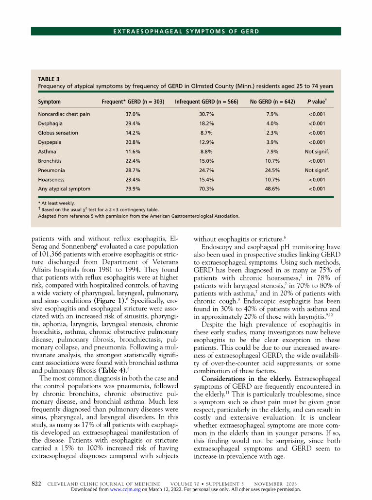
TABLE 3Frequency of atypical symptoms by frequency of GERD in Olmsted County (Minn.) residents aged 25 to 74 years
Symptom Frequent* GERD (n = 303) Infrequent GERD (n = 566) No GERD (n = 642) P value†
Noncardiac chest pain 37.0% 30.7% 7.9% < 0.001
Dysphagia 29.4% 18.2% 4.0% < 0.001
Globus sensation 14.2% 8.7% 2.3% < 0.001
Dyspepsia 20.8% 12.9% 3.9% < 0.001
Asthma 11.6% 8.8% 7.9% Not signif.
Bronchitis 22.4% 15.0% 10.7% < 0.001
Pneumonia 28.7% 24.7% 24.5% Not signif.
Hoarseness 23.4% 15.4% 10.7% < 0.001
Any atypical symptom 79.9% 70.3% 48.6% < 0.001
* At least weekly.† Based on the usual χ2 test for a 2 × 3 contingency table.Adapted from reference 5 with permission from the American Gastroenterological Association.
on March 12, 2022. For personal use only. All other uses require permission.www.ccjm.orgDownloaded from

PathophysiologyProposed mechanisms of extraesophageal symptoms.Two possible mechanisms have been proposed asunderlying GERD-related extraesophageal symptoms:• Microaspiration of gastric contents into extra-
esophageal structures during reflux episodes• Stimulation by the gastric refluxate of a vagal
reflex arc extending from the esophageal body tothe bronchopulmonary and laryngeal systems. Both mechanisms have been supported by clini-
cal and laboratory data documenting the injuriouseffects of esophageal acid on extraesophageal struc-tures. Studies using dual-probe esophageal pH mon-itoring seem to support the reflex arc theory, where-as ambulatory pH studies of patients with suspectedextraesophageal complications have demonstratedacid reflux to the proximal esophagus and beyond.3
With regard to the first mechanism, physiologicprotective mechanisms normally prevent refluxatefrom entering the pharyngeal and laryngeal space tocause symptoms and tissue damage. A disturbancein any known, or perhaps unknown, protective fac-tor could possibly account for the production ofextraesophageal symptoms.12
Regarding the second mechanism, embryologicstudies show that the esophagus and bronchial treeshare a common embryonic origin, having bothdeveloped from common tissue of the foregut.1 It istherefore not surprising that they also share a com-mon neural innervation via the vagus nerve.1
Acidification of the distal esophagus can stimulateacid-sensitive receptors that could conceivably pro-
duce noncardiac chest pain or interact with pul-monary bronchi and other upper airway structuresby a vagally mediated arc.12
Neither of these mechanisms is completelyunderstood, nor is its clinical relevance appreciatedin the absence of additional outcomes data andmore sensitive methods for detecting the movementof gastric refluxate.3
Defense mechanisms against extraesophagealsymptoms. Defense mechanisms protecting againstextraesophageal complications of GERD have beenorganized into a four-tier system (Table 5).3 Withinthis system, each defense mechanism occurs inascending order from the distal esophagus to thesupraesophageal region.
Junctional structures at the gastroesophagealinterface (tier 1) include the lower esophagealsphincter (LES), the crural diaphragm, the slingfibers, and the phrenoesophageal ligament. TheLES and the crural diaphragm are discussed in theprevious article in this supplement. The sling fibersof the stomach, arranged in a C-shaped fashion withthe open side toward the lesser curvature, serve as a“flap valve” to augment LES pressure. The phreno-esophageal ligament helps to anchor the cruralfibers to the LES segment.3
The esophageal body motor response (tier 2)includes primary and secondary peristalsis andesophageal body tone. The esophageal body clears
CLEVELAND CLINIC JOURNAL OF MEDICINE VOLUME 70 • SUPPLEMENT 5 NOVEMBER 2003 S23
D E VA U LT
LaryngealStenosis
BronchialAsthma
0 0.5 1 1.5 2 2.5 3 3.5 4
Odds Ratio
Laryngitis
Pharyngitis
Aphonia
Sinusitis TABLE 4Pulmonary disorders significantly associated withesophagitis or esophageal stricture*
Disorder Odds 95% Pratio CI value
Chronic bronchitis 1.28 1.22–1.34 0.0001
Bronchial asthma 1.51 1.43–1.59 0.0001
Chronic obstructive 1.22 1.16–1.27 0.0001pulmonary disease
Pulmonary fibrosis 1.36 1.25–1.48 0.0001
Bronchiectasis 1.26 1.09–1.47 0.0022
Pulmonary collapse 1.31 1.23–1.40 0.0001
Pneumonia 1.15 1.12–1.18 0.0001
* Following multivariate logistic regression analysis. Comparisonsare between hospitalized patients with erosive esophagitisor esophageal stricture and hospitalized controls.
Adapted from reference 6 with permission from the AmericanGastroenterological Association.
FIGURE 1. Risk of extraesophageal complications in hospitalizedpatients with erosive esophagitis or esophageal stricture comparedwith hospitalized controls. Each point represents an odds ratio,depicted with its 95% confidence interval, obtained from a multi-variate logistic regression analysis. Reprinted from reference 6 withpermission from the American Gastroenterological Association.
on March 12, 2022. For personal use only. All other uses require permission.www.ccjm.orgDownloaded from
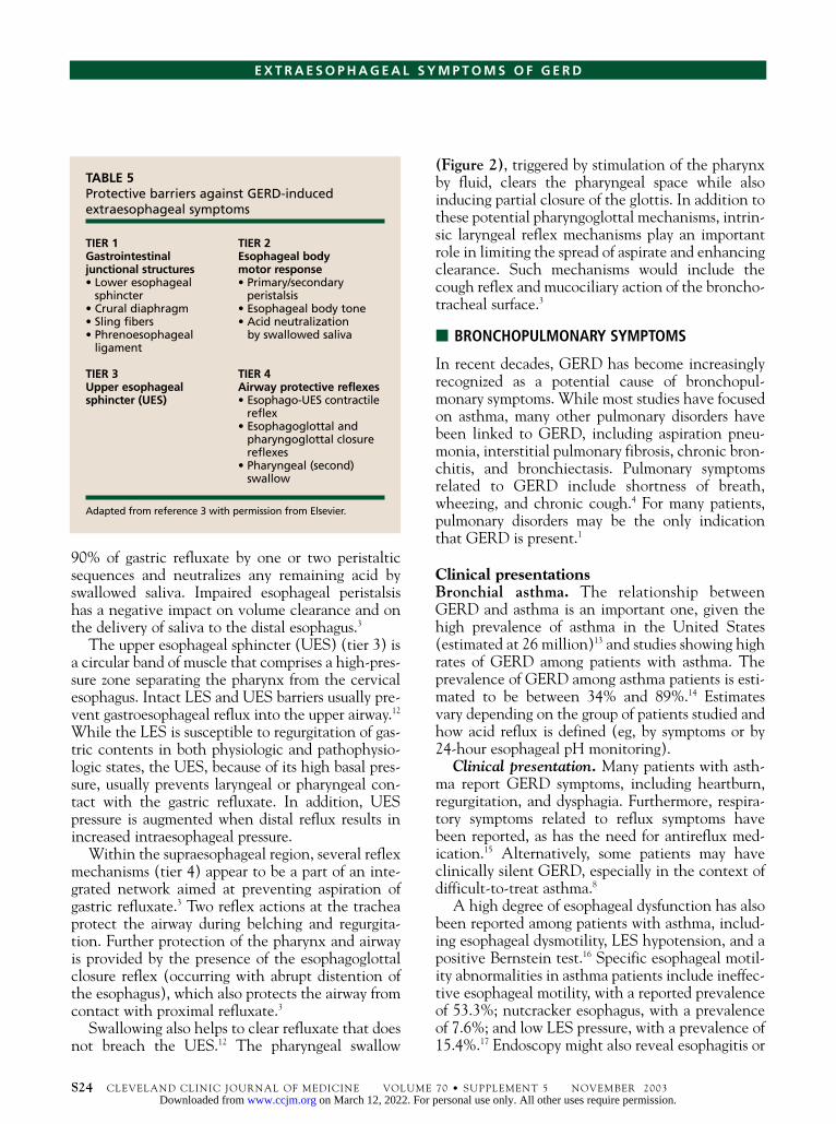
90% of gastric refluxate by one or two peristalticsequences and neutralizes any remaining acid byswallowed saliva. Impaired esophageal peristalsishas a negative impact on volume clearance and onthe delivery of saliva to the distal esophagus.3
The upper esophageal sphincter (UES) (tier 3) isa circular band of muscle that comprises a high-pres-sure zone separating the pharynx from the cervicalesophagus. Intact LES and UES barriers usually pre-vent gastroesophageal reflux into the upper airway.12
While the LES is susceptible to regurgitation of gas-tric contents in both physiologic and pathophysio-logic states, the UES, because of its high basal pres-sure, usually prevents laryngeal or pharyngeal con-tact with the gastric refluxate. In addition, UESpressure is augmented when distal reflux results inincreased intraesophageal pressure.
Within the supraesophageal region, several reflexmechanisms (tier 4) appear to be a part of an inte-grated network aimed at preventing aspiration ofgastric refluxate.3 Two reflex actions at the tracheaprotect the airway during belching and regurgita-tion. Further protection of the pharynx and airwayis provided by the presence of the esophagoglottalclosure reflex (occurring with abrupt distention ofthe esophagus), which also protects the airway fromcontact with proximal refluxate.3
Swallowing also helps to clear refluxate that doesnot breach the UES.12 The pharyngeal swallow
(Figure 2), triggered by stimulation of the pharynxby fluid, clears the pharyngeal space while alsoinducing partial closure of the glottis. In addition tothese potential pharyngoglottal mechanisms, intrin-sic laryngeal reflex mechanisms play an importantrole in limiting the spread of aspirate and enhancingclearance. Such mechanisms would include thecough reflex and mucociliary action of the broncho-tracheal surface.3
■ BRONCHOPULMONARY SYMPTOMS
In recent decades, GERD has become increasinglyrecognized as a potential cause of bronchopul-monary symptoms. While most studies have focusedon asthma, many other pulmonary disorders havebeen linked to GERD, including aspiration pneu-monia, interstitial pulmonary fibrosis, chronic bron-chitis, and bronchiectasis. Pulmonary symptomsrelated to GERD include shortness of breath,wheezing, and chronic cough.4 For many patients,pulmonary disorders may be the only indicationthat GERD is present.1
Clinical presentationsBronchial asthma. The relationship betweenGERD and asthma is an important one, given thehigh prevalence of asthma in the United States(estimated at 26 million)13 and studies showing highrates of GERD among patients with asthma. Theprevalence of GERD among asthma patients is esti-mated to be between 34% and 89%.14 Estimatesvary depending on the group of patients studied andhow acid reflux is defined (eg, by symptoms or by24-hour esophageal pH monitoring).
Clinical presentation. Many patients with asth-ma report GERD symptoms, including heartburn,regurgitation, and dysphagia. Furthermore, respira-tory symptoms related to reflux symptoms havebeen reported, as has the need for antireflux med-ication.15 Alternatively, some patients may haveclinically silent GERD, especially in the context ofdifficult-to-treat asthma.8
A high degree of esophageal dysfunction has alsobeen reported among patients with asthma, includ-ing esophageal dysmotility, LES hypotension, and apositive Bernstein test.16 Specific esophageal motil-ity abnormalities in asthma patients include ineffec-tive esophageal motility, with a reported prevalenceof 53.3%; nutcracker esophagus, with a prevalenceof 7.6%; and low LES pressure, with a prevalence of15.4%.17 Endoscopy might also reveal esophagitis or
S24 CLEVELAND CLINIC JOURNAL OF MEDICINE VOLUME 70 • SUPPLEMENT 5 NOVEMBER 2003
E X T R A E S O P H A G E A L S Y M P T O M S O F G E R D
TABLE 5Protective barriers against GERD-induced extraesophageal symptoms
TIER 1 TIER 2Gastrointestinal Esophageal body junctional structures motor response• Lower esophageal • Primary/secondary
sphincter peristalsis• Crural diaphragm • Esophageal body tone• Sling fibers • Acid neutralization • Phrenoesophageal by swallowed saliva
ligament
TIER 3 TIER 4Upper esophageal Airway protective reflexessphincter (UES) • Esophago-UES contractile
reflex• Esophagoglottal and
pharyngoglottal closurereflexes
• Pharyngeal (second) swallow
Adapted from reference 3 with permission from Elsevier.
on March 12, 2022. For personal use only. All other uses require permission.www.ccjm.orgDownloaded from

Barrett’s esophagus among patients with asthma,although most will not have esophagitis.7
Compared with normal controls, patients withasthma have a higher frequency of reflux symptoms,more frequent LES hypotension by manometry, andincreased esophageal acid contact times by 24-hourpH monitoring, which further supports the associa-tion between GERD and asthma.18
Pathogenesis. Bronchospasm is the hallmark ofasthma and occurs as a result of several different irri-tating stimuli to the bronchial airways. Acid refluxmay be the only trigger, or it may be one of manycontributing factors.1 Two possible pathophysiolog-ic mechanisms, referred to earlier in relation to allextraesophageal manifestations, have been pro-posed for GERD-induced asthma. While neither ofthese mechanisms is completely understood, bothappear to be involved in the relationship betweenGERD and asthma, and their relative effect variesamong patients.1 Both mechanisms might be activein some patients.18 Furthermore, both involve thevagus nerve and are blunted by vagotomy.
According to the reflex theory, stimulation ofacid-sensitive receptors by esophageal acid activatesa vagal response from the esophagus to the lung,which causes bronchoconstriction. Bronchocon-striction may, in fact, occur in all individuals as anormal protective mechanism in response to intra-esophageal acid perfusion.19 Peak expiratory flow
rates apparently return to normal after acid iscleared from the esophagus, although they do somore slowly among patients with asthma.
The reflux theory describes the microaspiration ofgastric contents into the bronchial tree, which caus-es direct irritation of the respiratory epithelium andstimulates inflammatory mediators.1 It is wellknown that mechanical stimulation of the upperairway or trachea can cause airway resistance.18
Bronchoconstriction in response to esophagealacidification has been demonstrated in both animalstudies20 and human studies.21 In animals, acidinstilled into the trachea predictably increased air-way resistance three to four times.20
More recently, investigators found an abruptdecrease in tracheal pH coinciding with broncho-constriction during episodes of gastroesophagealreflux in patients with asthma and typical GERDsymptoms (Figure 3).22 Further support for thereflux mechanism comes from a recent treatmentstudy showing that proximal acid reflux was a pre-dictor for improvement of asthma symptoms follow-ing aggressive acid suppression.23 A GERD-asthmacycle has been proposed, through which bron-chospasm promotes acid reflux, which promotes fur-ther bronchospasm. Asthma may also promoteGERD as a result of changes in esophageal physiol-ogy induced by asthma medications.1 A largeVeterans Administration-based study found, how-
CLEVELAND CLINIC JOURNAL OF MEDICINE VOLUME 70 • SUPPLEMENT 5 NOVEMBER 2003 S25
D E VA U LT
Hard Palate
Bolus
Nasopharynx
Soft Palate
Uvula
Oropharynx
Tongue
Laryngopharynx
Epiglottis
Larynx
Esophagus
Before Swallowing During Swallowing
FIGURE 2. Position of esophageal, laryngeal, and pharyngeal structures before and during swallowing.
on March 12, 2022. For personal use only. All other uses require permission.www.ccjm.orgDownloaded from

ever, that the GERD-asthma association is indepen-dent of bronchodilator use.7
Diagnosis. The patient’s history is an extremelyimportant part of the diagnosis of GERD-associatedasthma, despite the fact that approximately onethird of patients with asthma and esophageal dys-function do not have esophageal symptoms. Certainclinical clues can be helpful in identifying GERD-related asthma, as can selected tests (Table 6).1
Pulmonary symptoms suggesting reflux include noc-turnal cough, as well as worsening of asthma symp-toms after eating a large meal, drinking alcohol, orbeing in the supine position. GERD should be con-sidered in asthmatics who initially present in adult-hood, in those without an intrinsic component, andin those not responding to bronchodilator or steroidtherapy. An additional clue may be the develop-ment of reflux symptoms before the onset of asthma,or heartburn heralding an asthma attack.1
Esophageal tests that may be helpful in diagnosisinclude the barium esophagram, gastroesophagealscintigraphy, and prolonged esophageal pH moni-toring. The latter test, considered the gold standardfor GERD diagnosis, is the only esophageal test thatcan directly correlate acid reflux episodes withwheezing or other symptoms of bronchospasm.Nevertheless, confirming an esophageal cause forpulmonary symptoms using this test might stillprove difficult.1 Gastroesophageal scintigraphy has ahigh specificity, but it also has a low sensitivity,which limits its usefulness in adults.1
Irwin and colleagues8 found that they could usu-
ally determine the cause of difficult-to-controlasthma by using a systematic management protocol.While multiple factors were usually involved, thesingle most common contributory factor proved tobe GERD. Moreover, approximately two thirds ofaffected patients responded favorably to antirefluxtherapy. The researchers concluded that all diffi-cult-to-control asthma patients should be evaluatedfor GERD, even if GERD symptoms are minimal orabsent.8
Treatment. With regard to medical therapy,studies using proton pump inhibitors (PPIs) havehad more encouraging results than those usingantacids or histamine2-receptor antagonists. Thelatter have yielded inconsistent effects on asthmasymptoms and peak expiratory flow rates. A recentstudy23 using omeprazole to treat patients with asth-ma and GERD over 3 months showed that 73% ofpatients experienced marked alleviation of asthmasymptoms or increases in peak expiratory flow rate.Treatment reduced asthma symptoms by 57% after3 months (Figure 4). The patients most likely tobenefit from the therapy were those with frequent
S26 CLEVELAND CLINIC JOURNAL OF MEDICINE VOLUME 70 • SUPPLEMENT 5 NOVEMBER 2003
E X T R A E S O P H A G E A L S Y M P T O M S O F G E R D
TABLE 6Clinical clues and tests used in the diagnosis of GERD-associated asthma1
Clinical cluesAdult onset of asthma
No family history of asthma
Reflux symptoms preceding asthma onset
Wheezing worsened by meals, exercise, or supine position
Nocturnal cough or wheezing
Asthma worsened by theophylline or beta2-agonists
Asthma requiring prolonged systemic steroid therapy
Esophageal pH monitoringBest test for GERD-related asthma; > 50% of adults withasthma have abnormal acid reflux
Most episodes of wheezing do not occur during refluxepisodes, suggesting that multiple factors are involved
Barium studiesHelpful if they show hiatal hernia or reflux into proximalesophagus
Considerable variation in prevalence of esophagitis
Overnight gastroesophageal scintigraphyMore helpful in children than in adults
Uptake in chest (from stomach) suggests microaspiration
03300310 0350Time
pH
8
6
4
2
0
Tracheal pHEsophageal pH
FIGURE 3. Segment from the preoperative tracheal pH (brokenline) and esophageal pH (solid line) in a patient with asthma andGERD. Reprinted from reference 22 with permission from theSociety of Thoracic Surgeons.
on March 12, 2022. For personal use only. All other uses require permission.www.ccjm.orgDownloaded from

regurgitation or excessive proximal esophageal acidreflux. At least one third of patients needed 40 mgor more of omeprazole daily.
In a meta-analysis of placebo-controlled studiesto evaluate the effects of antireflux therapy on asth-ma control in patients with GERD, Field andSutherland24 found that antireflux therapy improvessymptoms and probably reduces the need for asthmamedication. Symptoms improved in 69% ofpatients, and medication use was reduced in 62%.However, lung function was not demonstrablyimproved in the majority of patients. Only 26%showed improvement in peak expiratory flow,whereas no patient showed improvement onspirometry. The researchers concluded that it wasnot yet possible to determine which asthma patientswill benefit from antireflux therapy.
Surgery is another treatment option, and onethat may enable patients to discontinue their asth-ma medications and decrease or discontinue steroidtherapy.1 In a combined analysis of 10 trials, 80% ofpatients experienced asthma improvement, morethan 50% of whom required no further asthma ther-apy.18 Factors identified as predictive of a positiveoutcome after antireflux surgery included onset ofGERD symptoms before respiratory symptoms, asth-ma improvement on medical therapy, and normalbaseline esophageal motility studies.25,26
Field and colleagues27 conducted a meta-analysisof 24 studies (spanning 30 years) examining theeffects of antireflux surgery on asthma. Like anti-reflux medical therapy, antireflux surgery improvedasthma symptoms and reduced medication require-ments, but it did not improve pulmonary function.GERD symptoms were improved in 90% of patients,asthma symptoms in 79% of patients, and asthmamedication use in 88% of patients. Only 27% ofpatients demonstrated improvement in pulmonaryfunction.
An algorithm can offer practical guidance on thediagnosis and management of possible extraesoph-ageal manifestations of GERD, including asthma.The algorithm presented in Figure 5 28 takes intoaccount the usefulness of both diagnostic testing,such as 24-hour ambulatory pH monitoring, andempiric therapy. If the patient’s clinical historystrongly suggests GERD, empiric PPI therapy isappropriate. If symptoms persist, 24-hour pH moni-toring (while the patient continues PPI therapy) isthe next step. Patients with an equivocal clinicalhistory for GERD should also undergo 24-hour pH
monitoring. If the results of the test are negative,additional diagnostic tests may be required.
Idiopathic pulmonary fibrosis. Repeated epi-sodes of gastric aspiration may provoke interstitialfibrosis. Restrictive lung disease resulting frominterstitial fibrosis has been shown in animal studiesto result from chronic acid reflux. In a prevalencestudy of GERD among subjects with definitive orpresumptive pulmonary fibrosis, both hiatal herniaand GERD were found to occur more frequentlyamong those with pulmonary fibrosis comparedwith controls.29 Important data among elderly sub-jects have shown a restrictive ventilatory defectamong individuals with GERD, in addition to lowvital capacity and forced expiratory flow rates.30,31
GERD has also been found to contribute to pul-monary fibrosis among patients with scleroderma,who often have severe GERD related to LEShypotension and esophageal body dysfunction.32
Chronic bronchitis. Patients with chronic bron-chitis have been shown in some studies to have amarkedly increased prevalence of GERD. In a studyof patients with chronic bronchitis and a history oftobacco use, 57% were found to have abnormalamounts of acid reflux.33
Aspiration pneumonia. Recurrent aspirationpneumonia is another pulmonary manifestation ofGERD. While an association between pneumoniaand GERD has been demonstrated by several stud-ies, the actual incidence of aspiration pneumoniadue to GERD is unknown. In a small pediatricstudy, Euler and colleagues34 reported a history ofrecurrent pneumonia in 95% of children with pul-monary disease and GERD. The pneumoniasreported in this study were slow to resolve, involvedmultiple lobes in most patients, and were persistentin four children with only right middle lobe in-volvement. A high prevalence of bronchitis or
CLEVELAND CLINIC JOURNAL OF MEDICINE VOLUME 70 • SUPPLEMENT 5 NOVEMBER 2003 S27
D E VA U LT
2 Months1 Month 3 Months
100
60
40
20
0
80
N = 22
30
43
57
Red
uct
ion
in A
sth
ma
Sym
pto
ms
in R
esp
on
der
Gro
up
(%
)
FIGURE 4. Reductions in asthma symptoms following treatmentwith omeprazole.23
on March 12, 2022. For personal use only. All other uses require permission.www.ccjm.orgDownloaded from

pneumonia has also been found among patientswith GERD and interstitial pulmonary fibrosis.35
Recurrent lung injury and pneumonia followingGERD can result from direct contact with causticgastric contents or aspiration of bacteria from the
upper digestive tract. Dual-probe pH monitoringwith the proximal probe positioned in the hypo-pharynx has indicated that patients with recurrentpneumonia have a higher incidence of reflux.Patients with pulmonary aspiration secondary to
S28 CLEVELAND CLINIC JOURNAL OF MEDICINE VOLUME 70 • SUPPLEMENT 5 NOVEMBER 2003
E X T R A E S O P H A G E A L S Y M P T O M S O F G E R D
FIGURE 5. Approach to diagnosing and managing GERD-related extraesophageal symptoms, including asthma. Reprinted from reference28 with permission.
Clinical Clues Strongly Suggest GERDHistory of heartburn, acid regurgitation;
symptoms worse after meals or when supine
Clinical Clues Equivocal No symptoms of heartburn
or regurgitation, or of steroid-dependent or adult-onset asthma
Treat with high-dose PPIfor at least 2 to 3 months
Continue therapy,titrate
Symptomsresolve
Continue therapy,titrate
Considersurgery
Normal(but symptomsstill suspicious)
AbnormalSymptomsresolve
Symptomspersist
POSSIBLEGERD-RELATED CONDITIONS
Perform 24-hour esophageal pH monitoring
Symptomspersist
Treat with PPI
Perform:• Barium esophagography• Endoscopy• Gastroesophageal scintigraphy
If abnormal
on March 12, 2022. For personal use only. All other uses require permission.www.ccjm.orgDownloaded from

GERD might also suffer from an esophageal motordysfunction affecting all three barriers to aspiration,namely, the LES, the esophageal pump mechanism,and the UES.36
■ LARYNGOPHARYNGEAL SYMPTOMS
GERD has been identified as a primary etiologicfactor in 10% to 20% of cases of persistent cough, inup to 80% of patients with difficult-to-managehoarseness, in 25% to 50% of patients with globussensation, and in a small but definite group ofpatients with laryngeal cancer.37,38 The relationshipbetween GERD and these disorders is thought to beso great by some otolaryngologists that they believeGERD may be the major cause of most inflammato-ry processes in this anatomic region.1 As many as50% of patients with GERD-related symptoms,however, do not have classic reflux symptoms, andthey primarily present with a cough or sore throat.2
The neuroanatomic proximity of the larynx tothe proximal esophagus makes it particularly vul-nerable to GERD.39 The most common laryngealabnormalities noted with GERD are erythema andedema of the cricoarytenoid folds and the posteriorportion of the true vocal cords, which are thehypopharyngeal regions closest to the proximalesophagus.40
More than 50% of patients with throat symptomsdue to acid reflux, however, have normal otolaryn-gologic findings. The most sensitive test for diag-nosing GERD-related otolaryngologic problems is24-hour esophageal pH monitoring with a dual pHprobe (Figure 6).2,28
PathophysiologyTwo main pathophysiologic mechanisms arebelieved to underlie the production of acid-relatedotolaryngologic symptoms. The first involves avagally mediated reflex, in which the stimulus isacid in the lower esophagus and the response ischronic repetitive throat clearing and coughing,leading to laryngeal symptoms and lesions. Thismechanism for hoarseness and other throat symp-toms is difficult to prove given limited evidence.38 Anumber of human and animal studies, however, dosuggest an important role for direct acid injury tothe vocal cord apparatus. These studies also suggestthat pepsin rather than acid is the primary injuriousagent, given that gastric contents having a pH of 4were able to markedly damage the laryngealmucosa.2
The pathophysiology of GERD-related laryngo-pharyngeal manifestations has been furtherexplained by motility and pH studies. Intermittentesophagopharyngeal reflux, occurring primarily atnight when UES pressures are low, appears to be themost likely mechanism by which GERD causes oto-laryngologic manifestations.1 Esophageal dysmotili-ty with poor acid clearance may be another con-tributing factor. Various researchers have reported ahigh incidence of esophageal dysfunction andesophageal motility disorders with a high incidenceof delayed acid clearance among patients with oto-laryngologic symptoms.28,41
Clinical presentationsThe most commonly associated clinical presenta-tions include hoarseness, chronic cough, throatclearing, globus, chronic laryngitis, and vocal cordgranulomas. Reflux laryngitis may be the mostprevalent laryngeal symptom.42 Less commonly seenin association with GERD are laryngeal and tra-cheal stenosis, laryngeal carcinoma, soreness in themouth, halitosis, sore throat, otalgia, chronic sinusi-tis, croup, stridor, dysphonia, and abnormal taste orloss of taste.1 (Symptoms affecting the oral cavityare discussed separately below.) Often, the medicalhistory alone does not suggest the presence ofGERD among persons with laryngopharyngealsymptoms, although a prevalence of 48% has beenreported for classic reflux symptoms among patientswith otolaryngologic manifestations.2
Hoarseness. Hoarseness caused by GERD occursin approximately 10% of all cases. Studies using 24-hour pH monitoring have been especially helpful inevaluating patients with unresponsive hoarseness,
CLEVELAND CLINIC JOURNAL OF MEDICINE VOLUME 70 • SUPPLEMENT 5 NOVEMBER 2003 S29
D E VA U LT
LaryngealCarcinoma
(n = 31)
Laryngitis(n = 48)
Globus(n = 24)
60
40
20
0
80
100
Laryngealor Tracheal
Stenosis(n = 32)
ChronicCough(n = 25)
Pati
ents
Wit
h A
bn
orm
alp
H S
tud
ies
(%)
Proximal (Pharyngeal) Reflux Distal Reflux Distal or Proximal Reflux
FIGURE 6. Results of proximal and distal esophageal pH moni-toring among patients with suspected GERD and extraesophagealsymptoms. Reprinted from reference 28 with permission.
on March 12, 2022. For personal use only. All other uses require permission.www.ccjm.orgDownloaded from

among whom acid reflux was found in 55% to 79%of cases.40
Chronic cough. Chronic cough is distinguishedfrom transient acute cough by an arbitrary durationof greater than 3 weeks. Based on an algorithmdeveloped by Irwin and colleagues43 to determinethe cause of chronic cough, GERD was the third-leading cause of chronic cough (after sinus condi-tions and asthma), accounting for 21% of cases(Figure 7).14
Globus sensation. Globus sensation may be asso-ciated with GERD in 25% to 50% of cases.40 De-scribed as an almost constant perception of a lumpin the throat, regardless of swallowing, it is moreprominent between meals and generally disappearsat nighttime. Increased UES pressure might be thecause, but this is unconfirmed.
Chronic laryngitis and sore throat. As many as60% of cases of chronic laryngitis and sore throathave been associated with acid reflux, which caus-es symptoms as well as erythema of the posteriorvocal cords, contact ulceration, vocal cord polyps,granuloma formation, and subglottic stenosisamong patients who have had prior endotrachealintubation.40
The most common laryngeal abnormalities seenwith GERD-related disease include edema and ery-thema of the posterior third of the vocal cords, aswell as edema, erythema, and epithelial hypertrophyof the posterior glottis (Figure 8).1 Paradoxically,overt esophagitis is absent among most affectedpatients. Taking into account the available data,Wong and colleagues42 have devised a suggestedalgorithm for the diagnosis of suspected reflux laryn-gitis (Figure 9). The first step is to rule out other
causes of hoarseness. If hoarseness is present formore than 4 weeks, an otolaryngologic consultationis appropriate.
In a study using dual-probe ambulatory pH mon-itoring, proximal esophageal acid exposure wasfound to be significantly increased among subjectswith persistent laryngeal symptoms (dysphonia,cough, globus sensation, frequent throat clearing, orsore throat).44 Nocturnal proximal esophageal acid-ification might play a particularly important role. Itwas present in over half of affected patients but innone of the control patients.
Recovery from chronic laryngitis has beenreported in patients receiving antireflux therapy ina graduated approach to treatment.45,46 The “step-up” approach to GERD, however, has been sup-planted by an emphasis on initial PPI therapy inmost centers. Wong and colleagues42 evaluated ninemethodologically diverse studies using antirefluxmedications to treat reflux laryngitis. They foundthat overall symptom improvement rates among thestudies ranged from 50% to 90%.42 Based on theavailable data, Wong and colleagues have recom-mended empiric PPI therapy for 2 to 3 months, asshown in the algorithm in Figure 9.42
Laryngeal cancer. An association betweenchronic GERD and laryngeal cancer has beenreported in four separate case series among patientswithout the typical risk factors of cigarette smokingor excessive alcohol intake.47
■ ORAL CAVITY SYMPTOMS
The effects of GERD on the mouth and salivaryglands can result in water brash, which is the spon-
S30 CLEVELAND CLINIC JOURNAL OF MEDICINE VOLUME 70 • SUPPLEMENT 5 NOVEMBER 2003
E X T R A E S O P H A G E A L S Y M P T O M S O F G E R D
FIGURE 8. Mild posterior reflux laryngitis as viewed duringupper endoscopy. Epithelial hypertrophy is apparent from theirregular appearance of the interarytenoid space between thevocal cords. In addition, the posterior portion of the vocal cordsnearest the esophageal opening appears edematous. Reprintedfrom reference 1 with permission from Elsevier.
AsthmaPND GERD
75
50
25
0
100
ChronicBronchitis
Bronchi-ectasis
Misc PND, Asthma, and/or
GERD
Cau
ses
of
Co
ug
h (
%)
86
45 5
2124
41
PND = postnasal drip.
FIGURE 7. The frequency and spectrum of 131 causes of chroniccough persisting for more than 3 months. Reprinted from refer-ence 14 with permission from the American College of ChestPhysicians.
on March 12, 2022. For personal use only. All other uses require permission.www.ccjm.orgDownloaded from

taneous appearance of high volumes of saliva in themouth, caused by a vagally mediated reflux initiat-ed by esophageal acid. Other effects include gingivi-tis and dental erosions, both of which are caused bydirect contact of gums and teeth with acidic reflux-ate. Additional clinical presentations involving theoral cavity can include mouth ulcers, otitis/otalgia,and chronic sinusitis.
In a study examining the prevalence of gastro-esophagopharyngeal acid reflux among patientswith chronic sinusitis, the prevalence of pharyngealreflux of gastric acid was significantly higher amongadults with chronic sinusitis unresponsive to con-ventional therapy compared with controls.48
A number of studies have evaluated the relation-ship between GERD episodes and the loss of toothstructure due to dental erosion. As seen in a cross-sectional study evaluating loss of tooth structure asmeasured by the Tooth Wear Index (TWI), adultsdiagnosed with GERD had significantly higher TWIscores compared with controls (P = 0.004).49 Thesefindings are consistent with those of many studiesindicating that dental erosion is the predominantoral lesion associated with GERD. Clinicians shouldrecognize that these lesions usually progress slowlyover many years and are not detected by the patient,physician, or dentist until significant damage hasoccurred to the teeth and overall masticatory sys-tem. Preventive measures include control of GERD,as well as referral to a dentist for prompt diagnosisand treatment of oral lesions.50
■ CONCLUSIONS
The relationship between GERD and extraesopha-geal symptoms can be elusive, and classic esopha-geal symptoms are often absent. A high index of sus-picion is necessary to make a diagnosis. Cliniciansneed to be aware of the possibility of reflux-relatedconditions. Acid reflux should be considered if signs
of GERD are present, if extraesophageal symptomsare unexplained, or if these symptoms are refractoryto treatment. While antireflux therapy is oftenhelpful, response to treatment is less predictablethan it is for typical GERD. Awareness of the rela-tionship between GERD and related pulmonary andotolaryngologic symptoms is a crucial first step inresolving troubling and usually chronic symptoms.
CLEVELAND CLINIC JOURNAL OF MEDICINE VOLUME 70 • SUPPLEMENT 5 NOVEMBER 2003 S31
D E VA U LT
FIGURE 9. Algorithm for the evaluation and treatment ofhoarseness. (1) ENT evaluation should be done early in the ill-ness. (2) Consider stopping therapy if there is low suspicion ofreflux, or consider continuing therapy if there is high suspicion ofreflux; use gastric probe to document acid supression; use pha-ryngeal probe. (3) Resume treatment, but try to decrease to thelowest dose necessary; consider surgery if long-term treatment isneeded. (4) If daytime reflux, increase PPI; if nighttime reflux, adda histamine2-receptor antagonist; for medical treatment failure,cautiously consider surgery. Reprinted from reference 42 withpermission from the American College of Gastroenterology.
NormalNo recurrence Recurrence Abnormal
Refer to ENT physicianObserve Rx (3) Increase dose (4)
PPI BID for 2 to 3 months
CLINICAL SUSPICIONRule out other causes (1)
pH monitoring (2)Stop Rx
Not improvedImproved
ENT = ear, nose, and throat.
■ REFERENCES1. Richter JE. Extraesophageal presentations of gastroesophageal
reflux disease. Semin Gastrointest Dis 1997; 8:75–89.2. Koufman JA. The otolaryngologic manifestations of gastro-
esophageal reflux disease (GERD). A clinical investigation of 225patients using ambulatory 24-hour monitoring and an experimen-tal investigation of the role of acid and pepsin in the developmentof laryngeal injury. Laryngoscope 1991; 101:1–78.
3. Hogan WJ, Shaker R. Supraesophageal complications of gastro-esophageal reflux. Dis Mon 2000; 46:195–232.
4. Al-Sabbagh G, Wo JM. Supraesophageal manifestations of gastroe-sophageal reflux disease. Semin Gastrointest Dis 1999; 10:113–119.
5. Locke GR III, Talley NJ, Fett SL, Zinsmeister AR, Melton LJIII. Prevalence and clinical spectrum of gastro-esophageal reflux:a population-based study in Olmsted County, Minnesota.Gastroenterology 1997; 112:1448–1456.
6. El-Serag HB, Sonnenberg A. Comorbid occurrence of laryngealor pulmonary disease with esophagitis in United States militaryveterans. Gastroenterology 1997; 113:755–760.
7. Sontag SJ, O’Connell S, Khandelwal S, et al. Most asthmaticshave gastroesophageal reflux with or without bronchodilator ther-apy. Gastroenterology 1990; 99:613–620.
8. Irwin RS, Curley FJ, French CL. Difficult to control asthma.Contributing factors and outcome of a systematic managementprotocol. Chest 1993; 103:1662–1669.
on March 12, 2022. For personal use only. All other uses require permission.www.ccjm.orgDownloaded from
9. Larrain A, Carrasco E, Galleguillos F, et al. Medical and surgi-cal treatment of nonallergic asthma associated with gastroe-sophageal reflux. Chest 1991; 99:1330–1335.
10. Sontag SJ, Schnell TG, Miller TQ, et al. Prevalence of esophagi-tis in asthmatics. Gut 1992; 33:872–876.
11. Richter JE. Gastroesophageal reflux disease in the older patient:presentation, treatment, and complications. Am J Gastroenterol2000; 95:368–373.
12. Fennerty MB. Extraesophageal gastroesophageal reflux disease:presentations and approach to treatment. Gastroenterol Clin1999; 28:861–873.
13. American Lung Association. Prevalence based on revised NationalHealth Interview Survey. Available at: www.lungusa.org/data/data_102000.html. Accessed January 16, 2003.
14. Harding SM, Richter JE. The role of gastroesophageal reflux inchronic cough and asthma. Chest 1997; 111:1389–1402.
15. Field SK, Underwood M, Brant R, et al. Prevalence of gastro-esophageal reflux symptoms in asthma. Chest 1996; 109:316–322.
16. Kjellen G, Brundin A, Tibbling L, et al. Oesophageal functionin asthmatics. Eur J Respir Dis 1981; 62:87–94.
17. Fouad YM, Katz PO, Hatlebakk JG, Castell DO. Ineffectiveesophageal motility: the most common motility abnormality inpatients with GERD-associated respiratory symptoms. Am JGastroenterol 1999; 94:1464–1467.
18. Richter JE. Gastroesophageal reflux disease and asthma: the twoare directly related. Am J Med 2000; 108(suppl 4a):153S–158S.
19. Schan CA, Harding SM, Haile JM, et al. Gastroesophagealreflux-induced bronchoconstriction: an intra-esophageal acidinfusion study using state of the art technology. Chest 1994;106:731–737.
20. Tuchman DN, Boyle JT, Pack AI, et al. Comparison of airwayresponses following tracheal or esophageal acidification in the cat.Gastroenterology 1984; 87:872–881.
21. Mansfield LE, Stein MR. Gastroesophageal reflux and asthma: apossible reflex mechanism. Ann Allergy 1978; 41:224–226.
22. Donnelly RJ, Berrisford RG, Jack CI, et al. Simultaneous tra-cheal and esophageal pH monitoring: investigating reflux-associ-ated asthma. Ann Thorac Surg 1993; 56:1029–1034.
23. Harding SM, Richter JE, Guzzo MR, et al. Asthma and gastro-esophageal reflux: acid suppressive therapy improves asthma out-come. Am J Med 1996; 100:395–405.
24. Field SK, Sutherland LR. Does medical antireflux therapyimprove asthma in asthmatics with gastroesophageal reflux?Chest 1998; 114:275–283.
25. Perrin-Fayolle M, Gormond F, Braillon G, et al. Long-termresults of surgical treatment for gastroesophageal reflux in asth-matic patients. Chest 1989; 96:40–45.
26. DeMeester TR, Bonavina K, Iascone C, et al. Chronic respira-tory symptoms and occult gastroesophageal reflux: a prospectiveclinical study and results of surgical therapy. Ann Surg 1990;211:337–345.
27. Field SK, Gelfand GA, McFadden SD. The effects of antirefluxsurgery on asthmatics with gastroesophageal reflux. Chest 1999;116:766–774.
28. Richter JE. Extraesophageal presentations of gastroesophagealreflux disease: the case for aggressive diagnosis and treatment.Cleve Clin J Med 1997; 64:37–45.
29. Mays EE, Dubois JJ, Hamilton GB. Pulmonary fibrosis associat-ed with tracheobronchial aspiration. Chest 1976; 69:512–515.
30. Raiha IH, Ivaska K, Sourander B. Pulmonary function in gastro-oesophageal reflux disease of elderly people. Age Aging 1992;21:368–373.
31. Raiha I, Manner R, Hietanen E, et al. Radiographic pulmonarychanges of gastro-oesophageal reflux disease in elderly patients.Age Aging 1992; 21:250–255.
32. Johnson DA, Drane WE, Curran J, et al. Pulmonary disease inprogressive systemic sclerosis. Arch Intern Med 1989; 149:589–593.
33. Ducolone A, Vandevenne A, Jouin H, et al. Gastroesophagealreflux in patients with asthma and chronic bronchitis. Am RevRespir Dis 1987; 135:327–332.
34. Euler AR, Byrne WJ, Ament ME, et al. Recurrent pulmonarydisease in children: a complication of gastroesophageal reflux.Pediatrics 1979; 63:47–51.
35. Tardif C, Nouveu TA, Denis P, et al. Surgical treatment of gas-troesophageal reflux in ten patients with severe asthma.Respiration 1989; 56:110–115.
36. Patti MG, Debas HT, Pellegrini CA. Esophageal manometry and24-hour pH monitoring in the diagnosis of pulmonary aspirationsecondary to gastroesophageal reflux. Am J Surg 1992; 163:401–406.
37. Gaynor EB. Otolaryngologic manifestations of gastroesophagealreflux. Am J Gastroenterol 1991; 86:801–805.
38. Champion GL, Richter JE. Atypical presentations of gastro-esophageal reflux disease: chest pain, pulmonary, and ear, nose,throat manifestations. Gastroenterologist 1993; 1:18–33.
39. Toohill RJ, Kuhn JC. Role of refluxed acid in pathogenesis oflaryngeal disorders. Am J Med 1997; 103(5A):100S–106S.
40. Ohman L, Olofsson J, Tibbling L, et al. Esophageal dysfunctionin patients with contact ulcer of the larynx. Ann Otol RhinolLaryngol 1983; 92:228–230.
41. Ossakow SJ, Etta G, Cotturi T, et al. Esophageal reflux and dys-motility as the basis of persistent cervical symptoms. Ann OtolRhinol Laryngol 1987; 96:387–392.
42. Wong RKH, Hanson DG, Waring PJ, Shaw G. ENT manifesta-tions of gastroesophageal reflux. Am J Gastroenterol 2000; 95(8suppl):S15–S22.
43. Irwin RS, Corrao WM, Pratter MR. Chronic persistent cough inthe adult: the spectrum and frequency of causes and successful out-come of specific therapy. Am Rev Respir Dis 1981; 123:413–417.
44. Jacob P, Kahrilas PJ, Herzon G. Proximal esophageal pH-metryin patients with “reflux laryngitis.” Gastroenterology 1991;100:305–310.
45. Kamel PL, Hanson D, Kahrilas PJ. Prospective trial of omepra-zole in the treatment of posterior laryngitis. Am J Med 1994;96:321–326.
46. Hanson DG, Kamel PL, Kahrilas PJ. Outcomes of anti-refluxtherapy in the treatment of chronic laryngitis. Ann Otol RhinolLaryngol 1995; 104:550–555.
47. Ward PH, Hanson DG. Reflux as an etiologic factor of carcino-ma of the laryngopharynx. Laryngoscope 1988; 98:1195–1199.
48. Ulualp SO, Toohill RJ, Hoffman R, Shaker R. Possible rela-tionship of gastroesophagopharyngeal acid reflux with pathogen-esis of chronic sinusitis. Am J Rhinol 1999; 13:197–202.
49. Gregory-Head BL, Curtis DA, Kim L, Cello J. Evaluation ofdental erosion in patients with gastroesophageal reflux disease. JProsthet Dent 2000; 83:675–680.
50. Lazarchik DA, Filler SJ. Dental erosion: predominant oral lesionin gastroesophageal reflux disease. Am J Gastroenterol 2000; 95(8suppl):S33–S38.
S32 CLEVELAND CLINIC JOURNAL OF MEDICINE VOLUME 70 • SUPPLEMENT 5 NOVEMBER 2003
E X T R A E S O P H A G E A L S Y M P T O M S O F G E R D
on March 12, 2022. For personal use only. All other uses require permission.www.ccjm.orgDownloaded from