Embed Size (px)
Citation preview

EXTRACTION, PURIFICATION AND CHARACTERIZATION OF A
GALACTOMANNAN FROM PROSOPIS JULIFLORA (SW.) DC. SEED
POLYSACCHARIDES
*Himani Bhatia1, P.K. Gupta
2 and P.L. Soni
3
1CMR Institute of Technology, Bangalore-37
2,3Chemistry Division, Forest Research Institute, Dehradun-248006, India
E-mail: [email protected] (*Corresponding Author)
Abstract: Prosopis juliflora (Sw.) DC. an Indian fast growing and spreading tree of which
pods, leaves are extensively used for various applications. The seeds contain about 22%
endosperm and possess the characteristics of becoming a potential source of seed gum. The
structural aspects of the galactomannans have been determined for a better understanding of
its properties. The purified seed polysaccharide has been characterized as a pure
galactomannan having a mannose-galactose ratio of 1:1.4 and the average molecular weight
(Mw) is 10.7 x 10⁵
D. Partial hydrolysis of the polysaccharide furnished one hepta-(I), one
octa-(II) and nona-(III) saccharides. Hydrolysis of oligosaccharide I, II and III followed by
GLC analysis furnished D-galactose and D-mannose in the ratio 3:4, 3:5 and 5:4 respectively.
Methylation analysis, periodate oxidation, smith degradation and NMR studies confirm that
the gum has the basic structure of legume galactomannans with a main chain of (1�4) -
linked �-D-mannopyranosyl units to which galactose units are attached at O-6.
Keywords: Prosopis juliflora (Sw.) DC; Galactomannan; Polysaccharide; Oligosaccharide;
Structure; Galactopyranose; Mannopyranose.
1. Introduction
The seed galactomannans (Dea & Morrison, 1975), commonly known as seed gums
belongs to very important class of neutral polysaccharides which find widespread
applications in industries (Whistler, 1990) and interaction studies. These vegetable products
are found in seeds of leguminous plants as cell wall storage component and energy reserve
which is produced in large amounts for international consumption. They are used as such or
as modified derivatives. In mixture with some other polysaccharides, they show very
interesting rheological properties. They are used in the fields of paper, textile,
pharmaceuticals, cosmetics, food and oil recovery industries. Current international trend
demands the introduction of alternative source of seed gums. Galactomannans have the
fundamental structure consisting of a main chain of �-(1�4) -D-mannopyranose units
substituted by single �-D-galactopyranose units at O-6, although there are few deviations
International Journal of Science, Environment ISSN 2278-3687 (O)
and Technology, Vol. 2, No 4, 2013, 708 – 724
Received June 15, 2013 * Published August 2, 2013 * www.ijset.net

709 Himani Bhatia, P.K. Gupta and P.L. Soni
from this basic structure. They differ from each other in mannose: galactose ratio and fine
structure regarding distribution of galactose branches on the main chain, thereby causing
variations in solubility, rheology and other properties. P. juliflora commonly known as
mesquite, vilayti babul, vilayti kikar belongs to family leguminosae sub family (mimosoidae).
It is a large shrub to a small evergreen tree. Usually tree attains a height of 9-12m and a girth
of 90cm. Under favourable conditions the tree attains a height of 18m (Tewari, 1995). The
seeds of the plant are found as weed in Rajasthan. The seeds contain about 22% endosperm
which is a rich source of galactomannan polysaccharide. In view of this high content of
endosperm gum, it was subjected to structural characterization. However, few reports are
there for galactose/mannose ratio in prosopis juliflora such as (Pinto Vieira et. al., 2007)
reported NMR study of galactomannan from the seeds of prosopis juliflora, (Figueiredo,
1983) reported chemical structure from defatted flour, (Cruz Alcedo, 1999) reported
production and characterization of prosopis seed galcatomannan and (Buckeridge et. al.,
1995) reported galactose/mannose ratio in various seed galactomannans. This is the first
report on the detailed structural elucidation of a galactomannan isolated from the seeds of
Prosopis juliflora (Sw.) DC.
2. Results and Discussion
2.1. Isolation and Purification
The seeds of Prosopis juliflora were collected from Jodhpur, Rajasthan. The seeds
were harvested in the month of May/ June. The moisture content of seeds was found to be
11%. The seeds were air dried in shade to moisture content of 0.09%. Endosperm (20 g) was
isolated by removing seed coat and germ by impact grinding using high speed domestic
grinder. Seed coat was soaked in cold water for 3h and endosperm was isolated from swollen
seed coat by separating the seed coat with the help of forceps and was stirred vigorously in
distilled water (1000 ml) for 5h at room temperature and centrifuged to remove water
insoluble impurities. The supernatant solution was poured into three times its volume of
ethanol with constant stirring. The polysaccharide was precipitated out in the form of a fluffy
precipitate. The polysaccharide was purified by fractionation from 0.8% aqueous solution by
increasing the concentration of ethanol stepwise; nearly 90% of the initial weight
precipitation at an ethanol contraction between 22 and 26% (wt. of ethanol/wt. of solution);
no significant precipitation was obtained at lower or higher concentration (upper limit 50%).
It was filtered under vacuum and dried in vacuum desiccator at room temperature.

������������������������������������������������Extraction, Purification and Characterization of a …….. 710
The polysaccharide (18.0g) so obtained was deionised by passing the aqueous solution
successively through the columns of freshly regenerated cation [Dowex-50W-X8] and anion
[Seralite-SRA-400] exchange resins in the ratio 1:2 (w/w Ratio of polysaccharide to cation
/anion exchange resins). The columns were washed with distilled water until the washings
showed a negative Molisch test for carbohydrates. The combined eluents were concentrated
to small volume [1/4th
] and subjected to further purification by dialysis. In the process, the
concentrated product was transferred into a cellophane bag and dialyzed for 72h in running
water. The dialyzed product was concentrated and re-precipitated with a large volume of
ethanol to obtain finally the pure polysaccharide. It was kept overnight, alcohol was decanted
off, and the precipitated polysaccharide was dried by treating with solvent ether, acetone and
absolute ethanol thereby removing inorganic impurities. It was filtered and lyophilized at -
40oC to obtain finally the pure polysaccharide in the form of a white amorphous powder
(16.1g).
2.2. Galactomannan Characterization
The purified galactomannan was a white, fibrous material. It appeared to be
homogenous by exclusion chromatography by passing it through sephadex G-150 and had [�]
D16+1120 (c 0.1%, H2O), ash content 0.1%, nitrogen 0.33%, easily water soluble, pH 6.90,
free from methoxyl, sulphur, uronic acid, and did not reduce Fehling solution. The molecular
weight of polysaccharide was found 10.7×10⁵D.The ratio of the constituent sugars was
determined by complete hydrolysis of the polysaccharide with sulphuric acid (2N, 18h)
followed by GLC Jannson et. al. (1976) (corresponding alditol acetate derivative) using
column ECNSS-M (3%). The polysaccharide has been characterized as a pure galactomannan
comprising D-galactopyranosyl and D-mannopyranosyl units.
Prosopis juliflora seed endosperm polysaccharide upon partial hydrolysis with dilute
sulphuric acid (0.05N, 3h) furnished a mixture of oligosaccharides along with
monosaccharides. Preliminary paper chromatographic examination of the hydrolyzates
revealed the presence of three oligosaccharides along with 2 monosaccharides, D-galactose
and D-mannose. The Rgal values of oligosaccharides were 0.31, 0.19 and 0.11 respectively in
the solvent system S2. From this mixture, the oligosaccharides were separated by preparative
chromatography on whatmann No.3 mm sheets and each oligosaccharide eluted separately
and the elutes combined to isolate pure polysaccharide. The homogeneity of the
oligosaccharides was checked by paper chromatography using organic solvent systems S1, S2

711 Himani Bhatia, P.K. Gupta and P.L. Soni
and S3 and spray reagents R1 and R2. The degree of polymerization of three oligosaccharides
corresponds to one hepta, one octa and one nonasaccharides.
2.3. Structural Studies
The purified polysaccharide and oligosaccharides were converted into its fully
methylated derivative using two successive Hakomori methylations (Hakomori, 1964)
followed by two subsequent purdie (Purdie& Irvine, 1903) methylations for complete
etherification. They were hydrolysed, converted to alditol acetate derivatives and analysed by
GLC. Alditol acetates of the hydrolyzed material were prepared by the method of (Jannson et
al., 1976). Sodium borohydride (0.020g) was added to hydrolyzates, and the mixture was
kept for 18h at room temperature. The mixture was neutralized by slow addition of dilute
acetic acid (6ml), and concentrated to dryness in vacuum rotator at 400C. Sodium was
removed by passing it through cation exchange resin (Dowex-50 W-X8). Boric acid was
removed by co distillations, in the vacuum rotator with methanol (3 × 5ml). The residue was
treated with redistilled acetic anhydride and pyridine, 1:1 (4ml) and refluxed for 6h. Toluene
(6ml), which gave an azeotrope with acetic anhydride, was added and the mixture was
distilled as above, until the rate of distillation decreases. A new portion of toluene (6ml) was
added and the solution was concentrated to dryness. It was dissolved in water (10ml) and the
acetylated sugars separated by shaking with dichloromethane (4 × 25ml). Traces of water
present in dichloromethane were removed by adding anhydrous sodium sulphate followed by
filtration and washing with dichloromethane before concentration. The proportions of
resultant sugars obtained are presented in Table-1.
Table-1: Relative retention time and Approximate molar proportion of the different partially
methylated sugars obtained from Prosopis Juliflora methylated polysaccharide.
* Retention Time of the methylated sugars is with respect to 1, 5-di-O-acetyl-2, 3, 4,
6-tetra-O-methyl-D-glucitol.
Isolation of 2, 3, 6- tri-O-methyl- D- mannose (4moles) and 2, 3-di-O-methyl- D-
mannose (10 moles) indicated that the main chain is composed of �- (1�4)- linkages and the
Sugars Rt* Approximate
molar
proportion
Nature of
linkages
2, 3, 4, 6-tetra-O-methyl-D-galactose 1.20 10 -C1
2, 3, 6-tri-O-methyl-D-mannose 2.20 4 -C1,-C4
2, 3-di-O-methyl-D-mannose 4.83 10 -C1,-C4,-C6

������������������������������������������������Extraction, Purification and Characterization of a …….. 712
polymer is branched. The presence of 2, 3, 4, 6-tetra-O-methyl galactose (10moles) showed
that the non-reducing single galactose units are attached to the branched mannose units
through �-(1�6) - glycosidic linkages. On the basis of the methylation analysis, it can be
concluded thatthe gum possesses the basic structure of a galactomannan having a main chain
of (1�4)-linked mannopyranosyl units with single side chains of galactopyranosyl units
attached to the main chain through (1�6)-linkages. By methylation analysis, the galactose:
mannose ratio was found to be 1:1.4, which is in close agreement with the results of chemical
analysis.
Evidence supporting the presence of 1�4 and 1�6 linkages in the framework of
polysaccharide has been obtained from the results of periodate oxidation (Abdel-Akher&
Smith, 1951; Bobbit, 1956; Dayer, 1956; Halsall et al. 1947; Malprade, 1928; Rankin &
Jeanes, 1954).The gum consumed 1.43 mol of periodate per hexosyl unit, with concomitant
liberation of 0.43 mol of formic acid per hexosyl unit. Completion of periodate reaction with
reference to uptake of periodate required 8days as the reaction was carried out at40C to avoid
overoxidation. Moreover, the oxidation of mannopyranosyl units is slow owing to hemiacetal
formation (Painter et al. 1979; Ishak & Painter, 1973). The periodate oxidation results are in
good agreement with the theoretical values of the proposed galactomannan structure based on
methylation analysis. The IR spectrum of the purified polysaccharide absorption bands at 814
and 871cm-1
indicating the presence of �- linked D-galactopyranosyl and �- linked D
mannopyranosyl units respectively (Barker et al. 1956).Resonances of the anomeric protons
in 1 H NMR spectroscopy are well separated and identified. The doublet at � 5.4 (H-1 of D-
galactopyranosyl units) (J1,2~Hz) and the singlet at � 5.1 (�-D mannopyranosyl units) (Gupta
et. al., 1987) indicate that the D-galactopyranosyl and D-mannopyranosyl units in the
polymer could have the 4C1conformation (Kapoor & Chaubey, 2001) (Duss et. al., 2000)
(Fig.1).

713 Himani Bhatia, P.K. Gupta and P.L. Soni
Fig.1 1H NMR Spectrum of Polysaccharide
Structural aspects of the galactomannans were also analysed by 13
C NMR
spectroscopy (Fig.2). The gum forms viscous solutions and successful spectroscopy was
possible only after sonication.
Fig.2 13
C NMR Spectrum of Polysaccharide
Resonances of all carbon atoms are fully resolved and well identified. Spectra are in
close agreement with those reported for other galactomannans (Table-2).
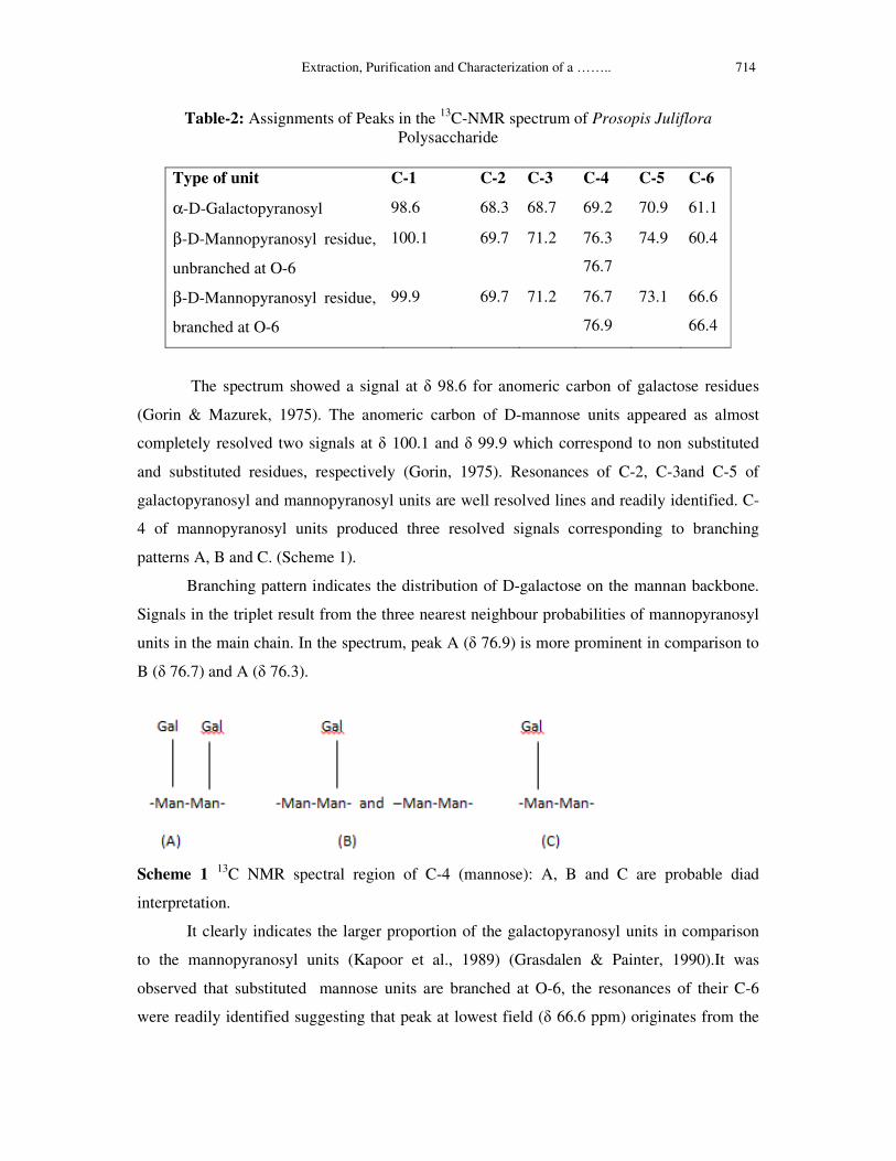
������������������������������������������������Extraction, Purification and Characterization of a …….. 714
Table-2: Assignments of Peaks in the 13
C-NMR spectrum of Prosopis Juliflora
Polysaccharide
The spectrum showed a signal at � 98.6 for anomeric carbon of galactose residues
(Gorin & Mazurek, 1975). The anomeric carbon of D-mannose units appeared as almost
completely resolved two signals at � 100.1 and � 99.9 which correspond to non substituted
and substituted residues, respectively (Gorin, 1975). Resonances of C-2, C-3and C-5 of
galactopyranosyl and mannopyranosyl units are well resolved lines and readily identified. C-
4 of mannopyranosyl units produced three resolved signals corresponding to branching
patterns A, B and C. (Scheme 1).
Branching pattern indicates the distribution of D-galactose on the mannan backbone.
Signals in the triplet result from the three nearest neighbour probabilities of mannopyranosyl
units in the main chain. In the spectrum, peak A (� 76.9) is more prominent in comparison to
B (� 76.7) and A (� 76.3).
Scheme 1 13
C NMR spectral region of C-4 (mannose): A, B and C are probable diad
interpretation.
It clearly indicates the larger proportion of the galactopyranosyl units in comparison
to the mannopyranosyl units (Kapoor et al., 1989) (Grasdalen & Painter, 1990).It was
observed that substituted mannose units are branched at O-6, the resonances of their C-6
were readily identified suggesting that peak at lowest field (� 66.6 ppm) originates from the
Type of unit C-1 C-2 C-3 C-4 C-5 C-6
α-D-Galactopyranosyl 98.6 68.3 68.7 69.2 70.9 61.1
β-D-Mannopyranosyl residue,
unbranched at O-6
100.1
69.7 71.2 76.3
76.7
74.9 60.4
β-D-Mannopyranosyl residue,
branched at O-6
99.9 69.7 71.2 76.7
76.9
73.1 66.6
66.4

715 Himani Bhatia, P.K. Gupta and P.L. Soni
C-6 resonance of the intermediate unit from groups of three contiguous, substituted mannosyl
residues (Triad I, Scheme 2) and peak at (� 66.4ppm) originates due to superposition of
signals from triads wherein two contiguous mannosyl units are substituted (Triad II, Scheme
2) (Manzi et al., 1986).
Scheme 2 13
C NMR spectral region of C-6 (mannose): Triad I and II are probable
interpretation.
Structure of oligosaccharide-I
It was found to be a heptasaccharide [�] + 1120 (c 0.05%, H2O), m.p. 200-202
0C (d),
that was homogenous and gave single spot [Rgal 0.31] using organic solvent S2 and spray
reagents R1 and R2 upon chromatographic examination. Upon complete acid hydrolysis, it
gave D- galactose and D- mannose on paper chromatogram in solvent systems S1, S2 and S3.
The alditol acetates of the hydrolysates of oligosaccharide on GLC analysis under conditions
C-1 showed two peaks corresponding to D-galactose and D- mannose in the molar ratio of
3:4.
The oligosaccharide was completely methylated by Hakomori method (Hakomori,
1964) followed by Purdie (Purdie & Irvine, 1904) for complete etherification. Complete
methylation was confirmed by IR spectrum of the methylated heptasaccharide which showed
complete absence of –OH band (3590-3225cm-1
). It was hydrolyzed and transformed into its
alditol acetates according to the method of Jansson et al. (1976). GLC of the resulting alditol
acetate under conditions C1, furnished 2, 3, 4, 6- tetra-O-methyl-D-galactose, 2, 3-di-O-
methyl-D-mannose, 2, 3, 4, 6-tetra-O-methyl-D-mannose in the molar ratio of 3:3:1,
respectively.

������������������������������������������������Extraction, Purification and Characterization of a …….. 716
On the basis of methylation study it was found that 2, 3-di-O-methyl-D-mannose and
2, 3, 4, 6- tetra-O-methyl-D-galactose indicate the presence of 1�4 and 1�6 linkages in this
heptasaccharide. These results suggest that the oligosaccharide has the fundamental structural
pattern having β- (1→4) main chain of D-mannose units and side chain of single α-(1→6)-
D-galactose residues.
On the basis of above discussion, the heptasaccharide may be assigned the following
plausible structure (Fig.3).
Fig. 3 Oligosaccharide- I
Structure of oligosaccharide-II
It was found to be an octasaccharide [�] + 860 (c 0.05%, H2O), m.p. 218-220
0C (d),
that was homogenous and gave single spot [Rgal 0.19] using organic solvent S2 and spray
reagents R1 and R2 upon chromatographic examination. Upon complete acid hydrolysis, it
gave D- galactose and D- mannose on paper chromatogram in solvent systems S1, S2 and S3.
The alditol acetates of the hydrolysate of oligosaccharide on GLC analysis under conditions
C-1 showed two peaks corresponding to D-galactose and D- mannose in the molar ratio of
3:5.
The oligosaccharide was completely methylated by Hakomori method (Hakomori,
1964) followed by Purdie (Purdie & Irvine, 1904) for complete etherification. Complete
methylation was confirmed by IR spectrum of the methylated heptasaccharide which showed
complete absence of –OH band (3590-3225cm-1
). It was hydrolysed and transformed into its
alditol acetates according to the method of Jansson et al. (1976). GLC of the resulting alditol
acetate under conditions C1, furnished 2,3,4,6- tetra-O-methyl-D-galactose, 2,3-di-O-methyl-
D-mannose, 2,3,4,6-tetra-O-methyl-D-mannose, 2,3,6-tri-O-methyl-D-mannose in the molar
ratio of 3:3:1:1 respectively.
On the basis of methylation study it was found that 2, 3-di-O-methyl-D-mannose, 2, 3,
6-tri-O-methyl-D-mannose and 2, 3, 4, 6- tetra-O-methyl-D-galactose indicate the presence of
�

717 Himani Bhatia, P.K. Gupta and P.L. Soni
1�4 and 1�6 linkages in this octasaccharide. These results suggest that the oligosaccharide
has the fundamental structural pattern having β- (1→4) main chain of D-mannose units and
side chain of single α- (1→6)-D-galactose residues. On the basis of above discussion, the
octasaccharide may be assigned the following plausible structure (Fig.4).
Fig.4 Oligosaccharide- II
Structure of oligosaccharide-III
It was found to be a nonasaccharide [�] + 1290 (c 0.05%, H2O), m.p. 240-242
0C (d),
that was homogenous and gave single spot [Rgal 0.11] using organic solvent S2 and spray
reagents R1 and R2 upon chromatographic examination. Upon complete acid hydrolysis, it
gave D- galactose and D- mannose on paper chromatogram in solvent systems S1, S2 and S3.
The alditol acetates of the hydrolysates of oligosaccharide on GLC analysis under conditions
C-1 showed two peaks corresponding to D-galactose and D- mannose in the molar ratio of
5:4.
The oligosaccharide was completely methylated by Hakomori method (Hakomori,
1964) followed by Purdie (Purdie & Irvine, 1904) for complete etherification. Complete
methylation was confirmed by IR spectrum of the methylated heptasaccharide which showed
complete absence of –OH band (3590-3225cm-1
). It was hydrolyzed and transformed into its
alditol acetates according to the method of Jansson et al. (1976). GLC of the resulting alditol
acetate under conditions C1, furnished 2,3,4,6- tetra-O-methyl-D-galactose, 2,3-di-O-methyl-
D-mannose, 2,3,4,6-tetra-O-methyl-D-mannose in the molar ratio of 4:4:1 respectively.
On the basis of methylation study it was found that 2,3-di-O-methyl-D-mannose and
2,3,4,6- tetra-O-methyl-D-galactose indicate the presence of 1�4 and 1�6 linkages in this
nonasaccharide. These results suggest that the oligosaccharide has the fundamental structural
pattern having β- (1→4) main chain of D-mannose units and side chain of single α- (1→6)-D-
galactose residues. On the basis of above discussion, the nonasaccharide may be assigned the
following plausible structure (Fig.5).

������������������������������������������������Extraction, Purification and Characterization of a …….. 718
Fig.5 Oligosaccharide-III
3. Experimental
3.1. Analytical Methods
Evaporations were conducted under diminished pressure at < 450 (bath). Descending
p.c. was done on whatman no. 1 and 3MM papers using 1-butanol-pyridine-water (6:4:3).
Sugars were detected by aniline hydrogen phthalate (Patridge, 1949) and alkaline silver
nitrate (Trevelyan, 1950). Constituent sugars were determined as the alditol acetates by GLC
on ECNSS-M (3%) on Gas Chrom Q (100-120 mesh) packed into 5' X 1/8" stainless steel
column in case of methylated sugars and BPX-70 capillary column with 0.22mm X 0.25µm
in case of completely hydrolysed polysaccharides. The operating conditions employed for
different sugar samples are listed below:
(C1) Column temperature 1700C, nitrogen flow rate 35-40 ml/min
(C2) Column temperature 2100C, nitrogen flow rate 2 ml/min
Authentic standards viz. 1, 5-di-O-acetyl-2, 3, 4, 6-tetra-O-methyl-D-glucitol, in the
case of methylated sugars and a mixture of mannose and galactose in the case of alditol
acetates of completely hydrolyzed oligosaccharides and polysaccharides were also run
simultaneously under the same conditions to give the values of relative retention times Rt.
N.M.R spectra were recorded on Bruker Avance II 400 instrument NMR spectrometer at 400
MHz. For 1H-NMR, the galactomannan sample was firstly exchanged in D2O by repeated
evaporations of 1mg/ml solution and finally dissolved in high quality D2O (99.96%D). Proton
spectra were obtained at 800C under conditions of quantitative analysis. For
13C-NMR
spectra, 20mg/ml galactomannan was prepared inD2O at 700C by continuous stirring for 6h
followed by sonication for 10min. The solution was filtered and filled in NMR tube. Optical
rotation was determined on Autopol-II, automatic polarimeter (Rudolph research, Flanders,
New Jersey) at 589nm, D-lines of sodium.

719 Himani Bhatia, P.K. Gupta and P.L. Soni
3.2. Isolation and purification of the polysaccharide
The endosperm of the seeds of Prosopis juliflora contains the seed galactomannan.
Cold water extraction of endosperm and continuous stirring with EtOH followed by
centrifugation yielded a crude polysaccharide (~81%). Polysaccharide was purified by ion
exchange followed by dialysis.
3.3. Investigation of the Polysaccharide
(a) Gel filtration: Gel permeation chromatography was performed on sephadex G-150
columns (superfine grade), Pharmacia column (56/70) using 0.02% solution of sodium azide
as eluent [void volume (Vo) 40 ml; total column volume (Vt) 140 ml]. The flow rate was
maintained at 0.5ml/min by using minipuls 2 peristaltic pumps and Gilson fraction collector
(Model-202) programmed in time mode. Fractions (2ml) each were collected and monitored
with anthrone sulphuric acid reagent (McCready et al., 1950) at 660nm using Chemito UV-
Vis spectrophotometer UV-2500. The graph of the absorbance v/s fractions collected was
plotted (Fig.6).
Fig.6 GPC elution pattern of P.juliflora seed polysaccharide on sephadex G-150 [void
volume (Vo) 40 ml; total column volume (Vt) 140 ml]
(b) Sugar composition: The constituent sugar analysis was carried out after complete
hydrolysis with 2N H2SO4 for 18h at 1000C. The sugars were separated by descending paper
(Whatmann No. 1) chromatography using 6:4:3 butanol- pyridine-water. The chromatograms
were sprayed with aniline hydrogen phthalate and alkaline silver nitrate. Constituent sugars
were determined as alditol acetates by GLC. Polysaccharide upon partial acid hydrolysis was
0
0.1
0.2
0.3
0.4
0.5
0.6
1
23
45
67
89
11
1
13
3
15
5
17
7
19
9
22
1
24
3
26
5
28
7
Fraction no.
Ab
so
rba
nc
e (
66
0 n
m)

������������������������������������������������Extraction, Purification and Characterization of a …….. 720
heated with sulphuric acid (0.05N, 100ml) on steam bath at 1000C for 3h. The mixture of
oligosaccharides and monosaccharide was resolved into its components by preparative
chromatography on whatmann No.3mm filter paper sheets using solvent system S2. The
strips corresponding to individual oligosaccharides were eluted with water, elutes were
concentrated separately, to obtain the three oligosaccharides. Homogeneity of the
oligosaccharides was checked by paper chromatography in solvent system S1, S2, S3 using R1
and R2 as spray reagent.
(c) Molecular Weight Determination: Polysaccharide solution (1 gm in 100 ml distilled
water) was prepared in 100 ml volumetric flask. It’s dilutions of 0.75, 0.50 and 0.25 in 50 ml
volumetric flask were made. The flow time of solvent (deionized water) was determined by
using an Ubbelohde viscometer. Viscometer was cleaned with suitable solvent and dry, clean,
filtered air blowed through viscometer to remove traces of solvent. Sample was introduced
into viscometer by pipetting ~10 ml. Viscometer was placed in constant-temperature bath
(250C) and allow 10 minutes for viscometer to thermally equilibrate. Efflux time is measured
by allowing the sample to flow freely. Efflux time was recorded in seconds to hundredths and
repeated. Efflux time measurement at least three times was repeated.
(d) Methylation Analysis: The polysaccharide (0.0550 gm) was dissolved in distilled
dimethyl sulphoxide (5ml) by magnetic stirring and warming at 45-500C in an inert
atmosphere (N2). The reaction mixture was stirred for 4 h till the evolution of hydrogen
ceased. After evaporation of the chloroform extracts to dryness, the residues were hydrolyzed
with formic acid (90%, 10ml) for 1h on steam bath at 100oC, the solutions evaporated and
treated with aqueous sulphuric acid (0.13 M, 15ml) for 18h on a steam bath. The partially
methylated compound was subjected to Purdie’s methylation by dissolving it in methyl
iodide (5 ml) with stirring under inert atmosphere and silver oxide (0.50 g) was added
periodically in 4 h and reaction was allowed for 6 h. This process was repeated two times on
the successive days. The completely methylated polysaccharide processed further as above,
and obtained in the syrup form with the yield as (0.0265g).The hydrolysate was neutralized
(BaCO3), concentrated, reduced (sodium borohydride) and transformed into alditol acetate.
GLC of partially methylated sugars was done on ECNSS-M (3%) column under condition
C1.
Oligosaccharide-I (0.0283g), Oligosaccharide- II (0.0292g), Oligosaccharide- III
(0.0321g) was methylated completely by Hakomori method (Hakomori, 1964) using sodium
hydride- dimethyl sulphoxide followed by Purdie (Purdie & Irvine, 1904) for complete

721 Himani Bhatia, P.K. Gupta and P.L. Soni
etherification. Each methylated oligosaccharide was recovered by chloroform extraction.
After evaporation of the chloroform extracts to dryness, the residues were hydrolyzed with
formic acid (90%, 10ml) for 1h on steam bath at 100oC, the solutions evaporated and treated
with aqueous sulphuric acid (0.13 M, 15ml) for 18h on a steam bath. The partially
methylated compound was subjected to Purdie’s methylation by dissolving it in methyl
iodide (5 ml) with stirring under inert atmosphere and silver oxide (0.50 g) was added
periodically in 4 h and reaction was allowed for 6 h. This process was repeated two times on
the successive days. The methylated oligosaccharide processed further as above, and
obtained in the syrup form with the yield as oligo-I (0.0123g), oligo-II (0.0132g), oligo-III
(0.0171g).
3.4 Periodate Oxidation: To a solution of galactomannan polysaccharide (0.0510 g in 25
ml) in water an aqueous solution of sodium metaperiodate (0.2g in 50ml) was added and the
volume of the resultant solution was made up to 100ml. A blank solution of sodium
metaperiodate (0.2g in 100 ml) was also prepared. These were kept in dark at room
temperature (40C) for 192 h. To determine periodate consumed, an aliquot (5ml) of the
periodate reaction mixture was added to a solution containing distilled water (20ml),
potassium iodide (20%, 2ml) and sulphuric acid (0.5N, 3ml). The liberated iodine was
immediately titrated with 0.1N sodium thiosulphate solution using starch as an indicator
(Bobbitt, 1956; Malaprade, 1928; Rankin &Jeans, 1954).
Acknowledgements
This work is supported by Forest Research Institute (FRI), Dehradun. The authors are
thankful to Director (FRI), Head, Chemistry Division, (FRI), Dehradun for providing
laboratory facilities. The authors are also thankful to Director, Punjab University, Chandigarh
for N.M.R studies. Thanks are also due to Dr. Vikas Rana (Scientist-C) Rain Forest Research
Institute, Jorhat for support and discussion.
References
[1] Abdel-Akher, M., & Smith, F. (1951).The repeating unit of glycogen. Journal of the
American Chemical Society, 73, 994-996.
[2] Barker, S. A., Bourne, E. J., & Whiffen, O. H. (1956). Use of infrared analysis in the
determination of carbohydrate structure. In D. Glick (Ed.). Methods of Biochemical
Analysis (Vol.3, pp. 213-245). New York: Interscience Publisher, Inc.

������������������������������������������������Extraction, Purification and Characterization of a …….. 722
[3] Bobbitt, J.M. (1956). Periodate oxidation of carbohydrates. In M.L. Wolfrom & R.S.
Tipson (Eds.). Advances in Carbohydrate Chemistry and Biochemistry, (Vol.11, pp. 1-
41).New York: Academic Press.
[4] Buckeridge, M., Panegassi, V. R., Rocha, D.C., &Dietrich, S. M. C. (1995). Seed
galactomannan in the classification and evolution of leguminosae, phytochemistry, 38,
871-875.
[5] Cruz Alcedo, G. E. (1999). Production and characterization of prosopis seed
galactomannan, a research dissertation.
[6] Dayer, J.R. (1956). Use of periodate oxidation in biochemical analysis In D. Glick (Ed.).
Methods of Biochemical Analysis (Vol.3, pp. 111-152). New York: Interscience
Publisher. Inc.
[7] Dea, ICM., & Morrison, A. (1975). Chemistry and interactions of seed galactomannans.
In M.L. Wolfrom & R.S. Tipson (Eds.). Advances in carbohydrate chemistry and
Biochemistry (Vol.31, pp. 241-312). New York: Academic Press.
[8] Duss, J.Q., Gotfredson, C.H., & Bock, K. (2000). Carbohydrate structural determination
by NMR spectroscopy: Modern methods and limitations. Chemical Reviews, 100(12),
4589-4614.
[9] Figueiredo, Antonio de, A. (1983). Extraction, identification and characteristics of the
polysaccharide from the mesquite beans (Prosopis juliflora DC). Cienc. Tecnol.
Ailment, 3 (1), 82- 90.
[10] Gorin, P.A.J., & Mazurek, M. (1975). Further Studies on the Assignment of Signals
in13
C Magnetic Resonance Spectra of Aldoses and Derived Methyl Glycosides.
Canadian Journal of Chemistry, 53, 1212-1223.
[11] Gorin, P.A.J. (1975). Assignment of signals of the carbon-13 magnetic resonance
spectrum of a selected polysaccharide: Comments on methodology. Carbohydrate
Research, 39, 3-10.
[12] Grasdalen, H., & Painter, T. (1980). N.M.R studies of composition and sequence in
legume-seed galactomannans. Carbohydrate Research, 81, 59-66.
[13] Gupta, D.S., Jain, B., Bajpai, K.S., & Sharma, S.C. (1987). Structure of galactomannan
from Cassia alata seed. Carbohydrate Research, 162, 271-276.
[14] Hakomari, S.I. (1964). A rapid permethylation of glycolipid, and polysaccharide
catalysed by methylsulfinyl carbanion in dimethyl sulphoxide. Journal of Biochemistry,
55, 205-208.

723 Himani Bhatia, P.K. Gupta and P.L. Soni
[15] Halsall, T. G., Hirst, E.L., & Jones, J.K.N. (1947). Oxidation of carbohydrates by the
periodate ion. Journal of Chemical Society, 1427-1432.
[16] Houwink, R. (1940). Zusammenhang zwischen viscosimetrisch und osmotisch
bestimmten Polymerisations gradenbei Hochpolymeren. Journal für Praktische Chemie,
157, 15-18.
[17] Ishak, M. F., & Painter T.J. (1973). The anomalous periodate oxidation limit of guaran.
Acta Chem. Scan, 27, 1268- 1276.
[18] Jannson, P.E., Kenne, L., Liedgren, H., Lindberg, B., & Lonngren, J. (1976). A practical
guide to methylation analysis of carbohydrates. Journal of chemical communication, 8,
1-76 [University of Stockholm].
[19] Kapoor, V.P., & Chaubey, Manjoosha. (2001). Structure of galactomannan from the
seeds of Cassia Angustifolia Vahl. Carbohydrate Research, 332, 439-444.
[20] Kapoor, V.P., Sen, A.K., & Farooqui, M.I.H. (1989). Structure of Dhaincha
galactomannan from the seeds of Sesbania bispinosa. Indian Journal of Chemistry, 28 B,
928-933.
[21] Lapasin, R., & Pricl, S. (1995). Rheology of polysaccharide systems In Rheology of
Industrial Polysaccharides, (Chapt.4, pp. 250-477) Blackie Academic and Professional,
London,
[22] Mc Cready, R.M., Guggolz, J., Silviera, V., & Owens, H.S. (1950). Determination of
starch and amylase in vegetables. Analytical Chemistry, 22, 1156-1158.
[23] Malaprade, L. (1928). Action of polyalcohol on periodic acid, analytical application.
Bulletin de la Societe Chimique de France, 43, 683-696.
[24] Manzi, A.E., Cerezo, A.S., & Shoolery, J.N. (1986).High resolution 13
C-N.M.R.
spectroscopy of legume-seed galactomannans. Carbohydrate Research, 148, 189-197.
[25] Painter, T.J., Gonzalez, J.J., & Hemmer, P.C. (1979). The distribution of D- galactose
residues in guaran and locust bean gum. Carbohydrate Research, 69, 217-226.
[26] Partridge, S.M. (1949). Aniline hydrogen phthalate as a spraying reagent for
chromatography of sugars. Nature, 164, 443.
[27] Pinto Vieira, I.G.P., Francisca Noe lia Pereira Mendes, F.N.P., Gallao, & Sousa de
Brioto, E. (2007). N.M.R study of galactomannans from the seeds of mesquite tree
(Prosopis juliflora (Sw.) DC). Food Chemistry, 101, 70-73.
[28] Purdie, T., & Irvine, J.C. (1903). The stereoisomeric tetramethyl methylglucosides and
tetramethyl glucose. Journal of Chemical Society, 85, 1049-1070.

������������������������������������������������Extraction, Purification and Characterization of a …….. 724
[29] Rankin, J.C., & Jeans, A. (1954). Evaluation of the periodate oxidation method for
structural analysis of dextrans. Journal of American Chemical Society, 76, 4435-4441.
[30] Tewari, D.N. (1995). Vilayti Babul (Prosopis juliflora), Indian council of forestry
research and education, Publication, Dehradun.
[31] Trevelyan, W.E., Procter, D.P., & Harrison, J.S. (1950). Detection of sugars on paper
chromatograms. Nature, 166, 444-445.
[32] Whistler, R.L. (1990). Introduction to Industrial Gums. In Whistler, R.L., & Be Miller, J.
N. (3rd Eds.). Industrial gums: Polysaccharides and their derivatives (pp. 5-7) New York:
Academic Press.