Embed Size (px)
Citation preview

Extraction, purification and analysis of histonesDavid Shechter1, Holger L Dormann1, C David Allis1 & Sandra B Hake1,2
1The Laboratory of Chromatin Biology, The Rockefeller University, New York, NY, USA. 2Present address: Department for Molecular Biology, Adolf-Butenandt Institute,Ludwig-Maximilians-University, Schillerstrasse 44, 80336 Munich, Germany. Correspondence should be addressed to S.B.H. ([email protected]).
Published online 7 June 2007; doi:10.1038/nprot.2007.202
Histone proteins are the major protein components of chromatin, the physiologically relevant form of the genome (or epigenome) in
all eukaryotic cells. Chromatin is the substrate of many biological processes, such as gene regulation and transcription, replication,
mitosis and apoptosis. Since histones are extensively post-translationally modified, the identification of these covalent marks on
canonical and variant histones is crucial for the understanding of their biological significance. Many different biochemical techniques
have been developed to purify and separate histone proteins. Here, we present standard protocols for acid extraction and salt
extraction of histones from chromatin; separation of extracted histones by reversed-phase HPLC; analysis of histones and their
specific post-translational modification profiles by acid urea (AU) gel electrophoresis and the additional separation of non-canonical
histone variants by triton AU(TAU) and 2D TAU electrophoresis; and immunoblotting of isolated histone proteins with modification-
specific antibodies.
INTRODUCTIONChromatin, a macromolecular complex composed of DNA andprotein, is the heritable material of eukaryotic cells. The repeatingunit structure of chromatin is the nucleosome, in which DNA iswrapped around a core octamer unit of dimers of the four histoneproteins (H2A, H2B, H3 and H4 and/or their variant isoforms)(Fig. 1) and further assembled into the larger chromatin fiberincluding the linker histone H1. The canonical histone proteins andthe less abundant histone variants are subject to an extensive arrayof post-translational modifications (PTMs), including Lys and Argmethylation; Lys acetylation, ubiquitylation, SUMOylation, andADP-ribosylation; Ser, Thr, and Tyr phosphorylation; and citrulli-nation of methyl-Arg residues1–4. Accumulating evidence suggeststhat the combination of modifications on nucleosomes and thepresence or absence of various histone variants contribute to theencoding of epigenetic information, the ultimate regulation of geneexpression5. Therefore, the study of histone proteins, and the PTMsthat they carry, has become increasingly important as moreinvestigations are conducted into the ‘epigenetic signatures’ ofthese important chromosomal proteins.
Histones were among the first proteins studied, in part becauseof their relative ease of isolation. Albrecht Kossel coined the term‘histon’ in 1884 when he isolated acid-soluble proteins from birderythrocyte nuclei (reviewed in ref. 6). This had followed FriedrichMiescher’s pioneering work in 1874 demonstrating the existence ofthe alkali-labile and acid-insoluble material he termed ‘nuclein’,later identified as DNA7. In retrospect, these early investigations,utilizing simple acid and base extractions, demonstrated that thetwo discrete components of the genetic material were nucleic acid
and protein and suggested the use of acid extraction for histonepreparation.
After Avery’s demonstration of DNA as the genetic material8 andWatson and Crick’s structure of DNA9, the study of histones andchromatin moved slowly as more attention was paid to the nucleicacid component of the genetic material. Some work on histonesthrough the 1950s utilized high-salt extraction of chromatin toisolate the histones. Simple fractionation of the histone populationusing a variety of dilute acids was attempted by a number ofresearch groups. More extensive characterization ensued and his-tone extraction from various cell types suggested different popula-tions of histones existed in different tissues10, foreshadowing thesignificant role histones and their variants have in epigeneticregulation. In 1967 Johns11 introduced the concept of separating
p
uor
G g
n ih si l
bu
P eru ta
N 700 2©
nat
ure
pro
toco
ls/
moc.er
ut an.
ww
w//:ptt
h
Histone Variants
H2AH2A
H2BH2B
H3H3
H4H4
H1H1
H2A.XH2A.Z
H2A.BbdMacroH2A
TestisH2B (mammals)
No known variants
many, especially
Post-translational modifications
Ser/Thr/Tyr phosphorylationLys/Arg methylation
Lys acetylationLys ubiquitylaitonLys sumoylation
Glu ADP-ribosylation
embryonic isoforms
H3.1 (mammals)H3.2, H3.3CENP-AFigure 1 | Depiction of histones in the nucleosome. Core histone proteins
H2A (orange), H2B (green), H3 (blue) and H4 (red) and a 147-bp DNA strand
(silver and red) are shown in the 1.9-A nucleosome core particle structure31
(as rendered by the software package VMD32) and in a cartoon diagram to
clearly illustrate the positions of the core histone proteins. The N-terminal
tails of the histone proteins are noted with arrows; these tails are the location
of the majority of the post-translational modifications (PTMs) of histones. The
histone proteins, the major known histone variants33 and the range of PTMs
found on these histone proteins are noted below. The functions of histone
modifications have been recently reviewed34.
NATURE PROTOCOLS | VOL.2 NO.6 | 2007 | 1445
PROTOCOL
histone proteins for analysis on acidic polyacrylamide gels, andPanyim and Chalkley12 developed the acid urea (AU) gel, whichcleanly separated histones, histone variants and differently mod-ified histone isoforms (such as acetylated and phosphorylatedhistones) on the basis of differences in their charge. Meanwhile,Allfrey and coworkers’13 discovery of acetylated and methylatedhistones in 1964 led them presciently to hypothesize that histonemodifications, notably acetylation, methylation and phosphoryla-tion, may regulate gene expression. Finally, the discoveries in 1996that a histone acetyltransferase14 and a histone deacetylase15 wereknown transcriptional regulators prefaced an explosion in histoneand chromatin research in the past decade16. These advances alldepend on a reliable and effective isolation of histone proteins.
To study the function of histones, histone variants and particu-larly histone modifications, many laboratories, including our own,routinely begin by acid extraction of histones from cells or nuclei,further purifying them by reversed-phase HPLC (RP-HPLC),a method that was used first for the analysis of histones in 1983,(ref. 17). Both the extracted and HPLC-purified histones are easilyanalyzed by a variety of techniques: AU gel analysis; immuno-blotting with modification-specific antibodies after separation byTris–Gly or Tris–tricine SDS–polyacrylamide gel electrophoresis(SDS-PAGE); and mass spectroscopy (MS) to determine theidentity and abundance of variants and PTMs and also to searchfor novel modifications18. The still-relevant utility of AU gelanalysis was recently demonstrated by the identification of‘long-distance’ linkage between acetylation and methylation19. Inthese experiments, di- and tri-methylation on histone H3K4 werespecifically found on slower-migrating H3 bands on an AU gel,indicating that H3K4 methylation was linked to H3 acetylation.
Model systems and cellular sources of histonesThe following protocols include our standardized approach tohistone extraction, purification and analysis. The techniquesfocus on isolation from mammalian cell culture, but the proceduresfollowing nuclei extraction are identical for yeast, Tetrahymenathermophila, Xenopus laevis egg extracts and other model systems inwhich nuclei are extractable. Please see the referenced methods forisolation of yeast nuclei20, Tetrahymena nuclei21, and Xenopus eggextract22 and Xenopus nuclear isolation23. Histones can also beextracted from animal tissue or plant cells following isolation oftheir nuclei, although those procedures may differ from thosepresented here, so be sure to follow an established nuclear isolationprocedure for that particular cell type. After successful nuclear orchromatin isolation, follow our protocols for purification andanalysis of histone proteins. Alternatively, a cruder histone pre-paration may be obtained simply by placing the entire cell or tissuein dilute acid; in that case, follow our protocols disregarding thechromatin isolation steps. As histones are among the most highlyconserved eukaryotic proteins, their biochemical behavior isremarkably similar across many species and model organisms, sothe majority of these protocols are of general utility.
In sum, the following techniques (outlined in Fig. 2) arestandard approaches for the experimental study of histone proteins.The extraction techniques are robust, and at least for routine cellculture of HeLa cells, yeast or Tetrahymena, the source material ischeap, so optimization of the described techniques is recom-mended to suit particular needs. We do not specifically addressroutine SDS-PAGE analysis, staining and SDS-PAGE gel western
blotting, as these are standard techniques, although our experiencedictates that adjustments are sometimes needed (e.g., electrophore-tic transfer of proteins is routine from SDS-PAGE and AU gels, butis diminished from triton AU (TAU) gels, often requiring increasedtransfer times or currents). A wealth of modification-specifichistone antibodies for immunoblotting are now available from anumber of suppliers, including Millipore (formerly Upstate),Abcam and LP Bio. Most of these antibodies work across a rangeof species, although investigators should check with the supplier toconfirm that it is true for a particular antibody of interest. Asepitopes can be masked by neighboring PTMs, investigators areencouraged to use independent techniques (MS, analytical gelelectrophoresis, etc) to verify positive or negative antibody results.
Experimental design and considerationsMost of the histone extraction work has been based on solubility ofhistones in acids, either dilute HCl or H2SO4, conditions underwhich most other nuclear proteins and nucleic acids will precipi-tate24. Note that the acid extraction method, when applied to plantcells, slime molds and some fungi (including the yeast Saccharo-myces cerevisiae), may produce a histone preparation that containslarge amounts of carbohydrates and polyphenols that readily cross-link and oxidize the histone proteins.
For the potential discovery of novel PTMs, it would be wise toconsider infrequently utilized histone extraction techniques, suchas high-salt extraction, in place of the typical acid extraction,as some histone modifications may be quite acid-labile25 (such asHis phosphorylation)26. Furthermore, acid and high salt maydifferentially extract histones from various chromatin environ-ments, such as heterochromatin and euchromatin. Utilization of
p
uor
G g
n ih si l
bu
P eru ta
N 700 2©
nat
ure
pro
toco
ls/
moc.er
ut an.
ww
w//:ptt
h
Cells
Nuclei or chromatin
Histones
Acid-extraction
High-saltextraction
Reversed-phaseHPLC
SDS-PAGE analysisAcid-urea gel analysisImmunoblotting with modification-specific antibodies
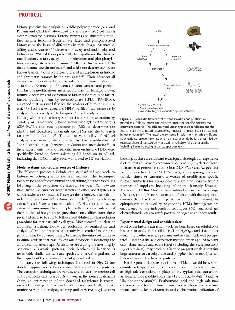
Figure 2 | Schematic flowchart of histone isolation and purification
procedures. Cells are grown and collected under the specific experimental
conditions required. The cells are lysed under hypotonic conditions and the
intact nuclei are collected; alternatively, nuclei or chromatin can be obtained
by other methods35. The nuclei are extracted in acidic or high-salt conditions
to selectively remove histones, which can subsequently be further purified by
reversed-phase chromatography or used immediately for other analysis,
including immunoblotting and mass spectroscopy.
1446 | VOL.2 NO.6 | 2007 | NATURE PROTOCOLS
PROTOCOL
both approaches may lead to a more complete analysis ofthe histones in a particular sample. Therefore we also present aless commonly used histone isolation strategy using high-saltextraction27.
High-salt extraction is a useful alternative to acid extraction for afew reasons. First of all, a neutral pH is maintained and any acid-labile histone modifications should therefore remain. Second, acidextraction and subsequent TCA-precipitation occasionally produceinsoluble material that may contain histones, reducing the yield ofthe preparation. Free histones, which are not incorporated intochromatin and might therefore represent a pool of either newlysynthesized histones or histones ejected from chromatin, can beisolated and analyzed for pre- or post-incorporation specificmodifications. Finally, salt extraction can differentially extractH2A and H2B (at greater than 1 M NaCl) and H3 and H4 (atgreater than 1.5 M NaCl)27. For downstream applications in whichmaintenance of neutral pH is crucial, such as determination ofacid-labile modifications, ensure that those methods are performedat a neutral pH. In these cases, histones should be chromatogra-phically separated by gel filtration or ion-exchange chromatogra-phy, instead of RP-HPLC. Alternatively, perform RP-HPLC inthe absence of trifluoroacetic acid (TFA), but expect some peakbroadening.
RP-HPLC, which separates molecules on the basis of hydropho-bicity, is a high-resolution method ideally suited for histoneproteins. Acid-extracted or salt-extracted histones can be purifiedto near-homogeneity in one run on a standard C8 or C18 column,using an acetonitrile gradient. The method presented is optimizedfor separation of total histones. If further separation betweenindividual histone proteins is required, the fractions can be re-loaded onto the column and separated using an RP-HPLC methodwith a shallower gradient, typically improving the separation.
SDS-PAGE (using standard Tris–Gly or alternatively using Tris–tricine (see http://www.natureprotocols.com/2006/06/23/tricinesdspage.php) is very useful for separation of the different histones.However, it does not allow for the separation of histone isoformswith different acetylation levels or phosphorylation status. This isaccomplished by AU gel electrophoresis, a method that separateshistones according to the individual charges introduced by acetyla-tion and phosphorylation. TAU and 2D TAU gel electrophoresisfurther separates certain histone variants, such as some H2Avariants and the H3 variants H3.1, H3.2 and H3.3, by the bindingof the detergent Triton X-100 to hydrophobic regions of theproteins.
In AU gel electrophoresis, proteins are denatured by a highconcentration of urea. Since urea denatures without affecting thecharge of proteins (as opposed to the ionic detergent SDS), thecharge of migrating proteins is determined solely by the number ofprotonated groups under the acidic running conditions of theprotocol. Acetylation reduces the positive charge of a Lys, whilephosphorylation introduces an additional negative charge. There-fore, these modifications greatly affect the migration of histones inAU gel electrophoresis, allowing for separation of the differentmodified isoforms.
For optimal resolution, we run long AU gels (40 cm). Alterna-tively, for fast separation we run short AU gels, using a standardminigel system (such as the BioRad Protean III system). Here, wepresent detailed protocols for preparing and running long andshort AU gels as well as short TAU gels. Long AU gels consist of aseparating gel section and a stacking gel section, which are pouredwithout gel-loading wells. Only after the gel has been pre-runovernight are the wells for sample loading cast. Short AU and TAUgels, on the other hand, have no stacking gel. They are cast in onestep, and no pre-running step is required.
MATERIALSREAGENTS.100� Phosphatase inhibitor cocktails I and II (Calbiochem, cat. nos. 524624
and 524625).10� Dulbecco’s PBS (acid extraction) (Invitrogen, cat. no. 14200-075).NuPAGE 10–20% SDS gel (Invitrogen, cat. no. EC6136).Histones from calf thymus (Roche, cat. no. 223565).Recombinant histone H4 produced in Escherichia coli (Millipore, cat.
no. 14-697).0.2 M (0.4 N) sulfuric acid (H2SO4) (acid extraction) ! CAUTION Has
corrosive properties. Avoid skin contact and swallowing of H2SO4. H2SO4
should be used with appropriate safety measures, such as protective gloves,glasses and clothing and adequate ventilation.
.100% TCA (acid extraction) 500 g, add 227 ml ddH2O for 100% (wt/vol),keep at 4 1C indefinitely (Fisher Scientific, cat. no. A322) ! CAUTIONCorrosive and causes severe burns. Inhalation may cause lung damage.
.100% ice-cold acetone (acid extraction) ! CAUTION Acetone can damage themucosa of the mouth and can irritate and damage skin. Acetone should beused with appropriate safety measures, such as protective gloves, glasses andclothing, and adequate ventilation. Store acetone in explosion- or flame-proof freezer.
.NP40 substitute (acid extraction) (Fluka, cat. no. 74385)
.Sodium butyrate (acid extraction) (Sigma, cat. no. B5887)
.2-Mercaptoethanol (acid extraction and RP-HPLC) (EMD Chemicals,cat. no. 6010)
.Acetonitrile, synthesis grade or better (RP-HPLC) (Fisher, cat. no. BA170-4)
.TFA (RP-HPLC) (Sigma, cat. no. 302031)! CAUTION Highly corrosive. It isextremely destructive to the upper respiratory tract, eyes, and skin, andshould not be disposed of into sewage systems as it is highly toxic to aquaticorganisms. TFA is also volatile; dispense in a hood.
.Urea (AU/TAU gels) (Sigma, cat. no. U0631)
.N,N,N¢,N¢-tetramethylethylenediamine (TEMED) (AU gels)
.10% (wt/vol) ammonium persulfate (APS) in ddH2O (AU/TAU gels)
.Glacial acetic acid (AU/TAU gels)
.Triton X-100 (TAU gels) (Sigma, cat. no. T8787)
.0.2% (wt/vol) Pyronin Y dye (AU/TAU gels) (Sigma, cat. no. P9172) inddH2O
.Methanol (AU/TAU gels and SDS-PAGE and AU/TAU western blotting)
.Short TAU gel lane run with histones (2D TAU gels) (prepared as describedin Steps 58–59)
.0.125 M Tris–Cl, pH 8.8 (2D TAU gels)
.0.7% Acetic acid (AcOH) (AU/TAU western blotting)
.Phenylmethanesulfonylfluoride (PMSF)
.Sucrose
.DTT
.KCl
.HEPES
.Pierce SilverSNAP II (Pierce)
.Magnesium chloride (MgCl2)
.Glycerol
.Brilliant Blue G-250 (Serva, cat. no. 17524)
EQUIPMENT.50-ml Falcon tubes (acid extraction and AU/TAU gels).15-ml Falcon tubes (AU/TAU gels).1.5-ml tubes (acid extraction, RP-HPLC and AU/TAU gels).Dialysis tubing with low molecular weight cut-off (e.g., 3,250 MWCO,
Spectra/Por 133 110) and plastic clamps (acid extraction/high-salt extraction).Lyophilizer or SpeedVac (Savant) with vacuum pump (RP-HPLC and
AU/TAU gels)
p
uor
G g
n ih si l
bu
P eru ta
N 700 2©
nat
ure
pro
toco
ls/
moc.er
ut an.
ww
w//:ptt
h
NATURE PROTOCOLS | VOL.2 NO.6 | 2007 | 1447
PROTOCOL
.1-ml glass Hamilton syringe with metal needle (#1001) (RP-HPLC)
.Glass plates (AU gels); for long gels we use 40 � 20 cm2 plates; for small gelsa regular minigel system such as the Hoefer Mighty Small (Hoefer) or theBioRad Protean III system (BioRad) and spacers (0.75 mm)
.Plastic combs (AU/TAU gels) (see EQUIPMENT SETUP)
.Gel electrophoresis apparatus (AU/TAU gels)
.Rocking platform (AU/TAU gels, SDS-PAGE and AU/TAU western blotting)
.Stir plate (AU/TAU gels)
.0.45-mm filters (Millipore)
.Gel chamber (BioRad or NuPAGE) (2D TAU gels)
.Pre-cast 2D-well gels (2D TAU gels) (e.g., Invitrogen NuPAGE Tris–Gly10–20% 2D well gels, cat. no. EC6136)
.Small plastic containers (AU/TAU gels and SDS-PAGE and AU/TAU westernblotting)
.Polyvinylidene difluoride (PVDF) membrane (various pore sizes) (westernblotting) or nitrocellulose membrane
.Whatman 3 MM paper (SDS-PAGE and AU/TAU western blotting)
.Wet-transfer chamber (western blotting)
.Power supply (500 V/500 mA) (SDS-PAGE and AU/TAU gels and SDS-PAGEand AU/TAU western blotting)
.HPLC system with pumps, UV detector and fraction collector, and 5-mlsample loop, such as Beckman System Gold (Beckman)
.4.6-mm-diameter C8 column (Perkin-Elmer Aquapore, cat. nos. RP-300,0711-0059)
REAGENT SETUPAppropriate cell culture or nuclei source For mammalian cellculture, 106 cells produce an adequate quantity of histones for routineanalysis of total histones by western blotting; 107 would be preferablefor MS analysis. m CRITICAL Wash cells well with PBS to remove serumproteins and aspirate completely so medium does not interfere withhypotonic lysis.Hypotonic lysis buffer (acid and salt extraction) Make a 10-ml solutioncontaining 10 mM Tris–Cl pH 8.0, 1 mM KCl, 1.5 mM MgCl2 and 1 mM DTTand chill on ice. Add protease and phosphatase inhibitors described below to a1� concentration just before use in lysis of cells.253 solution of complete protease inhibitor cocktail One tablet (Roche, cat.no. 1697 498) dissolved in 2 ml of ddH2O (can be stored for up to 12 weeks at�20 1C). m CRITICAL Add DTT, PMSF (final concentration of 1 mM) andprotease and phosphatase inhibitors just before use.Extraction buffer (high-salt extraction) Prepare 10 ml of a solutioncontaining 10 mM HEPES pH 7.9, 10 mM KCl, 1.5 mM MgCl2, 0.34 Msucrose, 10% glycerol. Add NP40 or NP40 substitute to 0.2% forcell lysis. Add protease and phosphatase inhibitors to 1� from stock
solutions. Add sodium butyrate to 10 mM to inhibit histone deacetylaseactivity.No-salt buffer (high-salt extraction) Prepare 10 ml of a solution containing3 mM EDTA, 0.2 mM EGTA.High-salt solubilization buffer (high-salt extraction) Prepare 10 ml of asolution containing 50 mM Tris–Cl pH 8.0, 2.5 M NaCl, 0.05% NP40.Dialysis solution (high-salt extraction) Prepare 2 l of 10 mM Tris–Cl pH 8.0,chilled to 4 1C.
Solvent A (RP-HPLC) (5% acetonitrile, 0.1% TFA) Prepare 1 l by addition of50 ml acetonitrile to 950 ml of ddH2O. Add 1 ml TFA using a syringe in a fumehood.
Solvent B (RP-HPLC) (90% acetonitrile, 0.1% TFA) Prepare 1 l by addition of100 ml of ddH2O to 900 ml of acetonitrile. Add 1 ml TFA using a syringe in afume hood.
Solvent C (RP-HPLC) (90% acetonitrile, no TFA) Prepare 1 l by addition of100 ml of ddH2O to 900 ml of acetonitrile.Note: Solvents A to C for RP-HPLC should be filtered using 0.45-mm filters.
60%:0.4% acrylamide:bisacrylamide solution (AU/TAU gels) Consists of250 g acrylamide in 417 ml ddH2O. Stir overnight and add 1.67 g bisacrylamide.Filter the solution and store in dark container protected from light. ! CAUTIONAcrylamide and bisacrylamide are potent neurotoxins. Use appropriate safetymeasures, such as protective gloves and safety goggles, and handle underadequate ventilation.Running buffer (AU/TAU gels) 5% AcOH.
Protamine sulfate solution (AU/TAU gels) (Sigma, cat. no. P4505, grade IIIfrom Herring) Should be prepared fresh at 25 mg ml�1 in ddH2O and stored in500-ml aliquots at �80 1C.Amido black solution (SDS-PAGE and AU/TAU western blotting) Prepare asolution containing 0.5% amido black dye (JT Baker, cat. no. A586-03) in 10%isopropanol and 10% AcOH.Coomassie brilliant blue solution (AU/TAU gels) 0.1% (wt/vol) Brilliant BlueG-250 (serva), 50% methanol, 10% AcOH. Solubilize Brilliant Blue G-250powder in methanol, then add AcOH and finally ddH2O. Filter solution andstore at room temperature (20–25 1C).Silver staining (AU/TAU gels) We use the Pierce SilverSNAP II kitfor silver staining. Alternatively, researchers may use homemadesilver stains28.Transfer buffer (AU/TAU western blotting) 0.7% AcOH
EQUIPMENT SETUPPlastic combs Make a ‘one-finger’ comb by cutting a piece comprising one‘finger’ from a plastic comb as is commonly used for pouring SDS-PAGE gels(Fig. 3). Save for future use (2D TAU gels).
PROCEDUREAcid extraction of histones � TIMING 2 d1| Preparation of nuclear extracts � TIMING Day 1, 1–1.5 h. Collect tissue culture cells (5 � 106 cells ml�1) in 50-ml tubesand pellet (10 min, 300g).Note: For the alternative method of high-salt extraction see Box 1.
2| Discard supernatant, wash cell pellet with 10 ml PBS and spin again (10 min, 300g). Again, discard supernatant.’ PAUSE POINT Cell pellet can be flash-frozen in liquid nitrogen and stored at �80 1C indefinitely.
3| Re-suspend cell pellet in 1 ml hypotonic lysis buffer and transfer to 1.5-ml tube.m CRITICAL STEP Steps 4–15 are all performed at 4 1C. This procedure can be scaled up to accommodate larger cell culture volumes,but more tubes may be necessary.
4| Incubate for 30 min on rotator at 4 1C to promote hypotonic swelling of cells and lysis by mechanical shearing duringrotation.
5| Pellet the intact nuclei by spinning in cooled tabletop centrifuge: 10,000g, 10 min, 4 1C.
6| Entirely discard supernatant with pipette and re-suspend nuclei in 400 ml 0.4 N H2SO4.m CRITICAL STEP Nuclei have to be re-suspended very well, with no clumps left in solution. If necessary, vortex the solution untilclumps are dissolved.
7| Incubate on rotator for at least 30 min or overnight.
8| Spin samples in cooled tabletop centrifuge to remove nuclear debris: 16,000g, 10 min.
p
uor
G g
n ih si l
bu
P eru ta
N 700 2©
nat
ure
pro
toco
ls/
moc.er
ut an.
ww
w//:ptt
h
1448 | VOL.2 NO.6 | 2007 | NATURE PROTOCOLS
PROTOCOL
9| Transfer the supernatant containing histones into a fresh 1.5-ml tube.Note: Proceed either by TCA-precipitation of histones (Steps 10–17) or by dialysis and lyophilization (Steps 18–22).
10| TCA-precipitation of histones � TIMING Day 2, 1.5 h. Add 132 ml TCA drop by drop to histone solution and invert the tubeseveral times to mix the solutions (final concentration of TCA is 33%).m CRITICAL STEP The histones precipitate in TCA. The solution will appear milky over time.
11| Incubate on ice for 30 min.’ PAUSE POINT This step can be extended to an overnight incubation.
12| Pellet histones by spinning in cooled tabletop centrifuge: 16,000g, 10 min at 4 1C.
13| Carefully remove supernatant with pipette and wash histone pellet with ice-cold acetone without disturbing it. Acetone isused to remove acid from the solution without dissolving the protein pellet.
14| Spin in microcentrifuge 16,000g, 5 min at 4 1C.
15| Repeat Steps 13 and 14.
16| Carefully remove all of the supernatant with pipette and air-dry histone pellet for 20 min at room temperature.
17| Dissolve histone pellet in appropriate volume of ddH2O (typically 100 ml, scale with quantity of cellular source) and transferinto fresh 1.5-ml tube. Go to Step 22.m CRITICAL STEP Histones will be visible as a ‘smear’ on the tube wall. To dissolve the histones, pipette up and down the tube wall.Occasionally an insoluble pellet remains, which can be removed by spinning for 10 min at 16,000g at 4 1C. As this insoluble pelletmay contain histones, an alternative to TCA precipitation follows.
18| Dialysis and lyophilization � TIMING Typically 2 d.(Do not continue from Step 17, only from Step 9.) Dialyzesupernatant from acid extraction in dialysis tubing withlow molecular weight cut-off against ddH2O for 2 h toovernight. 2-Mercaptoethanol (e.g., 10 ml of 100% in 1 lddH2O) may be added to prevent oxidation. Instead ofddH2O, 2.5% AcOH may be used for dialysis to ensurehistone solubility.m CRITICAL STEP Ensure that a low molecular weight(approximately 3,000) cut-off dialysis membrane is used, ashistones are small proteins and may be lost in higher weightcut-off membranes.’ PAUSE POINT Dialysis can continue overnight.
19| Carefully remove dialyzed solution from membrane andfreeze at �80 1C in a 15-ml Falcon tube or in multiple 1.5-mlcentrifuge tubes.
20| Load frozen histone solution into a SpeedVac (1.5-mltubes with opened cap) or a lyophilizer (15-ml tubes with ahole punched through the cover) and apply a vacuum.m CRITICAL STEP Do not apply heat.
21| After complete solvent removal (anywhere from 3 h toovernight), remove tubes from lyophilizer and either store thepowder at �20 1C or �80 1C or dissolve in appropriate volumeof ddH2O (approximately 100 ml).’ PAUSE POINT Histones can be stored frozen at �20 1C or�80 1C indefinitely.
22| Separate 1, 3 and 5 ml of the histone solution on 15%SDS/PAGE gel and stain with Coomassie Brilliant Blue solutionto characterize concentration of the histones before runningSDS-PAGE gels (Fig. 3) or performing SDS-PAGE western
p
uor
G g
n ih si l
bu
P eru ta
N 700 2©
nat
ure
pro
toco
ls/
moc.er
ut an.
ww
w//:ptt
h
0.4
N H 2SO 4
–
extra
cted
2.5
M N
aCl-
extra
cted
H1
H3
H2B
H2A
H4
15% SDS-PAGE
Figure 3 | Coomassie Blue–stained SDS gel with acid- and salt-extracted
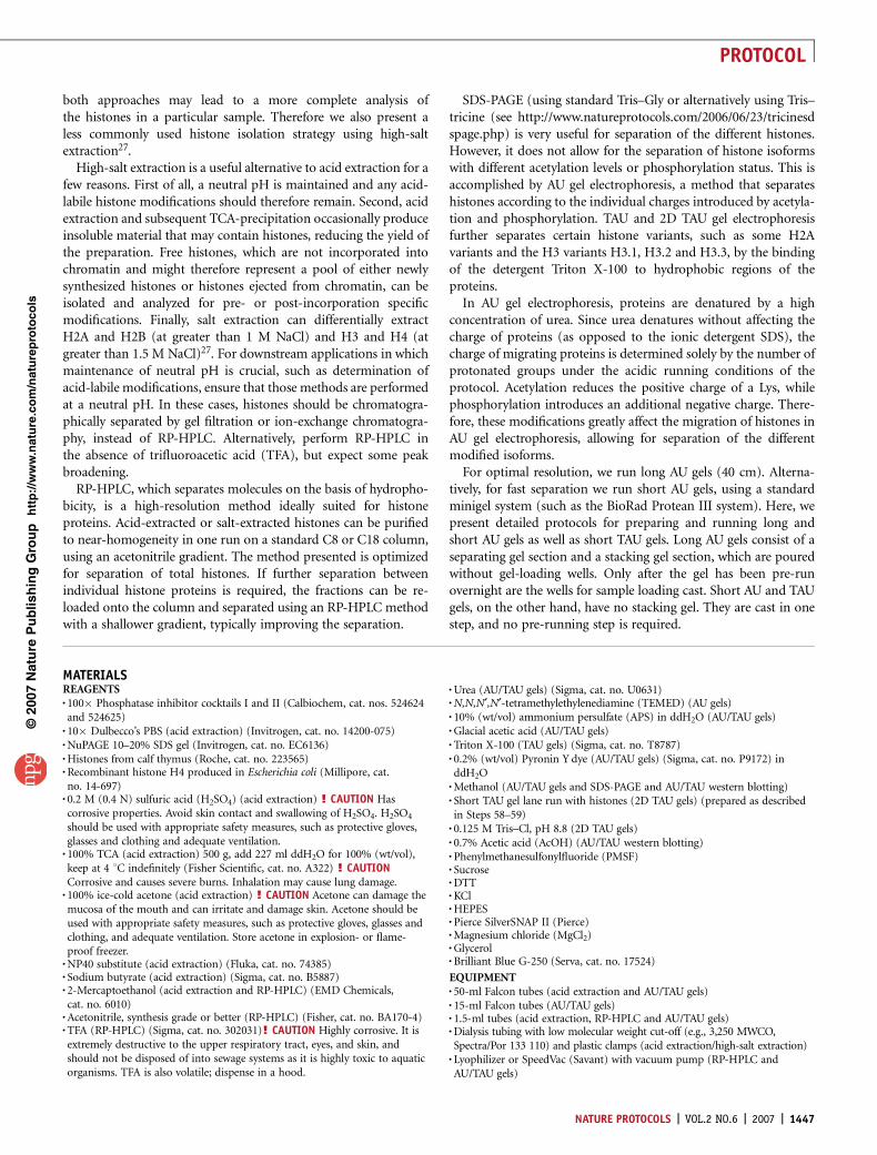
histones. Two microliters of acid-extracted and 2 ml of salt-extracted histones
from HeLa cells were run on a 15% SDS–polyacrylamide gel electrophoresis
(SDS-PAGE) gel and stained with Coomassie Blue dye. The locations of the
linker histone protein H1 and the core histone proteins H3, H2B, H2A and H4
are noted. A similar banding pattern is observed for most mammalian species’
histone proteins.
NATURE PROTOCOLS | VOL.2 NO.6 | 2007 | 1449
PROTOCOL
blotting, RP-HPLC (see Step 23, Fig. 4), AU, TAU (see Step 30, Fig. 5) or 2D TAU (see Step 53, Fig. 6) gelelectrophoresis. As a positive control, commercially available histone preparations (e.g., histones from calf thymus from Roche,cat. no. 223565, 10 mg lyophilized) can be used as a concentration standard. For the analysis of post-translational histonemodifications by western blotting, recombinant histones can be used as negative controls, as these do not contain any modifi-cations (e.g., recombinant histone H4 produced in Escherichia coli from Millipore, cat. no. 14-697, 1 mg lyophilized), As acontrol for good transfer of histones out of the gel onto the membrane, the membrane can be stained with amido black solution.? TROUBLESHOOTING
RP-HPLC purification of histones � TIMING 5–6 h, including column washing and storage23| Dissolve histones (either acid- or salt-extracted from 107 cells) in 200 ml ddH2O.
24| Load histone sample into sample loop with a glass Hamilton syringe and inject sample onto column to initiate theprogrammed run (see Box 2, Table 1 and Fig. 4).m CRITICAL STEP Do not use a plastic syringe, as contaminants from the plastic may interfere with subsequent MS.
25| Switch sample loop back from ‘inject’ position to ‘load’ position before the 5-min mark to prevent a delay in the gradient.
26| Watch chromatogram during run and check to ensure that fraction collector is working properly.
27| After the run finishes, the fractions can be frozen at �80 1C and lyophilized in a SpeedVac for 4 h or overnight.Lyophilized fractions can be stored at �80 1C dried or they can be re-dissolved in 100 ml ddH2O. The addition of a traceamount of 2-mercaptoethanol (e.g., 5 ml of 0.1 M 2-mercaptoethanol) to the fractions prevents the oxidative damage ofhistones during and after lyophilization.
p
uor
G g
n ih si l
bu
P eru ta
N 700 2©
nat
ure
pro
toco
ls/
moc.er
ut an.
ww
w//:ptt
h BOX 1 | HIGH-SALT EXTRACTION OF HISTONES � TIMING 4–6 H
1. Re-suspend 107 cells in 1 ml extraction buffer with 0.2% NP40. Incubate for 10 min on ice, occasionally rotating.m CRITICAL STEP Steps 1–16 are all performed at 4 1C. The main downside to salt extraction is that cellular modifying enzymes and proteasescan remain active for longer than in the acid-extraction protocol. It is therefore crucial that protease inhibitors, phosphatase inhibitors andhistone deacetylase inhibitors are included in the extraction buffers if analysis of modifications is desired.2. Spin in 4 1C cooled microcentrifuge: 6,500g, 5 min.3. Remove supernatant (the cytoplasm) completely and carefully with a 1-ml pipette.4. Wash pellet (the nuclei) in 1 ml extraction buffer (without NP40) by re-suspending the pellet and incubating on ice for 1 min.5. Spin in 4 1C cooled microcentrifuge: 6,500g, 5 min.6. Completely remove supernatant.7. Lyse the nuclei by re-suspending the nuclei pellet in 1 ml no-salt buffer and vortexing intermittently for 1 min (10 s on, 10 s off).8. Incubate on a rotator at 4 1C for 30 min.9. Spin and pellet chromatin and nuclear debris in microcentrifuge: 6,500g, 5 min.10. Remove supernatant containing the nucleoplasm and save if analysis of unincorporated histones is desired.Note: The chromatin pellet containing the DNA and histones should appear somewhat glassy.11. Re-suspend the chromatin pellet in 400 ml of High-Salt Solubilization buffer and vortex for 2 min.m CRITICAL STEP Chromatin should be re-suspended very well, with no clumps left. The solublization buffer volume can be increased to improvethe final yield; however, the resulting solution of histones will be more dilute.12. Incubate on rotator at 4 1C for 30 min.13. Spin and pellet DNA and nuclear debris in microcentrifuge: 16,000g, 10 min.14. Cut 5 cm of 3,500 MWCO dialysis tubing, pre-wet and close on one end with clamp. Transfer supernatant containing extracted histones intodialysis tubing.m CRITICAL STEP Ensure that there is adequate empty space in the tubing for a two- to threefold increase in volume of the solution afterdialysis. Clamp off the top of the tubing, such that there are a few centimeters’ slack of tubing past the clamp for ease of removal later, and putinto a 1-l beaker with dialysis solution in cold room with stir bar. Stir slowly, with modest movement of the tubing, for 1 h.15. Change dialysis solution: Remove tubing with gloved hand on clamp, discard solution and add remaining 1 l of fresh dialysis buffer. Stir for 1 h.m CRITICAL STEP 2 h of dialysis should bring the histone solution to 100–200 mM NaCl. If lower salt concentrations are required, continuedialysis for longer time with additional buffer changes.? TROUBLESHOOTING Some protein may precipitate after rapid dialysis against no salt. Histones remain in solution, but it is possible that somehistone protein will be lost. If precipitation occurs, a step-dialysis against 1, 0.5 and 0.2 M NaCl may be performed to limit protein precipitationowing to rapid change in ionic strength.16. Remove solution from dialysis tubing carefully: First, put a 15-ml conical tube on ice with the cap off. Remove dialysis tubing from flask andcarefully open one clamp. Insert top end of tubing into conical tube and squeeze out entire solution with gloved hands.’ PAUSE POINT Histone solutions can be stored frozen at �20 1C or �80 1C.17. See Step 22 of the acid-extraction protocol for SDS–polyacrylamide gel electrophoresis analysis of recovered histones.
1450 | VOL.2 NO.6 | 2007 | NATURE PROTOCOLS
PROTOCOL
’ PAUSE POINT Histone fractions canbe stored frozen at �80 1C.
28| Run 5 ml of each re-dissolved frac-tion corresponding to peaks on the chro-matogram on a 15% SDS-PAGE gel andCoomassie stain to determine the abun-dance of individual histones (Fig. 4).? TROUBLESHOOTING
29| Continue with MS or other analysis.
AU/TAU gel electrophoresis � TIMINGLong AU gel: 2 d; short AU/TAU gel: 2 h30| Pour separating gel � TIMING 2–3 h (long AU gel) or 1 h (short AU/TAUgel). Set up gel plates in gel-pouringapparatus.
31| Mix components of separating gelexcept for TEMED and 10% APS accord-ing to Table 2. Stir until urea is comple-tely dissolved. Do not apply heat todissolve urea. For long AU gels, degasthe solution under vacuum to preventthe formation of air bubbles during poly-merization. This step is not necessaryfor small gels.
p
uor
G g
n ih si l
bu
P eru ta
N 700 2©
nat
ure
pro
toco
ls/
moc.er
ut an.
ww
w//:ptt
h
100
90
80
70
60
50
40
30
20
10
0
Percent solvent B
H1
H2B
H2A
H4
H2AH3.2
H3.3
H3.1
min
2,000
1,800
1,600
1,400
1,200
1,000
UV
214
abso
rban
ce (
mA
U)
800
600
400
200
0
0 10 20 30 40 50 60 70 80 90 100 110 120 130 140
H1 H2B H2A H4 H2A H3.2/H3.3 H3.1 Coomassie-stained gel
36kD
2216
6
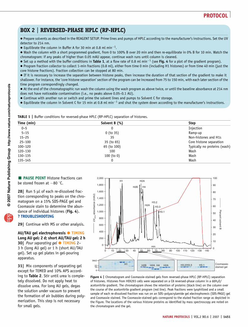
Figure 4 | Chromatogram and Coomassie-stained gels from reversed-phase HPLC (RP-HPLC) separation
of histones. Histones from HEK293 cells were separated on a C8 reversed-phase column in a ddH2O/
acetonitrile gradient. The chromatogram shows the retention of proteins (black line) on the column over
the course of the acetonitrile gradient program (red line). Peak fractions were lyophilized and a small
sample of each re-dissolved fraction was run on an SDS–polyacrylamide gel electrophoresis (SDS-PAGE) gel
and Coomassie stained. The Coomassie-stained gels correspond to the eluted fraction range as depicted in
the Figure. The locations of the various histone proteins as identified by mass spectroscopy are noted on
the chromatogram and the gel.
TABLE 1 | Buffer conditions for reversed-phase HPLC (RP-HPLC) separation of histones.
Time (min) Solvent B (%) Step
0–5 0 Injection5–15 0 (to 35) Ramp-up
15–25 35 Non-histones and H1s25–100 35 (to 65) Core histone separation
100–120 65 (to 100) Typically no proteins (wash)120–130 100 Wash130–135 100 (to 0) Wash135–145 0 Wash
BOX 2 | REVERSED-PHASE HPLC (RP-HPLC)
� Prepare solvents as described in the REAGENT SETUP. Prime lines and pumps of HPLC according to the manufacturer’s instructions. Set the UVdetector to 214 nm.� Equilibrate the column in Buffer A for 30 min at 0.8 ml min�1.� Wash the column with a short programmed gradient, from 0 to 100% B over 20 min and then re-equilibrate in 0% B for 10 min. Watch thechromatogram: if any peaks of higher than 0.05 mAU appear, continue wash runs until column is cleaned.� Set up a method with the buffer conditions in Table 1, at a flow rate of 0.8 ml min�1 (see Fig. 4 for a plot of the gradient program).� Program fraction collector to collect 1-min fractions (0.8 ml), either from time 0 min (including H1 histones) or from time 40 min (just thecore histone fractions). Fraction collection can be stopped at 80 min.� If it is necessary to increase the separation between histone peaks, then increase the duration of that section of the gradient to make itshallower. For instance, the ‘core histone separation’ section of the program can be increased from 75 to 150 min, with each later section of thetime program correspondingly changed.� At the end of the chromatographic run wash the column using the wash program as above twice, or until the baseline absorbance at 214 nmdoes not have noticeable contamination (i.e., no peaks above 0.05–0.1 AU).� Continue with another run or switch and prime the solvent lines and pumps to Solvent C for storage.� Equilibrate the column in Solvent C for 15 min at 0.8 ml min�1 and shut the system down according to the manufacturer’s instructions.
NATURE PROTOCOLS | VOL.2 NO.6 | 2007 | 1451
PROTOCOL
m CRITICAL All components of AU/TAU gels that contain urea (gel solutions and sample buffer) have to be prepared freshlyeach time, because the rapid degradation of urea at any pH above 8 leads to rapid carbamylation modification of histones.
32| Add TEMED and 10% APS and mix gently to start polymerization.
33| Pour separating gel. For long AU gels pour to 3 cm below top of plate. Overlay with ddH2O and allow 1 h to polymerize. Forshort AU/TAU gels pour gel to the top, insert comb and allow to polymerize for 20–30 min.Note: For preparation of long AU gels continue with Step 34. For short AU/TAU gels continue at Step 45.
34| Pour stacking gel (long AU gels only) � TIMING 2–3 h. Pour off ddH2O from the top of the polymerized gel. Residual ddH2Omay be removed by absorbing it with a Whatman 3 MM paper.
35| Mix components of stacking gel except for TEMED and 10% APS according to the table below. Stir until urea is completelydissolved.
36| Add TEMED and 10% APS to start polymerization. Pour stacking gel to 1.5 cm below the edge of the plate, overlay withddH2O and allow to polymerize for at least 2 h.
37| Pre-run gel � TIMING 8–10 h. Pre-running the gel with AU sample buffer removes residual radicals from the polymeriza-tion process, which might otherwise react with proteins of the sample and alter their migration characteristics. Set up gel inrunning chamber (running buffer: 5% AcOH). Use a syringe to rinse the top of the gel and wash away residual unpolymerizedacrylamide.
38| Load 500 ml of AU sample buffer (prepared as shown below) on top of the stacking gel.
39| Pre-run the gel at 300 V overnight (6–12 h).m CRITICAL STEP Histones are positively charged under the acidic running conditions of AU gel electrophoresis and thus, unlike inSDS-PAGE gels, run toward the negative end. It is therefore crucial to switch electrode leads.? TROUBLESHOOTING
40| Remove gel from running chamber.
41| Pour loading wells � TIMING 1–2 h. Mix components of gel for loading wells (according to the table below) except forTEMED and 10% APS. Vortex until urea is dissolved.
p
uor
G g
n ih si l
bu
P eru ta
N 700 2©
nat
ure
pro
toco
ls/
moc.er
ut an.
ww
w//:ptt
h
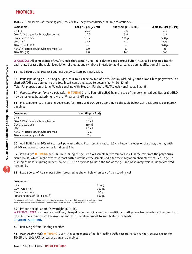
TABLE 2 | Components of separating gel (15% 60%:0.4% acryl:bisacrylamide/6 M urea/5% acetic acid).
Component Long AU gel (70 ml) Short AU gel (10 ml) Short TAU gel (10 ml)
Urea (g) 25.2 3.6 3.660%:0.4% acrylamide:bisacrylamide (ml) 17.5 2.5 2.5Glacial acetic acid 3.5 ml 500 ml 500 mldH2O (ml) 28.7 4.1 3.7310% Triton X-100 — — 370 mlN,N,N¢,N¢-tetramethylethylenediamine (ml) 420 60 6010% APS (ml) 980 140 140
Component Long AU gel (5 ml)
Urea 1.8 g60%:0.4% acrylamide:bisacrylamide 0.5 mlGlacial acetic acid 250 mldH2O 2.8 mlN,N,N¢,N¢-tetramethylethylenediamine 30 ml10% ammonium persulfate 70 ml
Component
Urea 0.36 g0.2% Pyronin Y 100 mlGlacial acetic acid 50 mlProtamine sulfatea (25 mg ml�1) 500 mlaProtamine, a small, highly cationic protein, serves as a scavenger for radicals during pre-running and as a blockingagent to reduce non-specific association of proteins with the gel matrix during the actual run of the sample.
1452 | VOL.2 NO.6 | 2007 | NATURE PROTOCOLS
PROTOCOL
42| Add TEMED and 10% APS to initiate polymerization.
43| Pour acrylamide solution up close to the edge of the plate. Insert comb.m CRITICAL STEP For long AU gels, the comb must be inserted deep enough that the teeth of the comb are pushed approximately1 mm into the stacking gel.? TROUBLESHOOTING
44| Allow gel to polymerize for 1 h.
Preparation of samples and running the gel45| Sample preparation � TIMING 0.5 h. Dry samples down to protein pellet in SpeedVac.Note: Since the differently charged isoforms of histones are separated, it is necessary to load more sample per lane than forSDS-PAGE.
46| Prepare AU sample buffer by mixing the components given shown below and vortexing until urea is dissolved.
47| Dissolve samples in 10 ml (long AU gels) or 5 ml (short AU/TAU gels) AU sample buffer.m CRITICAL STEP Do not boil the samples, as this will cause the urea to react covalently with the histone amines (carbamylation),and thereby change the migration behavior of the proteins.
48| Run the gel � TIMING Long AU gels: 0.5 h handling, 30 h running. Short AU or TAU gels: 0.5 h handling, 0.5–1.5 hrunning. Transfer gel into running chamber (running buffer: 5% AcOH).
49| Remove comb.m CRITICAL STEP If the ‘fingers’ between the wells are displaced from the stacking gel during comb removal, reposition them(we use a thin spatula for this); otherwise the sample will leak between loading wells.
50| Right before sample loading, flush wells with a syringe to remove excess urea and unpolymerized acrylamide.
51| Load samples.m CRITICAL STEP Put sample buffer in lanes without sample. For 2D TAU gels leave one lane empty (i.e., add no sample buffer)between samples in order to later identify sample lane.
52| For long AU gels, run samples into stacking gel at 200 V for 1–2 h, then run gel at 400 V for another 30–32 h (at thispoint, mammalian histone H4 will be close to the bottom of the gel, see Fig. 6). For short AU/TAU gels, run samples for0.5–1.5 h at 200 V.m CRITICAL STEP Histones are positively charged under the acidic running conditions of AU/TAU gels. Therefore, unlike SDS-PAGEgels, the proteins run toward the negative end. Remember to switch electrode leads.? TROUBLESHOOTING
53| Disassemble gel after the run and mark gel for orientation. AU/TAU gels can be stained with Coomassie Brilliant Bluesolution or by silver staining. Alternatively, the gel can be transferred to a nitrocellulose or PVDF membrane for western blotting(see protocol in Box 3). The gel can also be run in a second dimension to further differentiate the histone isoforms (2D TAU,proceed with Steps 54–66).
p
uor
G g
n ih si l
bu
P eru ta
N 700 2©
nat
ure
pro
toco
ls/
moc.er
ut an.
ww
w//:ptt
h
Component
Urea 0.36 g0.2% Pyronin Y 100 mlGlacial acetic acid 50 mlProtamine sulfatea (25 mg ml�1) 500 mlaProtamine, a small, highly cationic protein, serves as a scavenger for radicals during pre-running and as a blockingagent to reduce non-specific association of proteins with the gel matrix during the actual run of the sample.
Component Long AU gel (5 ml)
Urea 1.8 g60%:0.4% acrylamide:bisacrylamide 1.25 mlGlacial acetic acid 250 mldH2O 2.05 mlN,N,N¢,N¢-tetramethylethylenediamine 60 ml10% ammonium persulfate 140 ml
NATURE PROTOCOLS | VOL.2 NO.6 | 2007 | 1453
PROTOCOL
2D TAU/SDS gel � TIMING Approximately 3 h54| 2D TAU gels can be run with a homemade SDS-PAGE gel as second dimension (Fig. 5) or with a pre-cast commercial gelwith a ‘2D’ well, such as the NuPAGE 10–20% SDS gel (Invitrogen, cat. no. EC6136). If you are using the pre-cast second-dimension gel, skip to Step 58.
55| Preparation of 15% SDS-PAGE gels � TIMING 1 h. Clean glass plates and assemble using standard recipes.m CRITICAL STEP Use wider spacers to obtain a thicker SDS-PAGE gel than the first-dimension TAU gel, which makes transfer of theTAU gel slice on top of it easier.
56| Carefully mix all components for the separation gel, avoiding bubble formation, and pour acrylamide to a level of 4 cmbelow the top of the short plate, overlay with ddH2O or ethanol and let polymerize for 20 min.
57| Discard ddH2O and carefully mix all components for the stacking gel. Pour acrylamide to a level of 2 cm below the top ofthe short plate, overlay with ddH2O and let polymerize for 20 min.
p
uor
G g
n ih si l
bu
P eru ta
N 700 2©
nat
ure
pro
toco
ls/
moc.er
ut an.
ww
w//:ptt
h
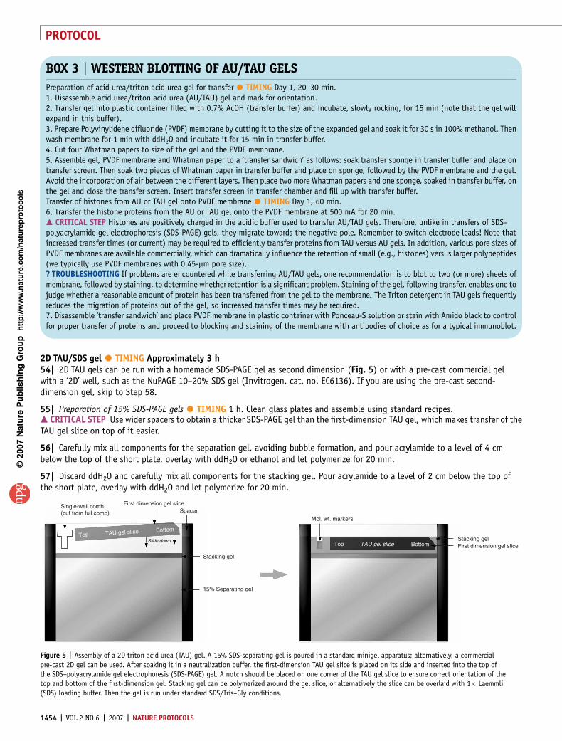
BOX 3 | WESTERN BLOTTING OF AU/TAU GELS
Preparation of acid urea/triton acid urea gel for transfer � TIMING Day 1, 20–30 min.1. Disassemble acid urea/triton acid urea (AU/TAU) gel and mark for orientation.2. Transfer gel into plastic container filled with 0.7% AcOH (transfer buffer) and incubate, slowly rocking, for 15 min (note that the gel willexpand in this buffer).3. Prepare Polyvinylidene difluoride (PVDF) membrane by cutting it to the size of the expanded gel and soak it for 30 s in 100% methanol. Thenwash membrane for 1 min with ddH2O and incubate it for 15 min in transfer buffer.4. Cut four Whatman papers to size of the gel and the PVDF membrane.5. Assemble gel, PVDF membrane and Whatman paper to a ‘transfer sandwich’ as follows: soak transfer sponge in transfer buffer and place ontransfer screen. Then soak two pieces of Whatman paper in transfer buffer and place on sponge, followed by the PVDF membrane and the gel.Avoid the incorporation of air between the different layers. Then place two more Whatman papers and one sponge, soaked in transfer buffer, onthe gel and close the transfer screen. Insert transfer screen in transfer chamber and fill up with transfer buffer.Transfer of histones from AU or TAU gel onto PVDF membrane � TIMING Day 1, 60 min.6. Transfer the histone proteins from the AU or TAU gel onto the PVDF membrane at 500 mA for 20 min.m CRITICAL STEP Histones are positively charged in the acidic buffer used to transfer AU/TAU gels. Therefore, unlike in transfers of SDS–polyacrylamide gel electrophoresis (SDS-PAGE) gels, they migrate towards the negative pole. Remember to switch electrode leads! Note thatincreased transfer times (or current) may be required to efficiently transfer proteins from TAU versus AU gels. In addition, various pore sizes ofPVDF membranes are available commercially, which can dramatically influence the retention of small (e.g., histones) versus larger polypeptides(we typically use PVDF membranes with 0.45-mm pore size).? TROUBLESHOOTING If problems are encountered while transferring AU/TAU gels, one recommendation is to blot to two (or more) sheets ofmembrane, followed by staining, to determine whether retention is a significant problem. Staining of the gel, following transfer, enables one tojudge whether a reasonable amount of protein has been transferred from the gel to the membrane. The Triton detergent in TAU gels frequentlyreduces the migration of proteins out of the gel, so increased transfer times may be required.7. Disassemble ‘transfer sandwich’ and place PVDF membrane in plastic container with Ponceau-S solution or stain with Amido black to controlfor proper transfer of proteins and proceed to blocking and staining of the membrane with antibodies of choice as for a typical immunoblot.
Top TAU gel slice Bottom
Single-well comb(cut from full comb)
Mol. wt. markers
First dimension gel sliceSpacer
Slide downTop TAU gel slice Bottom
Stacking gel
15% Separating gel
Stacking gelFirst dimension gel slice
Figure 5 | Assembly of a 2D triton acid urea (TAU) gel. A 15% SDS-separating gel is poured in a standard minigel apparatus; alternatively, a commercial
pre-cast 2D gel can be used. After soaking it in a neutralization buffer, the first-dimension TAU gel slice is placed on its side and inserted into the top of
the SDS–polyacrylamide gel electrophoresis (SDS-PAGE) gel. A notch should be placed on one corner of the TAU gel slice to ensure correct orientation of the
top and bottom of the first-dimension gel. Stacking gel can be polymerized around the gel slice, or alternatively the slice can be overlaid with 1� Laemmli
(SDS) loading buffer. Then the gel is run under standard SDS/Tris–Gly conditions.
1454 | VOL.2 NO.6 | 2007 | NATURE PROTOCOLS
PROTOCOL
58| Preparation of TAU gel lanes for 2Dseparation � TIMING Day 1, 1 h.After the TAU separation of the histonesamples is complete (see Step 53),cut out the lanes where the samplewas run.m CRITICAL STEP Sample lanes canbe identified by the faint red colorof the loading buffer. Use fresh razorblades to cut out the sample lane.Also, cut out a small corner from thebottom of the TAU gel slice to identifytop and bottom of gel slice beforeassembly.
59| Transfer TAU sample lanes carefullyto small plastic containers (a pipette-tipbox lid works well) filled with 0.125 MTris–Cl, pH 8.8, and rock slowly for5 min on rocking platform. Replacethree times with fresh 0.125 M Tris–Cl,pH 8.8 solution.m CRITICAL STEP By equilibrating thegel slices in Tris–Cl buffer, the acid in thegel will be neutralized.
60| Carefully transfer the gel slicehorizontally between the glass plates(see depiction in Fig. 5). Leave someroom to the left and right of the glassplate and to the separating gel.m CRITICAL STEP It is tricky to place the gel slice in between the glass plates without breaking it. Therefore, ensure that thesecond-dimension gel is thicker than the first-dimension gel (i.e., 1-mm-thick SDS gel for a 0.75-mm-thick TAU gel). Try to transferit on the top of the glass plate and then gently push the gel slice with both thumbs or a metal spatula down between the twoglass plates by applying soft pressure to different parts of the gel slice. Start pushing on one end of the gel slice and move on tothe other end of the gel slice. Repeat these steps until the gel slice is pushed in between the glass plates. Prepare 2 ml offresh stacking gel and pour it very carefully between the glass plates, thereby covering the TAU gel slice. To avoid bubbles,hold the glass plates at an angle and pour in more acrylamide solution to flush out bubbles. The TAU gel should be completelycovered in stacking gel.? TROUBLESHOOTING
61| (Only for ‘homemade’ gels; skip to Step 65 if using a pre-cast gel). Insert ‘one-finger’ comb into one side of the gel before itpolymerizes. This well will be used to load a protein marker in order to later identify the molecular weight of the proteins and todetermine the appropriate protein running distance (cross-reference with histone SDS gel in Fig. 3).
62| Let the gel polymerize for 30 min.
63| Fit assembled 2D TAU gel into chamber and fill carefully with Tris–Gly SDS running buffer without disturbing the TAU gel slice.
64| Remove comb finger carefully and flush out marker well with Tris–Gly SDS running buffer to remove bubbles and non-poly-merized acrylamide.
65| Load protein molecular weight marker in marker well.
66| Run the gel as usual using Tris–Gly SDS running buffer.Note: Disassemble glass plates, remove TAU gel slice and stacking gel, and place separating gel carefully in plastic containerfilled with Coomassie Brilliant Blue solution. Alternatively silver-stain gel or proceed to transfer (regular SDS-PAGE transfer pro-tocols) to perform western blotting techniques.
p
uor
G g
n ih si l
bu
P eru ta
N 700 2©
nat
ure
pro
toco
ls/
moc.er
ut an.
ww
w//:ptt
h
Short triton-acid urea Long acid-urea(hydrophobic- and
charge-based separation)(charge-based separation)
H2A
H3.2
H2B
H4 H4
H3.3
H1
H2A H2B
H3
H1 H1
H3Amido black α-H4 α-Acetyl H4
H2AH2B
H4
H3.1H2A
Untre
ated
+But
yrat
e
Untre
ated
+But
yrat
e
Untre
ated
+But
yrat
e
Untre
ated
+But
yrat
e
H1
H3.1 H3.2
H2A and H2A-variants
H3.3H2B
H4
First dimension: triton-acid urea
Sec
ond
dim
ensi
on: S
DS
a b c
d
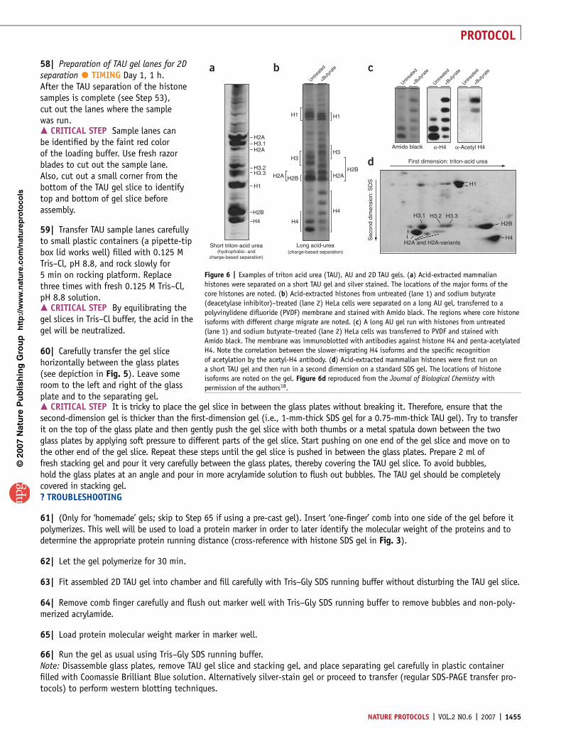
Figure 6 | Examples of triton acid urea (TAU), AU and 2D TAU gels. (a) Acid-extracted mammalian
histones were separated on a short TAU gel and silver stained. The locations of the major forms of the
core histones are noted. (b) Acid-extracted histones from untreated (lane 1) and sodium butyrate
(deacetylase inhibitor)–treated (lane 2) HeLa cells were separated on a long AU gel, transferred to a
polyvinylidene difluoride (PVDF) membrane and stained with Amido black. The regions where core histone
isoforms with different charge migrate are noted. (c) A long AU gel run with histones from untreated
(lane 1) and sodium butyrate–treated (lane 2) HeLa cells was transferred to PVDF and stained with
Amido black. The membrane was immunoblotted with antibodies against histone H4 and penta-acetylated
H4. Note the correlation between the slower-migrating H4 isoforms and the specific recognition
of acetylation by the acetyl-H4 antibody. (d) Acid-extracted mammalian histones were first run on
a short TAU gel and then run in a second dimension on a standard SDS gel. The locations of histone
isoforms are noted on the gel. Figure 6d reproduced from the Journal of Biological Chemistry with
permission of the authors18.
NATURE PROTOCOLS | VOL.2 NO.6 | 2007 | 1455
PROTOCOL
� TIMINGCoomassie staining and destaining: 4 h,Silver staining: 1 h, andTransfer: 1 h
? TROUBLESHOOTINGStep 22 (acid extraction)If histones are contaminated with a large background of other cellular proteins visible on a Coomassie-stained gel, it is possiblethat the hypotonic lysis was not completely effective. In that case, we recommend that the lysis buffer volume be increased anda Dounce homogenizer be used after initial hypotonic swelling. The cells should also be freeze-thawed before lysis.
Step 28 (RP-HPLC)If poor separation is encountered between histone proteins, first ensure that TFA was added to the solvents, as low pH ensuressharp peaks. H2A and H4 frequently have somewhat overlapping retention on C8 columns, so those eluted fractions can be re-injected onto the C8 column and run with a shallower gradient ramp to increase the separation.
Step 39 (AU/TAU gels)Note that the ability of Triton X-100 detergents to bind to hydrophobic regions of the proteins is strongly influenced by theoxidation state of the proteins. If separation of histones by TAU gel electrophoresis is compromised by high levels of histoneoxidation (i.e., smearing and poor band resolution), additional pre-running steps with 2.5 M cysteamine can be applied to‘scavenge’ oxidative free radicals from the gel29. For an additional, discontinuous AU/TAU gel system that systematicallyincorporates anti-oxidation practices, see ref. 30.
Step 43 (AU/TAU gels)Use freshly prepared 10% APS to ensure good polymerization. Keep the 60% acrylamide stock in an amber bottle away fromlight to prevent degradation of the stock.
Step 52 (AU/TAU gels)It can be difficult to determine the extent of migration of histone proteins through AU and TAU gels without prior experience.Five to ten micrograms of cytochrome c protein can be used as a marker, as it appears brownish while run. It typically migratesnear histone H4. However, it tends to become diffuse and difficult to see as the gel is run, so hold a white paper behind the gelto visualize it.
Step 60 (2D TAU gels)When using precise SDS gels for the second dimension, an alternative to casting the first-dimension gel slice in stacking gel andallowing it to polymerize is just to layer 1� SDS (Laemmli) loading buffer, as the pre-cast gels do not require a stack.
ANTICIPATED RESULTSAcid- or high-salt extraction of histones from nuclei or chromatin typically results in a tremendous enrichment of histoneproteins. Histones are very abundant proteins, usually present in cells in equal mass with the DNA6. Investigators following theprotocols presented should be able to routinely isolate quantities of purified histones for many follow-up experiments, includingimmunoblotting and MS.
Figure 3 illustrates a Coomassie-stained SDS-PAGE gel showing what is expected from acid or salt extractions of humannuclei. Notice that most of the proteins present are core histone proteins, running in a banding pattern typical for histones(between 11 and 17 kDa). Many isoforms of histone H1 proteins run between 25 and 35 kDa. Also notice that in the high-saltextraction more high molecular weight proteins are apparent. These are likely chromatin-associated proteins that are acid-inso-luble, so they only appear when extracted in neutral conditions. Note that histones from different species and different celltypes may have somewhat different banding patterns, but there are usually three discrete bands of H2A, H2B and H3 with alower H4 band.
Figure 6 demonstrates the relative benefits of the TAU gel and the long AU gel. Histones run on a short TAU or AU gel willnot exhibit the same pronounced separation of charge-dependent isoforms that the long AU gel will. But the short gel can beturned on its side and run in a second dimension with an SDS-PAGE gel, separating the proteins on the basis of size, whichallows explicit identification of protein variants. Furthermore, the non-ionic detergent Triton X-100 (and other such non-ionicdetergents in this series) binds to hydrophobic residues and retards the mobility of certain histones (in particular, histonesin the H2A and H3 families). Note that the ability of these detergents to bind to hydrophobic regions of the proteins isstrongly influenced by the oxidation state of the proteins. Thus, efforts are needed to keep the histone samples reduced byincubation with charged and neutral reducing agents, including the addition of cysteamine to the AU/TAU running buffers
p
uor
G g
n ih si l
bu
P eru ta
N 700 2©
nat
ure
pro
toco
ls/
moc.er
ut an.
ww
w//:ptt
h
1456 | VOL.2 NO.6 | 2007 | NATURE PROTOCOLS
PROTOCOL
(‘scavenging’ oxidative free radicals) (see TROUBLESHOOTING tip for Step 39 for more details). The long AU gel, while moretime-consuming to run, allows for unambiguous separation of acetylated- or phosphorylated-histone proteins.
Finally, Figure 4 shows a typical HPLC purification of acid-extracted histones. The chromatogram and stained gels show howmuch separation can be expected between each histone.
ACKNOWLEDGMENTS We are grateful to all the previous and current members ofthe Allis lab who contributed to the development of these techniques. D.S. is therecipient of a fellowship from the Irma T. Hirschl Trust, H.L.D. is supported by apredoctoral fellowship from the Boehringer Ingelheim Foundation and S.B.H. issupported by the Deutsche Forschungsgemeinschaft (DFG).
COMPETING INTERESTS STATEMENT The authors declare no competing financialinterests.
Published online at http://www.natureprotocols.comRights and permissions information is available online at http://npg.nature.com/reprintsandpermissions
1. Cheung, P., Allis, C.D. & Sassone-Corsi, P. Signaling to chromatin through histonemodifications. Cell 103, 263–271 (2000).
2. Khan, A.U. & Krishnamurthy, S. Histone modifications as key regulators oftranscription. Front. Biosci. 10, 866–872 (2005).
3. Iniguez-Lluhi, J.A. For a healthy histone code, a little SUMO in the tail keeps theacetyl away. ACS Chem. Biol. 1, 204–206 (2006).
4. Thompson, P.R. & Fast, W. Histone citrullination by protein arginine deiminase: isarginine methylation a green light or a roadblock? ACS Chem. Biol. 1, 433–441(2006).
5. Nightingale, K.P., O’Neill, L.P. & Turner, B.M. Histone modifications: signallingreceptors and potential elements of a heritable epigenetic code. Curr. Opin. Genet.Dev. 16, 125–136 (2006).
6. Van Holde, K.E. Chromatin (Springer-Verlag, New York, 1989).7. Miescher, F. Ueber die chemische Zusammensetzung der Eiterzellen. Med. Chem.
Unters. 4, 441–460 (1871).8. Avery, O.T., MacLeod, C.M. & McCarty, M. Studies of the chemical nature of the
substance inducing transformation of pneumococcal types. Induction oftransformation by a deoxyribonucleic acid fraction isolated from pneumococcustype III. J. Exp. Med. 79, 137–158 (1944).
9. Watson, J.D. & Crick, F.H. Molecular structure of nucleic acids; a structure fordeoxyribose nucleic acid. Nature 171, 737–738 (1953).
10. Stedman, E. Cell specificity of histones. Nature 166, 780–781 (1950).11. Johns, E.W. The electrophoresis of histones in polyacrylamide gel and their
quantitative determination. Biochem. J. 104, 78–82 (1967).12. Panyim, S. & Chalkley, R. High resolution acrylamide gel electrophoresis of
histones. Arch. Biochem. Biophys. 130, 337–346 (1969).13. Allfrey, V.G., Faulkner, R. & Mirsky, A.E. Acetylation and methylation of histones
and their possible role in the regulation of RNA synthesis. Proc. Natl. Acad. Sci.USA 51, 786–794 (1964).
14. Brownell, J.E. et al. Tetrahymena histone acetyltransferase A: a homolog toyeast Gcn5p linking histone acetylation to gene activation. Cell 84, 843–851(1996).
15. Taunton, J., Hassig, C.A. & Schreiber, S.L. A mammalian histone deacetylaserelated to the yeast transcriptional regulator Rpd3p. Science 272, 408–411(1996).
16. Goldberg, A.D., Allis, C.D. & Bernstein, E. Epigenetics: a landscape takes shape.Cell 128 (2007).
17. Gurley, L.R., Prentice, D.A., Valdez, J.G. & Spall, W.D. High-performanceliquid chromatography of chromatin histones. J. Chromatogr. 266, 609–627(1983).
18. Hake, S.B. et al. Expression patterns and post-translational modificationsassociated with mammalian histone H3 variants. J. Biol. Chem. 281, 559–568(2006).
19. Taverna, S.D. et al. Long-distance combinatorial linkage between methylationand acetylation on histone H3 N termini. Proc. Natl. Acad. Sci. USA 104,2086–2091 (2007).
20. Garcia, B.A. et al. Organismal differences in post-translational modifications inhistones H3 and H4. J. Biol. Chem. 282, 7641–7655 (2007).
21. Taverna, S.D., Coyne, R.S. & Allis, C.D. Methylation of histone h3 atlysine 9 targets programmed DNA elimination in tetrahymena. Cell 110,701–711 (2002).
22. Smythe, C. & Newport, J.W. Systems for the study of nuclear assembly, DNAreplication, and nuclear breakdown in Xenopus laevis egg extracts. Methods CellBiol. 35, 449–468 (1991).
23. Shechter, D., Costanzo, V. & Gautier, J. ATR and ATM regulate the timing of DNAreplication origin firing. Nat. Cell Biol. 6, 648–655 (2004).
24. Murray, K. The acid extraction of histones from calf thymusdeoxyribonucleoprotein. J. Mol. Biol. 15, 409–419 (1966).
25. Chen, C.C., Smith, D.L., Bruegger, B.B., Halpern, R.M. & Smith, R.A. Occurrenceand distribution of acid-labile histone phosphates in regenerating rat liver.Biochemistry 13, 3785–9 (1974).
26. Matthews, H.R. & Huebner, V.D. Nuclear protein kinases. Mol. Cell Biochem. 59,81–99 (1984).
27. von Holt, C. et al. Isolation and characterization of histones. Methods Enzymol.170, 431–523 (1989).
28. Chevallet, M., Luche, S. & Rabilloud, T. Silver staining of proteins inpolyacrylamide gels. Nat. Protoc. 1, 1852–1858 (2006).
29. Kaufman, P.D. Triton-acetic acid-urea (TAU) gel electrophoresis of histones. Bio.Protocol, http://www.bio.com/protocolstools/protocol.jhtml?id¼p2055.
30. Bonner, W.M., West, M.H. & Stedman, J.D. Two-dimensional gel analysis ofhistones in acid extracts of nuclei, cells, and tissues. Eur. J. Biochem. 109, 17–23(1980).
31. Davey, C.A., Sargent, D.F., Luger, K., Maeder, A.W. & Richmond, T.J. Solventmediated interactions in the structure of the nucleosome core particle at 1.9 Aresolution. J. Mol. Biol. 319, 1097–1113 (2002).
32. Humphrey, W., Dalke, A. & Schulten, K. VMD: visual molecular dynamics. J. Mol.Graph 14, 33–38–27–28 (1996).
33. Bernstein, E. & Hake, S.B. The nucleosome: a little variation goes a long way.Biochem. Cell Biol. 84, 505–517 (2006).
34. Kouzarides, T. Chromatin modifications and their function. Cell 128, 693–705(2007).
35. Wysocka, J. et al. WDR5 associates with histone H3 methylated at K4 and is essen-tial for H3 K4 methylation and vertebrate development. Cell 121, 859–872 (2005).
p
uor
G g
n ih si l
bu
P eru ta
N 700 2©
nat
ure
pro
toco
ls/
moc.er
ut an.
ww
w//:ptt
h
NATURE PROTOCOLS | VOL.2 NO.6 | 2007 | 1457
PROTOCOL