Embed Size (px)
Citation preview

The Pennsylvania State University
The Graduate School
Department of Physiology
EXTRACTION AND BINDING CHARACTERIZATION OF HYALURONIC ACID
BINDING PROTEINS FROM BOVINE NASAL CARTILAGE
A Thesis in
Physiology
by
Bret M. Weaver
2009 Bret M. Weaver
Submitted in Partial Fulfillment
of the Requirements
for the Degree of
Master of Science
August 2009

The thesis of Bret M. Weaver was reviewed and approved* by the following:
Herbert H. Lipowsky
Professor and Head Dept. of Bioengineering Thesis Advisor
William Hancock
Associate Professor of Bioengineering
Donna Korzick
Professor of Kinesiology Chair, Physiology Program
*Signatures are on file in the Graduate School

iii
ABSTRACT
The current project was undertaken to optimize the extraction of hyaluronic-acid binding
proteins (HABPs) from bovine nasal cartilage (BNC). An incubation pull-down assay was
devised by mixing various concentrations of trypsinized BNC with a constant amount of
functionalized sepharose beads conjugated with hyaluronic acid (HA). Following incubation of
BNC digests with the HA-derived beads, a fractional binding profile was established for a range
of digest-to-bead proportions. Elution of the beads with 2M NaCl and 4M GCl showed most
HABP binding activity coming off with 2M NaCl. Total protein and fractional binding values
were determined with Bradford assay and SDS-PAGE electrophoresis, respectively. From these
results, a mass elution scheme was prepared to follow a step-wise washing pattern proportional to
ionic strength. This allowed protein yield and specific binding amounts to be calculated.
Conjugation efficiency of the derived sepharose beads was determined by measuring the
difference in refractive index (RI) between the supernatant and the initial solution concentration
of added 64kDa HA. The refractive indices of a stock solution of HA were used to construct a
standard curve to calculate HA (w/v) % of the supernatant. Calculation of HABP binding values
was determined using the protein concentrations eluted with 2M NaCl as they were found to be
statistically more significant than the 4M GCl rinses (P<0.01).
A minimum of three percent conjugation was calculated from a standard conjugation
assay with non-degraded 64kDa HA and functionalized sepharose beads. Trypsinized HABPs
from BNC were found to have a MWw of 53.4 kDa, a Kd of 7.235x10⁻⁸M and a total binding-site
concentration ([TBS]) of 1.775x10⁻⁶M with most of the binding activity residing in the 47kDa
link protein. Total specific binding activity gave a yield of 1.18% deduced from the mass-elution
procedure.

iv
The current project was focused around methods and results previously reported
(Tengblad, 1978). Using conventional liquid affinity chromatography, specific binding activity
was washed out in the 4M GCl fraction. Two species are commonly found to bind to HA after
being extracted from trypsinized BNC: a 45kDa link protein and a 90kDa proteoglycan monomer.
Three bands were analyzed with the current incubation pull-down method that contributed to the
overall calculation of the weighted molecular-weight average: 97kDa, 65kDa and 47kDa. This
approach was derived from previous reports, and MW differences were due to the higher
concentration of acrylamide used to run the reducing PAGE. Liquid chromatography remains to
be the best procedure in order to enhance the purity of the sample for proper in-vivo
experimentation and interpretation of conjugative biochemical results.
v
TABLE OF CONTENTS
LIST OF FIGURES ............................................................................................................ viii
LIST OF TABLES .............................................................................................................. x
ACKNOWLEDGEMENTS ................................................................................................ xi
Chapter 1 Background and Introduction ............................................................................. 1
1.1 Identifying the Source
1.1.1 Fig. 1: Cartoon schematic of Aggrecan production and turnover.
1.2 Bio- and Physiochemical Association of Hyaluronic Acid
1.3 The HABP Binding Domain and its Relationship with HABP Isoforms.
1.3.1 Impetus for Molecular Studies
1.3.2 Fig 2: Rate Association Binding Kinetics Between HA and HABPs
1.4 Traditional Methods and Parameters Used to Isolate and Characterize Binding
Fractions
1.4.1 CsCl gradient
1.4.2 Column Chromatography and Combined Enzymatic Analysis
1.4.2.1 Relevant Distillation from Previous Work
1.5 Foundation for the Current Project
Chapter 2 Materials and Methods ....................................................................................... 13
2.1 Tissue Processing
2.1.1 Fig 3: Solubilized Protein Levels of BNC through 30hr Incubation with 4M GCl
2.2 Bead Saturation
2.2.1 Table 1: Fractional Volume Contribution by Added Buffer Related to
Incubation Substituents
2.2.2 Fig 4: Standard Curve of dRI in Terms of HA Fraction (w/v)

vi
2.2.3 Fig 5: Standard Curve of dRI in Terms of HA Fraction (w/v) corrected to
zero intercept.
2.3 Hyaluronic Acid Binding Protein (HABP) Binding Assay
2.3.1 Fig. 6: Flow scheme shown for initial tissue extraction and binding assay
2.3.2 HABP analysis
2.3.2.1 Fig 7: Procedure to determine individual band intensities
2.4 Mass Elution Scheme
2.4.1 Fig 8: Mass elution profile following several volumes of selective ionic conditions
Chapter 3 Results ............................................................................................................... 28
3.1 Initial Tissue Processing
3.1.1 Fig 9: 15% Reducing PAGE of Trypsinized Exract
3.2 Bead Saturation and HA Conjugation
3.2.1 Fig 10: Conjugation Percentage of Amino-Derived Sepharose Beads with
Hyaluronic Acid (HA)
3.2.2 Fig 11: Natural log conversion of the conjugation profile in terms of Bead % (v/v)
and umol HA added
3.2.3 Fig 12: Plot of natural log conversion profile within selected linear range.
3.3 HABP Binding Assay
3.3.1 Fig 13: Elution plot between the incubation concentration of trypsinized extract
and the total protein found between the unbound, and 2M NaCl and 4M GCl
elution fractions
3.3.2 Fig 14: Fractional saturation profile of trypsinized extract plotted in terms of the
incubation concentration
3.3.3 Fig 15: 15% reducing PAGE of the 2M NaCl TE elution assay
vii
3.3.4 Fig 16: Net intensity profile of 2M NaCl elution at 1.509 IC for the trypsinized
extract
3.3.5 Table 2: Comparison of Average and Specific Binding Fraction of 47kDa Species
3.3.6 Fig 17: 4M GCl elution assay of trypsinized extract
3.4 Mass Elution Scheme
3.4.1 Fig 18: A280 Elution profile of mass elution scheme
Chapter 4 Conclusion and Discussion ................................................................................. 45
4.1 Putting It All Together
4.2 Interpreting the Calculations: Binding Values and Saccharide Dependency
4.3 Statistical Methods
References ………… .......................................................................................................... 54

viii
LIST OF FIGURES
1.1.1 Figure 1: Cartoon schematic of Aggrecan production and turnover.
(Lark et.al., 1997)
1.3.2 Figure 2: Rate Association Binding Kinetics Between HA and HABPs
2.1.1 Figure 3: Solubilized Protein Levels of BNC through 30hr Incubation with 4M
GCl
2.2.2 Figure 4: Standard Curve of dRI in Terms of HA Fraction (w/v)
2.2.3 Figure 5: Standard Curve of dRI in Terms of HA Fraction (w/v) corrected to zero
intercept.
2.3.1 Figure 6: Flow scheme shown for initial tissue extraction and binding assay
2.3.2.1 Figure 7: Procedure to determine individual band intensities
2.4.1 Figure 8: Mass elution profile following several volumes of selective ionic
conditions
3.1.1 Figure 9: 15% SDS-Reducing PAGE of Trypsinized Extract
3.2.1 Figure 10: Conjugation Percentage of Amino-Derived Sepharose Beads with
Hyaluronic Acid (HA)
3.2.2 Figure 11: Natural log conversion of the conjugation profile in terms of Bead %
(v/v) and umol HA added.
3.2.3 Figure 12: Plot of natural log conversion profile within selected linear range
3.3.1 Figure 13: Elution plot between the incubation concentration of trypsinized extract
and the total protein found between the unbound, and 2M NaCl and 4M
GCl elution fractions
3.3.2 Figure 14: Fractional saturation profile of trypsinized extract plotted in terms of the
incubation concentration

ix
3.3.3 Figure 15: 15% reducing PAGE of the 2M NaCl TE elution assay
3.3.4 Figure 16: Net intensity profile of 2M NaCl elution at 1.509 IC for the trypsinized
extract
3.3.6 Figure 17: 4M GCl elution assay of trypsinized extract
3.4.1 Figure 18: A280 Elution profile of mass elution scheme

x
LIST OF TABLES
2.2.1 Table 1: Fractional Volume Contribution by Added Buffer Related to Incubation
Substituents
3.3.5 Table 2: Comparison of Average and Specific Binding Fraction of 47kDa Species

xi
ACKNOWLEDGEMENTS
I would like to sincerely thank Dr. Lipowsky for his driving wisdom, patience and
financial support to conduct this project. He is my Yoda: “…you gotta keep your eye on the
donut, „cause in the end we‟ll all be pushing up daisies…” -Dr. Lipowsky

1
Chapter 1
Background and Introduction
1.1 Identifying the Source
Proteoglycan biochemistry and glycobiology is a rapidly growing field that is quickly
covering a vast array of scientific disciplines. Not only has its role been well established within
biochemical methodology, but the well appreciated physiochemical nature of proteoglycan
interactions deduced from such studies are currently being applied to in-vivo models. The
pericellular glycocalyx in its simplest form describes a meshwork of interconnected protein and
glycosaminoglycan components that aggregate and form an exchangeable communicative
environment between the intra and extracellular world. Many studies have been undertaken to
elucidate the relationship between specific players of the extracellular glycocalyx and
downstream intracellular events that effect a response to a local external cue. Syndecan is a
proteoglycan that is composed of a linear protein core glycosylated with GAG (Glycos-Amino-
Glycan) groups at selected Ser residues. The residues glycosylated depends on the primary
sequence structure of the protein. It is produced, shuttled and modified along the ER-Golgi
pathway to be secreted at the apical membrane site. This proteoglycan projects from the
percicellular surface and interacts with other players of the ECM environment, and many studies
have shown a direct link of syndecan-mediated signaling to manifest an intracellular response.
This includes both the integration of transcription-dependent signaling cascades and cytoskeleton
rearrangement, which leads into mechanotransducion. These studies are beyond the scope of the
current one, but are worth mentioning.

2
Syndecan proteoglycans have three distinct domains: a linear core to which both keratin
sulfate and chondroitin sulfate are O-covalently attached determining the overall heterogeneity of
the proteoglycan, and two areas that lack these substituted GAG groups for non-covalent
association with the surrounding glycocalyx. One is for binding HA link proteins to bridge its
connection with HA, and another at the N-terminal domain that binds with the HA linear chain
(Keiser, 1977). Together in associative conditions, the HA linear chain, link proteins and PG
subunits aggregate to form an extractable fraction known as aggrecan. This can be harvested
from cartilage such as bovine nasal septa that provided the source for extracting viable hyaluronic
acid binding proteins (HABPs). Shown below is the natural turnover cycle of aggrecan in human
articular cartilage (Fig 1). The same scheme can be applied in bovine species with the aggrecan
product located at the apex. The smaller arrows show degradation sites along the PG subunit
downstream from the HABP total binding area (N-termini of PG subunit and Link Protein (LP)).

3
1.1.1 Fig. 1: Cartoon schematic of Aggrecan production and turnover.
1.1.1 Fig 1: Cartoon schematic showing turnover cycle of aggrecan and its integrated
components between the chondrocyte and extracellular matrix. From: Lark et. al., 1997
The importance of trying to visualize a specific ECM player along the vascular lumen and its
plausible role to mediate an in-vivo response is growing with every report of diabetes and other
aberrant inflammatory conditions, both acute and chronic. Examples include sepsis and systemic
lupus erythematous. (SLE), respectively. In-vivo binding markers can be conjugated with
HABPs harvested from cartilage to help understand these diseases more specifically in order to
elucidate the normal dynamic intermolecular state along the vascular lumen endothelium.
4
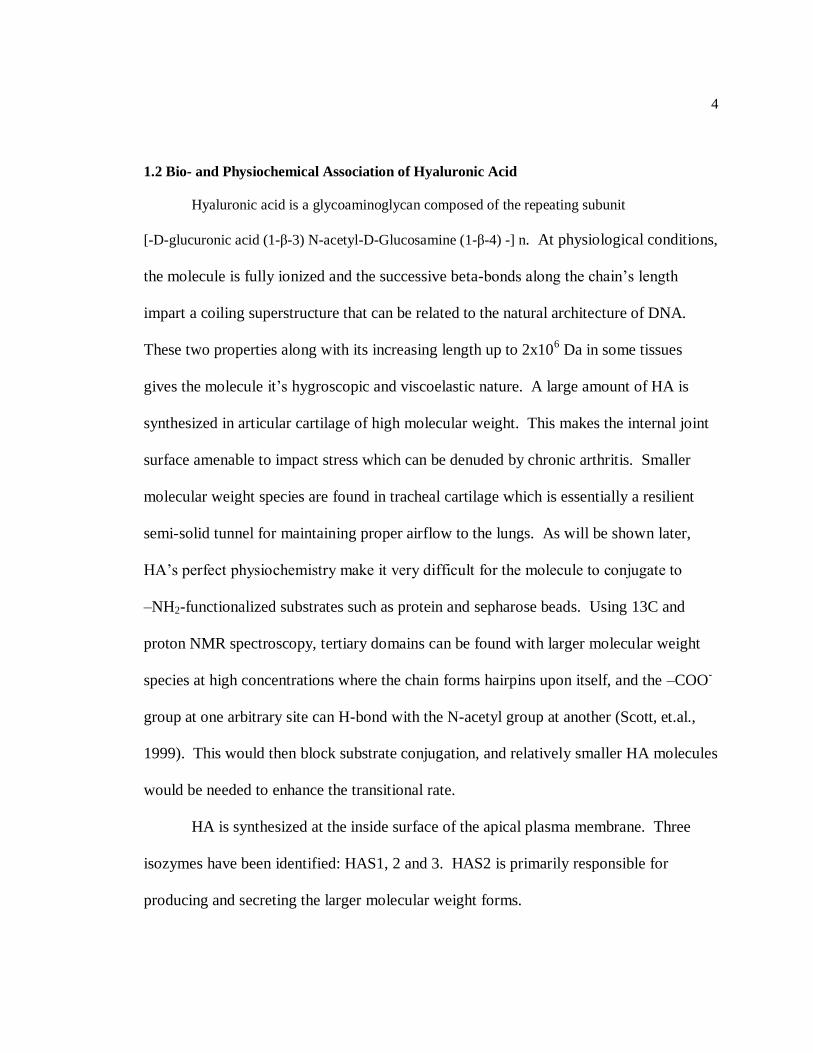
1.2 Bio- and Physiochemical Association of Hyaluronic Acid
Hyaluronic acid is a glycoaminoglycan composed of the repeating subunit
[-D-glucuronic acid (1-β-3) N-acetyl-D-Glucosamine (1-β-4) -] n. At physiological conditions,
the molecule is fully ionized and the successive beta-bonds along the chain‟s length
impart a coiling superstructure that can be related to the natural architecture of DNA.
These two properties along with its increasing length up to 2x10⁶ Da in some tissues
gives the molecule it‟s hygroscopic and viscoelastic nature. A large amount of HA is
synthesized in articular cartilage of high molecular weight. This makes the internal joint
surface amenable to impact stress which can be denuded by chronic arthritis. Smaller
molecular weight species are found in tracheal cartilage which is essentially a resilient
semi-solid tunnel for maintaining proper airflow to the lungs. As will be shown later,
HA‟s perfect physiochemistry make it very difficult for the molecule to conjugate to
–NH2-functionalized substrates such as protein and sepharose beads. Using 13C and
proton NMR spectroscopy, tertiary domains can be found with larger molecular weight
species at high concentrations where the chain forms hairpins upon itself, and the –COO-
group at one arbitrary site can H-bond with the N-acetyl group at another (Scott, et.al.,
1999). This would then block substrate conjugation, and relatively smaller HA molecules
would be needed to enhance the transitional rate.
HA is synthesized at the inside surface of the apical plasma membrane. Three
isozymes have been identified: HAS1, 2 and 3. HAS2 is primarily responsible for
producing and secreting the larger molecular weight forms.

5
This mechanism differs from the synthesis of other GAGs such as keratin sulfate,
dermatin sulfate and chondroitin sulfate which are produced and subsequently modified
against a protein core along the ER-Golgi secretory pathway. However, both types share
the source of UDP high energy donors from the uronic acid metabolic pathway that
begins with the modification of glucose-6-phosphate. These are sequestered from the
cytoplasm, and for the case of HA synthesis are directly added at their reducing end by
HAS at the inner leaflet of the periplasm. Inflammatory cytokines and various growth
factors have been implicated in regulating this initial flux, but HASs themselves seem to
be the constitutive ushers to provide a route for HA production and secretion. The supply
of glucose-6-phosphate is the initial rate-limiting step in glycolysis which primes other
pathways such as: the pentose-phosphate pathway that provides NADH2 reducing
equivalents for fatty acid production, glycogenesis in liver and muscle and the regulation
of gluconeogenesis in non-fed states. Simply put the ability of HA and other GAGs to be
produced and secreted depends on the availability of metabolic glucose and overall
energy balance.
1.3 The HABP Binding Domain and its Relationship with HABP Isoforms
For all functional and structural purposes, hyaluronic acid binding proteins are
virtually identical across the animal kingdom. Any differences among published reports
may be attributed to the way binding fragments are generated, isolated and treated for
analysis. The globular binding domains of HABPs and cartilage link proteins are non-
glycosylated. cDNA molecular methods show sequence homology between link proteins
and proteoglycan binding regions which are composed of a tandem array of alpha-helices
and beta-pleated sheets adjoined by disulfide cross-bridges (Doege et. al., 1986)

6
Favorable thermodynamic non-covalent association at the binding interface between
HABPs and hyaluronic acid would then be limited to the proper physiochemical
orientation of the protein binding site and repetitive GAG subunits of the HA polymer.
As discussed before, HA is composed of a predictable linear sequence of glucuronic acid
and N-acetyl glucosamine. Any variation in the presentation to the protein binding
surface would be due to MW and concentration at physiological pH. Optimal binding
occurs within the range of pH 5 and 7 (Tengblad, 1981). At the same solution conditions,
the HABP binding domain achieves its lowest Gibbs‟ free energy state. This ultimately
comes down to the primary sequence in protein structure, and the aliphatic non-polar and
polyanionic nature of HA must complementarily align with active site of HABPs.
When assessing the composition of isolated binding protein fractions from
trypsinized extracts, four analytical features are consistently shown: a relatively equal
amount of the basic amino-acid residues (BAA) Arg and Lys, an AA ratio between Cys
and those BAAs of approximately 1:3, a low amount of GAG leftovers after isolation as
determined by hexauronate analysis and a low amount of hydroxyproline content which
designates the presence of collagen. Acetylating reagents such as dansyl chloride show
eighty-percent binding inhibition compared with acetic anhydride in specifically targeting
primary amino groups presented by the amino acid residues Lys and Arg (Heinegaard
and Hascall, 1978). This is a highlighting mark with using cartilage as a source for
binding protein. Of course, some sulfated GAG remnants that co-precipitate with
variable Ser content are expected when initially extracting the binding fraction from the
whole tissue.

7
The HABP globular binding domain is best conceptualized in the context of the four
factors listed above in order to maintain a proper tertiary three dimensional form in
solution at optimal pH. This is clearly evident when analyzing the structural binding
properties of the link module (Kohda, et. al. 1996; Goetinck, et.al. 1987), and the
trypsinized fraction is well characterized (Heinegaard and Hascall, 1974).
1.3.1 Impetus for Molecular Studies
HABPs isolated from tissue extracts have been done using conventional biochemical
methods as will be explained in section 1.1.4. When the binding species is purified one can use
cDNA and amino-acid sequencing analysis, and interface those results with a queried database
that reference other isoforms and species-related HABPs. CD44, also known as the lymphocyte
homing receptor, is a surface glycoprotein primarily expressed on epithelial and myeloid cell-
types and has been shown to mediate T-cell activation, extravasation and margination, and
lamellipodia outgrowth. This effectively maturates the inflammatory response (Oliferenko et.al.,
2000; Turley et. al., 1991; Stamenkovic et. al., 1989; Siegelman et.al., 1999; Lesley et.al., 1994;
Stamenkovic et.al., 1991; Culty et.al., 1990; Roscic-Mrkic, 2003). CD44v (polymorphic splice
variants) are expressed in lymphoid tumor cells (Bartolazzi et.al., 1995). The expression
mechanism may depend on adjoining variable exon sites much like antibody and TCR secretion,
but the splicing-dependent machinery has not been well founded (Doege et.al., 1986). These
variable isoforms are present in the supernatant and on the cell surface of tumor cells, and are
probably responsible for disaggregating and disrupting the normal HA-PG arrangement to
increase spreading, invasion and metastasis. The loss of physical integrity and controlled
permeation properties along the endothelial glycocalyx that would result from this disruption can
be supported by the resulting intermolecular charge modification (Paul et. al., 2005) and shunted
equilibrium from HA and native HABPs (Fig. 2)

8
1.3.2 Fig 2: Rate Association Binding Kinetics Between HA and HABPs
[HA] + [BP] [HA:BP]
Rate1=kf[HA][BP] Rate2=kr[HA:BP]
At equilibrium: Rate1=Rate2=>Kd=[HA][BP]/[HA:BP]=kr/kf. Variant binding factors infiltrating
the glycocalyx and associating with HA increases Rate2 by decreasing the functional native
concentration of HA:BP. This leads to its dissociation and collapse of the luminal surface
endothelium. HA occupies approximately fifty percent of the glycocalyx volume (Henry et. al.,
1999), so its reasonable to assume a high probability of HA:BP break-down facilitated by these
variant isoforms. Generating hybrid cDNA constructs helps differentiate HA attachment, and
would therefore assess malignancy in pathological states (Bartolazzi et.al., 1995).
The non-pathological in-vivo HA-binding model initiates an autocrine-dependent
intracellular signaling cascade, and parallels the role of other HABP isoforms such as RHAMM
(Receptor for Hyaluronic Acid Mediated Motility) (Singleton et.al., 2002; Bourguignon et.al,
1999; Goueffic et.al., 2006). Together it seems CD44 and related isoforms effect and possibly
regulate the overall response to antigen clearance and tissue maintenance. HABPs harvested
from bovine nasal cartilage present similar amino-acid profiles and must maintain the same
proper three-dimensional fold at the binding surface. Any system used to express HA binding
antigens or isoforms needs to be put in context. If the species is glycosylated, yeast would be a
better model than bacteria (i.e. E.coli) unless the expressed protein itself is sufficient. Such
experiments depend on the targeted sequence, potency and requisite glycosylation pattern.
However, proper steps need to be followed to ensure LPS and related antigens are free of the
purified HABP binding fraction. This would otherwise lead to an unwanted inflammatory state in
in-vivo injection experiments.
k
r
kf
kr
9
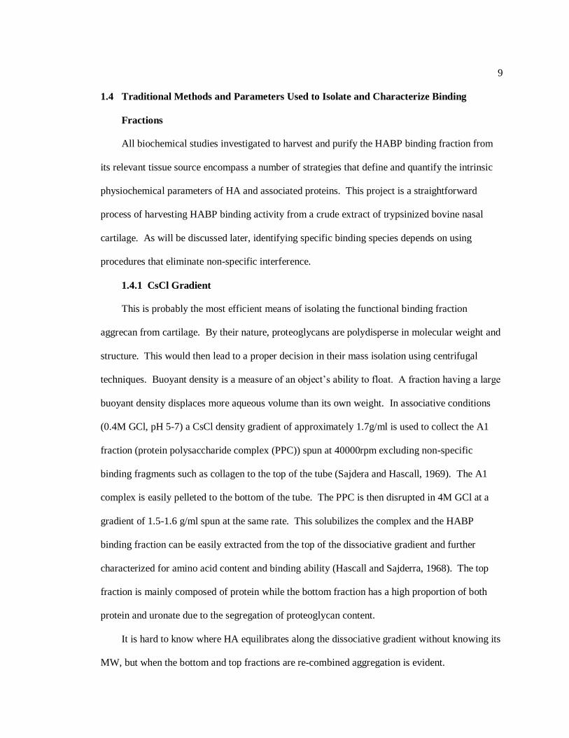
1.4 Traditional Methods and Parameters Used to Isolate and Characterize Binding
Fractions
All biochemical studies investigated to harvest and purify the HABP binding fraction from
its relevant tissue source encompass a number of strategies that define and quantify the intrinsic
physiochemical parameters of HA and associated proteins. This project is a straightforward
process of harvesting HABP binding activity from a crude extract of trypsinized bovine nasal
cartilage. As will be discussed later, identifying specific binding species depends on using
procedures that eliminate non-specific interference.
1.4.1 CsCl Gradient
This is probably the most efficient means of isolating the functional binding fraction
aggrecan from cartilage. By their nature, proteoglycans are polydisperse in molecular weight and
structure. This would then lead to a proper decision in their mass isolation using centrifugal
techniques. Buoyant density is a measure of an object‟s ability to float. A fraction having a large
buoyant density displaces more aqueous volume than its own weight. In associative conditions
(0.4M GCl, pH 5-7) a CsCl density gradient of approximately 1.7g/ml is used to collect the A1
fraction (protein polysaccharide complex (PPC)) spun at 40000rpm excluding non-specific
binding fragments such as collagen to the top of the tube (Sajdera and Hascall, 1969). The A1
complex is easily pelleted to the bottom of the tube. The PPC is then disrupted in 4M GCl at a
gradient of 1.5-1.6 g/ml spun at the same rate. This solubilizes the complex and the HABP
binding fraction can be easily extracted from the top of the dissociative gradient and further
characterized for amino acid content and binding ability (Hascall and Sajderra, 1968). The top
fraction is mainly composed of protein while the bottom fraction has a high proportion of both
protein and uronate due to the segregation of proteoglycan content.
It is hard to know where HA equilibrates along the dissociative gradient without knowing its
MW, but when the bottom and top fractions are re-combined aggregation is evident.

10
This can be followed up by chromatography, which will be discussed in 1.4.2. The top fraction is
relatively low in hydroxyproline, has about the same number of BAARs (Basic Amino Acid
Residues), low in Ser which rules out glycosylated protein, and shows a 1:3 to 1:4 ratio in
Cys:BAA content. This is systematic proof for the presence of HABPs harvested from bovine
nasal cartilage using simple associative-dissociative CsCl gradient centrifugation.
1.4.2 Column Chromatography and Combined Enzymatic Analysis
The proteoglycan constituents of intact cartilage are well-manifested by the sequential steps
needed to purify binding proteins. This project was mainly geared around prescribed procedures
that yield a specific binding fraction of 2.5% using affinity chromatography (Tengblad, 1978),
Immunochemistry experiments using antibodies against hyaluronidase-prepared fractions is
evidence for multiple binding antigens (Keiser, 1982). The observation of other studies showing
results pertaining to the complex heterogeneity of this type of tissue needs to be appreciated.
These concepts and useful biochemical strategies to analyze harvested tissue will be succinctly
discussed here. All literature sources that provide a knowledgeable foundation for this project
will be cited in detail among the list of references.
1.4.2.1 Relevant Distillation from Previous Work
The immunogenicity of PG fractions can be differentiated by the degree and extent to which
they are glycosylated. This presents the PG core protein surface in uniquely recognizable ways,
hence generating epitope variability. When these fractions are degraded by selective enzymes
such as Chondroitinase and Hyaluronidase that target specific glycosidic residues, surface epitope
expression becomes modulated and immunogenicity for the PG core components can be affected.
Kinetic analysis shows the initial rate of HA degradation favored by HAse endo-glycosidases, but
at increasing time intervals up to approximately 30mins, chondroitinase can be used to initially
select out sulfated GAGs in the extract before HA becomes largely degraded. This increases the
purity of the sample by keeping the functional binding complex intact.

11
The enzymatic discontinuity may be explained by the alternating differences in GAG sequence
between CS and HA. HA is essentially a glycopolymer made up of repeating glucose subunits,
while CS substitutes galactose at every other position along the chain. The axial vs. equatorial
hydroxyl group at the fourth carbon between galactose and glucose may present differing
stereoelectronic environments that increase or decrease the potential for nucleophilic attack at the
anomeric carbon. Better chromatographic resolution and fractional characterization of the HABP
derivatives from bovine nasal cartilage is achieved after degrading the initial crude extract with
chondroitinase before trypsinization (Heinegard and Hascall, 1974). Two MW species were
observed after the A1 fraction was isolated and treated from an associative CsCl gradient. The 90
kDa and a 45 kDa fractions eluted as separate peaks along a Sephadex G200 column under
dissociative conditions with the 90kDa fraction having a lower Kav. The eluted fractions were
appropriately named T-G200-2 and T-G200-3, corresponding with their respective Kav elution
values (Kav (45kDa)>Kav (90kDa)). The 90kDa band on a SDS reducing PAGE was also more
diffuse than the 45kDa band. This can be termed “PG spreading”. These two reasons point out
the buoyant density differences between glycans and pure HABPs. The heterogeneity of
substituted glycans give a lower buoyant density than pure HABPs in a dissociative CsCl
gradient. The tandem approach of using associative-dissociative CsCl gradient centrifugation to
initially extract the A1 aggregate before conjunctive enzymatic glycosidic degradation augments
specificity and leads to further procedures that characterize their functional capacity such as
ELISA, Western Blots and Immunoprecipitation. Competitive binding studies against a standard
preparation can be used to deduce the potency of the binding fraction prepared along this scheme.
Using reducing and alkylation agents to target Cys and BAA residues should be implemented to
interfere with the protein‟s native conformation and potential to bind to HA substrates.
Observing a decrease in binding ability after this treatment would be substantial to say a good
extract has been harvested.

12
1.5 Foundation for the Current Project
Four points can be made from the preceding introduction that provide a means of
developing an experiment to effectively analyze the yield of harvested HABPs from trypsinized
bovine nasal cartilage:
1. A defining presence of BAARs. This enables the use of a Bradford assay in
determining functional protein amount. Undisclosed results from an accompanying
experiment along with this project show increased BAAR liberation during HAse
incubation with crude extract in a time dependent fashion. Maximum absorbance was
found at 2hr out of an 8hr incubation period. Toward the end of this time frame, a
minimal absorbance was seen which can be explained by the increasing presence of
smaller HA derivatives that seem to saturate the enzymatic site under conditions of
low ionic strength (Asteriou et. al, 2005).
2. Multiple binding species. This has been shown with immunochemistry experiments
using agarose diffusion procedures to observe several precipitation lines between the
prepared fractions and antibodies against isolated binding antigens.
3. Characteristically low yield in specific binding activity after eluting bound substrate
with salts such as NaCl and GCl.
4. Molecular weight of HA. Tertiary domain organization of HMW forms under high
concentration may block the repetitive carboxyl group from conjugating to the amino
group of the selected substrate.

13
Chapter 2
Materials and Methods
2.1 Tissue Processing
Bovine nasal cartilage was extracted from the decapitated heads of slaughtered cows at
the PSU meat lab facility and had a net tissue weight of 60g. Stiff pliers were used to separate
associated membranes before weighing and processing of the cartilage. It was grated with a
Surform pocket plane and dissolved in 4M Guanidium Chloride(GCl)/0.5M Sodium Acetate
(NaAc); pH 5.8 at 4oC for 30 hr with constant stirring according to prescribed procedures
(Tengblad, 1978). One milliliter aliquots were taken at various time-points, spun and dialyzed.
The Bradford assay was used to monitor solubilized protein levels which were found to plateau at
30hr. [Fig 6, Step 1] (Fig 3).

14
2.1.1 Fig 3: Solubilized Protein Levels of BNC through 30hr Incubation with 4M GCl
Time (hr)
0 5 10 15 20 25 30 35
Concentr
ation (
mg/m
l)
0.0
0.2
0.4
0.6
0.8
1.0
1.2
1.4
Fig 3: Solubilized proteins levels were measured from 1ml aliquots at various time-
points through 30hr incubation with 4M GCl in triplicate. The results show the
maximum amount of protein coming off in solution at 30hr. .
GCl is a very dense salt, so the buffer was titrated with enough deionized water to
solubilize it before bringing the total solution to the appropriate volume. After filtering the
extract with pre-washed cheesecloth, it was maximally dialyzed (12 kDa MWCO, Sigma) in
deionized water for 24hr, then with 0.1MTris/0.1M NaAc; pH 7.3 for 12hr on a shaker table at
4oC. The buffered extract was frozen in 50ml parcels at -20
oC until further use[Fig 6, Step 2].
All tools and surfaces were sterile, and gloves were used at all times during this process.

15
2.2 Bead Saturation
Lyophilized hyaluronate (HA) from human umbilical cord (LIfecore Biomed, 64kDa)
was conjugated with NH2-functionalized sepharose beads (Affigel 102, BioRad, Hercules, CA)
according to established procedures (Underhill and Zhang). A total of 0.5g 64kDa HA was used
with 25ml of minimally saturated and equilibrated beads. A conjugation assay was performed to
assess the relative amounts of beads and HA needed to achieve maximum conjugation and bead
saturation.
A 2.4% HA (2.4g/100ml deionized water) stock solution was prepared for all procedures.
This was the highest concentration achievable using a 15 ml 10 MWCO filter (Millipore). The
limitation may be due to the high saturating potential of HA and its increase in viscosity
proportional to concentration. The beads were equilibrated in a 5ml syringe with a 0.2u filter
(Acrodisc, 0.2um HT Tuffryn Membrane;Pall, Gelman Laboratory) using 25mM phosphate
buffer, pH 5 according to package insert. Several washings with the buffer were required to
remove sodium azide in the stock preparation which was present as a bacteriostatic preservative.
Azide is a nucleophile, hence the beads must be thoroughly washed to ensure non-interference in
the conjugation process. For this same reason, Tris and Acetate buffers were not used in order to
prevent unwanted amino and acetyl substitution, respectively.
The conjugation buffer was prepared by the addition of the reagent carbodiimide (FW=
191.71, Sigma) to the phosphate equilibration buffer at a concentration of 5mM. When the beads
are in their compact and minimally saturated form, the concentration of surface-terminal amino
groups is approximated at 15 umol/ml. After equilibration, the beads were diluted 15x in the
same buffer to a concentration of 1umol/ml. One ml of the final solution was added to six
individual autoclaved glass tubes. The following amounts of HA were added to each tube from
the stock solution: 100mg, 50mg, 20mg, 10mg, 5mg, and 1mg.

16
It was suggested by the manufacturer the solvent fraction of the total conjugation reaction be no
greater than 0.3. Extra conjugation buffer was added to each reaction mixture to reduce the
viscosity from added HA while maintaining the appropriate solvent fraction (Table 1).
2.2.1 Table 1: Fractional Volume Contribution by Added Buffer Related to Incubation
Substituents
Amount HA Added
Substituent 1mg 5mg 10mg 20mg 50mg 100mg
64kDa HA; (from 2.367 % (w/v) stock 40ul 210ul 420 ul 840 ul 2.1 ml 4.2 ml
1 umol/mltNH2 Affigel (Dil 1/15x) 1ml 1ml 1ml 1ml 1 ml 1 ml
25 mM Phosphate Buffer; pH 4.7 300ul 360ul 420 ul 550 ul 1.93 ml 2.56 ml
Fractional Contribution of Buffer 0.22 0.23 0.23 0.23 0.38 0.33
All tubes were incubated at 37oC for 3hrs with constant shaking. The tubes were placed at an
angle with the bottom of the incubator to ensure mixing without displacing the majority of the
beads from the solution. If the inside surfaces of the tubes are largely coated with beads, then the
mixture is being shaken too rapidly and conjugation will not be complete. After incubation, the
tubes were cooled to room temperature and spun at 3000g for 30min. Refractive indices (RI)
were measured from 100ul of the supernatant using the Leica ABBE Mark II refractometer (Model
10480) and measurements from at least five aliquots were used to attain an average RI per tube.
The difference between these averaged readings and the conjugation buffer gave the change in RI
from the solvent due to the remaining HA in solution that was not conjugated. The amount of
HA left in the supernatant was determined from the a standard RI curve constructed from the
stock solution (Fig 4, 5).

17
2.2.2 Fig 4: Standard Curve of dRI in Terms of HA Fraction (w/v)
Standard Curve of the Difference in Refractive Index (dRI)from Buffer Alone as a Function of HA % with 95% Confidence Limitation
HA % (g/100mL)
0.0 0.2 0.4 0.6 0.8 1.0 1.2 1.4
dR
I
0.0000
0.0005
0.0010
0.0015
0.0020
0.0025
Fig 4: The difference in refractive index (dRI) plotted in terms of HA% (g/100ml).
R2=0.9987 and p<0.001.

18
2.2.3 Fig 5: Standard Curve of dRI in Terms of HA Fraction (w/v) corrected to zero
intercept.
HA % (g/100mL)
0.0 0.2 0.4 0.6 0.8 1.0 1.2 1.4
dR
I
0.0000
0.0005
0.0010
0.0015
0.0020
0.0025
Fig 5: Correction of standard dRI curve to zero interception. The corrected slope falls
within the deviation of individual values. Therefore, the equation: dRI=0.0015 ([HA%])
is a reliable model to predict the amount of HA in solution contributing to refractive
index.
The buffer with carbodiimide showed no difference in RI than with buffer alone. Conjugation
percentage was calculated with the following equation: [(Ci-Cs)/Ci]x100. Ci is the initial
reaction concentration of HA in w/v (g/100ml), and Cs is the concentration of free HA remaining
in the supernatant. Two conjugation profiles from the standard assay were created to assess the
completion of the mass conjugation procedure using 0.5g HA (Underhill and Zhang).

19
This is assuming reaction completion depends on both of two independent factors: the mol/mol
fraction between terminal amino groups present on the bead surface and HA in solution, and
percent bead concentration in the conjugation mixture. These conditions constitute an
entropically-dominant reactive state consistent with the physiochemical observations of HA
explained in section 1.2. The bead concentration was calculated from the final dilution factor of
the compact bead volume in each reaction tube.
2.3 Hyaluronic Acid Binding Protein (HABP) Binding Assay
An aliquot of untrypsinized frozen bovine nasal cartilage was thawed and concentrated
using 10 kea MWCO centrifuge tubes (Centrals) by spinning at 3000xg to minimal volume at
4oC[Fig 6, Step 3]. Half the extract was then digested with TPCK- treated trypsin (Sigma) at 2%
w/w for 3hr at 37oC with constant shaking. The reaction was stopped with soybean trypsin
inhibitor (STI, Sigma) at 2x the molar weight of trypsin added and incubated overnight at room
temperature with constant shaking[Fig 6, Step 4]. Both the trypsinized and untrypsinized extracts
(TE and UTE, respectively) were diluted in series with cold 0.1M Tris/0.1M NaAcetate; pH 7.3
on ice at relative concentrations of 1 to 0.2, 1 being the most concentrated [Fig 6,Step 5]. The
serial dilutions were done in 0.2 increments and corresponded to approximately 1.5mg/ml to
0.3mg/ml for TE, and 0.8mg/ml to 0.2mg/ml for UTE. All protein content measurements were
determined with the Bradford assay using the Shimadzu Pharma Spectrophotometer UV-1700,
and the accompanied Shimadzu UV Probe computer program. Standards were constructed with
the appropriate solvent or buffer to control for ionic strength.
A pilot assay was first performed to determine the proper amounts of extract and beads to
add to each binding incubation mixture. One-milliliter of saturated beads was added to undiluted
UTE aliquots of 0.1, 0.25, 0.5, 1, 2 and 4ml. Only the first three treatments eluted protein in a
proportional fashion.

20
This was attributed to both or one of two factors: 1.) an entropic effect of relative volumes
between the extract and beads used where higher yields were obtained with tighter vol:vol ratios
and 2.) possible bead aspiration when sucking off the supernatant. The final assay was conducted
with both the UTE and TE at their respective series concentration [Fig 6,Step 5]. This also
incorporated a blank incubation where only buffer was used. A constant volume ratio of 2ml of
extract was added to 0.5ml of conjugated beads.
Beads were equilibrated with cold 0.1M Tris/0.1M NaAcetate pH 7.3 as previously
mentioned. One-half milliliter aliquots were placed in separate ten milliliter autoclaved glass
capped tubes. All tubes were vortexed slightly and allowed to settle overnight at 4oC. The tubes
were then spun at 3000xg for 30min at 4oC and the unbound supernatant was collected
using sterilized Pasteur pipettes [Fig 6, Step 6]. Protein content was determined on the
supernatant with the Bradford assay using the Tris/Acetate buffer to conduct the standard.
A series of wash steps then took place where 2ml ice cold 2M NaCl was used to wash
each bead pellet, vortexed slightly and allowed to settle on ice for 30mins. The tubes
were spun again at 3000xg for 30min at 4oC, and protein content was determined on the
supernatant using a 2M NaCl Bradford standard [Fig 6, Step 7]. After this fraction was
collected, the beads were treated the same with the addition of ice cold 4M GCl [Fig 6,
Step 8]. The 4M GCl elution fractions needed to be washed and concentrated with cold 0.15M
NaCl in 0.5ml capped 10 MWCO eppindorf tubes (Fisher). They were spun at 3000xg to
minimal volume. This was done to eliminate excess GCl that would interfere with the Bradford
reagent, and to attain three replicate readings diluted from a workable concentration. According
to Beer‟s Law, absorbance is proportional to the concentration. The unbound fraction and 2M
NaCl eluted fractions were able to be read directly using the appropriate standard correcting for
background absorbance.

21
The 4M GCl fractions, however needed to be measured with a 0.15M NaCl Bradford standard,
and corrected for both background absorbance and the dilution factor used. In order to
standardize the 4M GCl elution results, values needed to be calculated in terms of a two-milliliter
elution volume. For example: if a measurement came from a 200ul concentrated sample that was
diluted by 1/5, the final reading would be: (observed)*5*(0.2/2). The following figure shows the
overall scheme to assay for the binding capacity of the trypsinized extract:

22
2.3.1 Fig.6: Flow scheme shown for initial tissue extraction and binding assay.
Bovine Nasal Cartilage
30hr incubation [Step 1]
Membrane Dialysis [Step 2]
Concentrate to Minimal volume [step 3]
Trypsinized Extract(TE) Saturated Sepharose Beads Untrypsinized Extract (UTE)
[Step 4]
Binding incubation and
dilution series
Binding Incubation and
dilution series [Step 5]
Unbound Fraction [Step 6] Unbound Fraction Cold 2M NaCl Elution
SDS Reducing PAGE and Image J Analysis
Second Fraction [Step 7] Cold 4M GCl Elution Second Fraction
SDS Reducing PAGE and ImageJ Analysis
Third Fraction [Step 8] Third Fraction
Fig. 6: Flow scheme shown for initial tissue extraction and binding assay. Details of
Affigel-102 Sepharose conjugation are left out for simplicity.
Membrane separation and grating

23
2.3.2 HABP analysis
To visualize the eluted HABPs, a 15% SDS reducing PAGE stained with Coomassie blue
R250 (Biolab, Hercules, CA) was used to empirically determine the fractional contribution of
each species. A picture of the gels was taken and analyzed with the ImageJ software program.
Images were taken with the Gel dot analyzer (Epichem Darkroom, UVP Bio Imaging Systems).
Band intensities were determined for the TE binding assay. This was done by drawing a
rectangle around each individual peak using SigmaPlot where each lateral side (width) was
limited to half of the maximum peak intensity (height) (Fig 7).

24
2.3.2.1 Fig 7: Procedure to determine individual band intensities
Net Intensity Profile of Tryp 2M NaCl Wash
Pixel Length
0 100 200 300 400 500
Ne
t In
ten
sity (
8 b
it)
0
20
40
60
80
100
Fig 7: Procedure to determine individual band intensities. As seen with the 47kDa MW
species, the band intensity was calculated by drawing a rectangle covering the
individual peak where the height was limited to the net intensity of the band and
the width to half of that value. The area of the rectangle (pixel length x net
intensity) is proportional to the integrated band intensity. Background noise
made it difficult to calculate the integrated intensity of the two bands running at
97 and 65 kDa, so a line was drawn tangent to the peak intersecting the ordinate in
order to approximate half the individual peak intensity.

25
The area of the peak is proportional to the integrated band intensity. The total intensity of each
individual lane was also determined using ImageJ. This is best conceptualized as the total area
under the curve representing the analyzed pixel length down the ordinate of the intensity profile
(Fig 7). The fractional peak intensity (integrated band intensity divided by the total intensity)
gave the relative contribution of each molecular weight species coming off in each fraction. All
values were corrected against the gel background. Determining the fractional peak intensities
also helped in calculating the weighted molecular weight average between all species analyzed in
each elution.
2.4 Mass Elution Scheme
An initial assay was conducted using 10ml of freshly concentrated TE incubated with
10ml conjugated beads. The total mixture was parceled into autoclaved glass tubes. Using both a
1:1 extract:bead volume ratio and glass tubes were needed to minimize bead loss while trying to
realize an enhanced yield en-masse. The incubation buffer used was 0.1MTris/0.1M NaAcetate,
pH 6.0. HABPs bind optimally between pH 5 and 7 (Tengblad, 1981). Earlier results showed
most of the eluted protein came off with the 2M NaCl rinse and subsequently insignificant levels
of protein were found in the 4M GCl elution fractions. With the current mass elution scheme, a
step-wise rinsing pattern was conducted with 1M, 1.5M and 2M NaCl followed up by 4M GCl.
The basis for this approach was to observe changes in specific binding activity as ionic strength
incrementally increased in a step-wise manner.
After the extract was added, the mixture was vortexed slightly and the beads were
allowed to settle on ice for 30mins. The tubes were then spun at 3000xg for 30mins at 4oC. Two
volumes each of 1M NaCl, 1.5M NaCl and 2M NaCl, and three volumes of 4M GCl were used to
wash the beads at separate steps, and all eluants were pooled. An A280 assay was used to
approximate the amount of total protein coming off the beads and an elution profile was created
(Fig 8).

26
2.4.1 Fig 8: Mass elution profile following several volumes of selective ionic conditions
1M N
aCl E1
1M N
aCl E2
1.5M
NaCl E
1
1.5M
NaCl E
2
2M N
aCl E1
2M N
aCl E2
4M G
Cl E1
4M G
Cl E2
4M G
Cl E3
A2
80 m
g/m
l
-0.1
0.0
0.1
0.2
0.3
0.4
0.5
0.6
0.7
2M NaCl E1 2M NaCl E2 4M GCl E1 4M GCl E2 4M GCl E3
-0.02
0.00
0.02
0.04
0.06
0.08
0.10
0.12
0.14
0.16
0.035mg/ml
Fig 8: Mass elution profile following several volumes of selective ionic conditions.
Upper panel shows scheme in its entirety from the first elution conditions.
Bottom panel shows elution profile magnified from the first volume elution of 2M
NaCl to the last volume elution of 4M GCl.

27
Separate A280 standards were not necessary during this process as in the binding assay. The
GCl fractions were treated as before. All pooled fractions were spun down, concentrated and
measurements were standardized to a 10ml elution volume.
After this initial assay, 200ml of trypsinized extract was processed en-masse. It was
necessary to do the extraction in a series of multiple cycles with the limited volume of saturated
beads available. GCl carried over if the beads were not thoroughly washed after each cycle.
Three volumes of cold deionized water were used to rinse the beads before resetting them with an
equal volume of 0.1MTris/0.1M NaAc, pH6. If a large amount of beads were used at one time
during the GCl elution step the beads were aspirated when drawing off the supernatant. This was
attributed to the increased viscosity of the 4M GCl solution and was confirmed by earlier studies
(Kawahara and Tanford, 1966). The incubation mixture was parceled into separate glass tubes
making sure the bead volume does not occupy a large fraction of the tubes. Based on the initial
elution profile, a two-step mass elution scheme was conducted. Elution volumes were added in
terms of the amount of molar salt needed to separate the desired protein product from a 1:1
bead:salt volume ratio. The profile followed 2 volumes of 1M NaCl, 2 volumes of 1.5M NaCl, 2
volumes of 2M NaCl and 3 volumes of 4M NaCl. With a 1:1 bead: salt ratio, 9 mol NaCl and 12
mol GCl per volume of conjugated beads were used to wash the bounded proteins in a two step
fashion. The targeted protein product came off in the GCl fraction as seen with the Micheles-
Menton plateau from 4M GCl E1 to 4M GCl E3 (Fig 8, lower panel) This fraction was pooled,
concentrated and washed with cold deionized water using 10 kDa MWCO tubes spun to minimal
volume at 3000xg at 4oC. This was done to wash out all the excess salt that may saturate and
damage the protein before being lyophilized or frozen for further use.

28
Chapter 3
Results
3.1 Initial Tissue Processing
Processed and grated cartilage tissue showed maximum protein levels after 30hr
incubation with 4M GCl [Fig 6, Step 1](Fig 3). After trypsinizing the cartilage extract, three
bands were prominently seen and analyzed for fractional binding contribution [Fig 9, Lane 5,
arrows]. A 15% SDS reducing PAGE was run on trypsinized bovine nasal cartilage. Any bands
migrating below 45kDa in the trypsinized lane was attributed to Soybean Trypsin Inhibitor (STI),
Trypsin or protein content not known to bind to hyaluronic acid. This was confirmed by previous
reports (Tengblad, 1978; Hascall and Heinegaard, 1974) Reducing conditions were proven to
unaffect migrating distance to any significant degree (Hascall and Heinegaard, 1974). The
relative Trypsin/STI amount added in Lane 6 was 1:2 in order to mimic the ratio added for BNC
treatment. This controlled for any intensity differences seen in the trypsinized extract (Lane 5).
No differences were seen.

29
97.4
65.2
45
31
21.5
14.4
3.1.1 Fig 9: 15% SDS-Reducing PAGE of Trypsinized Extract
Fig 9: Lanes labeled left to right. Lane 1: Molecular Weight Marker. Values in kDa shown at left.
Lane2: Blank Control. Lane 3: Concentrated Untrypsinzed Extract (CUE)=5.3mg/ml. Lane 4: CUE
diluted by ½ to enhance clarity. Lane 5: Trypsinized Extract. Arrows show bands analyzed for
fractional binding contribution. Concentration=1.6mg/ml. Lane 6: Trypsin/Soybean Trypsin
Inhibitor (STI) Control.
3.2 Bead Saturation and HA Conjugation
A standard curve was constructed to predict the difference in refractive index based on
the content of HA in solution (w/v, MW=64 kDa). Fig. 4 shows a linear curve (P<0.001,
R2=0.9987) within a 95% confidence interval and was subsequently corrected for zero
interception (Fig 5). The equation dRI=0.0015(HA%) was used to calculate the amount of HA
left in the supernatant post conjugation, and the formula [(Ci-Cx)/Ci ]x100 was used to calculate
conjugation percentage. Carbodiimide reagent was not shown to interfere with refraction (data
not shown). A conjugation curve was generated from varying amounts of HA against a constant
volume of amino-derived sepharose beads (see materials and methods).
Lane: 1 2 3 4 5 6
15% SDS-Reducing Page
Lane1: Marker
Lane2: Blank
Lane3: Concentrated Untrypsinized
Extract(CUTE)=5.3 mg/ml
Lane 4: ½ diluted CUTE
Lane 5: Trypsinized Extract(TE)=1.6
mg/ml
Lane 6: Trypsin/STI control

30
Conjugation percent was plotted in terms of the relative mole ratio between terminal amino
groups and HA (Fig. 10), and was shown to reach a maximum at approximately 50 (R2=0.9376).
3.2.1 Fig 10: Conjugation Percentage of Amino-Derived Sepharose Beads with
Hyaluronic Acid (HA)
Conjugation Profile of HA Derived Sepharose BeadsUsing Undegraded 64kDa HA
mol/mol tNH2/HA
0 10 20 30 40 50 60
Co
nju
gation
Pe
rce
nt
[(C
i-C
s)/
Ci]*
10
0
0
20
40
60
80
100
120
140
160
95% Confidence Band
95% Prediction Band
Fig 10: Conjugation percent profile of HA-Sepharose (Affi-gel 102). Red and blue lines
bound both a 95% prediction and confidence interval, respectively. Conjugation percent
is plotted in terms of the relative molar amounts between the amino groups and HA.
R2=0.9376.
The ratio deduced from the procedure to derive 25ml of equilibrated compact beads with 0.5g
64kDa HA was 48 (Underhill and Zhang). Though HA was treated for 3hr with testicular
hyaluronidase (materials and methods), this was an insignificant amount of time to achieve
complete degradation (Sakai et. al., 2007). It is therefore assumed HA largely maintained its
original MW form.

31
Plotting the information in terms of percent bead concentration (v/v) per umol HA, the same
curve is seen (results not shown). A natural log conversion of this profile was constructed, and
linearity fell between a ratio of 2 and 6 (Fig 11).
3.2.2 Fig 11: Natural log conversion of the conjugation profile in terms of bead %
(v/v) and umol HA added
Conjugation Profile In Terms of the Ratio of Bead v/v Percent and umol HA on a Natural Log Scale
ln (Bead%/umolHA)
-1 0 1 2 3 4 5 6 7
ln(C
on
j %
)
2.0
2.5
3.0
3.5
4.0
4.5
5.0
Fig 11: Natural log conversion of the conjugation profile in terms of bead % (v/v) and
umol HA added. Results are approximately linear within and including the in (Bead
%/umol HA) range of 2 and 6. Black line shows raw results. Red shows results used to
deduce a linear relationship.

32
3.2.3 Fig 12: Plot of natural log conversion profile within selected linear range .
Natural Profile Within Linear Range
ln (Bead %/umol HA)
2 3 4 5 6
ln (
Conj %
)
2.0
2.5
3.0
3.5
4.0
4.5
5.0
Fig 12: Plot of natural log conversion profile within selected linear range (Fig 11).
R2=0.999, 95% confidence. The corresponding equation used to calculate conjugation
percentage came to be: Conj% = 1.6382 (Bead % /umol HA)0.5132
. The Bead%/umol HA
ratio used in the mass incubation was 2.56, giving a conjugation percentage of
approximately 3.
Within this range, the results were re-plotted (Fig 12), and the ratio of bead % (v/v) per umol HA
was 2.56. The corresponding conjugation value based on this plot was 3 percent. Overall
conjugation efficiency would then be the product of two independent factors: mol/mol ratio and
bead percent (v/v) = (value attained with Fig 10)*(value attained with Fig 12) = (100%)*(3%) =
(100%)*(0.03) =3%. This will be explained more thoroughly in chapter 4.

33
3.3 HABP Binding Assay
All results for the untrypsinized extract will not be shown, but mentioned as final
IMAGE-J analysis from those gels were hard to interpret and resolve. The incubation
concentration (IC) prepared in the dilution series of both the UTE and TE were plotted against the
total protein coming off in the unbound, 2M NaCl and 4M GCl elution fractions (Fig 13).
3.3.1 Fig 13: Elution plot between the incubation concentration of trypsinized extract
and the total protein found between the unbound, and 2M NaCl and 4M GCl elution
fractions.
Linear Correlation Between the Incubation Concentration (IC)and Total Protein ([P]t) In Each Trypsinized Incubation Assay
IC (mg/ml)
0.0 0.2 0.4 0.6 0.8 1.0 1.2 1.4 1.6
[P]t a
vg m
g/m
l+
/- S
D
0.0
0.2
0.4
0.6
0.8
1.0
1.2
1.4
1.6
Fig 13: Elution plot between the incubation concentration of trypsinized extract and the
total protein found between the unbound, and 2M NaCl and 4M GCl elution fractions.
Curve plotted within 95% confidence and is linear after correction to origin (P<0.001).

34
A strong linear correlation was found for both preparations, but only Fig 13 shows the trypsinized
results (R2=0.987 and P<0.001). The corresponding blank measurements (y-intercept) for both
the UTE and TE before origin adjustment were 0.0313 mg/ml and 0.0364 mg/ml, respectively.
This suggests the used beads themselves did not contribute any significant amount of residual
protein that may interfere with the assay. The blank absorbance value calculated from the
Bradford standard curve was subtracted from all other measurements which were significantly
greater than the blank (P<0.01). A fractional saturation curve was prepared and observed in a
Michelis-Menton fashion, and maximum saturation was seen at 0.2855 with a Kd of 0.2460
mg/ml (Fig 14).

35
3.3.2 Fig 14: Fractional saturation profile of trypsinized extract plotted in terms of the
incubation concentration
Fractional Saturation Profile of Trypsinized ExtractFract Sat = [0.2855*IC (mg/ml)]/[(0.2460 mg/ml + IC(mg/ml)]
IC (mg/ml)
0.0 0.2 0.4 0.6 0.8 1.0 1.2 1.4 1.6
[P]b
t/IC
0.00
0.05
0.10
0.15
0.20
0.25
0.30
0.35
95% Confidence Band
95% Prediction Band
Fig 14: Fractional saturation profile of trypsinized extract plotted in terms of the
incubation concentration (IC). Complete fractional saturation reaches its maximum at
0.2855 with a Kd of 0.2460mg/ml. R2=0.9614.
The fractional values were calculated by dividing the corrected average of the elution fraction by
its respective IC. This was mostly contributed by the 2M NaCl elution series as the 4M GCl
elution measurements were statistically insignificant from the blank absorbance value. SDS-
PAGE was conducted and three bands were analyzed for their ability to bind to the gel (Fig 15,
Lane 7, Arrows).

36
3.3.3 Fig 15: 15% reducing PAGE of 2M NaCl TE elution assay
Fig 15: 15% reducing PAGE of 2M NaCl TE elution assay. Lane 1 shows molecular weight marker
with corresponding kDa values at left while lanes 2 through 7 show elution results of blank(Lane
2) through 1.509 mg/ml IC (Lane 7). Molecular weight species analyzed shown in Lane 7 at full
saturation.
Lane 1 2 3 4 5 6 7
97.4
65.2
45
31
21.5
14.4

37
3.3.4 Fig 16: Net intensity profile of 2M NaCl elution at 1.509 IC for the trypsinized
extract
Net Intensity Profile of Tryp 2M NaCl Wash
Pixel Length
0 100 200 300 400 500
Ne
t In
ten
sity (
8 b
it)
0
20
40
60
80
100
Fig 16: Net intensity profile of 2M NaCl elution at 1.509 IC for the trypsinized extract.
The three peaks (arrows) correspond to the molecular weight species analyzed for
binding activity: 97kDa, 65kDa and 47kDa. Pixel length is proportional to band
migration.
IMAGE-J analysis was performed on the 2M NaCl elution fraction of the 1.509 mg/ml
trypsinized incubation (IC) (Fig 15). At this IC, fractional saturation is equal to 0.2455 which is
less than 5 percent of the maximum fractional saturation of 0.2855 given by the model (Fig 14).
Therefore, maximum saturation can be assumed to be achieved at 1.509 mg/ml IC. This gives a
complete assessment of binding capacity for this assay. The net total protein intensity of the
individual lane was compared to the net integrated band intensity of the three separate molecular
weight species (Fig 16).
Pixel Length MW Band
~20 97 kDa
~100 65 kDa
~180 47 kDa

38
Net intensity was calculated using IMAGE-J by subtracting the background gel intensity from the
total intensity. All fractional values from the IMAGE-J measurements were calculated as the
individual corrected integrated intensity of the band divided by the total corrected intensity of the
lane (see materials and methods). The three separate molecular weight species analyzed ran at
97, 65 and 47kDa (Fig 15, Fig 16) giving fractional integrated intensities of 0.006, 0.06 and 0.15,
respectively. This gave a total integrated fraction of 0.22 and an intensity ratio of 1:10:25,
respectively. The weighted molecular weight average (MW W) was calculated to be 53.4 kDa.
Since this favors the 47kDa species, its individual contribution was analyzed and shown to be
consistent with the overall fractional binding trend (Table 2).

39
3.3.5 Table 2: Comparison of Average and Specific Binding Fraction of 47kDa Species
1 2 3 4 5 6 7
IC
(mg/ml)
Fr. Band
Intensity
(47 kDa
Band)
Protein Binding
Fraction
+/- SD
Specific Binding
Fraction (M)
Specific
Binding
Fraction
x10(-6)
M
Average Specific
Binding Fraction
(M, from MW W)
Average
Specific
Binding
Fraction
x10(-6) M
0 0 0 0 0 0 0
0.3018 0.1042 0.1425 +/-
2.9826e-3
9.5346x10(-8)
M
0.095346 9.52594x10(-8) M 0.0952594
0.6036 0.1455 0.2307 +/-
9.6671e-3
4.3108x10(-7)
M
0.43108 3.08440x10(-7) M 0.30844
0.9054 0.1506 0.2099 +/-
0.0183
6.0895x10(-7)
M
0.60895 4.20946x10(-7) M 0.420946
1.2072 0.1503 0.2536 +/-
0.0600
9.7902x10(-7)
M
0.97902 6.78113x10(-7) M 0.678113
1.509 0.1582 0.2292 +/-
0.0123
1.1642x10(-6)
M
1.1642 7.66086x10(-7) M 0.766086
Protein binding fraction: fractional protein content coming off in each elution. This is
calculated as [eluted protein amount/IC]. Specific binding fraction: amount of 47kDa
species (M) coming off in each fraction. This is calculated as [(Col1)(Col2)(Col3)/47kDa]. Average specific binding fraction: amount analyzed
protein coming off in each elution. This is calculated from the weighted molecular
weight average [(Col1)(TIF)(Col3)/MWw]. TIF=Total Integrated Fraction.

40
Total binding sites were calculated to be approximately 1.775x10⁻⁶
M at maximum saturation, and
was derived from both the weighted molecular weight average and the total integrated fraction at
full saturation. Kd is defined as the concentration of substrate occupying one-half of the total
binding sites, and the corresponding IC value provided by the model is 0.2460mg/ml (Fig 14).
With the methods used and the high amount of non-specific binding activity in the crude extract,
it is better to interpret substrate binding ability at the point of maximum saturation. So, one-half
of the total binding activity {1/2[(0.28)(0.22)]} corresponds to a Kd of 7.235x10⁻⁸
M. Though
protein levels were insignificant after the 4M GCl elution step, the bands analyzed for binding
activity were present to a minimal degree in this series (Fig 17). Interpretation of these results
will be explained in the discussion section.

41
3.3.6 Fig 17: 4M GCl elution assay of trypsinized extract
Fig 17: 4M GCl elution assay of trypsinized extract. Lane assignments are consistent
with Fig 15. Arrows show the presence of the analyzed molecular weight species in the
eluant of the 0.9054 mg/ml trypsinized IC.
97.4
65.2
45
31
21.5
14.4

42
3.4 Mass Elution Scheme
With the low binding capacity of the trypsinized extract and the propensity of the
derived beads to be largely displaced from the solution if incubated in relatively large volumes,
an equal volume ratio of trypsinized extract to derived beads was used in mass extraction and
allotted in separate tubes (see materials and methods). An initial elution scheme was devised to
follow and approximate the step-wise binding pattern with different molar salt solutions using
A280 analysis (Fig 18).

43
3.4.1 Fig 18: A280 Elution profile of mass elution scheme
1M N
aCl E1
1M N
aCl E2
1.5M
NaCl E
1
1.5M
NaCl E
2
2M N
aCl E1
2M N
aCl E2
4M G
Cl E1
4M G
Cl E2
4M G
Cl E3
A2
80 m
g/m
l
-0.1
0.0
0.1
0.2
0.3
0.4
0.5
0.6
0.7
2M NaCl E1 2M NaCl E2 4M GCl E1 4M GCl E2 4M GCl E3
-0.02
0.00
0.02
0.04
0.06
0.08
0.10
0.12
0.14
0.16
0.035mg/ml

44
Fig 18. A280 Elution profile of mass elution scheme. Derived and re-stated from Fig 8.
Elution steps are labeled on the ordinate with their respective molar salt values.
GCl=Guanidinium chloride. Bottom panel shows the highlighted portion in the upper
panel on a closer scale. Value of 0.035 mg/ml shows hyperbolic plateau in protein levels
through 4M GCl elution series. This corresponds to a 1.18% yield in specific binding
activity. Initial extract concentration was 2.9783 mg/ml after blank and Typing/STI
correction.
The initial extract concentration after correcting for the blank measurement, STI and Trypsin was
2.9783 mg/ml. Peak binding activity at the 4M GCl elution plateau was 0.035 mg/ml giving a
yield of 1.18% in specific binding activity. An initial hump was seen at the first elution volume
of 2M NaCl. This may be attributed to proteoglycan HABP subunits having the ability to bind to
the derived sepharose beads.

45
Chapter 4
Conclusion and Discussion
4.1 Putting it All Together
Perhaps the greatest concept taken away from this project and reading material
surrounding the basis to isolate and characterize HABPs from bovine nasal cartilage (BNC) is
heterogeneity. BNC has to be the perfect tissue to dissect and elucidate the complex inter-
relationship of proteoglycans and link-proteins. It is an easy and economical source for this goal,
and can be stored for long term use without any significant loss in binding function, though yields
are extremely low.
BNC is a highly tensile tissue, and its physiological function structurally initiates proper
laminar airflow to the lungs. Inhaling through the nose and exhaling out the mouth is a simple
example of breathing efficiency. The turbulence resulting from the large amount of air coming in
through the oral cavity decreases the potential for oxygen to reach the caudal part of the lungs
where the surface area to volume ratio of dense alveolar tissue is maximal. Nasal septa can be
thought of as a pillar to support the intake conduit to the trachea for directing smooth airflow,
thus resulting in maximum breathing efficiency and hemoglobin saturation. Glycoprotein and
GAG polymers cohesively interact with collagen to produce its characteristic mechanical
behavior seen in cartilage.
Targeting the extraction and isolation of HABPs from this tissue requires aggressive steps
to realize a practical yield of viable protein. This project was primed by previous work in
isolating the HA binding fraction of BNC using conventional liquid chromatographic methods
(Tengblad, 1978).

46
In this case, the binding yield was 2.5%, and 4M GCl was specifically administered as the
reversible denaturant to rinse off, collect and measure HABPs from HA-derived sepharose beads.
The yield using the current mass incubation scheme with trypsinized extract deduced from
the defined Micheles-Menton elution plateau with three successive volumes of 4M GCl was
calculated to be 1.18% (Fig 18, Lower Panel). Due to the large loss of binding activity after 2M
NaCl elution in the HABP binding assay, this scheme was devised to follow total protein coming
off the HA-derived beads through a step-wise increase in ionic strength. Successive equal-
volume washes from 1M to 1.5M NaCl E1 (first elution volume) initially showed a precipitous
decline in binding activity. This may be non-specific material coming off the beads such as
collagen, and C-terminal proteoglycan fragments left-over after BNC solubilization and
trypsinization. As the ionic strength is pushed up to 2M NaCl E2 (second elution volume), a
hump is observed (Fig 18, upper panel). At this stage, N-terminal HABP binding substituents of
proteoglycan trypsinized monomers are most likely being eluted. Keratin sulfate and chondroitin
sulfate GAG substitution along the protein core near the HABP binding domain is causing the
protein to spread out and interact more appreciably with the high molar NaCl content.
DEAE column fractions of BNC tryptic fragments with high buoyant density have been
shown to express a relatively high amount of Ser content, a favorable ratio between Cys and
BAARs to compose the binding domain, increased ionic selectivity upon elution and co-
immunoprecipitation against anti-bodies selective for proteoglycan fractions that were digested
with both chondroitinase and hyaluronidase. (Keiser et.al., 1971; Keiser and Hatcher, 1977;
Keiser, 1982; Keiser et.al., 1982). The high amount of non-specific material being excluded early
in the mass-elution scheme is consistent with binding saturation experiments using Sephadex-
G200 chromatography where the reactive proteoglycan complex is shifted to the void volume,
and a large amount of the remaining protein is contained in a fraction at the low end of the elution
range (Heinegaard and Hascall, 1974).

47
Buoyant density differences between PG HABP monomers and smaller more specific
HABPs in BNC have been shown using a procedure to label the aggregated A1 fraction
(Heinegaard and Hascall, 1974). PG monomers are found at the bottom of a dissociative CsCl
equilibrium gradient, but run at a high molecular weight in an SDS reducing PAGE stained with
Coomassie blue. The spreading pattern is imposed by Ser O-glycosylation of chondroitin and
keratin sulfate making the band appear diffuse in the gel. This increases the surface area of the
PG monomer which may be limiting its migration through the polyacrylamide gel. The
monomer‟s inherent density enables it to spin down to the higher end of the CsCl gradient.
Together, GAG substitution and its interaction with the PG monomer HA-binding domain
regulate its association with the aggregated A1 complex, and leads to the manipulation of solvent
conditions to select for specific binding activity. This will be explained in more detail later.
The A1 fraction is composed of the functional binding complex known as aggrecan, and
can be separated in an associative CsCl gradient with crude extract. The unifying substituent to
mediate aggregation between proteoglycan monomers and link proteins is hyaluronic acid.
Separation procedures prior to the work by Tengblad have used different solvent conditions in
order to solubilize functional binding fractions, also known as “link” fractions. Cartilage tissue
has been treated with KCl, MgCl2, GCl and CaCl2. 4M GCl has been found to produce the
maximal concentration of solubilized hexauronate (Sajdera and Hascall, 1969), and this project
re-affirmed this procedure with the 30hr incubation of grated cartilage tissue in the same solvent
(Fig 3) GCl has the ability to decrease the Tm of protein at high concentrations while stabilizing
or even enhancing it at lower concentrations (Mayer and Schmid, 1993). The loss in Gibb‟s free
energy of HABPs after dialyzing the extract in proper solution conditions can be comparable to
reclaiming a native state. GCl is a reversible denaturant allowing dissociation and reconditioning
of native complexes.

48
The non-Newtonian behavior of the proteoglycan A1 aggregate ascribes its high
sedimentation value and concentration-dependent viscosity, and can be appreciated by
sedimentation experiments. The functional aggregated complex seems to be separated more
efficiently using 4M GCl opposed to 3M MgCl2. The sedimentation profiles of the extract are
universally bi-modal, but when 3M MgCl2 is used as the extracting solvent, most of the
hexauronate spins down in the faster sedimenting mode (Sajdera and Hascall, 1969). Protein
yield and amino acid content between these two solution conditions are relatively similar. The
faster mode seen with the 4M GCl extract is the functional A1 complex, and represents roughly
twenty percent of total equilibrated content. When the solution is reduced with dithiothreitol, it
shifts to the lower modal value and loses its aggregation potential (Sajdera and Hascall, 1969).
This chemically modified PPC represents less than eight percent protein which is consistent with
low yields of the link fraction.. Cysteine is a necessary amino-acid to maintain the proper three-
dimensional fold of the HABP binding domain, and when it is reduced those bridges collapse
resulting in the protein to lose its integrity and drag in solution. Other experiments show that
modifying amino groups of HABP extracts lose a significant amount of binding activity
(Heinegaard and Hascall, 1979). Similar results using trypsinized binding fragments to saturate
added amounts of HA and collecting those functional binding complexes in the excluded volume
of Sephardic G200 columns show a relatively large amount of non-specific protein eluting in the
total volume of the chromatogram, discussed earlier (Heinegaard and Hascall, 1974). Roughly
thirty percent of the total protein was identified as both the PG HABP monomers (90 kDa
species) and the more specific link protein (45kDa). From the results given by Heinegaard and
Hascall, the specific yield contribution by both species is inferred to be 14% and 5%,
respectively.

49
This proportion agrees with the current project where maximum binding saturation of the
trypsinized extract was found at a total fraction of twenty-nine percent, the maximum value of the
PG monomer hump is five-percent and the value corresponding to specific binding activity is
1.18%. These low values engender the need of affinity column chromatography to exclude non-
specific remnants from the original tissue source.
After this sample was prepared, several biochemical measurements were made. A
spectromatogram was analyzed through the full UV range and found to be dirty. However, both
BCA and A280 measurements agreed well with a purchased stock preparation of lyophilized
HABPs. Using more specific methods to isolate native binding activity such as a western blot
with labeled HA-FITC, or competitive binding assays will help define a workable concentration
for preparing potent in-vivo injections.
The prominent bands analyzed ran at 97kDa, 64kDa and 47kDa. Three species were
chosen, because a possible discrepancy in the results by Tengblad show binding ability of a
68kDa and 45kDa species, but discuss the 90kDa species in place of the former. Other reports
suggest and show the presence of both the 90kDa HABP PG monomer and 45kDa link protein
(Tengblad, 1978; Heinegaard and Hascall, 1974). For these reasons the 97, 65 and 47kDa species
were analyzed. The 47kDa species is proposed to be the link protein, with a sixteen-percent
specific binding fraction determined by Image-J analysis (Table 2). Reports using frontal gel
chromatography show twenty-percent specific binding activity, and this low amount was justified
by both the different binding capabilities of link proteins within the complex and variable
heterogeneity of intact BNC (Lyon and Nieduszynski, 1983).
The way in which this project was conducted was a crude attempt at isolating viable
protein within the trypsinized extract having the inherent ability to bind to HA-derived sepharose
beads. For this reason, a pull down incubation method was used.

50
The initial HABP binding assay was essentially conducted in a two step process in order to
characterize binding ability and differential yield. After the beads were incubated overnight in
associative conditions, 2M NaCl then 4M GCl solvents were used to wash off protein remaining
on the surfaces of the beads. As discussed in the results section, there was an insignificant
amount of protein that retained its ability to bind to the beads when 4M GCl was used. This is
also shown in Fig 17. At high ICs, a decline in protein elution is observed. Proteoglycans, by
their nature, have the ability to self aggregate and this can be seen in Schleridien patterns and
centrifugation analysis (Sajdera and Hascall, 1969; Hascall and Heinegaard, 1974; Hascall and
Sajdera, 1969). Though the functional binding complex is retained in the A1 fraction after
associative CsCl centrifugation, much of the PG constituents are mixed in this complex and may
have interfered with the targeted protein‟s ability to bind to its substrate. An associative CsCl
gradient was not used in this project, so the harvested solution contained all known constituents
when pull-down incubation was initiated. As the IC reached it threshold, the PG monomers may
have induced non-specific aggregation with HABPs in the extract causing a significant amount of
protein to be washed off with 2M NaCl. This can be more appreciated in the mass elution
scheme (Fig 18).
The hump observed as the ionic strength gradually increases from 1M NaCl to 2M NaCl
before reaching the more specific elution volumes of 4M NaCl is most likely the PG HABP
monomers washing off from HA. The glycosylated GAG groups covalently attached to Ser
residues in these monomers may be destabilizing the thermodynamic requirement (“spreading”
phenomenon) to remain attached. This is highlighted by the abrupt decline in A280 at the second
elution volume of 2M NaCl (2M NaCl E2). Therefore, the low yield calculated by extrapolating
the value corresponding to the level of the Micheles-Menton plateau from successive 4M GCl
elutions should be partially explained by heterotypic interaction of PG HABP monomers. It can
also be due to the low conjugation percentage of HA-Sepharose derivitization.

51
Conjugation depended on both the relative concentrations between amino-derived beads
and HA, and HA MW. It was expressed in the introduction that the concentration and molecular
weight of HA in solution affects its surface-area presentation deduced by NMR spectroscopy.
This would lead to a deviation in its conjugation ability. IR spectra have been used to delineate
the absorbance differences between chondroitin sulfate and hyaluronic acid in terms of relative
hydration percentage (Servatty et. al, 2001). An additional experiment was conducted to confirm
this observation by controlling for high absorbance values seen at the low UV range in a diluting
mixture of 64 kDa HA with albumin. The same shifting behavior in the absorbance peak of HA
at relative dilution values was seen, but needs to be re-assessed. Hyaluronic acid‟s hygroscopic
nature and its ability to “open up” as the solution becomes more dilute gives a higher probability
for conjugation with increasing MW HA derivatives. This can be observed with increasing
solution viscosity as the concentration of HA increases. It would also provide a suitable
explanation that a 2.4% (w/v) limit was achieved with 64kDa HA when preparing the stock
solution, and would agree with other reports (Yanaki and Yamaguchi, 1990) The limitation of
HA conjugation with amino-derived sepharose beads was calculated at a low three-percent, and
can only be due to high molecular weight and the relative concentration factor between HA and
sepharose beads. The conjugation reaction is entropically-driven and the independent
contribution of these two factors weigh in the overall calculation of conjugation percentage. This
needs to be followed up by more specific methods to define HA MW, and its role in conjugation
efficiency.
Colorimetric procedures have been used to quantify HA MW in solution, and is directly
proportional to the reducing-end amount (Asteriou et.al., 2001) The time for HA degradation
prescribed for bead conjugation was three hours, and twelve hrs is needed to see a significant
decline in MW thus preventing autonomous HA association and tertiary aggregate formation.

52
A more suitable time-frame can be found to attain the optimal HA size in order to achieve a
definite maximum in conjugation leading to more reliable increases in specific binding yields.
4.2 Interpreting the Calculations: Binding Values and Saccharide Dependency
Eluted band intensities were carried over in a proportional fashion running at 65kDa in
the UTE 4M GCl elution assay (results not shown). This may represent untrypsinized material
having the ability to bind to HA, and controls for the validity of the binding assay itself.
Since these results overlap that of the TE, it also may provide proof of heterotypic interference by
trypsinzed PG monomers. Using the calculated MWw and SigmaPlot curve-fitting program, the
concentration of total binding sites ([TBS]) and Kd of trypsinized HABPs is 1.775x10⁻⁶M and
7.235x10⁻⁸
M, respectively. Scatchard analysis cannot be generated due to the inability of
molecular weight binding contributions to be resolved at the low end of the incubation assay, and
the exceptional amount of non-specific binding activity.
The average specific binding fraction of the 47kDa binding species was calculated to be
0.766x10⁻⁶
M at 1.509mg/ml IC (Table 2) showing most of the activity to be favor the link
protein. The molar content of HABPs between PG monomers and link proteins are essentially the
same at full saturation, but the amount of added HA needed to reach this point was 7.5x higher
with link proteins (Heinegaard and Hascall, 1974). The size of HA used in this report was
230kDa giving 583.3 disaccharide units, where the molecular weight of one glucuronic acid-
glucosamine unit is 394.3 g/mol. Link proteins were able to cover an average of 45 units, though
differences have been reported (Lyon and Niedusynski, 1983). There have been reports of
temperature affecting affinity (Lyon and Niedusynski, 1983), but conditions were kept constantly
cold in the current project except where measurements were being made. Together with reliable
data, this ensures a viable sample was prepared post mass preparation.

53
Nevertheless, when trying to enhance the purity of the sample and provide a good amount of
HABPs from BNC, traditional liquid affinity chromatography needs to be used in conjunction
with western-blot and ligand binding assays. Having all the amenable resources necessary to
achieve this end are more than welcome. This will establish a suitable starting point for in-vivo
preparation and analysis.
4.3 Statistical Methods
All measures of significance between the mean values of eluted protein and refractive
indices of post-conjugated supernatant, and their respective blank measurements were conducted
with the Minitab computer program (Student Version 14) using the student‟s t-test.
Curve-fitting and plot generation was done using Sigmaplot v10.0, which enabled statistical
analysis and interpretation.

References
Adamson, R.H. Permeability of frog mesenteric capillaries after partial pronase digestion of the
endothelial glycocalyx. Journal of Physiology. 428, 1-13, 1990.
Antonopoulous, C.A. et.al. Extraction and purification of proteoglycans from various types of
connective tissue. Biochimica et Biophysica Acta. 338, 108-119, 1974
Asteriou, T. et.al. An improved assay for the N-acetyl-D=glucosamine reducing ends of the
polysaccharides in the presence of proteins. Analytical Biochemistry. 293, 53-59, 2001.
Asteriou, T. et.al. Inhibition of hyaluronan hydrolysis catalyzed by hyaluronidase at high
substrate concentrations and low ionic strength. Matrix Biology. 25, 166-174, 2006.
Balazs, E.A. et. al. Transient intermediates in the radiolysis of hyaluronic acid. Radiation
Research. 31, 243-255, 1967.
Bartolazzi, A. et.al. Regulation of growth and dissemination of a human lymphoma by CD44
splice variants. Journal of Cell Science. 108, 1723-1733, 1995.
Bergqvist, Ann-Sofi et.al. Hyaluronan and its binding proteins in the epithelium and the
intraluminal fluid of the bovine oviduct. Zygote. 13, 207-218, 2005.

55
Bignami, A., Hosley, M. and Dahl, D. Hyaluronic acid and hyaluronic acid-binding proteins in
brain extracellular matrix. Anat. Embryol. 188, 419-433, 1993.
Bourguignon, L.Y.W. et.al. Rho-kinase (ROK) promotes CD443, 8-10-ankyrin interaction and
tumor cell migration in metastatic breast cancer cells. Cell Motility and the Cytoskeleton. 43,
269-287, 1999.
Brightman, M.W. and Kaya, M. Permeable endothelium and the interstitial space of brain.
Cellular and Molecular Neurobiology. 20(2), 111-130, 2000.
Cohen, M. et.al. Organization and adhesive properties of the hyaluronan pericellular coat of
chondrocytes and epithelial cells. Biophysical Journal. 85, 1996-2005,2003.
Culty, M. et.al. The hyaluronate receptor is a member of the CD44 (H-CAM) family of cell
surface glycoproteins. The Journal of Cell Biology. 111(6,Part 1), 2765-2774, 1990.
Day, A.J. et.al. Overexpression, purification and refolding of link module from human TSG-6 in
Escherichia coli: effect of temperature, media and mutagenesis on lysine misincorporation at
arginine AGA codons. Protein Expression and Purification. 8, 1-16, 1996.
Doege, K. et.al. Link protein cDNA sequence reveals a tandemly repeated protein structure.
Proc. Natl. Acad. Sci. 83, 3761-3765, 1986.

56
Engstrom-Laurent, A. and Hallgren, R. Circulating hyaluronic acid levels vary with physical
activity in healthy subjects and in rheumatoid arthritis patients. Arthritis and Rheumatism.
30(12), 1333-1338, 1987.
Estess, P. et.al. Interleukin 15 induces endothelial hyaluronan expression in-vitro and promotes
activated T cell extravasation through a CD44-dependent pathway in-vivo. J. Exp. Med. 190(1),
9-19, 1999.
Fraser, J.R.E., Laurent, T.C. and Laurent, U.B.C. Hyaluronan: its nature, distribution, functions
and turnover. Journal of Internal Medicine. 242, 27-33, 1997.
Genasetti, A. et.al. Hyaluronan and human endothelial cell behavior. Connective Tissue
Research. 49, 120-123, 2008.
Gerdin, B. and Hallgren, R. Dynamic role of hyaluronan (HYA) in connective tissue activation
and inflammation. Journal of Internal Medicine. 242, 49-55, 1997.
Goetinck, P.F. et. al. The tandemly repeated sequences of cartilage link protein contain the sites
for interaction with hyaluronic acid. The Journal of Cell Biology. 105, 2403-2408, 1987.
Goldstein, L.A. et.al. A human lymphocyte homing receptor, the hermes antigen, is related to
cartilage proteoglycan core and link proteins. Cell. 56, 1063-1072, 1989.
Goueffie, Y. et.al. Hyaluronan induces vascular smooth muscle cell migration through RHAMM-
mediated PI3K-dependent Rac-activation. Cardiovascular Research. 72, 339-348, 2006.

57
Gouverneur, M. et al. Fluid shear stress stimulates incorporation of hyaluronan into endothelial
cell glycocalyx. Am J Heart Circ Physiol. 290, H458-H462, 2006.
Greenwald, R.A. Oxygen radicals, inflammation, and arthritis: pathophysiological considerations
and implications for treatment. Seminars in Arthritis and Rheumatism. 20(4), 219-240, 1991.
Gregory, J.D. Short communication: multiple aggregation factors in cartilage proteoglycan.
Biochem. J. 133, 383-386, 1973
Hascall, V.C. and Heinegaard, D. Aggregation of cartilage proteoglycans I. The role of
hyaluronic acid. The Journal of Biological Chemistry, 249(13), 4232-4241, 1974
Hascall, V.C. and Sajdera, S.W. Proteinpolysaccharide complex from bovine nasal cartilage: the
function of glycoprotein in the formation of aggregates. The Journal of Biological Chemistry.
244(9), 2384-2396, 1969.
Hascall, V.C., and Heinegaard, D. Aggregation of cartilage proteoglycans II. Oligosaccharide
competitors of the proteoglycan hyaluronic acid interaction. The Journal of Biological
Chemistry.249(13), 4242-4249, 1974.
Heinegaard, D. Extraction, fractionation and characterization of proteoglycans from bovine
tracheal cartilage. Biochimica et Biophysica Acta. 181-192, 1972.

58
Heinegaard, D. and Hascall, V.C. Aggregation of cartilage proteoglycans III. Characteristics of
the proteins isolated from trypsin digests of aggregates. The Journal of Biological Chemistry.
249(13), 4250-4256, 1974.
Heinegaard, D.B. and Hascall, V.C. The effects of dansylation and acetylation on the interaction
between hyaluronic acid and the hyaluronic acid binding region of cartilage proteoglycans. The
Journal of Biological Chemistry. 254(3), 921-926, 1979,
Heldin. P., Laurent, T.C. and Heldin, C.H. Effect of growth factors on hyaluronan synthesis in
cultrured human fibroblasts. Biochem. J. 258, 919-922, 1989.
Henry, C.B.S. and Duling, B.R. Permeation of the luminal capillary glycocalyx is determined by
hyaluronan. The American Physiological Society. H508-H514, 1999.
Hoffman, P. Selective aggregation of proteoglycans with Hyaluronic Acid. The Journal of
Biological Chemistry. 254(23), 11854-11860, 1978.
Kawahara, K. and Tanford, C. Viscosity and density of aqueous solutions of urea and guanidine
hydrochloride. Journal of Biological Chemistry. 241(13), 3228-3232, 1966.
Keiser, H.D. Immunodiffusion studies of the tryptic fragments of bovine nasal-cartilage
proteoglycan monomer of high buoyant density. Biochem. J. 203, 691-698, 1982.

59
Keiser, H.D. and Hatcher, V.B. A comparison of bovine nasal cartilage proteoglycan core protein
produced by chondroitinase and hyaluronidase: the possible role of protease contaminants.
Connective Tissue Research. 5, 147-155, 1977.
Keiser, H.D., Adlersberg, J.B. and Steinman, H.M. Isolation and biochemical characterization of
the tryptic fragments of bovine nasal-cartilage proteoglycan monomer of high buoyant density.
Biochem. J. 203, 683-689, 1982.
Keiser, H.D., Shulman, H.J., and Sandson, J.I. Immunochemistry of cartilage proteoglycan.
Biochem. J. 126, 163-169, 1972.
Kohda, D. et.al. Solution structure of the link module: a hyaluronan-binding domain involved in
extracellular matrix stability and cell migration. Cell. 86, 767-776, 1996.
Lark, M.W. Aggrecan Degradation in Human Cartilage: Evidence for both Matrix
Metalloproteinase and Aggrecanase Activity in Normal, Osteoarthritic, and Rheumatoid Joints.
J. Clin. Invest. 100(1), 93-106, 1997.
Laurent, T.C. and Fraser, R.E. Hyaluronan. The FASEB Journal. 6, 2397-2404, 1992
Lesley, J., Schulte, R. and Hyman, R. Binding of hyaluronic acid to lymphoid cell lines is
inhibitied by monoclonal antibodies against Pgp-1. Experimental Cell Research. 187, 224-233,
1990.
Lesley, T. et.al. Hyaluronan binding function of CD44 is transiently activated on T cells during
an in-vivo immune response. J. Exp. Med. 180, 383-387, 1994.

60
Liu, N. et.al. Metastatin: a hyaluronan-binding complex from cartilage that inhibits tumor
growth. Cancer Research. 61, 1022-1028, 2001.
Luscombe, M. and Phelps, C.F. Action of degradative enzymes on the light fraction of bovine
septa protein polysaccharide. Biochem. J. 103, 103-109, 1967
Luscombe, M. and Phelps, C.F. The composition and physiochemical properties of bovine nasal-
septa protein-polysaccharide complex. Biochem. J. 102, 110-118, 1967
Lyon, M. and Nieduszynski, I.A. A study of equilibrium binding of link protein to hyaluronate.
Biochem. J. 213, 445-450, 1983
Mayr, L.M. and Schmid, F.X. Stabilization of a protein by guanidinium chloride. Biochemistry.
32, 7994-7998, 1993.
Melrose, J., Numata, Y. and Ghosh, P. Biotinylated hyaluronan: a versatile and highly sensitive
probe capable of detecting nanogram levels of hyaluronan binding proteins (hyaladherins) on
electroblots by a novel affinity detection procedure. Electrophoresis. 17, 205-212, 1996.
Mochizuki, S. et. al. Role of hyaluronic acid glycoaminoglycans in shear-induced endothelium-
derived nitric oxide release. Am J Physiol Heart Circ Physiol. 285, H722-H726, 2003.
Ohno, H. et.al. Calibration of the relative molecular mass of proteoglycan subunit by column
chromatography on Sepharose CL-2B. Biochem. J. 235, 553-557, 1986.

61
Oliferenko, S. et.al. Hyaluronic acid (HA) binding to CD44 activates Rac1 and induces
lamellipodia outgrowth. The Journal of Cell Biology. 148(6), 1159-1164, 2000.
Pahakis, M.Y. et.al. The role of endothelial glycocalyx components in mechanotransduction of
fluid shear stress. Biochem. Biophys. Res. Commun. 355(1), 228-233, 2007.
Pal, S., Doganges, P.T. and Schubert, M. The separation of new forms of the
proteinpolysaccharides of bovine nasal cartilage. The Journal of Biological Chemistry, 241(18),
4261-4266, 1966
Perlin, J.P. et. al. Structural data concerning the link proteins from bovine nasal cartilage
proteoglycan complex. FEBS Letters, 119(2), 333-336, 1980.
Reitsma, S., Staaf, D.W. and Vink, H. The endothelial glycocalyx: composition, functions, and
visualization. Eur J Physiol. 454, 345-359, 2007.
Roscic-Mrkic, B. et.al. RANTES (CCL5) uses the proteoglycan CD44 as an auxiliary receptor to
mediate cellular activation signals and HIV-1 enhancement. Chemokines, 102(4), 1169-1177,
2003.
Rosenberg, L. and Schubert, M. The proteinpolysaccharides of cartilage. Rheumatology. 3, 1-
60, 1970.

62
Rosenberg, L., Hellman, W., and Kleinschmidt, A.K. Molecular models of
proteinpolysaccharides from bovine nasal cartilage based on electron microscopic studies. The
Journal of Biological Chemistry. 245(16), 4123-4130, 1970.
Rosenberg, L., Pal. S., Beale, R., and Schubert, M. A comparison of proteinpolysaccharides of
bovine nasal cartilage isolated and fractionated by different methods. The Journal of Biological
Chemistry. 245(16), 4112-4122, 1970.
Sajdera, S.W. and Hascall, V.C. Proteinpolysaccharide complex from bovine nasal cartilage: a
comparison of low and high shear extraction procedures. The Journal of Biological Chemistry,
244(1), 77-87, 1969
Sakai, S. et. al. Matrix assisted laser desorption ionization-time of flight mass spectrometry
analysis of hyarluronan oligosaccharides. Analytica Chimica Acta. 593, 207-213, 2007.
Scott, J. and Heatley, F. Hyaluronan forms specific stable tertiary structures in aqueous solution:
A 13
C NMR study. Proc. Natl. Acad. Sci. 96, 4850-4855, 1999.
Servaty, R. et.al. Hydration of polymeric components of cartilage-an infrared spectroscopic study
on hyaluronic acid and chondroitin sulfate. International Journal of Biological Macromolecules.
28, 121-127, 2001.
Siegelman, M.H., DeGrendele, H.C. and Estess, P. Activation and interaction of CD44 and
hyaluronan in immunological systems. Journal of Leukocyte Biology. 66, 315-321, 1999.

63
Singleton, P.A. and Bourgulgnon, L.Y.W. CD44v10 interaction with Rho-kinase (ROK)
activates inositol 1,4,5-triphosphate (IP3) receptor mediated Ca2+
signaling during hyaluronan
(HA)-induced endothelial cell migration. Cell Motility and the Cytoskeleton. 83, 293-316, 2002.
Smith, J.W., Peters, T.J. and Serafini-Fracassini, A. Observations on the distribution of the
proteinpolysaccharide complex and collagen in bovine articular cartilage. J. Cell. Sci. 2, 129-
136, 1967.
Speziale, P. et.al. Interactions between different corneal proteoglycans. Biochem J. 173, 935-
939, 1978
Squire, J.M. et.al. Quasi-periodic substructure in the microvessel endothelial glycocalyx: a
possible explanation for molecular filtering? Journal of Structural Biology. 136, 239-255, 2001.
Stamenkovic, I. et.al. The hematopoietic and epithelial forms of CD44 are distinct polypeptides
with different adhesion potentials for hyaluronate-bearing cells. The EMBO Journal. 10(2), 343-
348, 1991.
Stamenkovic, I. et.al. A lymphocyte molecule implicated in lymph node homin is a member of
the cartilage link protein family. Cell. 56, 1057-1062, 1989.
Stern, R., Asari, A.A. and Sugahara, K.N. Hyaluronan fragments: an information –rich system.
European Journal of Cell Biology. 85, 699-715, 2006.

64
Tabata, T. et.al. Low molecular weight hyaluronan increases the uptaking of oxidized LDL into
monocytes. Endocrine Journal. 54(5), 685-693, 2007.
Taylor, K.R. et. al. Hyaluronan fragments stimulate endothelial recognition of injury through
TLR 4. The Journal of Biological Chemistry. 279(17), 17079-17084, 2004.
Tengblad, A. A comparative study of the binding of cartilage link protein and the hyaluronate-
binding region of the cartilage proteoglycan to hyaluronate-substituted Sepharose gel. Biochem. J.
199, 297-305, 1981.
Tengblad, A. Affinity chromatography on immobilized hyaluronate and its application to the
isolation of hyaluronate binding proteins from cartilage. Biochimca et Biophysica Acta, 578, 281-
289, 1979
Termeer, C. et.al. Oligosaccharides of hyaluronan activate dendritic cells via toll-like receptor 4.
J. Exp. Med. 195(1), 99-111, 2002.
Toole, B.P. Hyaluronan and its binding proteins, the hyaladherins. Current Opinion in Cell
Biology. 2, 839-844, 1990.
Tsiganos, C.P., Hardingham, T.E. and Muir, H. Proteoglycans of cartilage: an assessment of their
structure. Biochimica et Biophysica Acta. 229, 529-534, 1971.
Turley, E.A. et.al. Hyaluronan and a cell-associated hyaluronan binding protein regulate the
locomotion of ras-transformed cells. The Journal of Cell Biology. 112(5), 1041-1047, 1991.

65
Turley, E.A., Bowman, P. and Kytryk, M.A. Effects of hyaluronate and hyaluronate binding
proteins on cell motile and contact behavior. J. Cell. Sci. 78, 133-145, 1985
Turley, E.A., Brassel, P. and Moore, D. A hyaluronan-binding protein shows a partial and
temporally regulated codistributioni with actin on locomoting chick heart fibroblasts.
Experimental Cell Research. 187, 243-249, 1990.
Turner, M.R., Clough, G. and Michel, C.C. The effects of cationized ferritin and native ferritin
upon the filtration coefficient of single frog capillaries. Evidence that proteins in the endothelial
cell coat influence permeability. Microvascular research. 25, 205-222, 1983.
Underhill, C.B. and Zhang, L. Analysis of hyaluronan using biotinylated hyaluronan binding
proteins. Methods in Molecular Biology. 137, 441-447.
Underhill, C.B., Thurn, A.L. and Lacy, B.E. Characterization and identification of the
hyaluronate binding site from membranes of SV-3T3 cells. The Journal of Biological Chemistry.
260(13), 8128-8133, 1985.
van den Berg, B.M. et.al. The endothelial glycocalyx protects against myocardial edema.
Circulation Research. 92, 592-594,2003.
van Haaren, P.M.A. et. al. Charge modification of the endothelial surface layer modulates the
permeability barrier of isolated rat mesenteric small arteries. Am. J. Physiol. Heart Circ.Physiol.
289, H2503-H2507, 2005.

66
vanTeeffelen, Jurgen W.G.F. et.al. Hyaluronidase treatment of coronary glycocalyx increases
reactive hyperemia but not adenosine hyperemia in dog hearts. Am J Physiol Heart Circ Physiol.
289, H2508-H2513, 2005.
Yanaki, T. and Yamaguchi, T. Temporary network formation of hyaluronate under a
physiological condition. 1. Molecular-weight dependence. Biopolymers. 30, 415-425, 1990,
Yang, B., Yang, B.L. and Goetinck, P.F. Biotinylated hyaluronic acid as a probe for identifying
hyaluronic acid-binding proteins. Analytical Biochemistry. 228, 299-306, 1995.
Zhu, W. et al. Determination of kinetic changes of aggrecan-hyaluronan interactions in solution
from its rheological properties. J. Biomechanics. 27(5), 571-579, 199.