Embed Size (px)
Citation preview

Available online at www.sciencedirect.com
Acta Biomaterialia 5 (2009) 1–13
www.elsevier.com/locate/actabiomat
Review
Extracellular matrix as a biological scaffold material:Structure and function
Stephen F. Badylak *, Donald O. Freytes, Thomas W. Gilbert
McGowan Institute for Regenerative Medicine, University of Pittsburgh, Pittsburgh, PA 15219, USA
Received 23 June 2008; received in revised form 11 September 2008; accepted 15 September 2008Available online 2 October 2008
Abstract
Biological scaffold materials derived from the extracellular matrix (ECM) of intact mammalian tissues have been successfully used in avariety of tissue engineering/regenerative medicine applications both in preclinical studies and in clinical applications. Although it is rec-ognized that the materials have constructive remodeling properties, the mechanisms by which functional tissue restoration is achieved arenot well understood. There is evidence to support essential roles for both the structural and functional characteristics of the biologicalscaffold materials. This paper provides an overview of the composition and structure of selected ECM scaffold materials, the effects ofmanufacturing methods upon the structural properties and resulting mechanical behavior of the scaffold materials, and the in vivo deg-radation and remodeling of ECM scaffolds with an emphasis on tissue function.� 2008 Acta Materialia Inc. Published by Elsevier Ltd. All rights reserved.
Keywords: Extracellular matrix; Scaffold; Remodeling; Degradation; Ultrastructure
Contents
1. Introduction . . . . . . . . . . . . . . . . . . . . . . . . . . . . . . . . . . . . . . . . . . . . . . . . . . . . . . . . . . . . . . . . . . . . . . . . . . . . . . . 22. Composition of biological scaffold materials. . . . . . . . . . . . . . . . . . . . . . . . . . . . . . . . . . . . . . . . . . . . . . . . . . . . . . . . . 23. Structure of ECM biological scaffold materials . . . . . . . . . . . . . . . . . . . . . . . . . . . . . . . . . . . . . . . . . . . . . . . . . . . . . . . 34. Effect of processing upon structure and function of biological scaffold materials . . . . . . . . . . . . . . . . . . . . . . . . . . . . . . . 4
1742-7
doi:10.
* CoE-m
4.1. Decellularization . . . . . . . . . . . . . . . . . . . . . . . . . . . . . . . . . . . . . . . . . . . . . . . . . . . . . . . . . . . . . . . . . . . . . . . . 44.2. Hydration . . . . . . . . . . . . . . . . . . . . . . . . . . . . . . . . . . . . . . . . . . . . . . . . . . . . . . . . . . . . . . . . . . . . . . . . . . . . . 54.3. Dehydration . . . . . . . . . . . . . . . . . . . . . . . . . . . . . . . . . . . . . . . . . . . . . . . . . . . . . . . . . . . . . . . . . . . . . . . . . . . 54.4. Powdered ECM scaffolds . . . . . . . . . . . . . . . . . . . . . . . . . . . . . . . . . . . . . . . . . . . . . . . . . . . . . . . . . . . . . . . . . . 64.5. Gel form of ECM scaffolds . . . . . . . . . . . . . . . . . . . . . . . . . . . . . . . . . . . . . . . . . . . . . . . . . . . . . . . . . . . . . . . . 6
5. Hybrid scaffolds composed of ECM . . . . . . . . . . . . . . . . . . . . . . . . . . . . . . . . . . . . . . . . . . . . . . . . . . . . . . . . . . . . . . 76. Terminal sterilization . . . . . . . . . . . . . . . . . . . . . . . . . . . . . . . . . . . . . . . . . . . . . . . . . . . . . . . . . . . . . . . . . . . . . . . . . 87. Changes in mechanical behavior of ECM scaffolds during in vivo remodeling . . . . . . . . . . . . . . . . . . . . . . . . . . . . . . . . . 88. Role of mechanical loading on ECM remodeling . . . . . . . . . . . . . . . . . . . . . . . . . . . . . . . . . . . . . . . . . . . . . . . . . . . . . 99. Functions of solubilized/degraded biological scaffold materials . . . . . . . . . . . . . . . . . . . . . . . . . . . . . . . . . . . . . . . . . . 10
10. Summary. . . . . . . . . . . . . . . . . . . . . . . . . . . . . . . . . . . . . . . . . . . . . . . . . . . . . . . . . . . . . . . . . . . . . . . . . . . . . . . . . 10References. . . . . . . . . . . . . . . . . . . . . . . . . . . . . . . . . . . . . . . . . . . . . . . . . . . . . . . . . . . . . . . . . . . . . . . . . . . . . . . . 10
061/$ - see front matter � 2008 Acta Materialia Inc. Published by Elsevier Ltd. All rights reserved.
1016/j.actbio.2008.09.013
rresponding author. Tel.: +1 412 235 5144; fax: +1 412 235 5110.ail address: [email protected] (S.F. Badylak).

2 S.F. Badylak et al. / Acta Biomaterialia 5 (2009) 1–13
1. Introduction
Biological scaffold materials composed of extracellularmatrix (ECM) have been shown to facilitate the construc-tive remodeling of many different tissues in both preclinicalanimal studies and in human clinical applications. TheECM from which these scaffold materials are derived froma variety of tissues, including heart valves [1–7], blood ves-sels [8–11], skin [12], nerves [13,14], skeletal muscle [15],tendons [16], ligaments [17], small intestinal submucosa(SIS) [18–20], urinary bladder [21–23] and liver [24]. Themechanisms by which biological scaffold materials pro-mote site appropriate tissue reconstruction are not wellunderstood and there is legitimate controversy concerningthe relevant importance of the composition vs. structure ofthese materials. The composition of ECM scaffolds con-sists of a complex mixture of molecules that mediate struc-tural and/or biological properties. These molecules arearranged in unique three-dimensional (3-D) patterns thatare ideally suited to the tissue from which the ECM is har-vested. Typically, such scaffold materials are biodegradableunless processed in such a manner that irreversible cross-links are created between the resident molecules. The com-posite structure of these ECM molecules, as well as theirin vivo degradability, has marked effects upon the hostresponse and the remodeling events that determine theeventual clinical outcome. A partial list of commerciallyavailable products composed of extracellular matrix is
Table 1Commercially available biological scaffold materials
Product Company Ma
AlloDerm Lifecell HumAlloPatch� Musculoskeletal Transplant Foundation HumAxisTM dermis Mentor HumBard� Dermal Allograft Bard CadCuffPatchTM Arthrotek PorDurADAPTTM Pegasus Biologicals HorDura-Guard� Synovis Surgical BovDurasis� Cook SIS PorDurepair� TEI Biosciences FetaFasLata� Bard CadGraft Jacket� Wright Medical Tech HumOasis� Healthpoint PorOrthADAPTTM Pegasus Biologicals HorPelvicol� Bard PorPeri-Guard� Synovis Surgical BovPermacolTM Tissue Science Laboratories PorPriMatrixTM TEI Biosciences FetaRestoreTM DePuy PorStratasis� Cook SIS PorSurgiMendTM TEI Biosciences FetaSurgisis� Cook SIS PorSuspendTM Mentor HumTissueMend� TEI Biosciences FetaVascu-Guard� Synovis Surgical BovVeritas� Synovis Surgical BovXelmaTM Molnlycke ECMXenformTM TEI Biosciences FetaZimmer Collagen Patch� Tissue Science Laboratories Por
provided in Table 1 as a testament to the clinical relevanceof these concepts.
Although well-designed and informative studies havebeen conducted on a variety of ECM scaffold material,the most comprehensive studies regarding mechanical andstructural properties, macro- and ultrastructure and bio-logical activity have been reported for urinary bladdermatrix (UBM) and SIS.
The objective of this paper is to provide an overview ofstructure/function relationships within these two biologicalscaffold materials, and to extend these relationships toother biological scaffold materials when possible. In thecontext of this overview, the term ‘‘function” is used inthe broadest sense including biomechanical and physio-logic effects.
2. Composition of biological scaffold materials
ECM scaffolds consist of the structural and functionalmolecules secreted by the resident cells of each tissue andorgan from which they are prepared. Therefore, the specificcomposition and distribution of the ECM constituents willvary depending on the tissue source. The ECM scaffoldderived from porcine small intestinal submucosa (SIS–ECM) is the biological scaffold material that has been mostextensively characterized, and therefore will be used as aprototypical ECM scaffold. By dry weight, SIS–ECM scaf-fold is composed of greater than 90% collagen. The large
terial Processing Form
an skin Natural Dry sheetan fascia lata Natural Dry sheetan dermis Natural Dry sheet
averic human dermis Natural Dry sheetcine small intestinal submucosa (SIS) Cross-linked Hydrated sheetse pericardium Cross-linked Dry sheetine pericardium Cross-linked Hydrated sheetcine small intestinal submucosa (SIS) Natural Dry sheetl bovine skin Natural Dry sheetaveric fascia lata Natural Dry sheetan skin Natural Dry sheet
cine small intestinal submucosa (SIS) Natural Dry sheetse pericardium Cross-linked Dry sheetcine dermis Cross-linked Hydrated sheetine pericardium Cross-linked Dry sheetcine skin Cross-linked Hydrated sheetl bovine skin Natural Dry sheet
cine small intestinal submucosa (SIS) Natural Dry sheetcine small intestinal submucosa (SIS) Natural Dry sheetl bovine skin Natural Dry sheet
cine small intestinal submucosa (SIS) Natural Dry sheetan fascia lata Natural Dry sheet
l bovine skin Natural Dry sheetine pericardium Cross-linked Dry sheetine pericardium Cross-linked Hydrated sheet
protein, PGA, water Gell bovine skin Natural Dry sheet
cine dermis Cross-linked Hydrated sheet
S.F. Badylak et al. / Acta Biomaterialia 5 (2009) 1–13 3
majority of the collagen is type I, with minor amounts ofcollagen types (Col) III, IV, V and VI also present [25]. Uri-nary bladder matrix (UBM–ECM) also contains the samecollagen types as SIS–ECM, with greater amounts of ColIII being present, as well as Col VII. Col VII is an impor-tant component of the epithelial basement membrane thatdistinguishes this particular ECM scaffold from most otherECM scaffold materials [26]. SIS–ECM contains a varietyof glycosaminoglycans (GAGs), including heparin, heparansulfate, chondroitin sulfate and hyaluronic acid [27]. Theamount of GAGs remaining in a tissue after decellulariza-tion depends greatly on the method of decellularization.For example, ionic detergents are often used in the decell-ularization process and such detergents can remove GAGsfrom the ECM [28]. SIS–ECM has been shown to containadhesion molecules such as fibronectin and laminin [26,29],the proteoglycan decorin and the glycoproteins biglycanand entactin (unpublished data). Various growth factorsare also present in SIS–ECM, including transforminggrowth factor-b [30,31], basic fibroblast growth factor (b-FGF) [31,32] and vascular endothelial growth factor(VEGF) [33]. Several of these growth factors have beenshown to retain their bioactivity even after terminal steril-ization and long-term storage [30,32]. In summary, biolog-ical scaffolds composed of extracellular matrix have acomplex composition with a variety of diverse moleculesthat are perfectly suited to support the cellular processesnecessary for optimal function of the tissue and organ fromwhich they are harvested. The ability of an ECM harvestedfrom one tissue to function as a biological scaffold materialfor the same or different tissue may vary.
3. Structure of ECM biological scaffold materials
The ultrastructure and 3-D architecture of ECM scaf-folds can be largely preserved throughout processing stepsrequired for decellularization of the tissue if care is taken toavoid harsh chaotropic agents [26,34]. There is morpholog-ical evidence that scaffolds composed of ECM from specificorgans retain defining structures, such as the basementmembrane of the urinary bladder in UBM and the stratumcompactum of the small intestine [26]. Microscopic andultrastructural features of the matrix play important rolesin modulating the behavior of cells that contact the scaffoldby controlling the cells’ ability to migrate into the scaffold[26] or by influencing tissue specific cell phenotype [35,36].For example, an intact basement membrane can largelyprevent in vitro cell penetration into the underlying matrixand foster the formation of confluent cell populations thatcover the surface [26]. Alternatively, an irregular fibroussurface architecture can facilitate penetration of selectedcell types into the midsubstance of the ECM scaffold [26].The ECM can dramatically affect the differentiation path-way of human embryonic stem cells and selected progeni-tor cell populations [35,37,38].
The collagen fiber architecture of an ECM scaffoldplays a critical role in determining its biomechanical
behavior. The alignment and organization of collagenfibers are dependent on the function of the source tissuefrom which the ECM is derived. For example, the colla-gen fibers within a ligament or tendon are highly alignedalong the long axis of the tissue to provide the greatestresistance to strain in a load-bearing application. Thus,the use of ECM derived from tendons and ligaments isa logical choice for repair of structures, such as the ante-rior cruciate ligament [17,39,40]. The small intestinal sub-mucosa also has a characteristic collagen fiberorganization that is related to its native in vivo function.SIS–ECM has a preferred alignment along the native lon-gitudinal axis of the small intestine, and it appears thatthis preference is a composite of two populations of colla-gen fibers with their centroids shifted �30� from the lon-gitudinal axis of the intestine [34]. This spiral arrangementof collagen fibers with their adjacent smooth muscle celllayer allows the small intestine to constrict in a mannerthat promotes the efficient transport of a bolus of biomass(i.e. peristalsis). When the SIS–ECM is subjected to biax-ial mechanical testing, this preferred fiber orientationleads to an anisotropic biomechanical behavior, withgreater strength and tangent modulus along the preferredfiber direction [34].
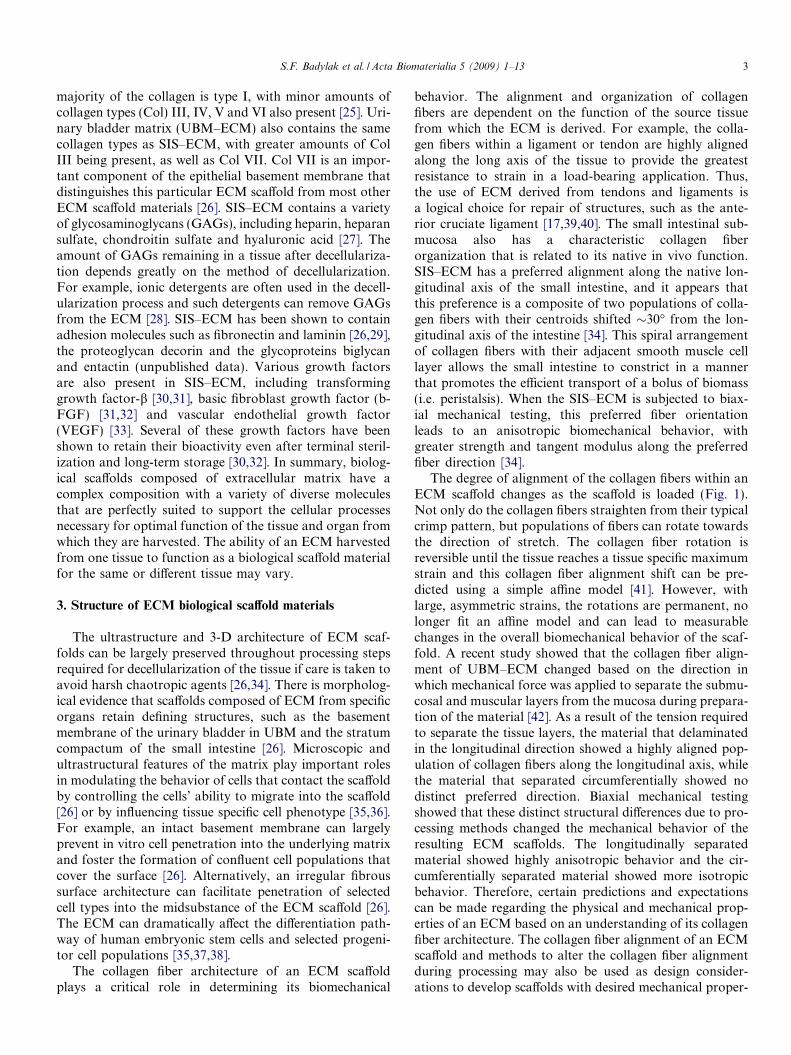
The degree of alignment of the collagen fibers within anECM scaffold changes as the scaffold is loaded (Fig. 1).Not only do the collagen fibers straighten from their typicalcrimp pattern, but populations of fibers can rotate towardsthe direction of stretch. The collagen fiber rotation isreversible until the tissue reaches a tissue specific maximumstrain and this collagen fiber alignment shift can be pre-dicted using a simple affine model [41]. However, withlarge, asymmetric strains, the rotations are permanent, nolonger fit an affine model and can lead to measurablechanges in the overall biomechanical behavior of the scaf-fold. A recent study showed that the collagen fiber align-ment of UBM–ECM changed based on the direction inwhich mechanical force was applied to separate the submu-cosal and muscular layers from the mucosa during prepara-tion of the material [42]. As a result of the tension requiredto separate the tissue layers, the material that delaminatedin the longitudinal direction showed a highly aligned pop-ulation of collagen fibers along the longitudinal axis, whilethe material that separated circumferentially showed nodistinct preferred direction. Biaxial mechanical testingshowed that these distinct structural differences due to pro-cessing methods changed the mechanical behavior of theresulting ECM scaffolds. The longitudinally separatedmaterial showed highly anisotropic behavior and the cir-cumferentially separated material showed more isotropicbehavior. Therefore, certain predictions and expectationscan be made regarding the physical and mechanical prop-erties of an ECM based on an understanding of its collagenfiber architecture. The collagen fiber alignment of an ECMscaffold and methods to alter the collagen fiber alignmentduring processing may also be used as design consider-ations to develop scaffolds with desired mechanical proper-
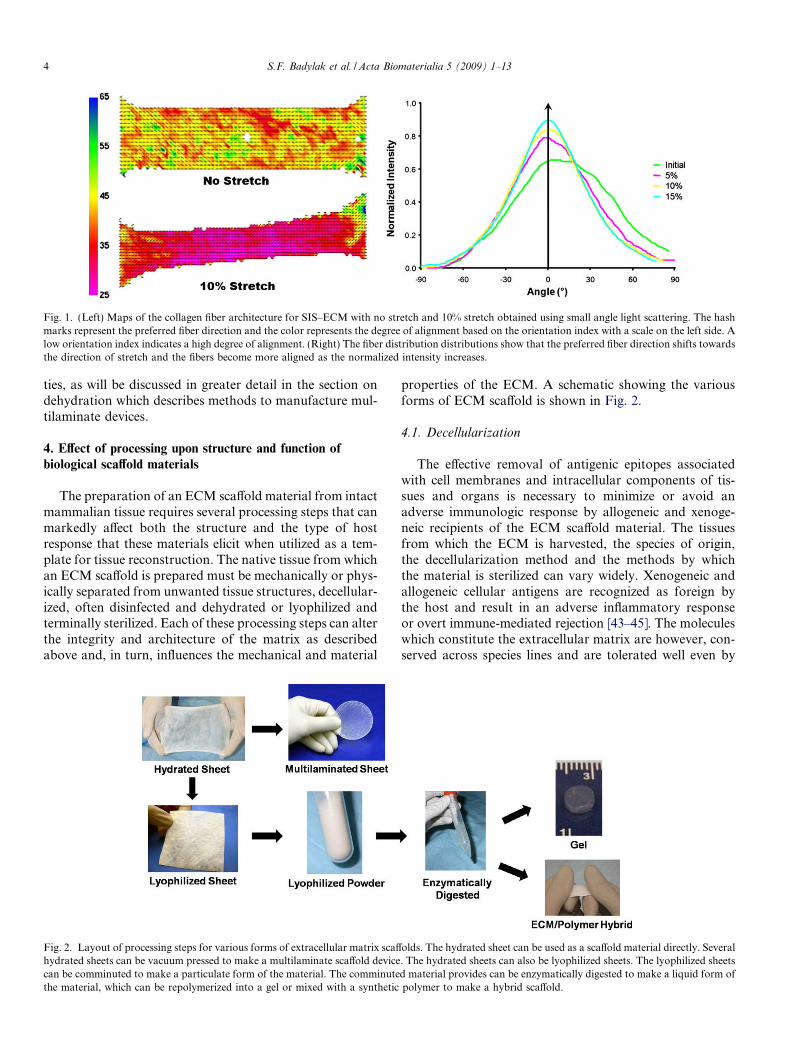
Fig. 1. (Left) Maps of the collagen fiber architecture for SIS–ECM with no stretch and 10% stretch obtained using small angle light scattering. The hashmarks represent the preferred fiber direction and the color represents the degree of alignment based on the orientation index with a scale on the left side. Alow orientation index indicates a high degree of alignment. (Right) The fiber distribution distributions show that the preferred fiber direction shifts towardsthe direction of stretch and the fibers become more aligned as the normalized intensity increases.
4 S.F. Badylak et al. / Acta Biomaterialia 5 (2009) 1–13
ties, as will be discussed in greater detail in the section ondehydration which describes methods to manufacture mul-tilaminate devices.
4. Effect of processing upon structure and function of
biological scaffold materials
The preparation of an ECM scaffold material from intactmammalian tissue requires several processing steps that canmarkedly affect both the structure and the type of hostresponse that these materials elicit when utilized as a tem-plate for tissue reconstruction. The native tissue from whichan ECM scaffold is prepared must be mechanically or phys-ically separated from unwanted tissue structures, decellular-ized, often disinfected and dehydrated or lyophilized andterminally sterilized. Each of these processing steps can alterthe integrity and architecture of the matrix as describedabove and, in turn, influences the mechanical and material
Fig. 2. Layout of processing steps for various forms of extracellular matrix scaffhydrated sheets can be vacuum pressed to make a multilaminate scaffold devicecan be comminuted to make a particulate form of the material. The comminutethe material, which can be repolymerized into a gel or mixed with a synthetic
properties of the ECM. A schematic showing the variousforms of ECM scaffold is shown in Fig. 2.
4.1. Decellularization
The effective removal of antigenic epitopes associatedwith cell membranes and intracellular components of tis-sues and organs is necessary to minimize or avoid anadverse immunologic response by allogeneic and xenoge-neic recipients of the ECM scaffold material. The tissuesfrom which the ECM is harvested, the species of origin,the decellularization method and the methods by whichthe material is sterilized can vary widely. Xenogeneic andallogeneic cellular antigens are recognized as foreign bythe host and result in an adverse inflammatory responseor overt immune-mediated rejection [43–45]. The moleculeswhich constitute the extracellular matrix are however, con-served across species lines and are tolerated well even by
olds. The hydrated sheet can be used as a scaffold material directly. Several. The hydrated sheets can also be lyophilized sheets. The lyophilized sheetsd material provides can be enzymatically digested to make a liquid form ofpolymer to make a hybrid scaffold.
S.F. Badylak et al. / Acta Biomaterialia 5 (2009) 1–13 5
xenogeneic recipients [46–49]. Certain antigens, such as thegalactosyl-a-1,3-galactose, have been shown to be presentin porcine SIS–ECM but fail to activate complement orbind IgM antibody, presumably because of the smallamount and widely scattered distribution of the antigen[50,51]. The ultimate goal of any decellularization protocolis to remove all cellular material without adversely affectingthe composition, mechanical integrity and eventual biolog-ical activity of the remaining ECM. Commonly used meth-ods of decellularization include a combination of physicaland chemical treatments. Sonication, agitation and freezingand thawing are commonly used methods to disrupt cellmembranes, release cell contents and facilitate the subse-quent rinsing and removal of cell remnants from theECM. The commonly used decellularization methodsappear to be insufficient to achieve complete decellulariza-tion, as most, if not all, ECM scaffold materials retain someDNA [52,53]. Although it seems logical that the decellular-ization process will by definition affect the structure andcomposition of the extracellular matrix, the intent of theprocess is the preservation of as much of the nativemechanical properties and biological properties of the ori-ginal ECM as possible. Some detergents used to facilitatedecellularization have been shown to disrupt collagen ofcertain tissues, thereby decreasing the mechanical strengthof the tissue, while the same detergent may have no effecton the collagen in another tissue [17,39]. Studies haveshown that removal of GAGs from the scaffold can havea negative effect on viscoelastic behavior of the scaffold,which is not surprising since water retention is one of themajor functional characteristics of GAGs within a tissue[54]. Therefore, the decellularization method requires opti-mization for each tissue to remove cellular material withoutcompromising the mechanical properties of the tissue.
4.2. Hydration
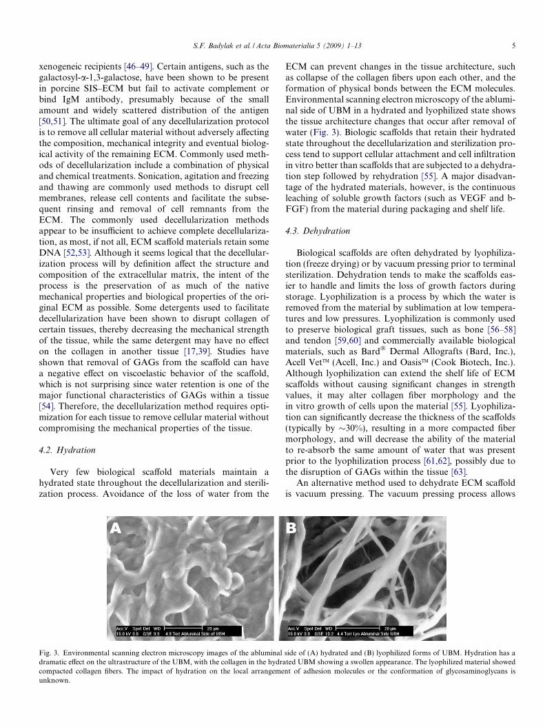
Very few biological scaffold materials maintain ahydrated state throughout the decellularization and sterili-zation process. Avoidance of the loss of water from the
Fig. 3. Environmental scanning electron microscopy images of the abluminaldramatic effect on the ultrastructure of the UBM, with the collagen in the hydracompacted collagen fibers. The impact of hydration on the local arrangemeunknown.
ECM can prevent changes in the tissue architecture, suchas collapse of the collagen fibers upon each other, and theformation of physical bonds between the ECM molecules.Environmental scanning electron microscopy of the ablumi-nal side of UBM in a hydrated and lyophilized state showsthe tissue architecture changes that occur after removal ofwater (Fig. 3). Biologic scaffolds that retain their hydratedstate throughout the decellularization and sterilization pro-cess tend to support cellular attachment and cell infiltrationin vitro better than scaffolds that are subjected to a dehydra-tion step followed by rehydration [55]. A major disadvan-tage of the hydrated materials, however, is the continuousleaching of soluble growth factors (such as VEGF and b-FGF) from the material during packaging and shelf life.
4.3. Dehydration
Biological scaffolds are often dehydrated by lyophiliza-tion (freeze drying) or by vacuum pressing prior to terminalsterilization. Dehydration tends to make the scaffolds eas-ier to handle and limits the loss of growth factors duringstorage. Lyophilization is a process by which the water isremoved from the material by sublimation at low tempera-tures and low pressures. Lyophilization is commonly usedto preserve biological graft tissues, such as bone [56–58]and tendon [59,60] and commercially available biologicalmaterials, such as Bard� Dermal Allografts (Bard, Inc.),Acell VetTM (Acell, Inc.) and OasisTM (Cook Biotech, Inc.).Although lyophilization can extend the shelf life of ECMscaffolds without causing significant changes in strengthvalues, it may alter collagen fiber morphology and thein vitro growth of cells upon the material [55]. Lyophiliza-tion can significantly decrease the thickness of the scaffolds(typically by �30%), resulting in a more compacted fibermorphology, and will decrease the ability of the materialto re-absorb the same amount of water that was presentprior to the lyophilization process [61,62], possibly due tothe disruption of GAGs within the tissue [63].
An alternative method used to dehydrate ECM scaffoldis vacuum pressing. The vacuum pressing process allows
side of (A) hydrated and (B) lyophilized forms of UBM. Hydration has ated UBM showing a swollen appearance. The lyophilized material showed
nt of adhesion molecules or the conformation of glycosaminoglycans is

Fig. 4. Multilaminate form of UBM shaped to match the gastroesophageal junction and the esophagus.
6 S.F. Badylak et al. / Acta Biomaterialia 5 (2009) 1–13
for the lamination of multiple sheets of ECM to increasethe strength and/or design-in specific mechanical behaviorbased upon knowledge of the collagen fiber architecture.For example, Restore� (DePuy Orthopaedics) is con-structed of 10 layers of SIS–ECM, with two layers orientedevery 72�. The resulting sheet provides strength at the timeof implantation that exceeds the strength of a native rota-tor cuff tendon tissue (i.e. the tissue for which it was orig-inally intended to function as a biological reinforcementscaffold) and imparts isotropy to natively anisotropic mate-rials. Lamination via vacuum pressing of ECM scaffoldsalso reduces the extensibility, and changes the ultrastruc-tural morphology of the resulting construct [22,64]. Vac-uum pressing is an effective method for constructing avariety of 3-D shapes of ECM scaffold materials (Fig. 4).
Although constructive in vivo remodeling has beenobserved with the use of hydrated, lyophilized and multila-minated forms of ECM scaffolds [65–71], ultrastructuralchanges that occur as a result of dehydration can affect cellattachment, in vivo degradation rate and cellular infiltra-tion [55]. The optimal configuration and method of pro-cessing of an ECM scaffold should be determined foreach clinical application.
4.4. Powdered ECM scaffolds
Lyophilized sheets of ECM can be comminuted into anECM powder or particulate form [23]. A particle formallows for the delivery of the ECM as a suspension via min-imally invasive techniques (e.g. needle injection) to the siteof interest or for the manufacture of 3-D scaffolds by com-paction methods. The particles present in comminutedECM retain the ultrastructural, 3-D surface characteristicsof the parent ECM sheet. Particle sizes ranging from 50 to200 lm in diameter can be reproducibly manufactured.Suspensions made from a particulate (powdered) form oflyophilized UBM have been successfully used as an inject-able scaffold for the treatment of urinary incontinence inpreclinical studies [72], but the needle size required toaccommodate passage of the particles is prohibitive (i.e.too large external diameter) for many clinical applications.Carriers such as glycerin are typically required to increase
the viscosity of a suspension of particles intended for clin-ical use. Acellular human dermal matrix has been investi-gated as a micronized form for injection into laryngealtissue, but tissue augmentation is not possible due to itsrapid in vivo degradation [73]. Powdered forms of ECMscaffolds may also be used for topical delivery or may becombined with synthetic polymers to create hybrid scaf-folds. Since a particulate form of such scaffold materialsare not expected to serve load-bearing functions, the phys-ical properties of the ECM, such as particle size, surfacearea and type of liquid carrier, are the relevant variablesthat affect the ease and convenience with which the mate-rial can be delivered to the intended site.
4.5. Gel form of ECM scaffolds
A liquid or gel form of ECM can further expand the clin-ical utility of an ECM scaffold by allowing the delivery ofthe material via minimally invasive methods to sites of inter-est. Stated differently, a gel form can be delivered to a site ofinterest in its pre-gel liquid state more readily than a suspen-sion of particles. The solubilized ECM can be delivered bycatheter or needle-based surgical techniques to irregularlyshaped anatomic sites. A gel form can also serve as a celldelivery vehicle when appropriate. The rheological proper-ties of the gel can be designed to be similar to those of thetissue that is being repaired. Ideally, the gel processingmethods would minimize or avoid purification steps thatcould remove or destroy the active growth factors andlow molecular weight peptides present in the native ECM,and the gel form would retain the native bioactivity of theparent ECM scaffold. Previous studies have shown that agel form of an ECM derived from SIS can be produced thatis able to support the growth and differentiation of a varietyof cells in vitro, but the preparation of this gel form requiredan aggressive collagen purification process that likelyresulted in the loss of bioactive molecules [74].
Recently, a preparation of a gel derived from UBM hasbeen reported in which no purification steps were neces-sary. Lyophilized UBM powder was enzymatically (i.e.pepsin) digested at low pH resulting in a viscous solution.A pepsin-digested form of UBM–ECM was able to self-
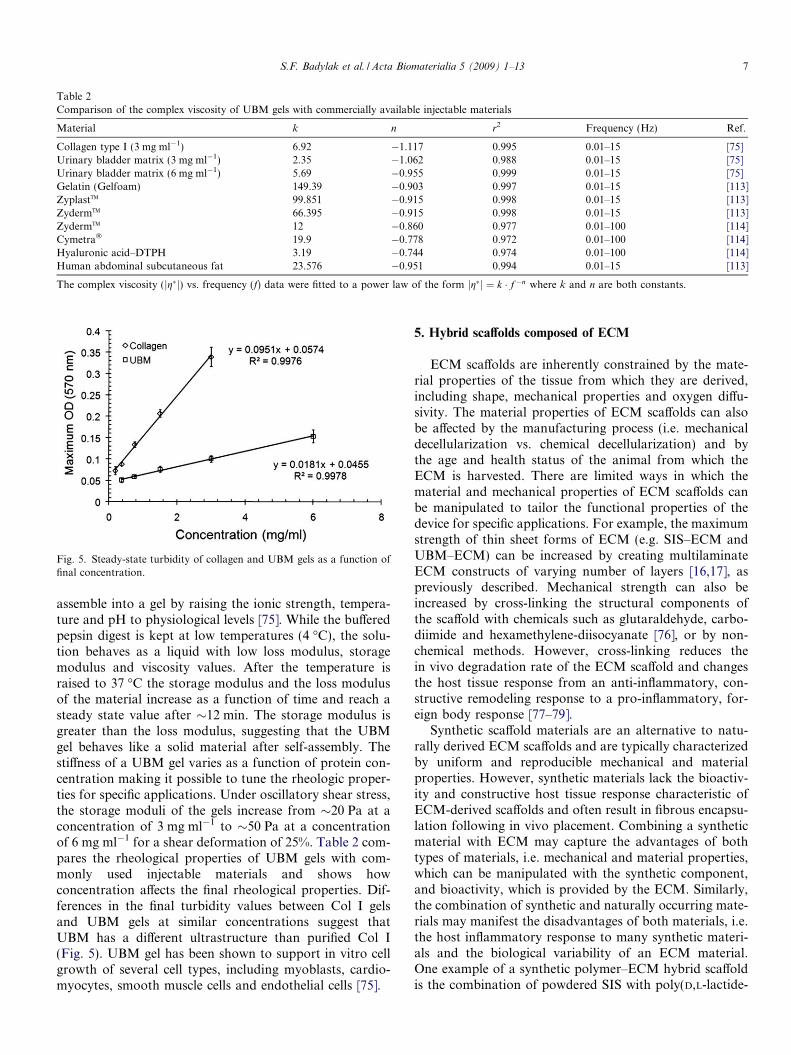
Table 2Comparison of the complex viscosity of UBM gels with commercially available injectable materials
Material k n r2 Frequency (Hz) Ref.
Collagen type I (3 mg ml�1) 6.92 �1.117 0.995 0.01–15 [75]Urinary bladder matrix (3 mg ml�1) 2.35 �1.062 0.988 0.01–15 [75]Urinary bladder matrix (6 mg ml�1) 5.69 �0.955 0.999 0.01–15 [75]Gelatin (Gelfoam) 149.39 �0.903 0.997 0.01–15 [113]ZyplastTM 99.851 �0.915 0.998 0.01–15 [113]ZydermTM 66.395 �0.915 0.998 0.01–15 [113]ZydermTM 12 �0.860 0.977 0.01–100 [114]Cymetra� 19.9 �0.778 0.972 0.01–100 [114]Hyaluronic acid–DTPH 3.19 �0.744 0.974 0.01–100 [114]Human abdominal subcutaneous fat 23.576 �0.951 0.994 0.01–15 [113]
The complex viscosity (jg�j) vs. frequency (f) data were fitted to a power law of the form jg�j ¼ k � f�n where k and n are both constants.
Fig. 5. Steady-state turbidity of collagen and UBM gels as a function offinal concentration.
S.F. Badylak et al. / Acta Biomaterialia 5 (2009) 1–13 7
assemble into a gel by raising the ionic strength, tempera-ture and pH to physiological levels [75]. While the bufferedpepsin digest is kept at low temperatures (4 �C), the solu-tion behaves as a liquid with low loss modulus, storagemodulus and viscosity values. After the temperature israised to 37 �C the storage modulus and the loss modulusof the material increase as a function of time and reach asteady state value after �12 min. The storage modulus isgreater than the loss modulus, suggesting that the UBMgel behaves like a solid material after self-assembly. Thestiffness of a UBM gel varies as a function of protein con-centration making it possible to tune the rheologic proper-ties for specific applications. Under oscillatory shear stress,the storage moduli of the gels increase from �20 Pa at aconcentration of 3 mg ml�1 to �50 Pa at a concentrationof 6 mg ml�1 for a shear deformation of 25%. Table 2 com-pares the rheological properties of UBM gels with com-monly used injectable materials and shows howconcentration affects the final rheological properties. Dif-ferences in the final turbidity values between Col I gelsand UBM gels at similar concentrations suggest thatUBM has a different ultrastructure than purified Col I(Fig. 5). UBM gel has been shown to support in vitro cellgrowth of several cell types, including myoblasts, cardio-myocytes, smooth muscle cells and endothelial cells [75].
5. Hybrid scaffolds composed of ECM
ECM scaffolds are inherently constrained by the mate-rial properties of the tissue from which they are derived,including shape, mechanical properties and oxygen diffu-sivity. The material properties of ECM scaffolds can alsobe affected by the manufacturing process (i.e. mechanicaldecellularization vs. chemical decellularization) and bythe age and health status of the animal from which theECM is harvested. There are limited ways in which thematerial and mechanical properties of ECM scaffolds canbe manipulated to tailor the functional properties of thedevice for specific applications. For example, the maximumstrength of thin sheet forms of ECM (e.g. SIS–ECM andUBM–ECM) can be increased by creating multilaminateECM constructs of varying number of layers [16,17], aspreviously described. Mechanical strength can also beincreased by cross-linking the structural components ofthe scaffold with chemicals such as glutaraldehyde, carbo-diimide and hexamethylene-diisocyanate [76], or by non-chemical methods. However, cross-linking reduces thein vivo degradation rate of the ECM scaffold and changesthe host tissue response from an anti-inflammatory, con-structive remodeling response to a pro-inflammatory, for-eign body response [77–79].
Synthetic scaffold materials are an alternative to natu-rally derived ECM scaffolds and are typically characterizedby uniform and reproducible mechanical and materialproperties. However, synthetic materials lack the bioactiv-ity and constructive host tissue response characteristic ofECM-derived scaffolds and often result in fibrous encapsu-lation following in vivo placement. Combining a syntheticmaterial with ECM may capture the advantages of bothtypes of materials, i.e. mechanical and material properties,which can be manipulated with the synthetic component,and bioactivity, which is provided by the ECM. Similarly,the combination of synthetic and naturally occurring mate-rials may manifest the disadvantages of both materials, i.e.the host inflammatory response to many synthetic materi-als and the biological variability of an ECM material.One example of a synthetic polymer–ECM hybrid scaffoldis the combination of powdered SIS with poly(D,L-lactide-
8 S.F. Badylak et al. / Acta Biomaterialia 5 (2009) 1–13
co-glycolide) to create tissue-engineered bone [80]. Anotherexample includes the combination of UBM with a poly(e-ster-urethane)urea (PEUU) to create an elastomeric hybridscaffold [81]. The combination of PEUU with UBM resultsin an elastomeric material with increased maximum stiff-ness, strength and strain when compared to lyophilizedUBM sheets. When implanted in a subcutaneous location,hybrid scaffolds degrade faster than purified PEUU butslower than lyophilized UBM sheets [81].
6. Terminal sterilization
Several studies have shown that terminal sterilization ofECM scaffolds can have a detrimental effect on themechanical properties of the scaffold. Recently, it wasshown that UBM-ECM had decreased uniaxial and biaxialmechanical properties after exposure to ethylene oxide(750 mg h�1), gamma irradiation (20 kGy) and electronbeam irradiation (22 kGy) [82]. Several studies have alsoinvestigated the effects of a wide range of gamma irradia-tion exposure on dermis ECM [83,84]. At low dosages ofgamma irradiation (<15 kGy), the strength and modulusof the scaffold increased, but the mechanical propertiesdecreased above 15 kGy in a dose-dependent manner[83]. These changes appear to be due to an increase in col-lagen cross-linking due to the low dose of irradiation thatlevels off after approximately 5 kGy, along with collagenchain scission that continues to increase with the irradia-tion dose [84]. The mechanisms for changes in mechanicalproperties following ethylene oxide and electron beam irra-diation have not been studied in detail.
7. Changes in mechanical behavior of ECM scaffolds during
in vivo remodeling
The mechanical behavior of ECM scaffolds changes dur-ing the process of in vivo remodeling [85], and such changesare dependent on factors such as the local tissue microenvi-ronment, the rate of scaffold degradation, forces presentwithin the mechanical environment, and the rate and extentto which the infiltrating cells deposit new ECM. The moststudied ECM scaffold during the in vivo remodeling pro-cess is SIS–ECM. In a canine model of Achilles tendonreconstruction, a segmental defect was created andreplaced with a tube of SIS–ECM (native geometry, notlaminated) [25]. The stifle joint was immobilized for5 weeks, but weight bearing was allowed. At 1 week afterimplantation, the strength of the tendon repaired withSIS–ECM was less than 100 N, or 10% of the originalstrength. As new collagenous connective tissue was formed,the strength of the remodeled SIS–ECM implant tissuegradually increased until the strength exceeded that of thenative musculotendinous junction and the insertion intothe calcaneus by 12 weeks after surgery [25]. In a goatmodel of anterior cruciate ligament reconstruction, aremodeled SIS–ECM scaffold showed a decreased strengthat 3 months after surgery, followed by an increase to a level
comparable to a patellar tendon autograft at 1 year [86]. Ina dog model of rotator cuff reconstruction, a 10-layer mul-tilaminate device (RestoreTM) was used to replace a com-pletely resected infraspinatus tendon, and the animalswere allowed unlimited cage activity after surgery [87].Although no mechanical data was presented for the earlyphase of remodeling, the failure loads of the tendonrepaired with SIS–ECM eventually increased by approxi-mately threefold from the time of repair, and were compa-rable to failure loads for rotator cuffs subjected to aprimary repair, despite having a cross-sectional area thatwas half the size [87]. In addition, when SIS–ECM wasused to repair the gap defect in the medial collateral liga-ment of a rabbit knee, the strength at failure and modulusof the ligament substance were increased at both 12 and26 weeks of healing compared to no treatment [88,89].The improved mechanical properties of the remodeled tis-sue compared to new host tissue that formed with no treat-ment is thought to be a result of decreased expression andsynthesis of Col V and various proteoglycans, increasedcollagen fibril diameter and increased collagen fiber align-ment in the ligament repaired with SIS–ECM [88,90,91].Clearly and logically, the structural changes that occur dur-ing in vivo remodeling of ECM scaffolds are associatedwith marked changes in scaffold strength.
The mechanical behavior of remodeled ECM scaffoldshas also been evaluated in a number of muscular tissueapplications. An eight-layer SIS–ECM device used torepair the abdominal body wall (skeletal muscle) of a dogdecreased to 50% of its initial strength by 10 days afterimplantation, but subsequently increased to twice its initialstrength by 24 months [85]. After repair of the canine uri-nary bladder (smooth muscle) with a three-layer SIS–ECM device [92], it was found that the compliance of theremodeled graft increased substantially compared to thedevice at the time of reconstruction (30-fold difference),to levels that were not statistically different from the nor-mal urinary bladder. Furthermore, the remodeled SIS–ECM showed contractility responses that were similar tothe normal bladder as a result of innervated muscle forma-tion. UBM–ECM has been used to repair canine esophagus(smooth muscle) and myocardium (cardiac muscle) [93,94].For the esophageal repair, the ECM graft rapidly remod-eled to approach the pressure-diameter response and com-pliance of the adjacent native esophagus within 90 days[93]. The newly formed esophageal tissue also showed peri-staltic activity, although the muscle contraction was asyn-chronous with the adjacent native esophagus [93]. In astudy of myocardial repair in a canine model, a single layerof ECM (�100 lm) remodeled and showed evidence ofcontractility and improved regional function of the heartwithin 8 weeks [94].
These examples show that in the short term afterimplantation, ECM scaffolds typically show a decrease instrength that is temporally associated with the rapidin vivo degradation of the scaffold [68]. Quantitative assess-ment of scaffold degradation has been performed in both

Fig. 6. Schematic of the mechanical contribution of an ECM scaffold overtime as it degrades and the mechanical contribution of the new host tissueas it forms during ECM remodeling in the presence of appropriatemechanical loading.
S.F. Badylak et al. / Acta Biomaterialia 5 (2009) 1–13 9
canine urinary bladder and Achilles tendon applications,with similar results in both cases. Degradation of SIS–ECM occurs rapidly, with nearly 50% of the scaffolddegraded by 1 month and complete degradation by3 months [68,95] (Fig. 6). These degradation kinetics pro-vide insight into the rate of new matrix production. Inthe early phase of remodeling, degradation occurs quiterapidly, before newly deposited ECM has the opportunityto fully organize, thus the initial decrease in scaffoldstrength. However, once the infiltrating cells have estab-lished residence and begin producing site-specific newECM, rapid scaffold remodeling occurs, with an increasein the strength and site-appropriate mechanical behavior[85,93] (Fig. 6). In the application of esophageal recon-struction, the graft became more compliant, while in theapplication of tendon or ligament reconstruction, the loadto failure increased.
8. Role of mechanical loading on ECM remodeling
In contrast to the fact that most preclinical studies withSIS–ECM successfully utilized early mobilization, someearly clinical applications of SIS–ECM used immobiliza-tion and non-weight-bearing conditions following surgeryto protect the scaffold material during the phase of rapidECM degradation. To determine the effects of immobiliza-tion on the remodeling of an SIS–ECM scaffold, a studywas performed in which an SIS–ECM scaffold was usedto repair a segmental defect in a rabbit Achilles tendonwith different immobilization protocols [96]. Rabbits wereseparated into five groups. In four groups, a 1.5 cm sectionof the Achilles tendon was surgically removed and repairedwith an SIS–ECM interpositional graft, after which thesurgically repaired limb was immobilized for 2 weeks.The first group of animals was sacrificed at the end of2 weeks as a control. The other three groups had their hind
limb braced to allow full range of motion, partial range ofmotion (60–90� of flexion) or no range of motion for anadditional 4 weeks, at which point all animals were sacri-ficed. In the control group, a sham operation was per-formed in which the Achilles tendon was exposed, but nodefect was created and no SIS–ECM material wasimplanted. The sham-operated hind limb was immobilizedfor 2 weeks prior to sacrifice. Histological analysis of thegroups with a partial or full range of motion showed densecollagenous connective tissue oriented along the longitudi-nal axis of the tendon. Spindle-shaped cells were distrib-uted throughout the tendon and oriented along thelongitudinal axis of the tendon. The only differencebetween the partial and full range of motion groups wasthat fewer cells were found in the center of the remodeledgraft with partial motion.
The SIS–ECM remodeling process showed an entirelydifferent morphologic response when no period of remobi-lization followed immobilization of the limb. In all ani-mals, there was histologic evidence of the SIS–ECMscaffold at the defect site with only limited deposition ofdisorganized new host connective tissue. The cellularitywas limited to the periphery of the graft, with almost nocells found in the middle of the device. In summary, activeloading of a remodeling ECM scaffold accelerates theremodeling process and results in the formation of arobust, site-appropriate tissue.
In vitro models are now being developed that willincrease our understanding of the role of mechanical load-ing in the constructive remodeling response observed withECM scaffolds in vivo. A recent study showed that cyclicuniaxial stretching of fibroblasts seeded on the SIS–ECMscaffold led to increases in the expression of Col I, whilethe expression of Col III decreased slightly [97]. In vitrostudies that have investigated the expression of Col I andCol III by fibroblasts seeded on silicone substrates haveshown that expression of both genes is increased inresponse to mechanical stimuli [98–100]. The SIS–ECMenvironment in the presence of mechanical loading appearsto facilitate a more normal Col III to Col I ratio [101] and amore normal distribution of collagen fibril diameters in thehealing tissue [102–104] compared to the collagen fibrildiameter that is present in the absence of mechanical load-ing. In a separate in vitro study that investigated the effectsof cyclic uniaxial stretching on fibroblasts seeded on SIS–ECM, the constructs showed increased stiffness, probablydue in part to new collagen synthesis and reorganizationof existing collagen [106]. The findings of these in vitrostudies may partially explain the improved mechanicalproperties that have been reported in in vivo studies inwhich physiological mechanical loading was allowed dur-ing the remodeling process [88,89]. Future studies mayprovide additional information on the effects of themechanical environment of other cell types that have beenshown to be important contributors to site-specific remod-eling, such as macrophages [78] and bone marrow-derivedcells [65,107,108].

10 S.F. Badylak et al. / Acta Biomaterialia 5 (2009) 1–13
9. Functions of solubilized/degraded biological scaffold
materials
Separate from the mechanical and structural functionsof biological scaffold materials are the biological activitiesassociated with the host tissue response. Biological scaf-folds composed of extracellular matrix have been shownto markedly affect angiogenesis, cell proliferation, cellmigration and cell differentiation. Such biological activi-ties are typically caused by cell signaling mechanisms thatinvolve soluble molecules. Scaffolds composed of ECMhave been shown to be rich in growth factors [31,33],bifunctional molecules such as fibronectin [29] and vari-ous types of collagen [25,26], among other structuraland functional molecules. More recently, degradationproducts of the parent ECM molecules have been shownto have significant biological activity themselves [109–112]. Stated differently, there is significant functionalactivity attributed to the degradation of the native scaf-fold structure and release of the inherent bioactive constit-uents. Unlike the mechanical and structural propertiesthat are dependent upon an intact 3-D structure, the bio-logical activities are in large part dependent upon just theopposite; that is, the degradation of the intact 3-Dstructure.
Processing methods that inhibit degradation of biologicalscaffolds, such as chemical cross-linking will significantlyalter its functional profile and therefore the host tissueresponse to the biological material. From this perspective,degradable biological scaffolds may be considered as con-trolled release devices for a variety of functional molecules.
The concept of functionality that is a result of scaffolddegradation by necessity implies that mechanical and struc-tural properties will be in a dynamic state. Accurate predic-tions of the biological functionality will depend upon anunderstanding of the rate of scaffold degradation, the com-position of the materials from which the biological scaf-folds are constructed, and the nature of degradationproducts and their local and systemic distribution follow-ing in vivo placement.
10. Summary
The consideration of structural and functional relation-ships of biological scaffolds includes an understanding ofthe 3-D architecture of biological materials, the biochemi-cal composition of such materials, the manufacturing pro-cesses involved in producing such materials, and, perhapsmost importantly, an understanding of the changes thatoccur with such materials following in vivo placementand host remodeling. Although this work largely describesthe structural and functional characteristics of SIS–ECMand UBM–ECM, the principles can apply to other ECM-based scaffold materials with variations depending on thetissue source of the ECM and processing methods. It isnow recognized that mammalian extracellular matrix rep-resents an excellent scaffold material suitable for many
therapeutic applications. The structural support and bio-logical signaling that allow ECM scaffolds to promote con-structive remodeling are likely the same characteristics thathave evolved for tissue homeostasis and repair and replace-ment following injury. The successful utilization of mam-malian ECM as a therapeutic device will depend in largepart upon our ability to understand and take advantageof the native structure/function relationships of the biolog-ical scaffold material.
References
[1] Bader A, Schilling T, Teebken OE, Brandes G, Herden T, SteinhoffG. Tissue engineering of heart valves – human endothelial cellseeding of detergent acellularized porcine valves. Eur J CardiothoracSurg 1998;14:279–84.
[2] Booth C, Korosis SA, Wilcox HE, Watterson KG, Kearney JN,Fisher J. Tissue engineering of cardiac valve prostheses I: develop-ment and histological characterization of an acellular porcinescaffold. J Heart Valve Dis 2002;11:457–62.
[3] Grauss RW, Hazekamp MG, Oppenhuizen F, van Munsteren CJ,Gittenberger-de-Groot AC, DeRuiter MC. Histological evaluationof decellularised porcine aortic valves: matrix changes due todifferent decellularisation methods. Eur J Cardiothorac Surg2005;27:566–71.
[4] Kasimir MT, Rieder E, Seebacher G, Silberhumer G, WolnerE, Weigel G. Comparison of different decellularization proce-dures of porcine heart valves. Int J Artif Organs 2003;26:421–7.
[5] Korossis SA, Booth C, Wilcox HE, Watterson KG, Kearney JN,Fisher J. Tissue engineering of cardiac valve prostheses II: biome-chanical characterization of decellularized porcine aortic heartvalves. J Heart Valve Dis 2002;11:463–71.
[6] Rieder E, Kasimir MT, Silberhumer G, Seebacher G, Wolner E,Simon P. Decellularization protocols of porcine heart valves differimportantly in efficiency of cell removal and susceptibility of thematrix to recellularization with human vascular cells. J ThoracCardiovasc Surg 2004;127:399–405.
[7] Schenke-Layland K, Vasilevski O, Opitz F, Konig K, Riemann I,Halbhuber KJ, et al. Impact of decellularization of xenogeneic tissueon extracellular matrix integrity for tissue engineering of heartvalves. J Struct Biol 2003;143(3):201–8, Sep.
[8] Conklin BS, Richter ER, Kreutziger KL, Zhong DS, Chen C.Development and evaluation of a novel decellularized vascularxenograft. Med Eng Phys 2002;24:173–83.
[9] Dahl SL, Koh J, Prabhakar V, Niklason LE. Decellularized nativeand engineering arterial scaffolds for transplantation. Cell Trans-plant 2003;12:659–66.
[10] Schmidt CE, Baier JM. Acellular vascular tissues: natural bioma-terials for tissue repair and tissue engineering. Biomaterials2000;21(22):2215–31.
[11] Uchimura E, Sawa Y, Taketani S, Yamanaka Y, Hara M, MatsudaH. Novel method of preparing acellular cardiovascular grafts bydecellularization with poly(ethylene glycol). J Biomed Mater Res A2003;67:834–7.
[12] Chen RN, Ho HO, Tsai YT, Sheu MT. Process development of anacellular dermal matrix (ADM) for biomedical applications. Bio-materials 2004;25:2679–86.
[13] Hudson TW, Liu SY, Schmidt CE. Engineering an improvedacellular nerve graft via optimized chemical processing. Tissue Eng2004;10:1346–58.
[14] Kim BS, Yoo JJ, Atala A. Peripheral nerve regeneration usingacellular nerve grafts. J Biomed Mater Res 2004;68A(2):201–9.
[15] Borschel GH, Dennis RG, Kuzon JWM. Contractile skeletal muscletissue-engineered on an acellular scaffold. Plast Reconstr Surg2004;113:595–602.
S.F. Badylak et al. / Acta Biomaterialia 5 (2009) 1–13 11
[16] Cartmell JS, Dunn MG. Effect of chemical treatment on tendoncellularity and mechanical properties. J Biomed Mater Res2000;49:134–40.
[17] Woods T, Gratzer PF. Effectiveness of three extraction techniques inthe development of a decellularized bone–anterior cruciate liga-ment–bone graft. Biomaterials 2005;26(35):7339–49, Dec.
[18] Badylak SF, Lantz GC, Coffey A, Geddes LA. Small intestinalsubmucosa as a large diameter vascular graft in the dog. J Surg Res1989;47(1):74–80.
[19] Badylak SF, Tullius R, Kokini K, Shelbourne KD, Klootwyk T,Voytik SL, et al. The use of xenogeneic small intestinal submucosaas a biomaterial for Achilles tendon repair in a dog model. Journalof Biomedical Materials Research 1995;29(8):977–85.
[20] Kropp BP, Eppley BL, Prevel CD, Rippy MK, Harruff RC, BadylakSF, et al. Experimental assessment of small intestinal submucosa asa bladder wall substitute. Urology 1995;46(3):396–400.
[21] Chen F, Yoo JJ, Atala A. Acellular collagen matrix as a possible ‘‘offthe shelf” biomaterial for urethral repair. Urology1999;54(3):407–10.
[22] Freytes DO, Badylak SF, Webster TJ, Geddes LA, Rundell AE.Biaxial strength of multilaminated extracellular matrix scaffolds.Biomaterials 2004;25(12):2353–61.
[23] Gilbert TW, Stolz DB, Biancaniello F, Simmons-Byrd A, BadylakSF. Production and characterization of ECM powder: implicationsfor tissue engineering applications. Biomaterials 2005;26(12):1431–5.
[24] Lin P, Chan WC, Badylak SF, Bhatia SN. Assessing porcine liver-derived biomatrix for hepatic tissue engineering. Tissue Eng2004;10(7–8):1046–53.
[25] Badylak SF, Tullius R, Kokini K, Shelbourne KD, Klootwyk T,Voytik SL, et al. The use of xenogeneic small intestinal submucosaas a biomaterial for Achilles tendon repair in a dog model. J BiomedMater Res 1995;29(8):977–85.
[26] Brown B, Lindberg K, Reing J, Stolz DB, Badylak SF. Thebasement membrane component of biologic scaffolds derived fromextracellular matrix. Tissue Eng 2006;12(3):519–26.
[27] Hodde JP, Badylak SF, Brightman AO, Voytik-Harbin SL.Glycosaminoglycan content of small intestinal submucosa: a bio-scaffold for tissue replacement. Tissue Eng 1996;2(3):209–17.
[28] Gilbert TW, Sellaro TL, Badylak SF. Decellularization of tissuesand organs. Biomaterials 2006;27(19):3675–83.
[29] Hodde JP, Record R, Tullius R, Badylak SF. Fibronectin peptidesmediate HMEC adhesion to porcine-derived extracellular matrix.Biomaterials 2002;23(8):1841–8.
[30] McDevitt CA, Wildey GM, Cutrone RM. Transforming growthfactor-beta1 in a sterilized tissue derived from the pig small intestinesubmucosa. J Biomed Mater Res A 2003;67(2):637–40.
[31] Voytik-Harbin SL, Brightman AO, Kraine MR, Waisner B, BadylakSF. Identification of extractable growth factors from small intestinalsubmucosa. J Cell Biochem 1997;67(4):478–91.
[32] Hodde JP, Ernst DM, Hiles MC. An investigation of the long-termbioactivity of endogenous growth factor in OASIS Wound Matrix. JWound Care 2005;14(1):23–5.
[33] Hodde JP, Record RD, Liang HA, Badylak SF. Vascular endothe-lial growth factor in porcine-derived extracellular matrix. Endothe-lium 2001;8(1):11–24.
[34] Sacks MS, Gloeckner DC. Quantification of the fiber architectureand biaxial mechanical behavior of porcine intestinal submucosa. JBiomed Mater Res 1999;46(1):1–10.
[35] Gong J, Sagiv O, Cai H, Tsang SH, Del Priore LV. Effects ofextracellular matrix and neighboring cells on induction of humanembryonic stem cells into retinal or retinal pigment epithelialprogenitors. Exp Eye Res 2008;86(6):957–65.
[36] Sellaro TL, Ravindra AK, Stolz DB, Badylak SF. Maintenance ofhepatic sinusoidal endothelial cell phenotype in vitro using organ-specific extracellular matrix scaffolds. Tissue Eng2007;13(9):2301–10.
[37] Hosokawa T, Betsuyaku T, Nishimura M, Furuyama A, Katagiri K,Mochitate K. Differentiation of tracheal basal cells to ciliated cells
and tissue reconstruction on the synthesized basement membranesubstratum in vitro. Connect Tissue Res 2007;48(1):9–18.
[38] Hosokawa T, Betsuyaku T, Odajima N, Suzuki M, Mochitate K,Nasuhara Y, et al. Role of basement membrane in EMMPRIN/CD147 induction in rat tracheal epithelial cells. Biochem BiophysRes Commun 2008;368(2):426–32.
[39] Cartmell JS, Dunn MG. Development of cell-seeded patellar tendonallografts for anterior cruciate ligament reconstruction. Tissue Eng2004;10(7–8):1065–75.
[40] Harrison RD, Gratzer PF. Effect of extraction protocols andepidermal growth factor on the cellular repopulation of decellular-ized anterior cruciate ligament allografts. J Biomed Mater Res A2005;75(4):841–54.
[41] Gilbert TW, Sacks MS, Grashow JS, Woo SL-Y, Badylak SF,Chancellor MB. Fiber kinematics of small intestinal submucosaunder biaxial and uniaxial stretch. J Biomech Eng2006;128(6):890–8.
[42] Gilbert TW, Wognum S, Joyce EM, Freytes DO, Sacks MS,Badylak SF. Collagen fiber alignment and biaxial mechanicalbehavior of porcine urinary bladder derived extracellular matrix.Biomaterials 2008; doi:10.1016/j.biomaterials.2008.08.022.
[43] Erdag G, Morgan JR. Allogeneic vs xenogeneic immune reaction tobioengineered skin grafts. Cell Transplant 2004;13(6):701–12.
[44] Gock H, Murray-Segal L, Salvaris E, Cowan P, D’Apice AJ.Allogeneic sensitization is more effective than xenogeneic sensitiza-tion in eliciting Gal-mediated skin graft rejection. Transplantation2004;77(5):751–3.
[45] Ross JR, Kirk AD, Ibrahim SE, Howell DN, Baldwin 3rd WM,Sanfilippo FP. Characterization of human anti-porcine ‘‘naturalantibodies” recovered from ex vivo perfused hearts–predominanceof IgM and IgG2. Transplantation 1993;55(5):1144–50.
[46] Bernard MP, Chu ML, Myers JC, Ramirez F, Eikenberry EF,Prockop DJ. Nucleotide sequences of complementary deoxyribonu-cleic acids for the pro alpha 1 chain of human type I procollagen.Statistical evaluation of structures that are conserved duringevolution. Biochemistry 1983;22(22):5213–23.
[47] Bernard MP, Myers JC, Chu ML, Ramirez F, Eikenberry EF,Prockop DJ. Structure of a cDNA for the pro alpha 2 chain ofhuman type I procollagen. Comparison with chick cDNA for proalpha 2(I) identifies structurally conserved features of the proteinand the gene. Biochemistry 1983;22(5):1139–45.
[48] Constantinou CDJ, Jimenez SA. Structure of cDNAs encoding thetriple-helical domain of murine alpha 2 (VI) collagen chain andcomparison to human and chick homologues. Use of polymerasechain reaction and partially degenerate oligonucleotide for genera-tion of novel cDNA clones. Matrix 1991;11(1):1–9.
[49] Exposito JY, D’Alessio M, Solursh M, Ramirez F. Sea urchincollagen evolutionarily homologous to vertebrate pro-alpha 2(I)collagen. J Biol Chem 1992;267(22):15559–62.
[50] McPherson TB, Liang H, Record RD, Badylak SF. Galalpha(1,3)Gal epitope in porcine small intestinal submucosa. Tissue Eng2000;6(3):233–9.
[51] Raeder RH, Badylak SF, Sheehan C, Kallakury B, Metzger DW.Natural anti-galactose alpha1,3 galactose antibodies delay, but donot prevent the acceptance of extracellular matrix xenografts.Transplant Immunol 2002 Jun;10(1):15–24.
[52] Derwin KA, Baker AR, Spragg RK, Leigh DR, Iannotti JP.Commercial extracellular matrix scaffolds for rotator cuff tendonrepair. Biomechanical, biochemical and cellular properties. J BoneJoint Surg Am 2006;88(12):2665–72.
[53] Gilbert TW, Freund JM, Badylak SF. Quantification of DNA inbiologic scaffold materials. J Surg Res 2008; doi:10.1016/j.jss.2008.02.013.
[54] Lovekamp JJ, Simionescu DT, Mercuri JJ, Zubiate B, Sacks MS,Vyavahare NR. Stability and function of glycosaminoglycans inporcine bioprosthetic heart valves. Biomaterials 2006;27(8):1507–18.
[55] Freytes DO, Tullius RS, Valentin JE, Stewart-Akers AM, BadylakSF. Hydrated versus lyophilized forms of porcine extracellular

12 S.F. Badylak et al. / Acta Biomaterialia 5 (2009) 1–13
matrix derived from the urinary bladder. J Biomed Mater Res A2008; doi:10.1002/jbm.a.31821.
[56] Burchardt H, Jones H, Glowczewskie F, Rudner C, Enneking WF.Freeze-dried allogeneic segmental cortical-bone grafts in dogs. JBone Joint Surg Am 1978;60(8):1082–90.
[57] Cornu O, Banse X, Docquier PL, Luyckx S, Delloye C. Effect offreeze–drying and gamma irradiation on the mechanical propertiesof human cancellous bone. J Orthop Res 2000;18(3):426–31.
[58] Jackson DW, Grood ES, Wilcox P, Butler DL, Simon TM, HoldenJP. The effects of processing techniques on the mechanical propertiesof bone–anterior cruciate ligament–bone allografts. An experimentalstudy in goats. Am J Sports Med 1988;16(2):101–5.
[59] Smith CW, Young IS, Kearney JN. Mechanical properties oftendons: changes with sterilization and preservation. J Biomech Eng1996;118(1):56–61.
[60] Toritsuka Y, Shino K, Horibe S, Nakamura N, Matsumoto N, OchiT. Effect of freeze-drying or gamma-irradiation on remodeling oftendon allograft in a rat model. J Orthop Res 1997;15(2):294–300.
[61] Hafeez YM, Zuki AB, Yusof N, Asnah H, Loqman MY, NoordinMM, et al. Effect of freeze-drying and gamma irradiation onbiomechanical properties of bovine pericardium. Cell Tissue Bank2005;6(2):85–9.
[62] Curtil A, Pegg DE, Wilson A. Freeze drying of cardiac valves inpreparation for cellular repopulation. Cryobiology1997;34(1):13–22.
[63] Gilbert TW, Gilbert S, Madden M, Reynolds SD, Badylak SF.Morphologic assessment of extracellular matrix scaffolds for patchtracheoplasty in a canine model. Ann Thorac Surg2008;86(3):967–74, discussion 967–974.
[64] Freytes DO, Rundell AE, Vande Geest J, Vorp DA, Webster TJ,Badylak SF. Analytically derived material properties of multilami-nated extracellular matrix devices using the ball-burst test. Bioma-terials 2005;26(27):5518–31.
[65] Zantop T, Gilbert TW, Yoder MC, Badylak SF. Extracellularmatrix scaffolds are repopulated by bone marrow-derived cells in amouse model of Achilles tendon reconstruction. J Orthop Res2006;24(6):1299–309.
[66] Ringel RL, Kahane JC, Hillsamer PJ, Lee AS, Badylak SF. Theapplication of tissue engineering procedures to repair the larynx. JSpeech Lang Hear Res 2006;49(1):194–208.
[67] Huber JE, Spievack A, Simmons-Byrd A, Ringel RL, Badylak S.Extracellular matrix as a scaffold for laryngeal reconstruction. AnnOtol Rhinol Laryngol 2003;112(5):428–33.
[68] Gilbert TW, Stewart-Akers AM, Simmons-Byrd A, Badylak SF.Degradation and remodeling of small intestinal submucosa in canineAchilles tendon repair. J Bone Joint Surg Am 2007;89(3):621–30.
[69] Bertone AL, Goin S, Kamei SJ, Mattoon JS, Litsky AS, WeisbrodeSE, et al. Metacarpophalangeal collateral ligament reconstructionusing small intestinal submucosa in an equine model. J BiomedMater Res A 2008;84(1):219–29.
[70] Badylak S, Kokini K, Tullius B, Whitson B. Strength over time of aresorbable bioscaffold for body wall repair in a dog model. J SurgRes 2001;99(2):282–7.
[71] Badylak S, Kokini K, Tullius B, Simmons-Byrd A, Morff R.Morphologic study of small intestinal submucosa as a body wallrepair device. J Surg Res 2002;103(2):190–202.
[72] Wood JD, Simmons-Byrd A, Spievack AR, Badylak SF. Use of aparticulate extracellular matrix bioscaffold for treatment of acquiredurinary incontinence in dogs. J Am Vet Med Assoc2005;226(7):1095–7.
[73] Lundy DS, Casiano RR, McClinton ME, Xue JW. Early results oftranscutaneous injection laryngoplasty with micronized acellulardermis versus type-I thyroplasty for glottic incompetence dysphoniadue to unilateral vocal fold paralysis. J Voice 2003;17(4):589–95.
[74] Brightman AO, Rajwa BP, Sturgis JE, McCallister ME, RobinsonJP, Voytik-Harbin SL. Time-lapse confocal reflection microscopy ofcollagen fibrillogenesis and extracellular matrix assembly in vitro.Biopolymers 2000;54(3):222–34.
[75] Freytes DO, Martin J, Velankar SS, Lee AS, Badylak SF.Preparation and rheological characterization of a gel form of theporcine urinary bladder matrix. Biomaterials 2008;29(11):1630–7.
[76] Ratner BD. Biomaterials science: an introduction to materials inmedicine. 2nd ed. Amsterdam: Elsevier Academic Press; 2004.
[77] Badylak SF, Valentin JE, Ravindra AK, McCabe GP, Stewart-Akers AM. Macrophage phenotype as a determinant of biologicscaffold remodeling. Tissue Eng Part A 2008; doi:10.1089/ten.tea.2007.0264.
[78] Badylak SF, Gilbert TW. Immune response to biologic scaffoldmaterials. Semin Immunol 2008;20(2):109–16.
[79] Valentin JE, Badylak JS, McCabe GP, Badylak SF. Extracellularmatrix bioscaffolds for orthopaedic applications. A comparativehistologic study. J Bone Joint Surg Am 2006;88(12):2673–86.
[80] Lee SJ, Lee IW, Lee YM, Lee HB, Khang G. Macroporousbiodegradable natural/synthetic hybrid scaffolds as small intestinesubmucosa impregnated poly(D,L-lactide-co-glycolide) for tissue-engineered bone. J Biomater Sci Polym Ed 2004;15(8):1003–17.
[81] Stankus JJ, Freytes DO, Badylak SF, Wagner WR. Hybridnanofibrous scaffolds from electrospinning of a synthetic biodegrad-able elastomer and urinary bladder matrix. J Biomater Sci Polym Ed2008;19(5):635–52.
[82] Freytes DO, Stoner RM, Badylak SF. Uniaxial and biaxialproperties of terminally sterilized porcine urinary bladder matrixscaffolds. J Biomed Mater Res B Appl Biomater 2008;84(2):408–14.
[83] Gouk SS, Lim TM, Teoh SH, Sun WQ. Alterations of humanacellular tissue matrix by gamma irradiation: histology, biomechan-ical property, stability, in vitro cell repopulation, and remodeling. JBiomed Mater Res B Appl Biomater 2008;84(1):205–17.
[84] Sun WQ, Leung P. Calorimetric study of extracellular tissue matrixdegradation and instability after gamma irradiation. Acta Biomater2008;4(4):817–26.
[85] Badylak SF, Kokini K, Tullius B, Whitson B. Strength over time ofa resorbable bioscaffold for body wall repair in a dog model. J SurgRes 2001;99(2):282–7.
[86] Badylak SF, Arnoczky S, Plouhar P, Haut R, Mendenhall V, ClarkeR, et al. Naturally occurring extracellular matrix as a scaffold formusculoskeletal repair. Clin Orthop 1999(367 Suppl.):s333–43.
[87] Dejardin LM, Arnoczky SP, Ewers BJ, Haut RC, Clarke RB.Tissue-engineered rotator cuff tendon using porcine small intestinesubmucosa. Histologic and mechanical evaluation in dogs. Am JSports Med 2001;29(2):175–84.
[88] Liang R, Woo SL-Y, Takakura Y, Moon DK, Jia F, AbramowitchSD. Long-term effects of porcine small intestine submucosa on thehealing of medial collateral ligament: a functional tissue engineeringstudy. J Orthop Res 2006;24(4):811–9.
[89] Musahl V, Abramowitch SD, Gilbert TW, Tsuda E, Wang JH-C,Badylak SF, et al. The use of porcine small intestinal submucosa toenhance the healing of the medial collateral ligament – a functionaltissue engineering study in rabbits. J Orthop Res 2004;22(1):214–20.
[90] Liang R, Woo SL, Nguyen TD, Liu PC, Almarza A. Effects of abioscaffold on collagen fibrillogenesis in healing medial collateralligament in rabbits. J Orthop Res 2008;26(8):1098–104.
[91] Woo SL-Y, Takakura Y, Liang R, Jia F, Moon DK. Treatment withbioscaffold enhances the fibril morphology and the collagen com-position of healing medial collateral ligament in rabbits. Tissue Eng2006;12(1):159–66.
[92] Kropp BP, Sawyer BD, Shannon HE, Rippy MK, Badylak SF,Adams MC, et al. Characterization of small intestinal submucosaregenerated canine detrusor: assessment of reinnervation in vitro,compliance and contractility. J Urol 1996;156(2 Pt. 2):599–607.
[93] Badylak SF, Vorp DA, Spievack AR, Simmons-Byrd A, Hanke J,Freytes DO, et al. Esophageal reconstruction with ECM and muscletissue in a dog model. J Surg Res 2005;128(1):87–97.
[94] Kochupura PV, Azeloglu EU, Kelly DJ, Doronin SV, Badylak SF,Krukenkamp IB, et al. Tissue-engineered myocardial patch derivedfrom extracellular matrix provides regional mechanical function.Circulation 2005;112(9 Suppl.):I144–9.

S.F. Badylak et al. / Acta Biomaterialia 5 (2009) 1–13 13
[95] Record RD, Hillegonds D, Simmons C, Tullius R, Rickey FA,Elmore D, et al. In vivo degradation of 14C-labeled small intestinalsubmucosa (SIS) when used for urinary bladder repair. Biomaterials2001;22(19):2653–9.
[96] Hodde JP, Badylak SF, Shelbourne KD. The effect of range ofmotion on remodeling of small intestinal submucosa (SIS) whenused as an Achilles tendon repair material in the rabbit. Tissue Eng1997;3(1):27–37.
[97] Gilbert TW, Stewart-Akers AM, Sydeski J, Nguyen TD, BadylakSF, Woo SL-Y. Gene expression by fibroblasts seeded on smallintestinal submucosa and subjected to cyclic stretching. Tissue Eng2007;13(6):1313–23.
[98] Kim SG, Akaike T, Sasagaw T, Atomi Y, Kurosawa H. Geneexpression of type I and type III collagen by mechanical stretch inanterior cruciate ligament cells. Cell Struct Funct 2002;27(3):139–44.
[99] Loesberg WA, Walboomers XF, van Loon JJ, Jansen JA. The effectof combined cyclic mechanical stretching and microgrooved surfacetopography on the behavior of fibroblasts. J Biomed Mater Res A2005;75(3):723–32.
[100] Yang G, Crawford RC, Wang JH-C. Proliferation and collagenproduction of human patellar tendon fibroblasts in response tocyclic uniaxial stretching in serum-free conditions. J Biomech2004;37(10):1543–50.
[101] Niyibizi C, Kavalkovich K, Yamaji T, Woo SL-Y. Type V collagenis increased during rabbit medial collateral ligament healing. KneeSurg Sports Traumatol Arthrosc 2000;8(5):281–5.
[102] Birk DE. Type V collagen: heterotypic type I/V collagen interactionsin the regulation of fibril assembly. Micron 2001;32(3):223–37.
[103] Birk DE, Mayne R. Localization of collagen types I, III and Vduring tendon development. Changes in collagen types I and III arecorrelated with changes in fibril diameter. Eur J Cell Biol1997;72(4):352–61.
[104] Lapiere CM, Nusgens B, Pierard GE. Interaction between collagentype I and type III in conditioning bundles organization. ConnectTissue Res 1977;5(1):21–9.
[106] Androjna C, Spragg RK, Derwin KA. Mechanical conditioning ofcell-seeded small intestine submucosa: a potential tissue-engineeringstrategy for tendon repair. Tissue Eng 2007;13(2):233–43.
[107] Badylak SF, Park K, Peppas N, McCabe G, Yoder M. Marrow-derived cells populate scaffolds composed of xenogeneic extracellularmatrix. Exp Hematol 2001;29(11):1310–8.
[108] Almarza AJ, Yang G, Woo SL, Nguyen T, Abramowitch SD.Positive changes in bone marrow-derived cells in response to cultureon an aligned bioscaffold. Tissue Eng Part A 2008;14(9):1489–95.
[109] Brennan EP, Reing J, Chew D, Myers-Irvin JM, Young EJ, BadylakSF. Antibacterial activity within degradation products of biologicalscaffolds composed of extracellular matrix. Tissue Eng2006;12(10):2949–55.
[110] Li F, Li W, Johnson S, Ingram D, Yoder M, Badylak SF. Low-molecular-weight peptides derived from extracellular matrix aschemoattractants for primary endothelial cells. Endothelium2004;11(3–4):199–206.
[111] Reing JE, Zhang L, Myers-Irvin J, Cordero KE, Freytes DO, Heber-Katz E, et al. Degradation products of extracellular matrix affect cellmigration and proliferation. Tissue Eng Part A 2008; doi:10.1089/ten.tea.2007.0425.
[112] Sarikaya A, Record R, Wu CC, Tullius B, Badylak SF, Ladisch M.Antimicrobial activity associated with extracellular matrices. TissueEng 2002;8(1):63–71.
[113] Chan RW, Titze IR. Viscosities of implantable biomaterials in vocalfold augmentation surgery. Laryngoscope 1998;108(5):725–31.
[114] Klemuk SA, Titze IR. Viscoelastic properties of three vocal-foldinjectable biomaterials at low audio frequencies. Laryngoscope2004;114(9):1597–603.