Embed Size (px)
Citation preview
RESEARCH ARTICLE
Extent of arterial calcification by conventional
vitamin K antagonist treatment
Selma HasificID1☯*, Kristian Altern Øvrehus1☯, Oke GerkeID
2☯, Jesper Hallas3☯,
Martin Busk4‡, Jess Lambrechtsen5‡, Grazina Urbonaviciene6‡, Niels Peter
Rønnow Sand7‡, Jens Steen Nielsen8,9‡, Louise Diederichsen10‡, Kenneth
Bruun Pedersen1‡, Rasmus Carter-StorchID5‡, Nivethitha Ilangkovan11‡, Hans Mickley1‡,
Lars Melholt Rasmussen12‡, Jes Sandal Lindholt13‡, Axel DiederichsenID1☯
1 Department of Cardiology, Odense University Hospital, Odense, Denmark, 2 Department of Nuclear
Medicine, Odense University Hospital, Odense, Denmark, 3 Clinical Pharmacology and Pharmacy,
University of Southern Denmark, Odense, Denmark, 4 Department of Cardiology, Little Belt Hospital Vejle,
Vejle, Denmark, 5 Department of Cardiology, Odense University Hospital Svendborg, Svendborg, Denmark,
6 Department of Cardiology, Regional Hospital Central Jutland Silkeborg, Silkeborg, Denmark, 7 Department
of Cardiology, Hospital South West Jutland Esbjerg, Esbjerg, Denmark, 8 DD2, Steno Diabetes Centre
Odense, Odense University Hospital, Odense, Denmark, 9 OPEN, Odense Patient Data Explorative
Network, Odense University Hospital, Odense, Denmark, 10 Department of Rheumatology, Odense
University Hospital, Odense, Denmark, 11 Department of Internal Medicine, Little Belt Hospital Kolding,
Kolding, Denmark, 12 Department of Clinical Biochemistry, Odense University Hospital, Odense, Denmark,
13 Department of Cardiothoracic and Vascular Surgery, Odense University Hospital,Odense, Denmark
☯ These authors contributed equally to this work.
‡ These authors also contributed equally to this work.
Abstract
Background and aims
Vitamin K antagonists (VKA) remain the most frequently prescribed oral anticoagulants
worldwide despite the introduction of non-vitamin K antagonist oral anticoagulants (NOAC).
VKA interfere with the regeneration of Vitamin K1 and K2, essential to the activation of coag-
ulation factors and activation of matrix-Gla protein, a strong inhibitor of arterial calcifications.
This study aimed to clarify whether VKA treatment was associated with the extent of coro-
nary artery calcification (CAC) in a population with no prior cardiovascular disease (CVD).
Methods
We collected data on cardiovascular risk factors and CAC scores from cardiac CT scans
performed as part of clinical examinations (n = 9,672) or research studies (n = 14,166) in the
period 2007–2017. Data on use of anticoagulation were obtained from the Danish National
Health Service Prescription Database. The association between duration of anticoagulation
and categorized CAC score (0, 1–99, 100–399,�400) was investigated by ordered logistic
regression adjusting for covariates.
Results
The final study population consisted of 17,254 participants with no prior CVD, of whom
1,748 and 1,144 had been treated with VKA or NOAC, respectively. A longer duration of
PLOS ONE
PLOS ONE | https://doi.org/10.1371/journal.pone.0241450 October 29, 2020 1 / 14
a1111111111
a1111111111
a1111111111
a1111111111
a1111111111
OPEN ACCESS
Citation: Hasific S, Øvrehus KA, Gerke O, Hallas J,
Busk M, Lambrechtsen J, et al. (2020) Extent of
arterial calcification by conventional vitamin K
antagonist treatment. PLoS ONE 15(10):
e0241450. https://doi.org/10.1371/journal.
pone.0241450
Editor: Pablo Garcia de Frutos, Institut
d’Investigacions Biomediques de Barcelona, SPAIN
Received: June 3, 2020
Accepted: October 14, 2020
Published: October 29, 2020
Copyright: © 2020 Hasific et al. This is an open
access article distributed under the terms of the
Creative Commons Attribution License, which
permits unrestricted use, distribution, and
reproduction in any medium, provided the original
author and source are credited.
Data Availability Statement: Authors cannot share
a de-identified dataset due to legal restrictions on
publishing patient information. The full data set
cannot be shared publicly because of the Danish
Data Protection Law. Data are available by request;
to request access to the full dataset, contact the
Danish Data Protection Agency by email: ouh.
Funding: This work was supported by Novo
Nordisk Foundation (NNF17OC0029268) (https://
novonordiskfonden.dk/en/) and Independent
VKA treatment was associated with higher CAC categories. For each year of VKA treat-
ment, the odds of being in a higher CAC category increased (odds ratio (OR) = 1.032, 95%
CI 1.009–1.057). In contrast, NOAC treatment duration was not associated with CAC cate-
gory (OR = 1.002, 95%CI 0.935–1.074). There was no significant interaction between VKA
treatment duration and age on CAC category.
Conclusions
Adjusted for cardiovascular risk factors, VKA treatment–contrary to NOAC—was associated
to higher CAC category.
Introduction
Vitamin K antagonists (VKA) have been the most widely prescribed anticoagulants since their
introduction in 1954. Around 75,500 Danes (1.3% of the population) were treated with VKA
in 2017 [1]. Despite the introduction of non-vitamin K antagonist oral anticoagulants
(NOAC), VKA are still the most frequently used anticoagulants worldwide because of its long
history, low price and broader therapeutic use compared to NOAC. The prevalence of cardio-
vascular disease (CVD) is increasing due to the aging population, and the use of anticoagulants
is expected to rise [2].
VKA inhibit the recycling of vitamin K, including phylloquinone (vitamin K1) and mena-
quinone (vitamin K2). They are essential for activation of functional clotting factors II, VII, IX
and X, and γ-carboxylation of proteins involved in inhibition of arterial calcification, i.e.
matrix-Gla proteins (MGP), respectively. MGP is a potent local inhibitor produced in the vas-
cular smooth muscle cells in the vessel wall. It acts by inhibiting calcium crystal formation and
regulating bone morphogenetic protein 2, which is a growth factor responsible for osteogenic
differentiation [3]. Arterial calcifications caused by MGP-deficiency were originally shown in
MGP-null mice in 1997 [4]. As VKA interferes with the vitamin K-driven γ-carboxylation of
MGP, the balance of cellular calcium uptake and the mineralization process in bone and blood
vessels is impaired, subsequently promoting vascular calcification [5, 6]. In animal studies, the
inhibition of the vitamin K-dependent proteins by VKA resulted in arterial and soft tissue cal-
cification [5, 7]. It has also been suggested that VKA are associated with enhanced tissue calci-
fication including coronary artery calcification (CAC) in humans [6, 8–10]. As CAC is a
strong and independent predictor of CVD [11] this may be a truly unwanted side effect of
VKA.
Thus, the aim of this study is to clarify if VKA treatment, after adjustment for standard car-
diovascular risk factors, is associated with the presence and extent of CAC in a large patient
population with no prior cardiovascular disease.
Materials and methods
Study design and population
This is a multicenter, observational study. More than 24,000 cardiac computed tomography
(CT) scans were performed between 2007 and 2017 as part of clinical examinations at Odense
University Hospital, or as part of research studies at five centres (Odense, Svendborg, Vejle,
Esbjerg, Silkeborg). After approval from the Danish Data Protection Agency, traditional car-
diovascular risk factors and CAC scores were collected from the electronic patient databases at
Odense University Hospital, the Western Denmark Heart Registry [12], The National Danish
PLOS ONE Vitamin K antagonists and artery calcification
PLOS ONE | https://doi.org/10.1371/journal.pone.0241450 October 29, 2020 2 / 14
Research Fund Denmark (7090-00014B) (https://
dff.dk/en/front-page?set_language=en). The
funders had no role in study design, data collection
and analysis, decision to publish, or preparation of
the manuscript.
Competing interests: The authors have declared
that no competing interests exist.
Ablation Database [13], and research databases (DANCAVAS [14], DanRisk [15], IDA [16],
MYODAN [17], NOTICE [18], AMFAST [19], and Ilangkovan N et al [20].). The study partic-
ipants’ medication status since 2004 was collected from the Danish National Health Service
Prescription Database (DNHSPD) [21]. For each participant, all prescriptions before the CT
scan were identified in data from DNHSPD.
All individuals having a cardiac CT scan performed within the study period were included.
Subjects with missing patient ID numbers, examination date or CAC score were excluded. If a
participant had a clinical examination as well as a research examination performed, the latter
was selected as data on risk factors and CAC score were most complete for these. If more than
one CT scan was performed the latest was included ensuring the longest history of VKA expo-
sure. Further, participants below the age of 18 and patients with CVD were excluded.
Baseline characteristics were defined as follows; Diabetes mellitus was defined as known
diabetes, HbA1c�48.0 mmol/mol, or antidiabetic treatment within three months prior to the
CT-scan. Hypertension was defined as known hypertension, measured systolic blood pressure
�160 mmHg, diastolic blood pressure�100 mmHg [14], or antihypertensive treatment within
three months prior to the CT-scan. Hypercholesterolemia was defined as known hypercholes-
terolemia, a total cholesterol�5.0 mmol/L, LDL-cholesterol�3.0 mmol/L, or treatment with
statins within three months prior to the CT-scan. CVD was self-reported and defined as prior
myocardial infarction, coronary revascularization, stroke, or peripheral artery disease. Family
history of CVD was defined as a first degree relative (men age<55 years and women age <65
years) with history of CVD. The most widely recommended CKD-EPI Creatinine Equation
(2009) was used to estimate the glomerular filtration rate (eGFR) [22]. Chronic kidney disease
(CKD) was defined as eGFR less than 60 mL/min.
VKA included warfarin (ATC, B01AA03) and phenprocoumon (B01AA04), while NOAC
included apixaban (B01AF02), dabigatran (B01AE07), rivaroxaban (B01AF01) and edoxaban
(B01AF03). In keeping with our mechanistic understanding of how VKA might affect CAC,
our exposure measure was cumulative treatment duration, rather than focusing on ongoing
use at the time of CT scan or on cumulative dose. The number of days’ supply for a prescrip-
tion was not given in our data source. Instead, durations of VKA and NOAC treatment were
calculated from the assumption that a user of anticoagulants might have been treated in peri-
ods, for which reason treatment episodes were defined. Treatment episodes of VKA and
NOAC were built by assigning a treatment period to each prescription. If the treatment period
of one prescription covered the starting date of a following prescription on the same drug,
then these two prescriptions were thought to belong to the same, uninterrupted episode. Dif-
ferent rules were used for VKA and NOAC. For VKA, a treatment period of a prescription was
set to start on the date of dispensing and lasting for 100 days. The 100-day treatment period
was tested in a number of sensitivity analyses, ranging from 50 to 150 days. Hereby individual
dosage concerns were avoided. Treatment periods of NOAC prescriptions were calculated
with the assumption that the user took one tablet per day, two if it was apixaban or dabigatran.
Furthermore, a grace period of 30 days was added to each NOAC prescription to account for
irregular dispensing due to imperfect adherence or stockpiling, i.e., the total treatment period
assigned to a NOAC prescription was the number or half the number of tablets, depending on
the drug, plus 30 days. The last prescription in an episode was assigned the same duration as
all other prescriptions. The total duration of anticoagulant treatment for an individual was cal-
culated simply by adding the duration of episodes.
The outcome variable, CAC score, was measured in non-contrast CT scans using estab-
lished software (syngo.CT CaScoring–Siemens Healthcare). CAC score was assessed by sum-
ming the scores from all foci in the coronary arteries and expressed in Agatston units (AU)
PLOS ONE Vitamin K antagonists and artery calcification
PLOS ONE | https://doi.org/10.1371/journal.pone.0241450 October 29, 2020 3 / 14
[23]. The analyses were performed by expert physicians and skilled radiographers at the differ-
ent medical centers.
Statistics
Participant characteristics are tabulated by the four anticoagulation treatment groups of inter-
est; never users of VKA/NOAC, ever users of VKA, ever users of NOAC, ever users of both
VKA and NOAC. Variables are presented as n (%), mean ± standard deviation (SD) or median
with 25th and 75th percentiles where appropriate. Empirical histograms were used to evaluate if
a continuous variable followed a normal distribution. Means were compared by Student’s t-
test (two groups) or one-way ANOVA test (multiple groups), medians by nonparametric k-
sample test with continuity correction where available and categorical variables by chi-square
test.
Since the CAC score is highly right skewed and with excess zero-values it was categorized
for further analyses. The categorisation was based on commonly used cut-points: 0, 1–99, 100–
399 and�400 AU, corresponding to no, mild, moderate and severe atherosclerotic plaque bur-
den, respectively [24]. The association between the categorized CAC score and VKA was
investigated by ordered logistic regression in which all known cardiovascular risk factors and
possible confounders were included. The independent variables included in the model were:
age, gender, smoking, body mass index (BMI), diabetes mellitus, hypertension, hypercholes-
terolemia, family history of CVD, eGFR, VKA treatment duration and NOAC treatment dura-
tion. Moreover, propensity score adjustment was included in the model in a separate analysis.
As CAC score is associated with age test for interaction effects between age and treatment
duration of VKA as well as NOAC on CAC score was performed to assess whether age stratifi-
cation was necessary.
In all cases, two-tailed p-values <0.05 were considered to be statistically significant. All
analyses were performed by Stata statistical software (version 15, StataCorp, College Station,
Texas 77845 USA).
Results
Baseline characteristics
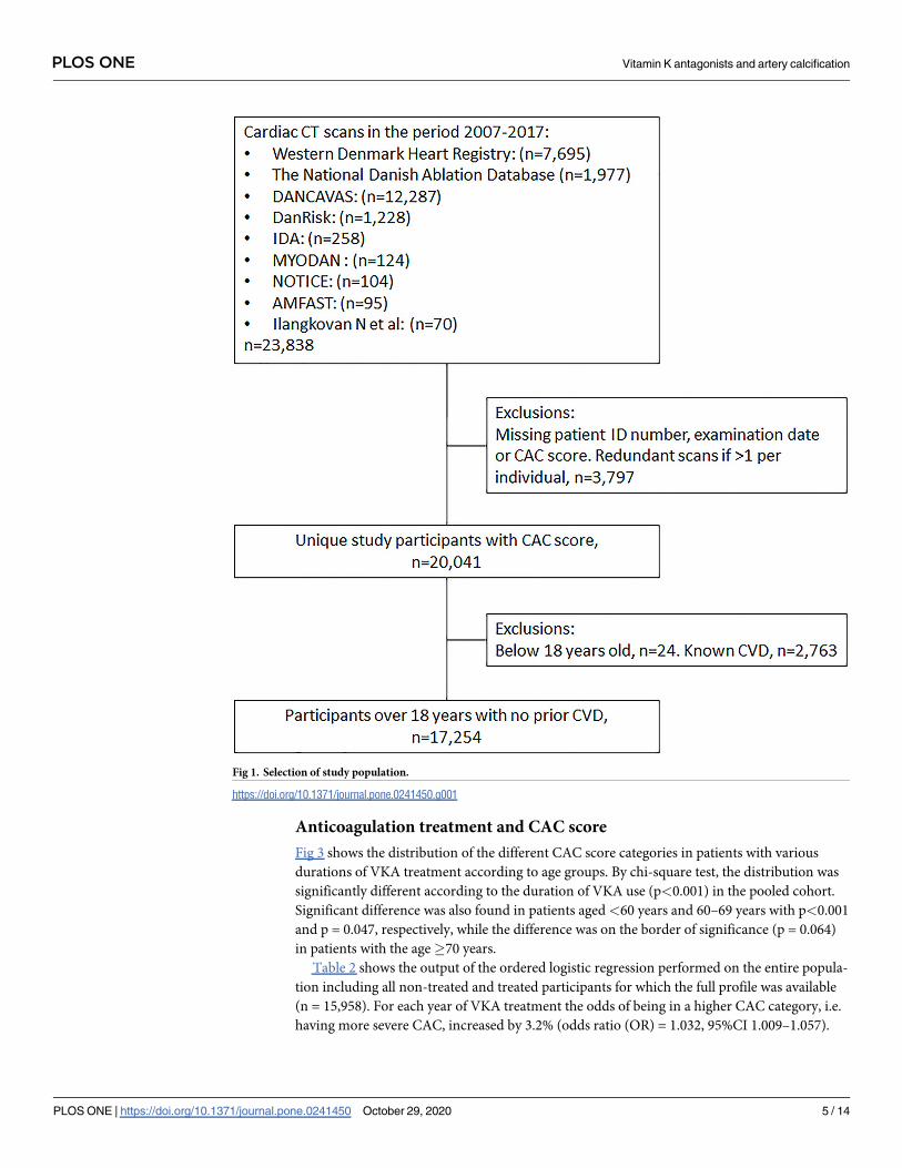
In total, data on 23,838 cardiac CT scans were collected. Due to missing data or individuals
with several CT scans, 3,797 were excluded. Furthermore, participants below the age of 18
(n = 24) and patients with known CVD (n = 2,763) were excluded. The final study population
included 17,254 participants (Fig 1).
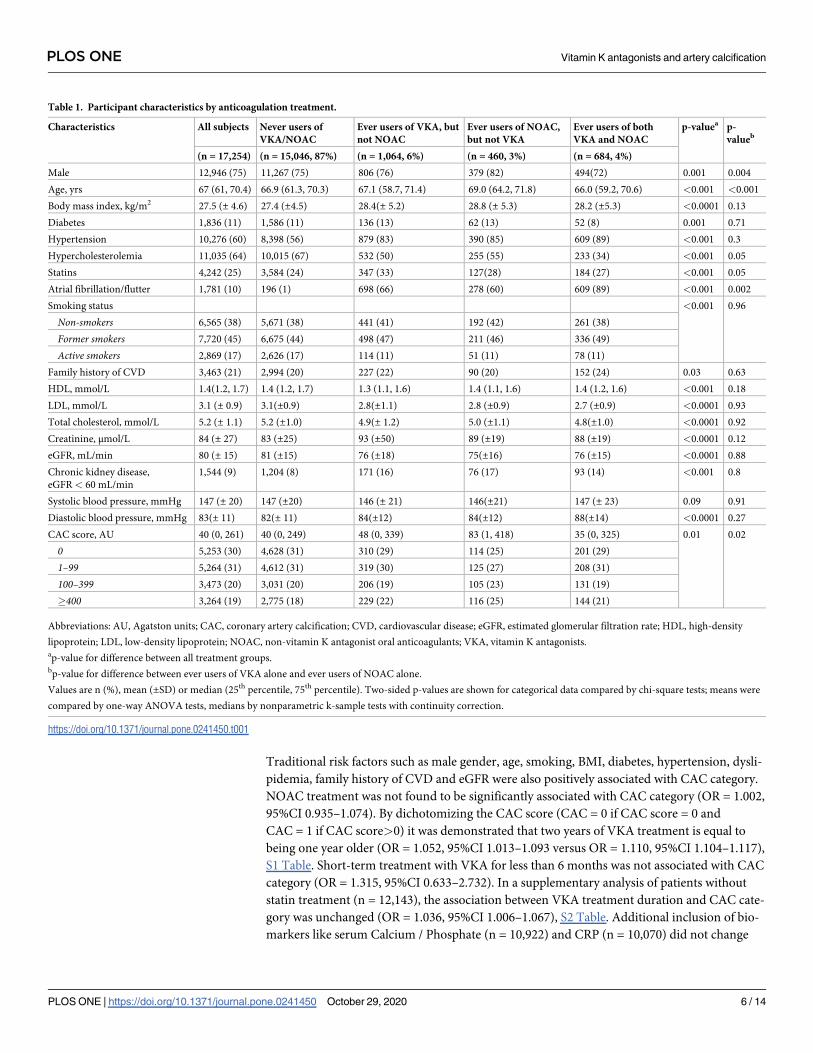
Table 1 describes the baseline characteristics of the study participants stratified by anticoa-
gulation treatment status. Overall, the participants had a median age of 67 years, and 75%
(n = 12,946) were men; 10% (n = 1,748) were ever users of VKA and 7% (n = 1,144) were ever
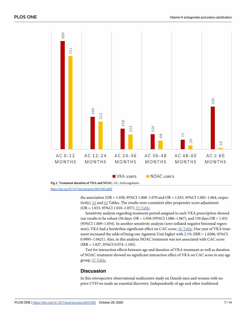
users of NOAC. Apart from long term treatment (�5years), the treatment duration of VKA
and NOAC was similar (Fig 2). For patients treated with anticoagulants, the median treatment
durations were 13.8 and 8.0 months with VKA and NOAC, respectively. When subjects treated
exclusively with either VKA (6%, n = 1,064) or NOAC (3%, n = 460) were contrasted in a
crude comparison, the VKA group had a smaller proportion of men (p = 0.004), were younger
(p<0.001), and had a lower median CAC score (48 versus 83, p = 0.02), but did not differ sig-
nificantly in any other parameters. By comparing the median CAC score in each of the three
treated groups with the non-treated group, the ever users of NOAC had a significantly higher
CAC score (p = 0.002), while VKA users and users of both VKA and NOAC did not differ
from the never users (p = 0.39, p = 0.79).
PLOS ONE Vitamin K antagonists and artery calcification
PLOS ONE | https://doi.org/10.1371/journal.pone.0241450 October 29, 2020 4 / 14
Anticoagulation treatment and CAC score
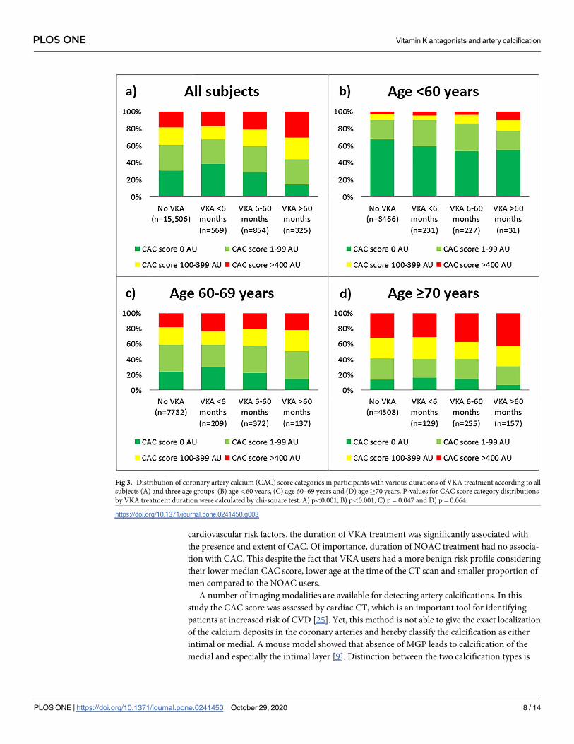
Fig 3 shows the distribution of the different CAC score categories in patients with various
durations of VKA treatment according to age groups. By chi-square test, the distribution was
significantly different according to the duration of VKA use (p<0.001) in the pooled cohort.
Significant difference was also found in patients aged<60 years and 60–69 years with p<0.001
and p = 0.047, respectively, while the difference was on the border of significance (p = 0.064)
in patients with the age�70 years.
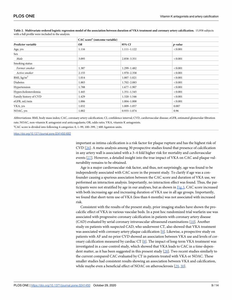
Table 2 shows the output of the ordered logistic regression performed on the entire popula-
tion including all non-treated and treated participants for which the full profile was available
(n = 15,958). For each year of VKA treatment the odds of being in a higher CAC category, i.e.
having more severe CAC, increased by 3.2% (odds ratio (OR) = 1.032, 95%CI 1.009–1.057).
Fig 1. Selection of study population.
https://doi.org/10.1371/journal.pone.0241450.g001
PLOS ONE Vitamin K antagonists and artery calcification
PLOS ONE | https://doi.org/10.1371/journal.pone.0241450 October 29, 2020 5 / 14
Traditional risk factors such as male gender, age, smoking, BMI, diabetes, hypertension, dysli-
pidemia, family history of CVD and eGFR were also positively associated with CAC category.
NOAC treatment was not found to be significantly associated with CAC category (OR = 1.002,
95%CI 0.935–1.074). By dichotomizing the CAC score (CAC = 0 if CAC score = 0 and
CAC = 1 if CAC score>0) it was demonstrated that two years of VKA treatment is equal to
being one year older (OR = 1.052, 95%CI 1.013–1.093 versus OR = 1.110, 95%CI 1.104–1.117),
S1 Table. Short-term treatment with VKA for less than 6 months was not associated with CAC
category (OR = 1.315, 95%CI 0.633–2.732). In a supplementary analysis of patients without
statin treatment (n = 12,143), the association between VKA treatment duration and CAC cate-
gory was unchanged (OR = 1.036, 95%CI 1.006–1.067), S2 Table. Additional inclusion of bio-
markers like serum Calcium / Phosphate (n = 10,922) and CRP (n = 10,070) did not change
Table 1. Participant characteristics by anticoagulation treatment.
Characteristics All subjects Never users of
VKA/NOAC
Ever users of VKA, but
not NOAC
Ever users of NOAC,
but not VKA
Ever users of both
VKA and NOAC
p-valuea p-
valueb
(n = 17,254) (n = 15,046, 87%) (n = 1,064, 6%) (n = 460, 3%) (n = 684, 4%)
Male 12,946 (75) 11,267 (75) 806 (76) 379 (82) 494(72) 0.001 0.004
Age, yrs 67 (61, 70.4) 66.9 (61.3, 70.3) 67.1 (58.7, 71.4) 69.0 (64.2, 71.8) 66.0 (59.2, 70.6) <0.001 <0.001
Body mass index, kg/m2 27.5 (± 4.6) 27.4 (±4.5) 28.4(± 5.2) 28.8 (± 5.3) 28.2 (±5.3) <0.0001 0.13
Diabetes 1,836 (11) 1,586 (11) 136 (13) 62 (13) 52 (8) 0.001 0.71
Hypertension 10,276 (60) 8,398 (56) 879 (83) 390 (85) 609 (89) <0.001 0.3
Hypercholesterolemia 11,035 (64) 10,015 (67) 532 (50) 255 (55) 233 (34) <0.001 0.05
Statins 4,242 (25) 3,584 (24) 347 (33) 127(28) 184 (27) <0.001 0.05
Atrial fibrillation/flutter 1,781 (10) 196 (1) 698 (66) 278 (60) 609 (89) <0.001 0.002
Smoking status <0.001 0.96
Non-smokers 6,565 (38) 5,671 (38) 441 (41) 192 (42) 261 (38)
Former smokers 7,720 (45) 6,675 (44) 498 (47) 211 (46) 336 (49)
Active smokers 2,869 (17) 2,626 (17) 114 (11) 51 (11) 78 (11)
Family history of CVD 3,463 (21) 2,994 (20) 227 (22) 90 (20) 152 (24) 0.03 0.63
HDL, mmol/L 1.4(1.2, 1.7) 1.4 (1.2, 1.7) 1.3 (1.1, 1.6) 1.4 (1.1, 1.6) 1.4 (1.2, 1.6) <0.001 0.18
LDL, mmol/L 3.1 (± 0.9) 3.1(±0.9) 2.8(±1.1) 2.8 (±0.9) 2.7 (±0.9) <0.0001 0.93
Total cholesterol, mmol/L 5.2 (± 1.1) 5.2 (±1.0) 4.9(± 1.2) 5.0 (±1.1) 4.8(±1.0) <0.0001 0.92
Creatinine, μmol/L 84 (± 27) 83 (±25) 93 (±50) 89 (±19) 88 (±19) <0.0001 0.12
eGFR, mL/min 80 (± 15) 81 (±15) 76 (±18) 75(±16) 76 (±15) <0.0001 0.88
Chronic kidney disease,
eGFR < 60 mL/min
1,544 (9) 1,204 (8) 171 (16) 76 (17) 93 (14) <0.001 0.8
Systolic blood pressure, mmHg 147 (± 20) 147 (±20) 146 (± 21) 146(±21) 147 (± 23) 0.09 0.91
Diastolic blood pressure, mmHg 83(± 11) 82(± 11) 84(±12) 84(±12) 88(±14) <0.0001 0.27
CAC score, AU 40 (0, 261) 40 (0, 249) 48 (0, 339) 83 (1, 418) 35 (0, 325) 0.01 0.02
0 5,253 (30) 4,628 (31) 310 (29) 114 (25) 201 (29)
1–99 5,264 (31) 4,612 (31) 319 (30) 125 (27) 208 (31)
100–399 3,473 (20) 3,031 (20) 206 (19) 105 (23) 131 (19)
�400 3,264 (19) 2,775 (18) 229 (22) 116 (25) 144 (21)
Abbreviations: AU, Agatston units; CAC, coronary artery calcification; CVD, cardiovascular disease; eGFR, estimated glomerular filtration rate; HDL, high-density
lipoprotein; LDL, low-density lipoprotein; NOAC, non-vitamin K antagonist oral anticoagulants; VKA, vitamin K antagonists.ap-value for difference between all treatment groups.bp-value for difference between ever users of VKA alone and ever users of NOAC alone.
Values are n (%), mean (±SD) or median (25th percentile, 75th percentile). Two-sided p-values are shown for categorical data compared by chi-square tests; means were
compared by one-way ANOVA tests, medians by nonparametric k-sample tests with continuity correction.
https://doi.org/10.1371/journal.pone.0241450.t001
PLOS ONE Vitamin K antagonists and artery calcification
PLOS ONE | https://doi.org/10.1371/journal.pone.0241450 October 29, 2020 6 / 14
the association (OR = 1.038, 95%CI 1.008–1.070 and OR = 1.033, 95%CI 1.002–1.064, respec-
tively), S3 and S4 Tables. The results were consistent after propensity score adjustment
(OR = 1.033, 95%CI 1.010–1.057), S5 Table.
Sensitivity analysis regarding treatment period assigned to each VKA prescription showed
our results to be robust (50 days: OR = 1.036 (95%CI 1.006–1.067), and 150 days OR = 1.031
(95%CI 1.009–1.054). In another sensitivity analysis (zero-inflated negative binomial regres-
sion), VKA had a borderline significant effect on CAC score, S6 Table. One year of VKA treat-
ment increased the odds of being one Agatston Unit higher with 2.1% (IRR = 1.0206, 95%CI
0.9995–1.0421). Also, in this analysis NOAC treatment was not associated with CAC score
(IRR = 1.037, 95%CI 0.974–1.105).
Test for interaction effects between age and duration of VKA treatment as well as duration
of NOAC treatment showed no significant interaction effect of VKA on CAC score in any age
group, S7 Table.
Discussion
In this retrospective observational multicenter study on Danish men and women with no
prior CVD we made an essential discovery. Independently of age and other traditional
Fig 2. Treatment duration of VKA and NOAC. AC, Anticoagulants.
https://doi.org/10.1371/journal.pone.0241450.g002
PLOS ONE Vitamin K antagonists and artery calcification
PLOS ONE | https://doi.org/10.1371/journal.pone.0241450 October 29, 2020 7 / 14
cardiovascular risk factors, the duration of VKA treatment was significantly associated with
the presence and extent of CAC. Of importance, duration of NOAC treatment had no associa-
tion with CAC. This despite the fact that VKA users had a more benign risk profile considering
their lower median CAC score, lower age at the time of the CT scan and smaller proportion of
men compared to the NOAC users.
A number of imaging modalities are available for detecting artery calcifications. In this
study the CAC score was assessed by cardiac CT, which is an important tool for identifying
patients at increased risk of CVD [25]. Yet, this method is not able to give the exact localization
of the calcium deposits in the coronary arteries and hereby classify the calcification as either
intimal or medial. A mouse model showed that absence of MGP leads to calcification of the
medial and especially the intimal layer [9]. Distinction between the two calcification types is
Fig 3. Distribution of coronary artery calcium (CAC) score categories in participants with various durations of VKA treatment according to all
subjects (A) and three age groups: (B) age<60 years, (C) age 60–69 years and (D) age�70 years. P-values for CAC score category distributions
by VKA treatment duration were calculated by chi-square test: A) p<0.001, B) p<0.001, C) p = 0.047 and D) p = 0.064.
https://doi.org/10.1371/journal.pone.0241450.g003
PLOS ONE Vitamin K antagonists and artery calcification
PLOS ONE | https://doi.org/10.1371/journal.pone.0241450 October 29, 2020 8 / 14
important as intima calcification is a risk factor for plaque rupture and has the highest risk of
CVD [26]. A meta-analysis among 30 prospective studies found that presence of calcification
in any artery wall is associated with a 3–4 fold higher risk for mortality and cardiovascular
events [27]. However, a detailed insight into the true impact of VKA on CAC and plaque vul-
nerability remains to be obtained.
Age is a major cardiovascular risk factor, and thus, not surprisingly, age was found to be
independently associated with CAC score in the present study. To clarify if age was a con-
founder causing a spurious association between the CAC score and duration of VKA use, we
performed an interaction analysis. Importantly, no interaction effect was found. Thus, the par-
ticipants were not stratified by age in our analyses, but as shown in Fig 3, CAC score increased
with both increasing age and increasing duration of VKA use in all age groups. Importantly,
we found that short-term use of VKA (less than 6 months) was not associated with increased
risk.
Consistent with the results of the present study, prior imaging studies have shown the pro-
calcific effect of VKA in various vascular beds. In a post hoc randomized trial warfarin use was
associated with progressive coronary calcification in patients with coronary artery disease
(CAD) evaluated by serial coronary intravascular ultrasound examinations [10]. Another
study on patients with suspected CAD, who underwent CT, also showed that VKA treatment
was associated with coronary artery plaque calcification [9]. Likewise, a prospective study on
patients with AF and no prior CVD showed an association between VKA use and levels of cor-
onary calcification measured by cardiac CT [8]. The impact of long-term VKA treatment was
investigated in a case-control study, which showed that VKA leads to CAC in a time-depen-
dent matter, as it has been suggested in this present study [28]. Two recent studies similarly to
the current compared CAC evaluated by CT in patients treated with VKA or NOAC. These
smaller studies had consistent results showing an association between VKA and calcification,
while maybe even a beneficial effect of NOAC on atherosclerosis [29, 30].
Table 2. Multivariate ordered logistic regression model of the association between duration of VKA treatment and coronary artery calcification. 15,958 subjects
with a full profile were included in the analysis.
CAC scorea (outcome variable)
Predictor variable OR 95% CI p-valueAge, yrs 1.116 1.111–1.122 <0.001
Sex
Male 3.095 2.858–3.351 <0.001
Smoking status
Former smoker 1.387 1.299–1.482 <0.001
Active smoker 2.155 1.970–2.358 <0.001
BMI, kg/m2 1.014 1.007–1.021 <0.001
Diabetes 1.883 1.702–2.083 <0.001
Hypertension 1.788 1.677–1.907 <0.001
Hypercholesterolemia 1.445 1.351–1.545 <0.001
Family history of CVD 1.429 1.320–1.546 <0.001
eGFR, mL/min 1.006 1.004–1.008 <0.001
VKA, yrs 1.032 1.009–1.057 0.007
NOAC, yrs 1.002 0.935–1.074 0.96
Abbreviations: BMI, body mass index; CAC, coronary artery calcification; CI, confidence interval; CVD, cardiovascular disease; eGFR, estimated glomerular filtration
rate; NOAC, non-vitamin K antagonist oral anticoagulants; OR, odds ratio; VKA, vitamin K antagonists.aCAC score is divided into following 4 categories: 0, 1–99, 100–399,�400 Agatston units.
https://doi.org/10.1371/journal.pone.0241450.t002
PLOS ONE Vitamin K antagonists and artery calcification
PLOS ONE | https://doi.org/10.1371/journal.pone.0241450 October 29, 2020 9 / 14
Thus, there are a number of smaller studies showing an association between VKA and
CAC. To extend our knowledge, prospective studies and randomized controlled trials with a
wide range of patients would be advantageous. A recent prospective, randomized, open-label
study compared the progression in coronary plaque in patients treated with either warfarin or
rivaroxaban. With a 1-year follow-up, warfarin was found to be significantly associated with
progression of total plaque volume after adjustment for cardiovascular risk factors [31]. Now
an on-going double-blinded, randomized, placebo-controlled trial investigates the difference
in progression of CAC score in patients with CAD randomized to either vitamin K2 supple-
mentation or placebo [32]. A similar on-going study seeks to examine if vitamin K2 supple-
mentation can slow down the calcification of aortic valves in patients with substantial aortic
valve calcification [33]. These studies will hopefully add knowledge on the importance of vita-
min K in tissue calcification.
The findings of the present and prior studies suggest that long-time use of VKA, together
with other risk factors, may enhance the progression of CAC significantly. NOAC do not
interfere with the vitamin K cascade, and were not associated with higher levels of CAC score.
Considering the aging population and the associated increased use of anticoagulant therapy in
patients with an increased thromboembolic risk, the observations in this study may have scien-
tific and pharmacologic interest.
Study limitations and strengths
A major limitation to our study is the lack of hard cardiovascular endpoints, which prevents us
from learning the true consequence of the higher CAC score by long-term VKA treatment.
The study is also limited by its observational design, why the cause and effect relationship
between VKA and CAC score might be open to interpretation. Furthermore, the medication
status was only evaluated since 2004, while it would have been advantageous to have a life-long
description of the VKA and NOAC use for a more accurate analysis. As NOAC is a relatively
new medical treatment, fewer patients were in long NOAC treatments and this might affect
our results.
However, in comparison with previous studies, this study has the largest sample size to
date. Due to the extensive numbers of descriptive data variables and a very large number of
participants we were able to adjust for traditional cardiovascular risk factors and use of medical
treatment. Data on risk factors as well as medication status were collected from databases and
registries with high validity [21]. Moreover, this study is the largest to investigate the effect of
both VKA and NOAC treatment duration on CAC, which minimizes the risk of confounding
by indication for the association between VKA and CAC. Another strength is that the partici-
pants were free of CVD, which reduced the impact of associated vascular disease on CAC,
thereby enhancing identification of potentially harmful effects of VKA.
Conclusions
Adjusted for cardiovascular risk factors, duration of VKA treatment, but not NOAC treat-
ment, was associated with the risk of a higher level of CAC score in adults with no prior CVD.
The procalcific effect of VKA is consistent with the findings in earlier imaging studies.
Supporting information
S1 Table. Dichotomized CAC score (CAC = 0 if CAC score = 0 and CAC = 1 if CAC
score>0) as outcome variable. Multivariate ordered logistic regression model of the associa-
tion between duration of VKA treatment and coronary artery calcification. 15,958 subjects
PLOS ONE Vitamin K antagonists and artery calcification
PLOS ONE | https://doi.org/10.1371/journal.pone.0241450 October 29, 2020 10 / 14
with a full profile were included in the analysis.
(DOCX)
S2 Table. Exclusion of patients in statin treatment. Multivariate ordered logistic regression
model of the association between duration of VKA treatment and coronary artery calcification.
12,143 subjects with a full profile were included in the analysis.
(DOCX)
S3 Table. Inclusion of serum calcium and phosphate in the model. Multivariate ordered
logistic regression model of the association between duration of VKA treatment and coronary
artery calcification. 10,922 subjects with a full profile were included in the analysis.
(DOCX)
S4 Table. Inclusion of CRP in the model. Multivariate ordered logistic regression model of
the association between duration of VKA treatment and coronary artery calcification. 10,070
subjects with a full profile were included in the analysis.
(DOCX)
S5 Table. Inclusion of propensity score adjustment in the model. Propensity score adjust-
ment in an ordered logistic regression model of the association between duration of VKA
treatment and coronary artery calcification. 15,958 subjects with a full profile were included in
the analysis.
(DOCX)
S6 Table. Sensitivity analysis using zero-inflated negative binomial regression. Zero-
inflated negative binominal regression model of the association between duration of VKA
treatment and coronary artery calcification. 15,958 subjects with a full profile were included in
the analysis.
(DOCX)
S7 Table. Interaction analysis in the ordered logistic regression model of the association
between duration of anticoagulation treatment and coronary artery calcification.
(DOCX)
S1 File. Declaration of interests.
(DOCX)
S2 File. Statement of originality.
(DOCX)
Acknowledgments
The authors thank Soren Moller, biostatistician and associate professor, PhD, OPEN–Odense
Patient data Explorative Network, Odense University Hospital for his help with data
management.
Author Contributions
Conceptualization: Selma Hasific, Kristian Altern Øvrehus, Axel Diederichsen.
Data curation: Selma Hasific, Kristian Altern Øvrehus, Axel Diederichsen.
Formal analysis: Selma Hasific, Kristian Altern Øvrehus, Oke Gerke, Jesper Hallas, Axel
Diederichsen.
Funding acquisition: Selma Hasific, Axel Diederichsen.
PLOS ONE Vitamin K antagonists and artery calcification
PLOS ONE | https://doi.org/10.1371/journal.pone.0241450 October 29, 2020 11 / 14
Investigation: Selma Hasific, Kristian Altern Øvrehus, Martin Busk, Jess Lambrechtsen, Gra-
zina Urbonaviciene, Niels Peter Rønnow Sand, Jens Steen Nielsen, Louise Diederichsen,
Kenneth Bruun Pedersen, Rasmus Carter-Storch, Nivethitha Ilangkovan, Hans Mickley,
Lars Melholt Rasmussen, Jes Sandal Lindholt, Axel Diederichsen.
Methodology: Selma Hasific, Kristian Altern Øvrehus, Oke Gerke, Jesper Hallas, Axel
Diederichsen.
Project administration: Selma Hasific, Kristian Altern Øvrehus, Axel Diederichsen.
Resources: Kristian Altern Øvrehus, Martin Busk, Jess Lambrechtsen, Grazina Urbonaviciene,
Niels Peter Rønnow Sand, Jens Steen Nielsen, Louise Diederichsen, Kenneth Bruun Peder-
sen, Rasmus Carter-Storch, Nivethitha Ilangkovan, Hans Mickley, Lars Melholt Rasmussen,
Jes Sandal Lindholt, Axel Diederichsen.
Supervision: Kristian Altern Øvrehus, Axel Diederichsen.
Validation: Selma Hasific, Kristian Altern Øvrehus, Oke Gerke, Jesper Hallas, Axel
Diederichsen.
Visualization: Selma Hasific, Kristian Altern Øvrehus, Axel Diederichsen.
Writing – original draft: Selma Hasific, Kristian Altern Øvrehus, Axel Diederichsen.
Writing – review & editing: Selma Hasific, Kristian Altern Øvrehus, Oke Gerke, Jesper Hallas,
Martin Busk, Jess Lambrechtsen, Grazina Urbonaviciene, Niels Peter Rønnow Sand, Jens
Steen Nielsen, Louise Diederichsen, Kenneth Bruun Pedersen, Rasmus Carter-Storch,
Nivethitha Ilangkovan, Hans Mickley, Lars Melholt Rasmussen, Jes Sandal Lindholt, Axel
Diederichsen.
References1. Medstat.dk. Sundhedsdatastyrelsen. www.medstat.dk Accessed 08 Dec 2018.
2. Disease burden and mortality estimates. Cause-specific mortality, 2000–2016. World Health Organiza-
tion. https://www.who.int/healthinfo/global_burden_disease/estimates/en/ Accessed 29 August 2020
3. Rennenberg RJ, Schurgers LJ, Kroon AA, Stehouwer CD. Arterial calcifications. J Cell Mol Med 2010
Sep; 14(9):2203–2210. https://doi.org/10.1111/j.1582-4934.2010.01139.x PMID: 20716128
4. Luo G, Ducy P, McKee MD, Pinero GJ, Loyer E, Behringer RR et al. Spontaneous calcification of arter-
ies and cartilage in mice lacking matrix GLA protein. Nature 1997 Mar 6; 386(6620):78–81. https://doi.
org/10.1038/386078a0 PMID: 9052783
5. Price PA, Faus SA, Williamson MK. Warfarin causes rapid calcification of the elastic lamellae in rat
arteries and heart valves. Arterioscler Thromb Vasc Biol 1998 Sep; 18(9):1400–7. https://doi.org/10.
1161/01.atv.18.9.1400 PMID: 9743228
6. Rennenberg RJ, van Varik BJ, Schurgers LJ, Hamulyak K, Cate HT, Vermeer C et al. Chronic coumarin
treatment is associated with increased extracoronary arterial calcification in humans. Blood 2010 Jun
17; 115(24):5121–3. https://doi.org/10.1182/blood-2010-01-264598 PMID: 20354170
7. Kruger T, Oelenberg S, Kaesler N, Schurgers LJ, van de Sandt AM, Bosor P et al. Warfarin induces car-
diovascular damage in mice. Arterioscler Thromb Vasc Biol 2013 Nov; 33(11):2618–24. https://doi.org/
10.1161/ATVBAHA.113.302244 PMID: 23990204
8. Weijs B, Blaauw Y, Rennenberg RJ, Shurgers LJ, Timmermans CC, Pison L et al. Patients using vita-
min K antagonists show increased levels of coronary calcification: an observational study in low-risk
atrial fibrillation patients. Eur Heart J 2011 Oct; 32(20):2555–2562. https://doi.org/10.1093/eurheartj/
ehr226 PMID: 21775389
9. Schurgers LJ, Joosen IA, Laufer EM, Chatrou ML, Herfs M, Winkens MH et al. Vitamin K-antagonists
accelerate atherosclerotic calcification and induce a vulnerable plaque phenotype. PLoS One 2012; 7
(8):e43229. https://doi.org/10.1371/journal.pone.0043229 PMID: 22952653
PLOS ONE Vitamin K antagonists and artery calcification
PLOS ONE | https://doi.org/10.1371/journal.pone.0241450 October 29, 2020 12 / 14
10. Andrews J, Psaltis PJ, Bayturan O, Shao M, Stegman B, Elshazly M et al. Warfarin Use Is Associated
With Progressive Coronary Arterial Calcification. J Am Coll Cardiol Img. 2018 Sep; 11(9):1315–1323.
https://doi.org/10.1016/j.jcmg.2017.04.010 PMID: 28734922
11. Hoffmann U, Massaro JM, D’Agostino RB Sr, Kathiresan S, Fox CS, O’Donell CJ. Cardiovascuar Event
Prediction and Risk Reclassification by Coronary, Aortic, and Valvular Calcification in the Framingham
Heart Study. J Am Heart Assoc 2016 Feb 22; 5(2).
12. Nielsen LH, Nørgaard BL, Tilsted HH, Sand NP, Jensen JP, Bøttcher M et al. The Western Denmark
Cardiac Computed Tomography Registry: a review and validation study. Clin Epidemiol 2014 Dec 31;
7:53–64. https://doi.org/10.2147/CLEP.S73728 PMID: 25657592
13. Catheterbased ablation of heart rythm disorders in Denmark. 2018. http://www.ablation.dk. Accessed
10 Dec 2018.
14. Diederichsen AC, Rasmussen LM, Søgaard R, Lambrechtsen J, Steffensen FH, Frost L et al. The Dan-
ish Cardiovascular Screening Trial (DANCAVAS): study protocol for a randomized controlled trial. Trials
2015 Dec 5; 16:554. https://doi.org/10.1186/s13063-015-1082-6 PMID: 26637993
15. Diederichsen SZ, Grønhøj MH, Mickley H, Gerke O, Steffensen FH, Lambrechtsen J et al. CT-detected
Growth of Coronary Artery Calcification in Asymptomatic Middle-Aged Subjects and Association With
15 Biomarkers. J Am Coll Cardiol Img. 2017 Aug; 10(8):858–866. https://doi.org/10.1016/j.jcmg.2017.
05.010 PMID: 28797406
16. Stidsen JV, Nielsen JS, Henriksen JE, Friborg SG, Thomsen RW, Olesen TB et al. Protocol for the spe-
cialist supervised individualised multifactorial treatment of new clinically diagnosed type 2 diabetes in
general practice (IDA): a prospective controlled multicentre open-label intervention study. BMJ Open
2017 Dec 10; 7(12):e017493. https://doi.org/10.1136/bmjopen-2017-017493 PMID: 29229652
17. Diederichsen LP, Diederichsen AC, Simonsen JA, Junker P, Søndergaard K, Lundberg IE et al. Tradi-
tional cardiovascular risk factors and coronary artery calcification in adults with polymyositis and derma-
tomyositis: a Danish multicenter study. Arthitis Care Res (Hoboken) 2015 May; 67(6):848–54. https://
doi.org/10.1002/acr.22520 PMID: 25418360
18. Pedersen KB, Madsen C, Sandgaard NCF, Diederichsen AC, Bak S, Brandes A. Subclinical atrial fibril-
lation in patients with recent transient ischemic attack. J Cardiovasc Electrophysiol. 2018 May; 29
(5):707–714. https://doi.org/10.1111/jce.13470 PMID: 29478291
19. Carter-Storch R, Møller JE, Christensen LE, Irmukhadenov A, Rasmussen LM, Pecini R et al. Postoper-
ative Reverse Remodeling and Symptomatic Improvement in Normal-Flow Low-Gradient Aortic Steno-
sis After Aortic Valve Replacement. Circ Cardiovasc Imaging. 2017 Dec; 10(12). https://doi.org/10.
1161/CIRCIMAGING.117.006580 PMID: 29222121
20. Ilangkovan N, Mogensen CB, Mickley H, Lassen TB, Lambrechtsen J, Sand NPR et al. Prevalence of
coronary artery calcification in a non-specific chest pain population in emergency and cardiology depart-
ments compared with the background population: a prospective cohort study in Southern Denmark with
12-month follow-up of cardiac endpoints. BMJ Open. 2018 Mar 3; 8(3):e018391. https://doi.org/10.
1136/bmjopen-2017-018391 PMID: 29502085
21. Johannesdottir SA, Horvath-Puho E, Ehrenstein V, Schmidt M, Pedersen L, Sørensen HT. Existing
data sources for clinical epidemiology: The Danish National Database of Reimbursed Prescriptions.
Clin Epidemiol. 2012; 4:303–13. https://doi.org/10.2147/CLEP.S37587 PMID: 23204870
22. Levey AS, Stevens LA, Schmid CH, Zhang YL, Castro AF 3rd, Feldman HI et al. CKD-EPI (Chronic Kid-
ney Disease Epidemiology Collaboration)., A new equation to estimate glomerular filtration rate. Ann
Intern Med 2009; 150: 604–12 https://doi.org/10.7326/0003-4819-150-9-200905050-00006 PMID:
19414839
23. Agatston AS, Janowitz WR, Hildner FJ, Zusmer NR, Viamonte M Jr, Detrano R. Quantification of coro-
nary artery calcium using ultrafast computed tomography. J Am Coll Cardiol 15(4):827–832 https://doi.
org/10.1016/0735-1097(90)90282-t PMID: 2407762
24. Erbel R, Budoff M. Improvement of cardiovascular risk prediction using coronary imaging: subclinical
atherosclerosis: the memory of lifetime risk factor exposure. Eur Heart J. 2012 May; 33(10):1201–13.
https://doi.org/10.1093/eurheartj/ehs076 PMID: 22547221
25. Budoff MJ, Gul KM. Expert review on coronary calcium. Vasc Health Risk Manag 2008; 4:315–324.
https://doi.org/10.2147/vhrm.s1160 PMID: 18561507
26. Mackey RH, Venkitachalam L, Sutton-Tyrrel K. Calcifications, arterial stiffness and atherosclerosis. Adv
Cardiol. 2007; 44:234–44. https://doi.org/10.1159/000096744 PMID: 17075212
27. Rennenberg RJ, Kessels AG, Schurgers LJ, van Engelshoven JM, de Leeuw PW, Kroon AA. Vascular
calcifications as a marker of increased cardiovascular risk: a meta-analysis. Vasc Health Risk Manag.
2009; 5(1):185–197. https://doi.org/10.2147/vhrm.s4822 PMID: 19436645
PLOS ONE Vitamin K antagonists and artery calcification
PLOS ONE | https://doi.org/10.1371/journal.pone.0241450 October 29, 2020 13 / 14
28. Unlu S, Sahinarslan A, Kilic HK, Gokalp G, Sezenoz B, Erbas Get al. Long-term vitamin-K antagonist
use and coronary artery calcification. Herz. 2020 Sep; 45(6):580–585. https://doi.org/10.1007/s00059-
018-4760-9 PMID: 30276478
29. Plank F, Beyer C, Friedrich G, Stuhlinger M, Hintringer F, Dichtl W et al. Influence of vitamin K antago-
nists and direct oral anticoagulation on coronary artery disease: A CTA analysis. Int J Cardiol. 2018 Jun
1; 260:11–15. https://doi.org/10.1016/j.ijcard.2018.03.019 PMID: 29530620
30. Win TT, Nakanishi R, Osawa K, Li D, Susaria SS, Jayawardena E et al. Apixaban versus warfarin in
evaluation of progression of atherosclerotic and calcified plaques (prospective randomized trial). Am
Heart J. 2019 Jun; 212:129–133. https://doi.org/10.1016/j.ahj.2019.02.014 PMID: 31002997
31. Lee J, Nakanishi R, Li D, Shaikh K, Shekar C, Osawa K et al. Randomized trial of rivaroxaban versus
warfarin in the evaluation of progression of coronary atherosclerosis. Am Heart J. 2018 Dec; 206:127–
130. https://doi.org/10.1016/j.ahj.2018.08.007 PMID: 30227941
32. Vossen LM, Schurgers LJ, van Varik BJ, Kietselaer BL, Vermeer C, Meeder JG et al. Menaquinone-7
Supplementation to Reduce Vascular Calcification in Patients with Coronary Artery Disease: Rationale
and Study Protocol (VitaK-CAC Trial). Nutrients 2015 Oct 28; 7(11):8905–8915. https://doi.org/10.
3390/nu7115443 PMID: 26516910
33. Lindholt JS, Frandsen NE, Fredgart MH,Øvrehus KA, Dahl JS, Møller JE et al. Effects of menaqui-
none-7 supplementation in patients with aortic valve calcification: study protocol for a randomized con-
trolled trial. BMJ Open 2018 Aug 23; 8(8):e022019. https://doi.org/10.1136/bmjopen-2018-022019
PMID: 30139903
PLOS ONE Vitamin K antagonists and artery calcification
PLOS ONE | https://doi.org/10.1371/journal.pone.0241450 October 29, 2020 14 / 14