Embed Size (px)
Citation preview
Extending the Spectrum of �-Dicarbonyl Compounds in Vivo*
Received for publication, March 7, 2014, and in revised form, August 25, 2014 Published, JBC Papers in Press, August 27, 2014, DOI 10.1074/jbc.M114.563593
Christian Henning‡, Kristin Liehr‡, Matthias Girndt§, Christof Ulrich§, and Marcus A. Glomb‡1
From the ‡Institute of Chemistry-Food Chemistry and the §Department of Internal Medicine II, Martin-Luther-UniversityHalle-Wittenberg, Kurt-Mothes-Strasse 2, 06120 Halle/Saale, Germany
Background: �-Dicarbonyls are central intermediates in the formation of advanced glycation end products (AGEs).Results: A quantitation method for the complete spectrum relevant in vivo was established.Conclusion: Non-enzymatic chemistry of glucose and L-ascorbic acid as precursors and �-dicarbonyl intermediates play animportant role in vivo.Significance: Knowledge of plasma levels of �-dicarbonyls is crucial to understand the complex pathways of AGE formation invivo.
Maillard �-dicarbonyl compounds are known as central inter-mediates in advanced glycation end product (AGE) formation.Glucose is the primary source of energy for the human body,whereas L-threo-ascorbic acid (vitamin C) is an essential nutrient,involved in a variety of enzymatic reactions. Thus, the Maillarddegradation of glucose and ascorbic acid is of major importance invivo. To understand the complex mechanistic pathways of AGEformation, it is crucial to extend the knowledge on plasma concen-trations of reactive key �-dicarbonyl compounds (e.g. 1-deoxyglu-cosone). With the present work, we introduce a highly sensitiveLC-MS/MS multimethod for human blood plasma based onderivatization with o-phenylenediamine under acidic conditions.The impact of workup and reaction conditions, particularly of pH,was thoroughly evaluated. A comprehensive validation providedthe limit of detection, limit of quantitation, coefficients of varia-tion, and recovery rates. The method includes the �-dicarbonyls1-deoxyglucosone, 3-deoxyglucosone, glucosone, Lederer’s glu-cosone, dehydroascorbic acid, 2,3-diketogulonic acid, 1-deoxypen-tosone, 3-deoxypentosone, 3,4-dideoxypentosone, pentosone, 1-de-oxythreosone, 3-deoxythreosone, threosone, methylglyoxal, glyoxal;the �-keto-carboxylic acids pyruvic acid and glyoxylic acid; and thedicarboxylicacidoxalicacid.Themethodwasthenappliedtotheana-lyses of 15 healthy subjects and 24 uremic patients undergoing hemo-dialysis. The comparison of the results revealed a clear shift in theproduct spectrum. In most cases, the plasma levels of target analyteswere significantly higher. Thus, this is the first time that a completespectrum of �-dicarbonyl compounds relevant in vivo has been estab-lished. The results provide further insights into the chemistry of AGEformation and will be helpful to find specific markers to differentiatebetween the various precursors of glycation.
Advanced glycation of proteins has been investigated inaging, diabetes, and nutrition (1–3). The Maillard reaction, anon-enzymatic process, is initiated when proteins are exposedto glucose or other carbohydrates. Through a series of reac-tions, it eventually yields the irreversible advanced glycation
end products (AGEs).2 There are multiple sources and mecha-nisms of AGE formation in vivo, involving oxidative and non-oxidative chemistry of reducing sugars, Schiff bases, Amadoriadducts, ascorbic acid and metabolic intermediates (4, 5).Because they are correlated to the severity of diabetic and ure-mic complications their clinical relevance was established(6 – 8).
AGE concentrations in vivo are markedly amplified in diabe-tes but also in non-diabetic uremia with a significant loss ofrenal clearance (9). Thus, besides substrate concentration, car-bonyl stress, as described by Baynes et al. (10), is an alternativeexplanation for the increase of chemical protein modificationsin various diseases. Carbonyl stress is caused by a generalizedincrease in the concentration of reactive carbonyl precursors ofAGEs. Among those, �-dicarbonyl compounds (�-DCs) play avery prominent role. Their relevance as key intermediates inAGE formation was extensively investigated in in vitro modelsystems (11–15).
Endogenous formation is supposed to be the predominantsource of �-DCs in human circulation (16). However, informa-tion on the physiological importance of exogenous dicarbonylintake is scarce and subject to recent investigations (17–19).Regarding this topic, foods and peritoneal dialysis fluids areconsidered to release considerable amounts of reactive �-DCs(20 –24).
For a comprehensive understanding of the complex formationpathways of AGEs in vivo, the analytical assessment of thecomplete �-DC spectrum is crucial. Nemet and Monnier (25)examined the L-threo-ascorbic acid (ASA, vitamin C) degradationproducts dehydroascorbic acid (DHA), 2,3-diketogulonic acid
* This work was supported by German Federal Ministry of Education andResearch (BMBF) Project 13N11798.
1 To whom correspondence should be addressed. Fax: 49-345-55-27341;E-mail: [email protected].
2 The abbreviations used are: AGE, advanced glycation end products; �-DC, �-dicar-bonylcompound;ASA,L-threo-ascorbicacid;DHA,dehydroascorbicacid;2,3-DKG,2,3-diketogulonicacid(L-threo-2,3-hexodiulosonicacid);3-DT,3-deoxythresosone(4-hydroxy-2-oxobutanal); GO, glyoxal; MGO, methylglyoxal (2-oxopropanal);3-DG,3-deoxyglucosone(3-deoxy-D-erythro-hexos-2-ulose);OPD,o-phenylenedi-amine; threosone, 3,4-dihydroxy-2-oxobutanal; 1-DG, 1-deoxyglucosone (1-de-oxy-D-erythro-hexo-2,3-diulose); DHA, dehydroascorbic acid; DHAP, dihydroxyac-etone phosphate; G3P, glyceraldehyde 3-phosphate; 1-DP, 1-deoxypentosone;3-DP, 3-deoxypentosone; 3,4-DDP, 3,4-dideoxypentosone (3,4-dideoxypentos-2-ulose);1-DT,1-deoxythresosone(1-hydroxy-2,3-butanedione);Q,quinoxaline;HDpatients, patients undergoing hemodialysis; 3-DGal, 3-deoxygalactosone; EtOAc,ethyl acetate; 3,4-DGE, 3,4-dideoxyglucosone-3-ene; MeOH, methanol; CID, colli-sion-induced dissociation; glucosone, D-arabino-hexos-2-ulose; Lederer’s glu-cosone, N6-(3,6-dideoxyhexos-2-ulos-6-yl)-L-lysine; pentosone, pentos-2-ulose.
THE JOURNAL OF BIOLOGICAL CHEMISTRY VOL. 289, NO. 41, pp. 28676 –28688, October 10, 2014© 2014 by The American Society for Biochemistry and Molecular Biology, Inc. Published in the U.S.A.
28676 JOURNAL OF BIOLOGICAL CHEMISTRY VOLUME 289 • NUMBER 41 • OCTOBER 10, 2014
by guest on January 3, 2020http://w
ww
.jbc.org/D
ownloaded from
(2,3-DKG), threosone, 3-deoxythresosone (3-DT), and xylosone inhuman lens. In human blood plasma, however, only glyoxal (GO),methylglyoxal (MGO), 3-deoxyglucosone (3-DG), and DHA havebeen described in detail so far (26–33). The implemented analyt-ical methods vary not only in the inevitable derivatization proce-dure but also in the chromatographic technique used. The com-mon alternative approach for the quantitation of DHA is themeasurement of the difference of ASA before and after a reductionstep (34, 35). Consequently, concentrations of published plasmalevels of healthy subjects differ in a wide range and are thus notcomparable (e.g. for GO from 220 to 1150 pmol/ml, for MGO from120 to 650 pmol/ml, and for DHA from 550 to 6800 pmol/ml(mean values)).
Glomb and Tschirnich compared common derivatizationapproaches (36). According to them, the use of aromatic o-di-amines (e.g. o-phenylenediamine (OPD)) is prerequisite for theanalysis of highly reactive �-DCs, such as 1-deoxyglucosone(1-DG). The detection of these short lived intermediates is lim-ited by the rate of condensation of the reagent with the carbonylmoiety. However, these trapping reagents impose high oxida-tive stress on the system investigated and could lead to artifactformation, especially of �-DCs that originate from oxidativepathways (e.g. D-arabino-hexos-2-ulose (glucosone)).
The work group of Thornalley (31) developed a reliablemethod for the detection of MGO as 6,7-dimethoxy-2-meth-ylquinoxaline in human plasma. They stressed the importanceof pH control to avoid the degradation of dihydroxyacetonephosphate (DHAP) and glyceraldehyde 3-phosphate (G3P) toMGO, both central intermediates in several metabolic path-ways in living organisms.
Although many methods for the detection of a few selected�-DCs were described already, a comprehensive methodincluding all relevant compounds in vivo has been missing up tonow. The particular analytical challenge in this regard is thevery diverging polarity of the target substances but also thebaseline separation of isomeric pairs with identical molecularmasses (e.g. 1-/3-deoxypentosone (1-/3-DP)).
We developed and validated a highly sensitive LC-MS/MS mul-timethod suitable for routine analysis based on the derivatizationwith OPD, including 15 �-dicarbonyl compounds and two �-keto-carboxylic acids. Both substance categories are hereafter referredto as �-DCs for reasons of simplicity. In detail, the method covers1-DG, 3-DG, glucosone, N6-(3,6-dideoxyhexos-2-ulos-6-yl)-L-lysine (Lederer’s glucosone), DHA, 2,3-DKG, 4,5-dihydroxy-2,3-pentanedione (1-DP), 4,5-dihydroxy-2-oxopentanal (3-DP),3,4-dideoxypentosone (3,4-DDP), pentos-2-ulose (pentosone), 1-de-oxythreosone (1-DT), 3-DT, threosone, MGO, ethanedial (glyoxal,GO), 2-oxopropanoic acid (pyruvic acid), oxoethanoic acid (glyoxylicacid), and ethanedioic acid (oxalic acid) and was used to screen aninitial set of plasma samples from 15 healthy human subjects and 24uremic patients undergoing hemodialysis (HD patients). The resultsare discussed with respect to the sources and mechanistic relation-ships of the �-DCs detected.
EXPERIMENTAL PROCEDURES
Materials and Plasma Samples—Chemicals of the highestgrade available were obtained from Sigma-Aldrich and Fisherunless otherwise indicated. NMR solvents were purchased from
ARMAR Chemicals (Leipzig/Doettingen, Germany). The qui-noxaline derivatives of the �-DCs will be marked hereafter withthe suffix “-Q”. 1-DG-Q, 3-DG-Q, glucosone-Q, Lederer’s glu-cosone-Q, 1-DP-Q, 3-DP-Q, 3,4-DDP-Q, pentosone-Q, 1-DT-Q,3-DT-Q, threosone-Q, pyruvic acid-Q, glyoxylic acid-Q, oxalicacid-Q, and 3-DG were synthesized according to our previouswork (37–39). The identities of target compounds were verified bynuclear magnetic resonance (NMR) experiments.
Written informed consent was obtained from all patients.The study was approved by the Ethics Committee of the Med-ical Faculty of the Martin Luther University Halle-Wittenberg.Blood samples were obtained from 15 healthy subjects withnormal renal function and 24 non-diabetic patients undergoinghemodialysis using EDTA as an anticoagulant (2 mg/ml wholeblood). In HD patients, samples were obtained predialysisbefore the midweek treatment session. Hemodialysis was per-formed three times weekly for 4 –5 h using polysulfone dialyz-ers. All patients were treated with bicarbonate hemodialysis(acid concentrate type 257, 8.4% sodium bicarbonate type 200,MTN Neubrandenburg GmbH, Neubrandenburg, Germany)with ultrapure water quality (by reverse osmosis and sterile fil-ters). Plasma was derived by centrifugation (3000 � g, 10 min,4 °C) within 20 min of collection and immediately subjected tothe assay procedure described below. HbA1c, creatinine, andC-reactive protein were measured by routine methods at thecentral laboratory of Martin-Luther-University Clinical Cen-ter, Halle (Saale, Germany).
2-(2�(R),3�(R),4�-Trihydroxybutyl)quinoxaline (3-DGal-Q)—3-Deoxy-D-threo-hexos-2-ulose (3-deoxygalactosone; 3-DGal)was synthesized according to the literature (40) with the excep-tion of the 3-DGal bis(benzoyl hydrazone) cleavage. Here,instead of benzaldehyde, sodium nitrite was used, following theprocedure of Henseke and Bauer (41). Purification of crude3-DGal-Q was achieved by preparative high performance liquidchromatography with ultraviolet detection (HPLC-UV). NMRresults were in line with those of Hellwig et al. (40).
3-(D-erythro-Glycerol-1-yl)-quinoxaline-2-carboxylic Acido-Aminoanilide (DHA Precursor-Q)—DHA (250 mg; 1.44 mmol)was suspended in methanol (25 ml), and OPD was added (308.5mg; 2.85 mmol). The reaction mixture was heated for 2 h at40 °C and allowed to cool. The precipitated solid was isolated byfiltration; washed with water, ethanol, and ether; and dried(yield: 282.6 mg, 76%). Recrystallization from ethanol gave yel-low needles.
1H NMR (500 MHz, DMSO-d6): � (ppm) � 3.44 –3.55 (m,2H), 4.03 (m, 1H), 5.42 (m, 1H), 6.63 (m, 1H), 6.8 (dd, J � 8.0, 1.4Hz, 1H), 7.0 (m, 1H), 7.36 (dd, J � 8.0, 1.5 Hz, 1H), 7.96 (m, 2H),8.17 (m, 1H), 8.23 (m, 1H). 13C NMR (100 MHz, DMSO-d6): �(ppm) � 63.3, 72.0, 74.5, 116.4, 116.6, 122.9, 126.4, 127.2, 128.9,129.5, 131.0, 132.0, 139.6, 141.2, 143.2, 147.4, 156.0, 165.1.HR-MS: m/z 393.09645 (found); m/z 393.09596 (calculated forC18H18O4N4K [M � K]�).
3-(D-erythro-Glycerol-1-yl)-quinoxaline-2-carboxylic �-Lac-tone (DHA-Q)—A suspension of precursor DHA-Q (500 mg) inwater (10 ml) was treated with 0.1 M hydrochloric acid (25 ml)and stirred at room temperature. The reaction process was fol-lowed by thin layer chromatography (TLC; EtOAc, UV detec-tion). The reaction mixture was extracted with EtOAc (3 �
�-Dicarbonyl Compounds in Vivo
OCTOBER 10, 2014 • VOLUME 289 • NUMBER 41 JOURNAL OF BIOLOGICAL CHEMISTRY 28677
by guest on January 3, 2020http://w
ww
.jbc.org/D
ownloaded from
35 ml), the organic layers were combined, and the solvent wasevaporated. The residue was purified by column chromatogra-phy (silica gel 60, 63–200 �m (Merck), EtOAc). Fractionsincluding the compound with Rf 0.24 (TLC: EtOAc, UV detec-tion) were collectively concentrated in vacuo to afford 180 mg(66%) of light yellow needles. NMR results were in line withthose of Nemet and Monnier (25). HR-MS: m/z 269.05301(found); m/z 269.05328 (calculated for C12H10O4N2Na [M �Na]�).
2,3-DKG—2,3-DKG as sodium salt was synthesized accord-ing to the method of Otsuka et al. (42). The product wasobtained as a white hygroscopic precipitate. It was character-ized by the LC-MS/MS method mentioned below afterderivatization with OPD and directly utilized for quinoxalinesynthesis.
3-((1S,2R)-1,2,3-Trihydroxypropyl)-quinoxaline-2-carboxylicAcid (2,3-DKG-Q)—The corresponding quinoxaline of 2,3-DKGwas obtained according to Nemet and Monnier (25). Purificationof the quinoxaline solution was done by preparative HPLC-UV(tR � 62 min). Fractions including the pure compound werecollectively evaporated under reduced pressure, dissolved inwater, and lyophilized to afford 2,3-DKG-Q as a light yellowpowder in quantitative yield. NMR data were in line with the liter-ature (25). HR-MS: m/z 263.06749 (found); m/z 263.06735 (calcu-lated for C12H11O5N2 [M � H]�).
Isotopically Labeled 2-Hydroxy-3-methyl-2,3-13C2-quinoxa-line (Pyruvic Acid-13C2-Q)—Pyruvic acid-13C2-Q was synthe-sized according to the method of Arun et al. (43). Briefly, asolution of OPD in distilled water (306.9 mg in 8 ml of ultrapurewater, 2.84 mM) was added to 1,2-13C2-pyruvic acid (250.0 mgin 8 ml ultrapure water, 2.84 mM) dropwise with constant stir-ring. The precipitated pale yellow colored compound was fil-tered, washed with water, and lyophilized. The crude samplewas recrystallized from 50% ethanol absolute (404.7 mg, 88%).NMR data were in line with the literature. HR-MS: m/z163.0779 (found); m/z 163.0776 (calculated for C7
13C2H8ON2[M � H]�).
Time Course of the OPD Reaction of 3-DG, MGO, GO, Glyox-ylic Acid, Pyruvic Acid, Oxalic Acid, DHA Precursor-Q, and2,3-DKG-Q under Assay Conditions—All compounds (20 �M)were incubated under assay conditions in amber glass vialsunder argon atmosphere at 22 °C in 0.4 M formate buffer (pH3.0) containing 0.55 mM OPD and 3.4 mM EDTA. Vials weretightly sealed by a screw cap with integrated polytetrafluoroeth-ylene/silicone septum. At various time points, aliquots of thereaction mixtures were subjected to LC-UV analysis asdescribed below. After 24 h, trifluoroacetic acid (TFA; 0.4 M)was added, and the incubation was continued for 1 h. Then pH3.0 was adjusted again with 4 M ammonium hydroxide. Theinjection volume was adjusted to account for the dilution of thereaction mixture. The yields were determined against theauthentic reference �-DC-Q standards.
Time Course of the OPD Reaction of ASA, DHA, 2,3-DKG,and 3-DG-Q under Assay Conditions—All compounds (20 �M)were incubated as described above over a prolonged timeperiod. No TFA was added after 24 h. At various time points,aliquots of the reaction mixtures were subjected to LC-UVanalysis as described below. The yields were determined
against the authentic reference �-DC-Q standards. Due tothe lack of authentic reference for the quinoxaline of 3,4-dideoxyglucosone-3-ene (3,4-DGE-Q), quantitation wasdone with 3-DGal-Q under the assumption of an equalextinction coefficient.
De Novo Formation of MGO-Q from G3P and DHAP withOPD under Various Assay Conditions—G3P and DHAP (50 and100 �M, which corresponds to a plasma concentration of 100and 200 �M, respectively) were incubated under assay condi-tions in amber glass vials under argon atmosphere at 22 °C invarious buffer solutions containing 0.55 mM OPD and 3.4 mM
EDTA. The buffers used were potassium phosphate buffer (0.4M, pH 7.0), sodium formate buffer (0.4 M, pH 3.0), and perchlo-ric acid (0.4 M, pH �0). Vials were tightly sealed by a screw capwith integrated polytetrafluoroethylene/silicone septum. Reac-tion mixtures were subjected to LC-UV analysis as describedbelow not only after 24 h but immediately after the addition ofOPD and after a 1-h incubation time to account for possibleMGO impurities of the starting materials. The yield of MGOwas determined against the authentic reference �-DC-Qstandards.
Assay of �-DCs in Plasma—Sodium formate buffer (2 M, pH3.0, 200 �l), the internal standard pyruvic acid-13C2-Q (1.25�M, 100 �l) and the derivatizing agent OPD (2.75 mM, 200 �l)were added to 500 �l of blood plasma. The sample was incu-bated for 24 h in the dark at room temperature under argonatmosphere. TFA (2 M, 250 �l) was added, and the incubationcontinued for 1 h under the same conditions. The pH of thesample was then adjusted to pH 3.0 with ammonium hydroxide(4 M, 415 �l). Water was added (85 �l) to give a total samplevolume of 1750 �l. The protein precipitate was separated bycentrifugation (16,000 � g and 20 °C). The supernatant (storageat �80 °C) was administered to LC-MS/MS analysis.
HPLC-UV—A Besta HD 2-200 pump (Wilhelmsfeld, Ger-many) was used at a flow rate of 15 ml/min. Elution of materialwas monitored by a UV detector (Jasco UV-2075 with a prepar-ative flow cell (Gross-Umstadt, Germany)). The detectionwavelength was 320 nm. Chromatographic separations wereperformed on a stainless steel column (KNAUER, 250 � 20mm, Eurospher-100 C18, 10 �m (Berlin, Germany)). Themobile phase used consisted of water (solvent A) and metha-nol/water (7:3 (v/v), solvent B). To both solvents (A and B) 0.8ml/liter formic acid was added. Samples were injected at 5% Bfor 2,3-DKG-Q and at 35% B for 3-DGal-Q and purified underisocratic conditions.
NMR Spectroscopy—NMR spectra were recorded on a VarianVXR 400 spectrometer operating at 400 MHz for 1H and 100MHz for 13C or on a Varian Unity Inova 500 instrument oper-ating at 500 MHz for 1H and 125 MHz for 13C, respectively.Chemical shifts are given relative to external SiMe4.
Accurate Mass Determination (High Resolution MS)—Thehigh resolution positive and negative ion electrospray ioniza-tion mass spectra (electrospray ionization-high resolution MS)were taken on a Bruker Apex III Fourier transform ion cyclo-tron resonance mass spectrometer (Bruker Daltonics, Billerica,MA) equipped with an Infinity cell, a 7.0-tesla superconductingmagnet, a radio frequency-only hexapole ion guide, and anexternal off-axis electrospray source (Apollo; Agilent, Santa
�-Dicarbonyl Compounds in Vivo
28678 JOURNAL OF BIOLOGICAL CHEMISTRY VOLUME 289 • NUMBER 41 • OCTOBER 10, 2014
by guest on January 3, 2020http://w
ww
.jbc.org/D
ownloaded from
Clara, CA). Nitrogen was used as a drying gas at 150 °C. Thesamples were dissolved in methanol, and the solutions wereintroduced continuously via a syringe pump at a flow rate of 120�l/h. The data were acquired with 256,000 data points andzero-filled to 1,024,000 by averaging 32 scans.
High Performance Liquid Chromatography with CoupledUltraviolet-Mass Spectrometry Detection (LC-UV-MS/MS)—The HPLC apparatus (Jasco, Gross-Umstadt, Germany) con-sisted of a pump (PU-2080 Plus) with degasser (LG-2080-02)and quaternary gradient mixer (LG-2080-04), a column oven(Jasco Jetstream II), an autosampler (AS-2057 Plus), and a UVdetector (UV-2075). Mass spectrometric detection was con-ducted on an API 4000 QTrap LC-MS/MS system (AppliedBiosystems/MDS Sciex, Concord, Canada) equipped with aturbo ion spray source using electrospray ionization in positivemode: sprayer capillary voltage, 4.5 kV; nebulizing gas flow, 50ml/min; heating gas, 60 ml/min at 550 °C; and curtain gas, 40ml/min.
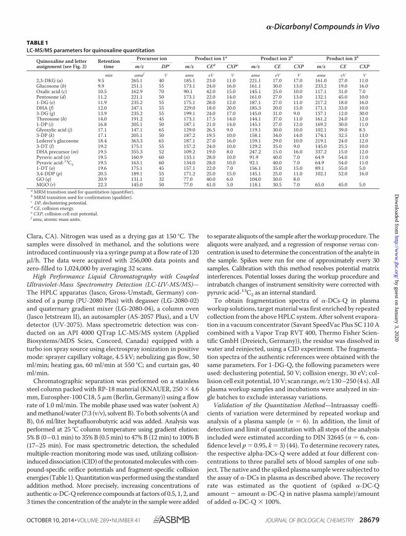
Chromatographic separation was performed on a stainlesssteel column packed with RP-18 material (KNAUER, 250 � 4.6mm, Eurospher-100 C18, 5 �m (Berlin, Germany)) using a flowrate of 1.0 ml/min. The mobile phase used was water (solvent A)and methanol/water (7:3 (v/v), solvent B). To both solvents (A andB), 0.6 ml/liter heptafluorobutyric acid was added. Analysis wasperformed at 25 °C column temperature using gradient elution:5% B (0–0.1 min) to 35% B (0.5 min) to 47% B (12 min) to 100% B(17–25 min). For mass spectrometric detection, the scheduledmultiple-reaction monitoring mode was used, utilizing collision-induced dissociation (CID) of the protonated molecules with com-pound-specific orifice potentials and fragment-specific collisionenergies (Table 1). Quantitation was performed using the standardaddition method. More precisely, increasing concentrations ofauthentic �-DC-Q reference compounds at factors of 0.5, 1, 2, and3 times the concentration of the analyte in the sample were added
to separate aliquots of the sample after the workup procedure. Thealiquots were analyzed, and a regression of response versus con-centration is used to determine the concentration of the analyte inthe sample. Spikes were run for one of approximately every 30samples. Calibration with this method resolves potential matrixinterferences. Potential losses during the workup procedure andintrabatch changes of instrument sensitivity were corrected withpyruvic acid-13C2 as an internal standard.
To obtain fragmentation spectra of �-DCs-Q in plasmaworkup solutions, target material was first enriched by repeatedcollection from the above HPLC system. After solvent evapora-tion in a vacuum concentrator (Savant SpeedVac Plus SC 110 Acombined with a Vapor Trap RVT 400, Thermo Fisher Scien-tific GmbH (Dreieich, Germany)), the residue was dissolved inwater and reinjected, using a CID experiment. The fragmenta-tion spectra of the authentic references were obtained with thesame parameters. For 1-DG-Q, the following parameters wereused: declustering potential, 50 V; collision energy, 30 eV; col-lision cell exit potential, 10 V; scan range, m/z 130 –250 (4 s). Allplasma workup samples and incubations were analyzed in sin-gle batches to exclude interassay variations.
Validation of the Quantitation Method—Intraassay coeffi-cients of variation were determined by repeated workup andanalysis of a plasma sample (n � 6). In addition, the limit ofdetection and limit of quantitation with all steps of the analysisincluded were estimated according to DIN 32645 (n � 6, con-fidence level p � 0.95, k � 3) (44). To determine recovery rates,the respective alpha-DCs-Q were added at four different con-centrations to three parallel sets of blood samples of one sub-ject. The native and the spiked plasma sample were subjected tothe assay of �-DCs in plasma as described above. The recoveryrate was estimated as the quotient of (spiked �-DC-Qamount � amount �-DC-Q in native plasma sample)/amountof added �-DC-Q � 100%.
TABLE 1LC-MS/MS parameters for quinoxaline quantitation
Quinoxaline and letterassignment (see Fig. 2)
Retentiontime
Precursor ion Product ion 1a Product ion 2b Product ion 3b
m/z DPc m/z CEd CXPe m/z CE CXP m/z CE CXP
min amuf V amu eV V amu eV V amu eV V2,3-DKG (a) 9.5 265.1 40 185.1 23.0 11.0 221.1 17.0 17.0 161.0 27.0 11.0Glucosone (b) 9.9 251.1 55 173.1 24.0 16.0 161.1 30.0 13.0 233.2 19.0 16.0Oxalic acid (c) 10.5 162.9 70 90.1 42.0 15.0 145.1 25.0 10.0 117.1 31.0 7.0Pentosone (d) 11.2 221.1 50 173.1 22.0 14.0 161.0 27.0 13.0 132.1 45.0 10.01-DG (e) 11.9 235.2 55 175.1 28.0 12.0 187.1 27.0 11.0 217.2 18.0 16.0DHA (f) 12.0 247.1 55 229.0 18.0 20.0 185.3 20.0 15.0 171.1 33.0 10.03-DG (g) 13.9 235.2 55 199.1 24.0 17.0 145.0 31.0 9.0 157.1 12.0 30.0Threosone (h) 14.0 191.2 45 173.1 17.5 14.0 144.1 37.0 11.0 161.2 24.0 12.01-DP (i) 16.8 205.1 50 187.1 21.0 14.0 145.1 27.0 12.0 169.2 30.0 11.0Glyoxylic acid (j) 17.1 147.1 65 129.0 26.5 9.0 119.1 30.0 10.0 102.1 39.0 8.53-DP (k) 17.1 205.1 50 187.2 19.5 10.0 158.1 34.0 14.0 174.1 32.5 13.0Lederer’s glucosone 18.4 363.3 65 187.1 27.0 16.0 159.1 29.0 10.0 219.1 24.0 12.03-DT (l) 19.2 175.1 55 157.2 24.0 10.0 129.2 35.0 9.0 145.0 25.5 10.0DHA precursor (m) 19.5 355.3 52 109.2 19.0 8.0 247.2 15.0 16.0 337.2 15.0 12.0Pyruvic acid (n) 19.5 160.9 60 133.1 28.0 10.0 91.9 40.0 7.0 64.9 54.0 11.0Pyruvic acid-13C2 19.5 163.1 60 134.0 28.0 10.0 92.1 40.0 7.0 64.9 54.0 11.01-DT (o) 19.6 175.1 45 157.1 22.0 7.0 156.1 35.0 15.0 89.1 55.0 5.03,4-DDP (p) 20.5 189.1 55 171.2 25.0 15.0 145.1 25.0 11.0 102.1 52.0 16.0GO (q) 20.9 131.1 32 77.0 40.0 6.0 104.0 30.0 8.0MGO (r) 22.3 145.0 50 77.0 41.0 5.0 118.1 30.5 7.0 65.0 45.0 5.0
a MRM transition used for quantitation (quantifier).b MRM transition used for confirmation (qualifier).c DP, declustering potential.d CE, collision energy.e CXP, collision cell exit potential.f amu, atomic mass units.
�-Dicarbonyl Compounds in Vivo
OCTOBER 10, 2014 • VOLUME 289 • NUMBER 41 JOURNAL OF BIOLOGICAL CHEMISTRY 28679
by guest on January 3, 2020http://w
ww
.jbc.org/D
ownloaded from
RESULTS
�-Dicarbonyl Compounds in Blood Plasma of HealthyHuman Subjects and Hemodialysis Patients—Plasma wasobtained from 15 healthy subjects with normal renal functionand no proteinuria and 24 uremic patients undergoing hemo-dialysis (HD patients). Details are given in Table 2. Normalrenal function was defined as a serum creatinine level below 102�mol/liter. To ensure the absence of inflammatory reactions,C-reactive protein had to be 5 �mol/liter or below for allhealthy subjects.
After derivatization of target �-DCs to the respective qui-noxalines and separation from the protein residue, the plasmasamples were subjected to the described LC-MS/MS method.All plasma workup samples of controls and HD patients wereeach analyzed in single batches to exclude interassay variations.Intraassay coefficients of variation were determined by inde-pendent analyses of three different blood samples for each sub-ject and were �10% in all cases. The results are presented inTable 3.
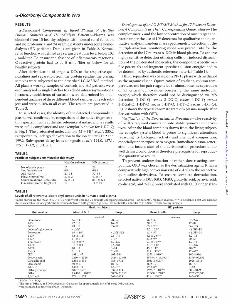
In selected cases, the identity of the detected compounds inplasma was confirmed by comparison of the native fragmenta-tion spectrum with authentic reference standards. The resultswere in full compliance and are exemplarily shown for 1-DG-Qin Fig. 1. The protonated molecular ion [M � H]� at m/z 235.2is expected to undergo dehydration to the ion at m/z 217.2 and199.2. Subsequent decay leads to signals at m/z 191.0, 187.1,175.1, 171.2, and 158.1.
Development of an LC-MS/MS Method for 17 Relevant Dicar-bonyl Compounds as Their Corresponding Quinoxalines—Thecomplex matrix and the low concentration of most target ana-lytes hamper the use of UV detectors for qualitative and quan-titative analysis. Tandem mass spectrometric detection in themultiple-reaction monitoring mode was prerequisite for thedetection of the 17 relevant �-DCs in blood plasma. To achievehighly sensitive detection utilizing collision-induced dissocia-tion of the protonated molecules, the compound-specific ori-fice potentials and fragment-specific collision energies had tobe determined by authentic reference material (Table 1).
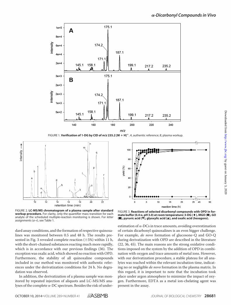
HPLC separation was based on a RP-18 phase with methanolas the organic eluent. Optimization of gradient, column tem-perature, and ion pair reagent led to almost baseline separationof all critical quinoxalines possessing the same molecularweight, which therefore could not be distinguished via MSdetection (1-DG-Q versus 3-DG-Q versus 4-DG-Q versus3-DGal-Q, 1-DP-Q versus 3-DP-Q, 1-DT-Q versus 3-DT-Q).Fig. 2 shows the typical chromatogram of a plasma sample afterderivatization with OPD.
Verification of the Derivatization Procedure—The reactivityof �-DCs required conversion into stable quinoxaline deriva-tives. After the blood sample is drawn from the living subject,the complex system blood is prone to significant alterationsregarding its biological activity and chemical composition,especially under exposure to oxygen. Immediate plasma gener-ation and instant start of the derivatization procedure underwell defined conditions is therefore prerequisite for reproduc-ible quantitative results.
To prevent underestimation of rather slow reacting com-pounds, OPD was chosen as the derivatization agent. It has acomparatively high conversion rate of �-DCs to the respectivequinoxaline derivatives. To ensure complete derivatization,selected native �-DCs (GO, MGO, glyoxylic acid, pyruvic acid,oxalic acid, and 3-DG) were incubated with OPD under stan-
TABLE 2Profile of subjects examined in this study
Healthy subjects HD patients
No. of participants 15 24Sex, female/male 6/9 6/18Age (years) 24–34 30–85HbA1c (mmol/mol) 37 � 2 40 � 17Serum creatinine (�mol/liter) 82 � 13 861 � 322C-reactive protein (mg/liter) �5 11 � 21
TABLE 3Levels of all relevant �-dicarbonyl compounds in human blood plasmaValues shown are the mean � S.D. of 15 healthy subjects and 24 patients undergoing hemodialysis (HD patients), replicate analyses, n � 3. Student’s t test was used forstatistical evaluation of significant differences between both groups: *, p � 0.01 versus healthy subjects; **, p � 0.001 versus healthy subjects.
QuinoxalineHealthy subjects HD patients
Mean � S.D. Range Mean � S.D. Range
pmol/ml pmol/mlGlucosone 46 � 11 28–67 96 � 49** 57–2761-DG 22 � 3 16–28 30 � 16 12–823-DG 43 � 5 35–56 65 � 20** 36–125Lederer’s glucosone �LOD 7.0 � 2.5a �LOD–13Pentosone 15 � 10a �LOD–32 11 � 5a �LOD–231-DP 3.6 � 1.2a 2.6–7.8 6.2 � 2.6a** 3.2–153-DP 11 � 2 9–17 33 � 9** 20–53Threosone 5.4 � 0.7a 4.2–6.6 9.0 � 3.5a** 4.2–191-DT 3.1 � 0.3 2.6–3.8 3.8 � 1.0* 2.0–6.63-DT 10 � 2 8–14 45 � 11** 28–75MGO 61 � 7 51–76 219 � 129** 42–617GO 491 � 47 405–564 1273 � 980** 400–4914Pyruvic acid 7250 � 2549 2818–12,038 35,874 � 19,080** 8399–97,935Glyoxylic acid 1264 � 353 783–1942 2031 � 608** 1026–3514Oxalic acid 40 � 21 18–82 36 � 12 16–703,4-DDP 6.0 � 1.0 4.4–7.7 19 � 8** 6–42DHA precursor 687 � 351b 337–1303 1925 � 1106b** 308–4829DHA 15,400 � 4019b 6809–23,967 12,538 � 7183b 2737–26,6802,3-DKG 1741 � 674c 567–2669 411 � 238c** 145–937
a LOD � � � LOQ.b The sum of DHA-Q and DHA precursor-Q account for approximately 38% of the true DHA content.c Values adjusted as described under “Disussion.”
�-Dicarbonyl Compounds in Vivo
28680 JOURNAL OF BIOLOGICAL CHEMISTRY VOLUME 289 • NUMBER 41 • OCTOBER 10, 2014
by guest on January 3, 2020http://w
ww
.jbc.org/D
ownloaded from
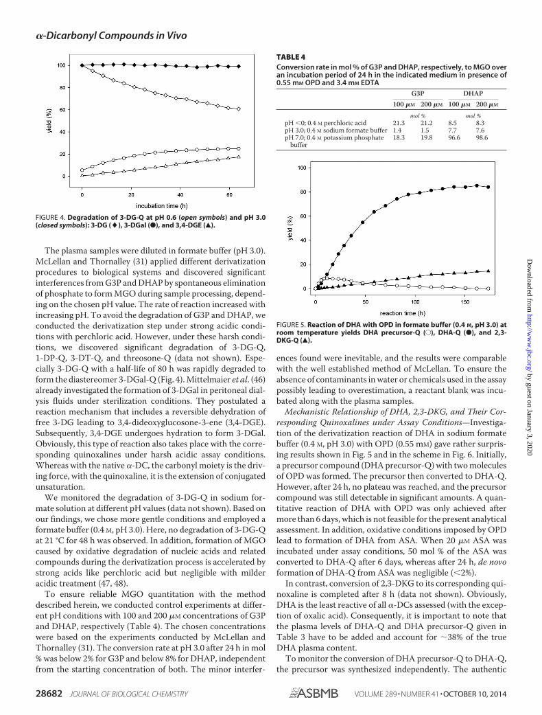
dard assay conditions, and the formation of respective quinoxa-lines was monitored between 0.5 and 48 h. The results pre-sented in Fig. 3 revealed complete reaction (�5%) within 11 h,with the short-chained substances reacting much more rapidly,which is in accordance with our previous findings (36). Theexception was oxalic acid, which showed no reaction with OPD.Furthermore, the stability of all quinoxaline compoundsincluded in our method was monitored with authentic refer-ences under the derivatization conditions for 24 h. No degra-dation was observed.
In addition, the derivatization of a plasma sample was mon-itored by repeated injection of aliquots and LC-MS/MS ana-lyses of the complete �-DC spectrum. Besides the risk of under-
estimation of �-DCs in trace amounts, avoiding overestimationof certain dicarbonyl quinoxalines is an even bigger challenge.For example, de novo formation of glucosone-Q and GO-Qduring derivatization with OPD are described in the literature(22, 36, 45). The main reasons are the strong oxidative condi-tions imposed on the system by the addition of OPD in combi-nation with oxygen and trace amounts of metal ions. However,with our derivatization procedure, a stable plateau for all ana-lytes was reached within the relevant incubation time, indicat-ing no or negligible de novo formation in the plasma matrix. Inthis regard, it is important to note that the incubation tookplace under argon atmosphere to minimize the impact of oxy-gen. Furthermore, EDTA as a metal ion-chelating agent waspresent in the assay.
FIGURE 1. Verification of 1-DG by CID of m/z 235.2 [M � H]�. A, authentic reference; B, plasma workup.
FIGURE 2. LC-MS/MS chromatogram of a plasma sample after standardworkup procedure. For clarity, only the quantifier mass transition for eachanalyte of the scheduled multiple-reaction monitoring is shown. For letterassignments (a–r), see Table 1.
FIGURE 3. Reactions of selected dicarbonyl compounds with OPD in for-mate buffer (0.4 M, pH 3.0) at room temperature: 3-DG (�), MGO (●), GO(f), pyruvic acid (�), glyoxylic acid (Œ), and oxalic acid (hexagons).
�-Dicarbonyl Compounds in Vivo
OCTOBER 10, 2014 • VOLUME 289 • NUMBER 41 JOURNAL OF BIOLOGICAL CHEMISTRY 28681
by guest on January 3, 2020http://w
ww
.jbc.org/D
ownloaded from
The plasma samples were diluted in formate buffer (pH 3.0).McLellan and Thornalley (31) applied different derivatizationprocedures to biological systems and discovered significantinterferences from G3P and DHAP by spontaneous eliminationof phosphate to form MGO during sample processing, depend-ing on the chosen pH value. The rate of reaction increased withincreasing pH. To avoid the degradation of G3P and DHAP, weconducted the derivatization step under strong acidic condi-tions with perchloric acid. However, under these harsh condi-tions, we discovered significant degradation of 3-DG-Q,1-DP-Q, 3-DT-Q, and threosone-Q (data not shown). Espe-cially 3-DG-Q with a half-life of 80 h was rapidly degraded toform the diastereomer 3-DGal-Q (Fig. 4). Mittelmaier et al. (46)already investigated the formation of 3-DGal in peritoneal dial-ysis fluids under sterilization conditions. They postulated areaction mechanism that includes a reversible dehydration offree 3-DG leading to 3,4-dideoxyglucosone-3-ene (3,4-DGE).Subsequently, 3,4-DGE undergoes hydration to form 3-DGal.Obviously, this type of reaction also takes place with the corre-sponding quinoxalines under harsh acidic assay conditions.Whereas with the native �-DC, the carbonyl moiety is the driv-ing force, with the quinoxaline, it is the extension of conjugatedunsaturation.
We monitored the degradation of 3-DG-Q in sodium for-mate solution at different pH values (data not shown). Based onour findings, we chose more gentle conditions and employed aformate buffer (0.4 M, pH 3.0). Here, no degradation of 3-DG-Qat 21 °C for 48 h was observed. In addition, formation of MGOcaused by oxidative degradation of nucleic acids and relatedcompounds during the derivatization process is accelerated bystrong acids like perchloric acid but negligible with milderacidic treatment (47, 48).
To ensure reliable MGO quantitation with the methoddescribed herein, we conducted control experiments at differ-ent pH conditions with 100 and 200 �M concentrations of G3Pand DHAP, respectively (Table 4). The chosen concentrationswere based on the experiments conducted by McLellan andThornalley (31). The conversion rate at pH 3.0 after 24 h in mol% was below 2% for G3P and below 8% for DHAP, independentfrom the starting concentration of both. The minor interfer-
ences found were inevitable, and the results were comparablewith the well established method of McLellan. To ensure theabsence of contaminants in water or chemicals used in the assaypossibly leading to overestimation, a reactant blank was incu-bated along with the plasma samples.
Mechanistic Relationship of DHA, 2,3-DKG, and Their Cor-responding Quinoxalines under Assay Conditions—Investiga-tion of the derivatization reaction of DHA in sodium formatebuffer (0.4 M, pH 3.0) with OPD (0.55 mM) gave rather surpris-ing results shown in Fig. 5 and in the scheme in Fig. 6. Initially,a precursor compound (DHA precursor-Q) with two moleculesof OPD was formed. The precursor then converted to DHA-Q.However, after 24 h, no plateau was reached, and the precursorcompound was still detectable in significant amounts. A quan-titative reaction of DHA with OPD was only achieved aftermore than 6 days, which is not feasible for the present analyticalassessment. In addition, oxidative conditions imposed by OPDlead to formation of DHA from ASA. When 20 �M ASA wasincubated under assay conditions, 50 mol % of the ASA wasconverted to DHA-Q after 6 days, whereas after 24 h, de novoformation of DHA-Q from ASA was negligible (�2%).
In contrast, conversion of 2,3-DKG to its corresponding qui-noxaline is completed after 8 h (data not shown). Obviously,DHA is the least reactive of all �-DCs assessed (with the excep-tion of oxalic acid). Consequently, it is important to note thatthe plasma levels of DHA-Q and DHA precursor-Q given inTable 3 have to be added and account for 38% of the trueDHA plasma content.
To monitor the conversion of DHA precursor-Q to DHA-Q,the precursor was synthesized independently. The authentic
FIGURE 5. Reaction of DHA with OPD in formate buffer (0.4 M, pH 3.0) atroom temperature yields DHA precursor-Q (E), DHA-Q (●), and 2,3-DKG-Q (Œ).
TABLE 4Conversion rate in mol % of G3P and DHAP, respectively, to MGO overan incubation period of 24 h in the indicated medium in presence of0.55 mM OPD and 3.4 mM EDTA
G3P DHAP100 �M 200 �M 100 �M 200 �M
mol % mol %pH �0; 0.4 M perchloric acid 21.3 21.2 8.5 8.3pH 3.0; 0.4 M sodium formate buffer 1.4 1.5 7.7 7.6pH 7.0; 0.4 M potassium phosphate
buffer18.3 19.8 96.6 98.6
FIGURE 4. Degradation of 3-DG-Q at pH 0.6 (open symbols) and pH 3.0(closed symbols): 3-DG (�), 3-DGal (●), and 3,4-DGE (Œ).
�-Dicarbonyl Compounds in Vivo
28682 JOURNAL OF BIOLOGICAL CHEMISTRY VOLUME 289 • NUMBER 41 • OCTOBER 10, 2014
by guest on January 3, 2020http://w
ww
.jbc.org/D
ownloaded from
reference DHA precursor-Q was then incubated under assayconditions (Fig. 7). A half-life of 5 h was observed. After 24 h,81% of DHA precursor-Q was converted to DHA-Q. Residualamounts of DHA precursor-Q (10% based on initial concentra-tion) remained and had to be considered for the calculation ofDHA concentration. Interestingly, also small amounts of 2,3-
DKG-Q were detected (7%). A reverse reaction of the quinoxa-lines to form free DHA with subsequent hydrolytic opening ofthe lactone ring system to yield 2,3-DKG and its correspondingquinoxaline 2,3-DKG-Q can be excluded. Thus, DHA-Q has tobe hydrolyzed slowly to form 2,3-DKG-Q directly. To furtherinvestigate the relationship of the two quinoxalines, 2,3-DKG-Q was incubated under assay conditions, including theprotein precipitation step with TFA after 24 h. The results inFig. 8 show conversion of 2,3-DKG-Q to DHA-Q, which wassignificantly triggered at the lower pH during the 1-h precipi-tation step. 2,3-DKG-Q yields 25% DHA-Q after the com-plete workup procedure. Obviously, there is a steady statebetween DHA-Q and 2,3-DKG-Q with preferred DHA-Q for-mation as the more stable product under acidic conditions.
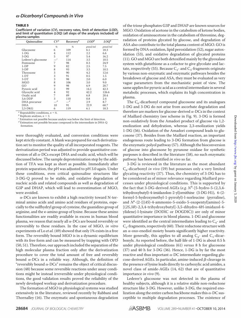
Validation of the Proposed Method—The limit of detectionand limit of quantitation with all steps of the analytical methodincluded were calculated as described under “ExperimentalProcedures.” The limit of detection differs from 0.5 to 42.2pmol/liter. The accuracy of the method was determined as therecovery rate at three different concentration levels and rangedbetween 82 and 120%. Repeatability was expressed as the coef-ficient of variation and was 10% or better. The complete valida-tion data are shown in Table 5.
DISCUSSION
Up to now, only GO, MGO, 3-DG, and DHA were quanti-tated in human plasma using established methods, although thespectrum of �-DCs in vivo is supposed to be far more complex.Major endogenous sources are the degradation of blood glu-cose and ascorbic acid. As a result, the corresponding degrada-tion products already identified in model experiments shouldalso be present in vivo. Beyond that, lipid peroxidation as well asenzymatic and non-enzymatic pathways irrespective of Mail-lard reactions contribute to the content of �-DCs. It is specu-lated that even exogenous sources like foods add to the plasmalevel of �-DCs.
Because derivatization with OPD is prone to both underesti-mation of �-DC levels and de novo formation during thederivatization procedure, recovery levels for each compound
FIGURE 6. Mechanistic relationship of DHA, 2,3-DKG, and their corre-sponding derivatization products.
FIGURE 7. Formation of DHA-Q (●) and 2,3-DKG-Q (Œ) starting from DHAprecursor-Q (E) under standard workup conditions.
FIGURE 8. Degradation of 2,3-DKG-Q (Œ) under standard workup condi-tions yields DHA-Q (●).
�-Dicarbonyl Compounds in Vivo
OCTOBER 10, 2014 • VOLUME 289 • NUMBER 41 JOURNAL OF BIOLOGICAL CHEMISTRY 28683
by guest on January 3, 2020http://w
ww
.jbc.org/D
ownloaded from
were thoroughly evaluated, and conversion conditions werekept strictly constant. A blank was prepared for each derivatiza-tion set to monitor the quality of all incorporated reagents. Thederivatization period was adjusted to provide quantitative con-version of all �-DCs except DHA and oxalic acid, which will bediscussed below. The sample deproteinization step by the addi-tion of TFA was kept as short as possible. Immediately afterprotein separation, the pH was adjusted to pH 3.0 again. Underthese conditions, even critical quinoxaline structures like3-DG-Q proved to be stable, and oxidative degradation ofnucleic acids and related compounds as well as degradation ofG3P and DHAP, which will lead to overestimation of MGO,were avoided.
�-DCs are known to exhibit a high reactivity toward N-ter-minal amino acids and amino acid residues of proteins, espe-cially to the sulfhydryl group of cysteine, the guanidino group ofarginine, and the �-amino group of lysine. Because these aminofunctionalities are readily available in excess in human bloodplasma, we reason that nearly all �-DCs are bound reversibly orirreversibly to these residues. In the case of MGO, in vitroexperiments of Lo et al. (49) showed that only 1% exists in a freeform. The reversibly bound MGO is in a dynamic equilibriumwith its free form and can be measured by trapping with OPD(50, 51). Therefore, our approach included the separation of thehigh molecular plasma fraction only after the derivatizationprocedure to cover the total amount of free and reversiblybound �-DCs in a reliable way. Although, the definition of“reversibly bound �-DCs” remains the object of critical discus-sion (48) because some reversible reactions under assay condi-tions might be instead irreversible under physiological condi-tions, the good validation results proved the reliability of thenewly developed workup and derivatization procedure.
The formation of MGO in physiological systems was studiedextensively in the literature, reviewed recently by Rabbani andThornalley (16). The enzymatic and spontaneous degradation
of the triose phosphates G3P and DHAP are known sources forMGO. Oxidation of acetone in the catabolism of ketone bodies,oxidation of aminoacetone in the catabolism of threonine, deg-radation of proteins glycated by glucose, and degradation ofASA also contribute to the total plasma content of MGO. GO isformed by DNA oxidation, lipid peroxidation (52), sugar autox-idation (53), and oxidative degradation of glycated proteins(11). GO and MGO are both detoxified mainly by the glyoxalasesystem with glutathione as a cofactor to give glycolate and lac-tate, respectively (55). Because C2- and C3-fragments originateby various non-enzymatic and enzymatic pathways besides thebreakdown of glucose and ASA, they must be evaluated as veryvague parameters from the mechanistic point of view. Thesame applies for pyruvic acid as a central intermediate in severalmetabolic processes, which explains its high concentration inplasma.
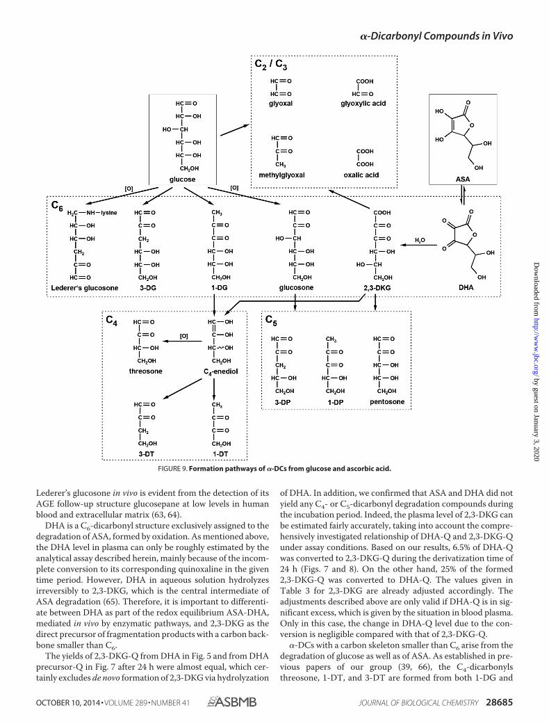
The C6-dicarbonyl compound glucosone and its analogues1-DG and 3-DG do not arise from ascorbate degradation andtherefore are markers for glucose-derived �-DCs in the contextof Maillard chemistry (see scheme in Fig. 9). 3-DG is formednon-oxidatively from the Amadori product of glucose via 1,2-enolization and dehydration, whereas 2,3-enolization yields1-DG (56). Oxidation of the Amadori compound leads to glu-cosone (37). Besides from the Maillard reaction, an importantendogenous route leading to 3-DG formation from glucose isthe enzymatic polyol pathway (57). Although the bioconversionof glucose into glucosone by pyranose oxidase for syntheticpurposes is described in the literature (58), no such enzymaticpathway has been identified in vivo so far.
3-DG is reviewed in the literature as the most abundantC6-dicarbonyl in vivo (59) but possesses only a very limitedglycating reactivity (37). Thus, the chemistry of 3-DG has tobe considered as of minor relevance regarding Maillard pro-cesses under physiological conditions. This is supported bythe fact that 3-DG-derived AGEs (e.g. N�-[5-hydro-5-(2,3,4-trihydroxybutyl)-4-imidazolon-2-yl]ornithine (3-DG-H1), 6-(2-formyl-5-hydroxymethyl-1-pyrrolyl)-l-norleucine (pyrraline),and N�-{2-{[(4S)-4-ammonio-5-oxido-5-oxopentyl]amino}-5-[(2S,3R)-2,3,4-trihydroxybutyl]-3,5-dihydro-4H-imidazol-4-ylidene}-l-lysinate (DODIC or DOGDIC)) are only of minorquantitative importance in blood plasma. 1-DG and glucosonewere identified as the central intermediates leading to C4- andC5-fragments, respectively (60). Their reductone structure withan �-oxo-enediol moiety boasts significantly higher reactivity.More generally, this applies to all analog C4- and C5-dicar-bonyls. As reported before, the half-life of 1-DG is about 0.5 hunder physiological conditions (61) versus 8 h for glucosone(37) and 40 h for 3-DG (36). Hence, 1-DG is by far the mostreactive and thus important �-DC intermediate regarding glu-cose-derived AGEs. In particular, amine-induced �-cleavage inthe presence of lysine leads directly to carboxylic acid amides, anovel class of amide-AGEs (14, 62) that are of quantitativeimportance in vivo (9).
Lederer’s glucosone was not detected in the plasma ofhealthy subjects, although it is a relative stable non-reductonestructure like 3-DG. However, unlike 3-DG, the required eno-lization along the entire carbon backbone makes this �-DC sus-ceptible to multiple degradation processes. The existence of
TABLE 5Coefficient of variation (CV), recovery rates, limit of detection (LOD),and limit of quantitation (LOQ) (all steps of the analysis included) ofplasma samples
Quinoxaline CVa Recoveryb LODb LOQb
% % pmol/ml pmol/mlGlucosone 6 109 6.1 18.31-DG 4 112 2.2 6.63-DG 5 97 5.4 16.2Lederer’s glucosone —c 114 3.5 10.5Pentosone 7 98 8.3 24.91-DP 9 82 2.5 7.53-DP 8 93 3.5 10.5Threosone 3 90 4.2 12.61-DT 3 91 0.5 1.53-DT 3 98 2.0 6.0MGO 8 106 3.0 9.0GO 9 98 6.9 20.7Pyruvic acid 2 99 14.1 42.3Glyoxylic acid 4 92 42.2 126.6Oxalic acid 7 89 6.8 20.43,4-DDP 3 110 0.8 2.4DHA precursor —d —d 2.9 8.7DHA 10 91 22.9 68.72,3-DKG 6 119 6.1 18.3
a Repeatability conditions, n � 6.b Replicate analyses, n � 3.c Estimation not possible because analyte was below the limit of detection.d Estimation not possible because compound is the intermediate in DHA-Q
formation.
�-Dicarbonyl Compounds in Vivo
28684 JOURNAL OF BIOLOGICAL CHEMISTRY VOLUME 289 • NUMBER 41 • OCTOBER 10, 2014
by guest on January 3, 2020http://w
ww
.jbc.org/D
ownloaded from
Lederer’s glucosone in vivo is evident from the detection of itsAGE follow-up structure glucosepane at low levels in humanblood and extracellular matrix (63, 64).
DHA is a C6-dicarbonyl structure exclusively assigned to thedegradation of ASA, formed by oxidation. As mentioned above,the DHA level in plasma can only be roughly estimated by theanalytical assay described herein, mainly because of the incom-plete conversion to its corresponding quinoxaline in the giventime period. However, DHA in aqueous solution hydrolyzesirreversibly to 2,3-DKG, which is the central intermediate ofASA degradation (65). Therefore, it is important to differenti-ate between DHA as part of the redox equilibrium ASA-DHA,mediated in vivo by enzymatic pathways, and 2,3-DKG as thedirect precursor of fragmentation products with a carbon back-bone smaller than C6.
The yields of 2,3-DKG-Q from DHA in Fig. 5 and from DHAprecursor-Q in Fig. 7 after 24 h were almost equal, which cer-tainly excludes de novo formation of 2,3-DKG via hydrolyzation
of DHA. In addition, we confirmed that ASA and DHA did notyield any C4- or C5-dicarbonyl degradation compounds duringthe incubation period. Indeed, the plasma level of 2,3-DKG canbe estimated fairly accurately, taking into account the compre-hensively investigated relationship of DHA-Q and 2,3-DKG-Qunder assay conditions. Based on our results, 6.5% of DHA-Qwas converted to 2,3-DKG-Q during the derivatization time of24 h (Figs. 7 and 8). On the other hand, 25% of the formed2,3-DKG-Q was converted to DHA-Q. The values given inTable 3 for 2,3-DKG are already adjusted accordingly. Theadjustments described above are only valid if DHA-Q is in sig-nificant excess, which is given by the situation in blood plasma.Only in this case, the change in DHA-Q level due to the con-version is negligible compared with that of 2,3-DKG-Q.
�-DCs with a carbon skeleton smaller than C6 arise from thedegradation of glucose as well as of ASA. As established in pre-vious papers of our group (39, 66), the C4-dicarbonylsthreosone, 1-DT, and 3-DT are formed from both 1-DG and
FIGURE 9. Formation pathways of �-DCs from glucose and ascorbic acid.
�-Dicarbonyl Compounds in Vivo
OCTOBER 10, 2014 • VOLUME 289 • NUMBER 41 JOURNAL OF BIOLOGICAL CHEMISTRY 28685
by guest on January 3, 2020http://w
ww
.jbc.org/D
ownloaded from
2,3-DKG via �-dicarbonyl cleavage with an C4-enediol as thereactive intermediate. Oxidation of the latter leads to threosone,whereas dehydration at C3 results in 3-DT. The enediol may alsoundergo isomerization to give 1-DT in an equivalent reaction. As aconsequence under deaeration, which represents the situation invivo, 3-DT was the prominent structure because the reductone1-DT has to be considered a much more reactive and, thus, shortlived intermediate.
It has to be noted that differentiation between l-threo-pen-tos-2-ulose (arising from ASA and often referred to asxylosone) and the C4-stereoisomer D-erythro-pentos-2-ulose(arising from glucose) was not possible. For the applied LC-MS/MS method, MRM parameters were determined withL-threo-pentos-2-ulose. In model incubations of glucose andlysine, a signal with retention time and mass transitions identi-cal to those of L-threo-pentos-2-ulose was detected (data notshown). By definition, with glucose as precursor, the detectedcompound has to be D-erythro-pentos-2-ulose. Thus, bothstructures coelute and are therefore summarized under theterm pentosone regardless of its origin. Yet, the plasma levels ofpentosone were comparatively high and did not fit into thepicture. Decarboxylation is a well established mechanism ofASA degradation and leads to C5-compounds, including pen-tosone (65, 67), but under physiological conditions, the forma-tion of C4-dicarbonyls from 2,3-DKG is favored (25, 39). Glu-cose-derived pentosone stems from glucosone by the samemechanism of hydrolytic �-cleavage as threosone from 1-DG asthe precursor. After isomerization and hydration, formic acid iscleaved off and gives an C5-1,2-enediol as the reactive interme-diate. Oxidation leads to pentosone, whereas water eliminationyields 3-DP or, after 2,3-enolization, 1-DP (60). Considering theneed for an oxidation step in order to obtain pentosone fromboth 2,3-DKG or glucosone, formation of 1-DP and 3-DPshould be favored. The results therefore strongly suggest anadditional alternative source for pentosone formation in vivo.
In Maillard chemistry, glyoxylic acid is assigned to disaccha-ride chemistry (38) but can also arise from oxidation of glyoxal(68) and degradation of DHA (65, 69). However, this cannotaccount solely for the plasma levels, which were 2.5-fold higherthan GO and in the same range as 2,3-DKG. An alternativesource is the degradation of hydroxyproline with the subse-quent glyoxylate metabolism in the human organism, which israther complex, involving several enzymatic and non-enzy-matic reactions, and has been subject to recent investigation(54).
Oxalic acid can originate via �-dicarbonyl fragmentation aswell as via oxidative �-DC cleavage from 2,3-DKG and is themain degradation product of the latter (39). In addition, oxalateis also part of the glyoxylate metabolism mentioned above.However, under assay conditions, the dicarboxylic acid oxalicacid is not converted to its corresponding quinoxaline. This isexpected, because at the chosen workup pH, the carboxylic acidgroups do not show sufficient carbonyl activity to react withOPD. Consequently, there must be alternative precursors foroxalic acid-Q formation to oxalic acid itself. 3,4-DDP is aknown intermediate of maltose degradation but was found inneither the glucose nor ASA reaction systems (38). Hence, theorigin of the detected 3,4-DDP-Q remains unknown.
The presented LC-MS/MS method provides for the first timethe opportunity to identify and quantitate the complete spec-trum of relevant �-DCs in plasma of healthy human subjects ina single chromatographic run. For 14 compounds, the plasmalevels were determined. Three compounds were below the limitof quantitation but were unequivocally identified. 10 sub-stances have not been reported for human plasma samplesbefore. To evaluate the clinical relevance of the assay describedherein, an initial set of 24 uremic patients undergoing hemodi-alysis was analyzed. Uremia is related to an increase in oxidativeand carbonyl stress and thus should lead to a clear shift in thedicarbonyl spectrum. Indeed, most �-DCs were considerablyhigher in HD patients. Glucose-derived glucosone exhibited a2-fold increase, which is expected under conditions of elevatedoxidative stress. In contrast, 1-DG does not require an oxida-tion step for its formation and remained nearly at the level ofhealthy subjects. Interestingly, plasma levels of 2,3-DKG wereconsiderably decreased in HD patients. This must be explainedby a significantly accelerated degradation via oxidative path-ways. GO and MGO are further compounds of published inter-est in regard to certain chronic diseases like uremia. A 3-foldelevation of GO and a 4-fold increase of MGO were observed inuremic plasma. This is in line with the literature (27, 29, 32),although the absolute values differ significantly, depending onthe respective study. The assessment of �-DC plasma levelsdepends strongly on the analytical approach, specifically onworkup conditions, derivatization procedure, and chromato-graphic method. Most importantly, the derivatization was con-ducted in the presence of protein to assess both free and revers-ibly bound �-DCs. Therefore, a direct comparison of theplasma levels of the present study and those reported previouslyis not possible.
Nevertheless, validation of the present method for the detec-tion of �-DC compounds in plasma has been carried out exten-sively regarding the formation of artifacts and mechanistic rela-tionships to exclude false quantitative data. In general, theelevated levels of �-DCs found explain the elevated levels ofAGEs in uremia. The newly developed method has now to beextended to follow-up studies with patients with various com-plications. The results for a wide range of highly reactive car-bonyl intermediates will help us to understand the complexmechanisms and factors that influence �-DC formation andconsequently open new perspectives regarding the formationand relevance of AGE chemistry in vivo.
Acknowledgment—We thank Dr. J. Schmidt (Leibnitz Institute ofPlant Biochemistry, Halle, Germany) for performing accurate massdetermination.
REFERENCES1. Monnier, V. M., Mustata, G. T., Biemel, K. L., Reihl, O., Lederer, M. O.,
Zhenyu, D., and Sell, D. R. (2005) Cross-linking of the extracellular matrixby the maillard reaction in aging and diabetes: an update on “a puzzlenearing resolution”. Ann. N.Y. Acad. Sci. 1043, 533–544
2. Baynes, J. W. (2001) The role of AGEs in aging: causation or correlation.Exp. Gerontol. 36, 1527–1537
3. Tessier, F. J., and Birlouez-Aragon, I. (2012) Health effects of dietary Mail-lard reaction products: the results of ICARE and other studies. Amino
�-Dicarbonyl Compounds in Vivo
28686 JOURNAL OF BIOLOGICAL CHEMISTRY VOLUME 289 • NUMBER 41 • OCTOBER 10, 2014
by guest on January 3, 2020http://w
ww
.jbc.org/D
ownloaded from
Acids 42, 1119 –11314. Degenhardt, T. P., Brinkmann-Frye, E., Thorpe, S. R., and Baynes, J. W.
(1998) Role of carbonyl stress in aging and age-related diseases. in TheMaillard Reaction in Foods and Medicine (O’Brien, J., ed) pp. 3–10, RoyalSociety of Chemistry, London
5. Hamada, Y., Araki, N., Koh, N., Nakamura, J., Horiuchi, S., and Hotta, N.(1996) Rapid formation of advanced glycation end products by interme-diate metabolites of glycolytic pathway and polyol pathway. Biochem. Bio-phys. Res. Commun. 228, 539 –543
6. Sell, D. R., Lapolla, A., Odetti, P., Fogarty, J., and Monnier, V. M. (1992)Pentosidine formation in skin correlates with severity of complications inindividuals with long-standing IDDM. Diabetes 41, 1286 –1292
7. McCance, D. R., Dyer, D. G., Dunn, J. A., Bailie, K. E., Thorpe, S. R., Baynes,J. W., and Lyons, T. J. (1993) Maillard reaction products and their relationto complications in insulin-dependent diabetes mellitus. J. Clin. Invest. 91,2470 –2478
8. Sugiyama, S., Miyata, T., Ueda, Y., Tanaka, H., Maeda, K., Kawashima, S.,Van Ypersele de Strihou, C., and Kurokawa, K. (1998) Plasma levels ofpentosidine in diabetic patients: an advanced glycation end product. J. Am.Soc. Nephrol. 9, 1681–1688
9. Henning, C., Smuda, M., Girndt, M., Ulrich, C., and Glomb, M. A. (2011)Molecular basis of maillard amide-advanced glycation end product (AGE)formation in vivo. J. Biol. Chem. 286, 44350 – 44356
10. Baynes, J. W., and Thorpe, S. R. (1999) Role of oxidative stress in diabeticcomplications: a new perspective on an old paradigm. Diabetes 48, 1–9
11. Glomb, M. A., and Monnier, V. M. (1995) Mechanism of protein modifi-cation by glyoxal and glycolaldehyde, reactive intermediates of the Mail-lard reaction. J. Biol. Chem. 270, 10017–10026
12. Glomb, M. A., and Lang, G. (2001) Isolation and characterization of glyox-al-arginine modifications. J. Agric. Food Chem. 49, 1493–1501
13. Smuda, M., and Glomb, M. A. (2013) Fragmentation pathways duringMaillard-induced carbohydrate degradation. J. Agric. Food Chem. 61,10198 –10208
14. Smuda, M., Voigt, M., and Glomb, M. A. (2010) Degradation of 1-deoxy-D-erythro-hexo-2,3-diulose in the presence of lysine leads to formation ofcarboxylic acid amides. J. Agric. Food Chem. 58, 6458 – 6464
15. Ahmed, N., and Thornalley, P. J. (2002) Chromatographic assay of glyca-tion adducts in human serum albumin glycated in vitro by derivatizationwith 6-aminoquinolyl-N-hydroxysuccinimidyl-carbamate and intrinsicfluorescence. Biochem. J. 364, 15–24
16. Rabbani, N., and Thornalley, P. J. (2012) Methylglyoxal, glyoxalase 1 andthe dicarbonyl proteome. Amino Acids 42, 1133–1142
17. Linden, T., Musi, B., Jarkelid, L., Forsback, G., Kjellstrand, P., Deppisch, R.,and Wieslander, A. (2001) Glucose degradation products in peritonealdialysis fluids may have both local and systemic effects: a study of residualfluid and mesothelial cells. Perit. Dial. Int. 21, 607– 610
18. Tauer, A., Bender, T. O., Fleischmann, E. H., Niwa, T., Jorres, A., andPischetsrieder, M. (2005) Fate of the glucose degradation products 3-de-oxyglucosone and glyoxal during peritoneal dialysis. Mol. Nutr. Food Res.49, 710 –715
19. Ankrah, N. A., and Appiah-Opong, R. (1999) Toxicity of low levels ofmethylglyoxal: depletion of blood glutathione and adverse effect on glu-cose tolerance in mice. Toxicol. Lett. 109, 61– 67
20. Degen, J., Hellwig, M., and Henle, T. (2012) 1,2-Dicarbonyl compounds incommonly consumed foods. J. Agric. Food Chem. 60, 7071–7079
21. Arribas-Lorenzo, G., and Morales, F. J. (2010) Analysis, distribution, anddietary exposure of glyoxal and methylglyoxal in cookies and their rela-tionship with other heat-induced contaminants. J. Agric. Food Chem. 58,2966 –2972
22. Gensberger, S., Mittelmaier, S., Glomb, M. A., and Pischetsrieder, M.(2012) Identification and quantification of six major �-dicarbonyl processcontaminants in high-fructose corn syrup. Anal. Bioanal. Chem. 403,2923–2931
23. Schalkwijk, C. G., Posthuma, N., ten Brink, H. J., ter Wee, P. M., andTeerlink, T. (1999) Induction of 1,2-dicarbonyl compounds, intermedi-ates in the formation of advanced glycation end-products, during heat-sterilization of glucose-based peritoneal dialysis fluids. Perit. Dial. Int. 19,325–333
24. Mittelmaier, S., Funfrocken, M., Fenn, D., Berlich, R., and Pischetsrieder,M. (2011) Quantification of the six major �-dicarbonyl contaminants inperitoneal dialysis fluids by UHPLC/DAD/MSMS. Anal. Bioanal. Chem.401, 1183–1193
25. Nemet, I., and Monnier, V. M. (2011) Vitamin C degradation products andpathways in the human lens. J. Biol. Chem. 286, 37128 –37136
26. Mirza, M. A., Kandhro, A. J., Memon, S. Q., Khuhawar, M. Y., and Arain,R. (2007) Determination of glyoxal and methylglyoxal in the serum ofdiabetic patients by MEKC using stilbenediamine as derivatizing reagent.Electrophoresis 28, 3940 –3947
27. Odani, H., Shinzato, T., Matsumoto, Y., Usami, J., and Maeda, K. (1999)Increase in three �,�-dicarbonyl compound levels in human uremic plas-ma: specific in vivo determination of intermediates in advanced Maillardreaction. Biochem. Biophys. Res. Commun. 256, 89 –93
28. Lapolla, A., Reitano, R., Seraglia, R., Sartore, G., Ragazzi, E., and Traldi, P.(2005) Evaluation of advanced glycation end products and carbonyl com-pounds in patients with different conditions of oxidative stress. Mol. Nutr.Food Res. 49, 685– 690
29. Lapolla, A., Flamini, R., Lupo, A., Arico, N. C., Rugiu, C., Reitano, R.,Tubaro, M., Ragazzi, E., Seraglia, R., and Traldi, P. (2005) Evaluation ofglyoxal and methylglyoxal levels in uremic patients under peritoneal dial-ysis. Ann. N.Y. Acad. Sci. 1043, 217–224
30. Knecht, K. J., Feather, M. S., and Baynes, J. W. (1992) Detection of 3-de-oxyfructose and 3-deoxyglucosone in human urine and plasma: evidencefor intermediate stages of the Maillard reaction in vivo. Arch. Biochem.Biophys. 294, 130 –137
31. McLellan, A. C., Phillips, S. A., and Thornalley, P. J. (1992) The assay ofmethylglyoxal in biological systems by derivatization with 1,2-diamino-4,5-dimethoxybenzene. Anal. Biochem. 206, 17–23
32. Nakayama, K., Nakayama, M., Iwabuchi, M., Terawaki, H., Sato, T.,Kohno, M., and Ito, S. (2008) Plasma �-oxoaldehyde levels in diabetic andnondiabetic chronic kidney disease patients. Am. J. Nephrol. 28, 871– 878
33. Lopez-Anaya, A., and Mayersohn, M. (1987) Ascorbic and dehydroascor-bic acids simultaneously quantified in biological fluids by liquid chroma-tography with fluorescence detection, and comparison with a colorimetricassay. Clin. Chem. 33, 1874 –1878
34. Margolis, S. A., Ziegler, R. G., and Helzlsouer, K. J. (1991) Ascorbic anddehydroascorbic acid measurement in human serum and plasma. Am. J.Clin. Nutr. 54, 1315S–1318S
35. Lykkesfeldt, J., Loft, S., Nielsen, J. B., and Poulsen, H. E. (1997) Ascorbicacid and dehydroascorbic acid as biomarkers of oxidative stress caused bysmoking. Am. J. Clin. Nutr. 65, 959 –963
36. Glomb, M. A., and Tschirnich, R. (2001) Detection of �-dicarbonyl com-pounds in Maillard reaction systems and in vivo. J. Agric. Food Chem. 49,5543–5550
37. Gobert, J., and Glomb, M. A. (2009) Degradation of glucose: reinvestiga-tion of reactive �-dicarbonyl compounds. J. Agric. Food Chem. 57,8591– 8597
38. Smuda, M., and Glomb, M. A. (2011) Novel insights into the Maillardcatalyzed degradation of maltose. J. Agric. Food Chem. 59, 13254 –13264
39. Smuda, M., and Glomb, M. A. (2013) Maillard degradation pathways ofvitamin C. Angew. Chem. Int. Ed. Engl. 52, 4887– 4891
40. Hellwig, M., Degen, J., and Henle, T. (2010) 3-deoxygalactosone, a “new”1,2-dicarbonyl compound in milk products. J. Agric. Food Chem. 58,10752–10760
41. Henseke,G.,andBauer,C. (1959)HeterocyclischeVerbindungen,V.Chinoxalin-synthesen mit Osonhydrazonen. Chem. Ber. 10.1002/cber.19590920236
42. Otsuka, M., Kurata, T., and Arakawa, N. (1986) Isolation and character-ization of an intermediate product in the degradation of 2,3-diketo-L-gulonic acid. Agric. Biol. Chem. 50, 531–533
43. Arun, V., Robinson, P. P., Manju, S., Leeju, P., Varsha, G., Digna, V., andYusuff, K. K. M. (2009) A novel fluorescent bisazomethine dye derivedfrom 3-hydroxyquinoxaline-2-carboxaldehyde and 2,3-diaminomaleoni-trile. Dyes Pigments 82, 268 –275
44. Materials Testing Standards Committee NA 062 of the Deutsches Institutfuer Normung (German Institute for Standardization) (1994) DIN 32645;Chemical Analysis: Decision Limit, Detection Limit and DeterminationLimit; Estimation in Case of Repeatability; Terms, Methods, Evaluation,
�-Dicarbonyl Compounds in Vivo
OCTOBER 10, 2014 • VOLUME 289 • NUMBER 41 JOURNAL OF BIOLOGICAL CHEMISTRY 28687
by guest on January 3, 2020http://w
ww
.jbc.org/D
ownloaded from
Beuth Verlag, Berlin45. Mittelmaier, S., Funfrocken, M., Fenn, D., Fichert, T., and Pischetsrieder,
M. (2010) Identification and quantification of the glucose degradationproduct glucosone in peritoneal dialysis fluids by HPLC/DAD/MSMS.J. Chromatogr. B Analyt. Technol. Biomed. Life Sci. 878, 877– 882
46. Mittelmaier, S., Funfrocken, M., Fenn, D., and Pischetsrieder, M. (2011)3-Deoxygalactosone, a new glucose degradation product in peritoneal di-alysis fluids: identification, quantification by HPLC/DAD/MSMS and itspathway of formation. Anal. Bioanal. Chem. 399, 1689 –1697
47. Chaplen, F. W., Fahl, W. E., and Cameron, D. C. (1996) Detection ofmethylglyoxal as a degradation product of DNA and nucleic acid compo-nents treated with strong acid. Anal. Biochem. 236, 262–269
48. Chaplen, F. W., Fahl, W. E., and Cameron, D. C. (1998) Evidence of highlevels of methylglyoxal in cultured Chinese hamster ovary cells. Proc. Natl.Acad. Sci. U.S.A. 95, 5533–5538
49. Lo, T. W., Westwood, M. E., McLellan, A. C., Selwood, T., and Thornalley,P. J. (1994) Binding and modification of proteins by methylglyoxal underphysiological conditions. A kinetic and mechanistic study with N-�-acety-larginine, N-�-acetylcysteine, and N-�-acetyllysine, and bovine serum al-bumin. J. Biol. Chem. 269, 32299 –32305
50. Dhar, A., Desai, K., Liu, J., and Wu, L. (2009) Methylglyoxal, protein bind-ing and biological samples: are we getting the true measure? J. Chro-matogr. B Analyt. Technol. Biomed. Life Sci. 877, 1093–1100
51. Chaplen, F. W., Fahl, W. E., and Cameron, D. C. (1996) Method for deter-mination of free intracellular and extracellular methylglyoxal in animalcells grown in culture. Anal. Biochem. 238, 171–178
52. Fu, M. X., Requena, J. R., Jenkins, A. J., Lyons, T. J., Baynes, J. W., andThorpe, S. R. (1996) The advanced glycation end product, N-�-(car-boxymethyl)lysine, is a product of both lipid peroxidation and glycoxida-tion reactions. J. Biol. Chem. 271, 9982–9986
53. Wells-Knecht, K. J., Zyzak, D. V., Litchfield, J. E., Thorpe, S. R., and Baynes,J. W. (1995) Mechanism of autoxidative glycosylation: identification ofglyoxal and arabinose as intermediates in the autoxidative modification ofproteins by glucose. Biochemistry 34, 3702–3709
54. Duarte, N. C., Becker, S. A., Jamshidi, N., Thiele, I., Mo, M. L., Vo, T. D.,Srivas, R., and Palsson, B. Ø. (2007) Global reconstruction of the humanmetabolic network based on genomic and bibliomic data. Proc. Natl. Acad.Sci. U.S.A. 104, 1777–1782
55. Thornalley, P. J. (1998) Glutathione-dependent detoxification of �-oxoal-dehydes by the glyoxalase system: involvement in disease mechanisms andantiproliferative activity of glyoxalase I inhibitors. Chem. Biol. Interact.111, 137–151
56. Niwa, T. (1999) 3-Deoxyglucosone: metabolism, analysis, biological activity, andclinical implication. J. Chromatogr. B Biomed. Sci. Appl. 731, 23–36
57. Tsukushi, S., Katsuzaki, T., Aoyama, I., Takayama, F., Miyazaki, T.,Shimokata, K., and Niwa, T. (1999) Increased erythrocyte 3-DG and AGEsin diabetic hemodialysis patients: role of the polyol pathway. Kidney Int.55, 1970 –1976
58. Karmali, A., and Coelho, J. (2011) Bioconversion of D-glucose into D-glu-cosone by glucose 2-oxidase from Coriolus versicolor at moderate pres-sures. Appl. Biochem. Biotechnol. 163, 906 –917
59. Vistoli, G., De Maddis, D., Cipak, A., Zarkovic, N., Carini, M., and Aldini,G. (2013) Advanced glycoxidation and lipoxidation end products (AGEsand ALEs): an overview of their mechanisms of formation. Free Radic. Res.47, 3–27
60. Glomb, M. A., Gobert, J., and Voigt, M. (2010) Dicarbonyls from Maillarddegradation of glucose and maltose. ACS Symp. Ser. 1042, 35– 44
61. Glomb, M. A., and Pfahler, C. (2000) Synthesis of 1-deoxy-D-erythro-hexo-2,3-diulose, a major hexose Maillard intermediate. Carbohydr. Res.329, 515–523
62. Glomb, M. A., and Pfahler, C. (2001) Amides are novel protein modifica-tions formed by physiological sugars. J. Biol. Chem. 276, 41638 – 41647
63. Biemel, K. M., Friedl, D. A., and Lederer, M. O. (2002) Identification andquantification of major maillard cross-links in human serum albumin andlens protein. Evidence for glucosepane as the dominant compound. J. Biol.Chem. 277, 24907–24915
64. Sell, D. R., Biemel, K. M., Reihl, O., Lederer, M. O., Strauch, C. M., andMonnier, V. M. (2005) Glucosepane is a major protein cross-link of thesenescent human extracellular matrix. Relationship with diabetes. J. Biol.Chem. 280, 12310 –12315
65. Shin, D. B., and Feather, M. S. (1990) The degradation of L-ascorbic acid inneutral solutions containing oxygen. J. Carbohydr. Chem. 9, 461– 469
66. Voigt, M., and Glomb, M. A. (2009) Reactivity of 1-deoxy-D-erythro-hexo-2,3-diulose: a key intermediate in the Maillard chemistry of hexoses.J. Agric. Food Chem. 57, 4765– 4770
67. Reihl, O., Lederer, M. O., and Schwack, W. (2004) Characterization anddetection of lysine-arginine cross-links derived from dehydroascorbicacid. Carbohydr. Res. 339, 483– 491
68. Rossner, J., Velisek, J., Pudil, F., and Davidek, J. (2001) Strecker degrada-tion products of aspartic and glutamic acids and their amides. Czech J.Food Sci. 19, 41– 45
69. Takagi, M., and Morita, N. (1987) Active oxygens and the peroxidation oflinoleic acid catalysed by degraded species of ascorbic acid. Bioelectro-chem. Bioenerg. 18, 171–178
�-Dicarbonyl Compounds in Vivo
28688 JOURNAL OF BIOLOGICAL CHEMISTRY VOLUME 289 • NUMBER 41 • OCTOBER 10, 2014
by guest on January 3, 2020http://w
ww
.jbc.org/D
ownloaded from
GlombChristian Henning, Kristin Liehr, Matthias Girndt, Christof Ulrich and Marcus A.
in Vivo-Dicarbonyl Compounds αExtending the Spectrum of
doi: 10.1074/jbc.M114.563593 originally published online August 27, 20142014, 289:28676-28688.J. Biol. Chem.
10.1074/jbc.M114.563593Access the most updated version of this article at doi:
Alerts:
When a correction for this article is posted•
When this article is cited•
to choose from all of JBC's e-mail alertsClick here
http://www.jbc.org/content/289/41/28676.full.html#ref-list-1
This article cites 67 references, 18 of which can be accessed free at
by guest on January 3, 2020http://w
ww
.jbc.org/D
ownloaded from