Embed Size (px)
Citation preview

Extending Microarray Technologyto Study Protein Function
Gavin MacBeathBauer Center for Genomics Research
Harvard University
MICROARRAYS: THE POWER OF MULTIPLEXING
Samples in 96-or 384-well plates
Slit width:~ 30 µmarrayer
print head
split pins
simultaneous analysis
cDNA microarrays
• quantitate relative abundance of >10,000
different mRNAs between two cell samples
repeated analysis
cDNA microarrays
• quantitate >10,000 mRNAs from cells under
>100 different conditions
• cluster genes that are co-regulated
MICROARRAYS: THE POWER OF MULTIPLEXING
Samples in 96-or 384-well plates
Slit width:~ 30 µmarrayer
print head
split pins
protein microarrays
• screen >1000 proteins simultaneously
simultaneous analysis repeated analysis
protein microarrays
• screen >1000 proteins with >1000 proteins
• screen >100 protein-protein interactions
with >10,000 small molecules (competition assay)
KEEP THE PROTEINS HAPPY
• hydrophilic surface
• amine-reactive chemistry
• constant hydration

OH
OH
OHEtO Si
OEtOEt
H2N (1)
Glass Slide
(2)
NO O
NO
O
O
O
O
(4)
NO O
NO
O
O
O
O
BSA-NHS Slide
(3)
BSA
O
O
O Si
Si
Si
O
O
OH
OH HN
NH2
HN
O
O
HN
HN
O
O
N
O
O
HN O
O
N
O
OBSA
O
O
O Si
Si
Si
O
O
OH
OH HN
NH2
HN
O
O
HN
HN
O
HN
O
BSA
HN
HN CO2
-H2N
H2N CO2-(2)
(1)
A
B
BSA-NHS SLIDES
OH
OH
OH
Glass Slide Aldehyde Slide
EtO Si OEtOEt
O
H
H2N
O SiO
SiO
OH
O
H
O
H
OSiOOH
O
H
N
O SiO
SiO
OH
N
H
H
OSiOOH
H BSAN
H2N
BSAH2N
(1)
(2)
A
B
ALDEHYDE SLIDES
PROTEIN MICROARRAYS
protein-protein
interactions
enzyme-substrate
interactions
protein-small molecule
interactions
protein-protein
interactions
Glass Slide
Fluorophore
PROTEIN-PROTEIN INTERACTIONS

p50
GST-FRB
0.50 µg/ml BODIPY-FL-IgG0.05 µg/ml Cy3-IΚBα0.50 µg/ml Cy5-FKBP12
Protein G
1 mm
100 nM rapamycin: - +
PROTEIN-PROTEIN INTERACTIONS
+1 cm
Column 109
Row
27
0.50 µg/ml BODIPY-FL-IgG0.50 µg/ml Cy5-FKBP12 + 100 nM rapamycin
PROTEIN-PROTEIN INTERACTIONS
TITRATION ON GLASS SLIDE
100
1,000
10,000
100,000
1,000,000
10,000,000
0.0001 0.001 0.01 0.1 1 10
[Cy5-FKBP12] (µg/ml)
Fluore
scence
units
GST-FRB2000 µg/ml1000 µg/ml500 µg/ml
150 pg/ml (12.5 pM)
PROTEIN MICROARRAYS
protein-protein
interactions
enzyme-substrate
interactions
protein-small molecule
interactions
enzyme-substrate
interactions

Radioactive
ATP ADP
Kinase
P
Glass Slide
ENZYME-SUBSTRATE INTERACTIONS IDENTIFYING SUBSTRATES OF PROTEIN KINASES
I-2
Elk1
Kemptide
1 mm
PKA CK II Erk2
PROTEIN MICROARRAYS
protein-protein
interactions
enzyme-substrate
interactions
protein-small molecule
interactions
protein-small molecule
interactions
FluorophoreBSA
SmallMolecule
BSA
Glass Slide
PROTEIN-SMALL MOLECULE INTERACTIONS

IDENTIFYING THE TARGETS OF SMALL MOLECULES
Streptavidin
(His)6-FKBP12
10 µg/ml A488-BSA-digoxigenin
5 µg/ml Cy3-BSA-AP1497
5 µg/ml Cy5-BSA-biotin
Anti-Dig-IgG
1 mm
PROTEIN MICROARRAYS
functionalgenomics
proteinprofiling
small moleculediscovery
subsets of related proteins
protein domains full-length proteins
• informatics (sequence motifs)• literature searches
• coiled coilsJohn Newman
• protocadherinsViara Grantcharova
• cancer proteinsJoshua LaBaer, Pascal Braun
• c. elegans developmentViara Grantcharova
FUNCTIONAL GENOMICS
Mediate homo-and hetero-dimerization
Found in:• structural proteins• motor proteins• transcription factors• membrane fusion proteins
In yeast, MULTICOIL predicts:• ~300 proteins with
2-stranded coiled-coils• ~250 proteins with
3-stranded coiled coils
COILED COILS
tropomyosinhomodimer
myc/maxheterodimer
J Mol Biol (1998) 281, 165. J Mol Biol (1986) 192, 111.

COILED-COIL SCREEN: YEAST TWO-HYBRID
Pilot Screen
Newman, Wolf & Kim (2000) PNAS 97, 13203
DNA Binding Domain Constructs
Act
ivat
ion
Dom
ain
Const
ruct
s
20 homotypic
19 heterotypic
3 intra-protein
1
68
1 68
Cy3-Gcn4p
Cy3-Met4p
Cy3-Met28p
Cy3-Spc42p
Cy3-Myc
Cy3-Mxi1
Cy3-Mad
Cy3-Max
COILED-COIL INTERACTIONS: PROTEIN MICROARRAYS
-
Met28pGcn4p Met4p Spc42p
MaxMadMyc Mxi1
John Newman
Prote
ins
1-7
2
Cy3-Met28p / Met4p
Cy3-Spc42p / Spc42p
PILOT COILED-COIL SCREEN: PROTEIN MICROARRAYS
John Newman
functionalgenomics
proteinprofiling
PROTEIN MICROARRAYS
small moleculediscovery

SCREENING METHOD 2
protein microarrays
SCREENING METHOD 2
wash and scan
protein microarrays
SCREENING METHOD 2
protein microarrays
SCREENING METHOD 2
protein microarrays
small moleculelibrary
each well containsall the labeled ligandsand a different small molecule

SCREENING METHOD 2
protein microarrays
SCREENING METHOD 2
protein microarrays
wash and scan
THE MICROARRAY WORLD
Glass slides
• 2.5 cm x 7.5 cm• >10,000 spots per plate• spacing varies
THE HTS WORLD
Microtiter plates
• 8.5 cm x 12.5 cm• 96, 384, or 1536 wells per plate• 9 mm, 4.5 mm, or 2.25 mm spacing
MICROARRAYS IN WELLS OF PLATES
silicone gasket
bottomless384-well plate
protein arrayson glass slides
strong adhesive
weaker,reversible,
water-tight seal

MULTIPLEXED SCREENING
• bottomless 384-well plate• 64 wells per slide x 4 slides per plate = 256 wells per plate• 256 wells per plate x 100 proteins per well = 25,600 assays per plate
proteinprofiling
functionalgenomics
PROTEIN MICROARRAYS
small moleculediscovery
ANTIBODY ARRAYS: SANDWICH ELISA
untreatedcells
lyse lyse
fractionate fractionate
treatedcells
1. apply unlabeled protein
2. detect with labeledsecondary antibody
Antigen
+ TfR + ErbB2 + EGFR
+ TfR+ ErbB2+ EGFR
anti-TfR capture mAb
anti-ErbB2 capture mAb
anti-EGFR capture mAb
detected with mixture of: anti-TfR mAb#2-CY3 anti-ErbB2 mAb#2-Alexa488 anti-EGFR mAb#2-CY5
ANTIBODY ARRAYS: MICRO-SANDWICH ASSAY
Flourescent 2' Antibodies
Capture Antibody

TfR
ErbB2
EGFR
0
1000
3000
2000
TfR
ErbB2
EGFR
0
1000
3000
2000
TfR
ErbB2
EGFR
0
1000
3000
2000
TfR
ErbB2
EGFR
0
1000
3000
2000
0
250
500
0
250
500
0
250
500
0
250
500
A-431squamouscarcinoma
SK-BR-3breast
carcinoma
MCF-7transfected
ErbB2
MCF-7breast
carcinoma
micro-arrayflow
cytometry
MICRO-SANDWICH DETECTIONTUMOR CELL PROFILING
glass slide
ANTIBODY ARRAYS: MICRO-SANDWICH ASSAY
antigen
PO4PO4 PO4
fluorophore
Ratiometric analyses of protein modification
- EGFR is phosphorylated in response to EGF- ErbB2 is phosphorylated by EGFR- EGFR is down-regulated- TfR remains unchanged
EGFREGFR
EGFR
EGFR
Erb
B2TfR
EGF
PHOSPHORYLATION AND REGULATION OF GROWTH FACTOR RECEPTORS
PO4 PO
4
RATIOMETRIC ANALYSES OF EXPRESSION AND PHOSPHORYLATION
Blue - Alexa488; Green - Cy3; Red - Cy5Capture and detection antibodies against different epitopes
InterrogateTfR expression
(internal reference)
TfR
α-TfR
α-TfR
InterrogateErbB2 expression +
Y-1248 phosphorylation
ErbB24
α-ErbB2
PO4
α-ErbB2α-pY1248
InterrogateEGFR expression +
Y-1068 phosphorylation
EGFRPO4
α-EGFR
PO4
α-EGFRα-pY1068
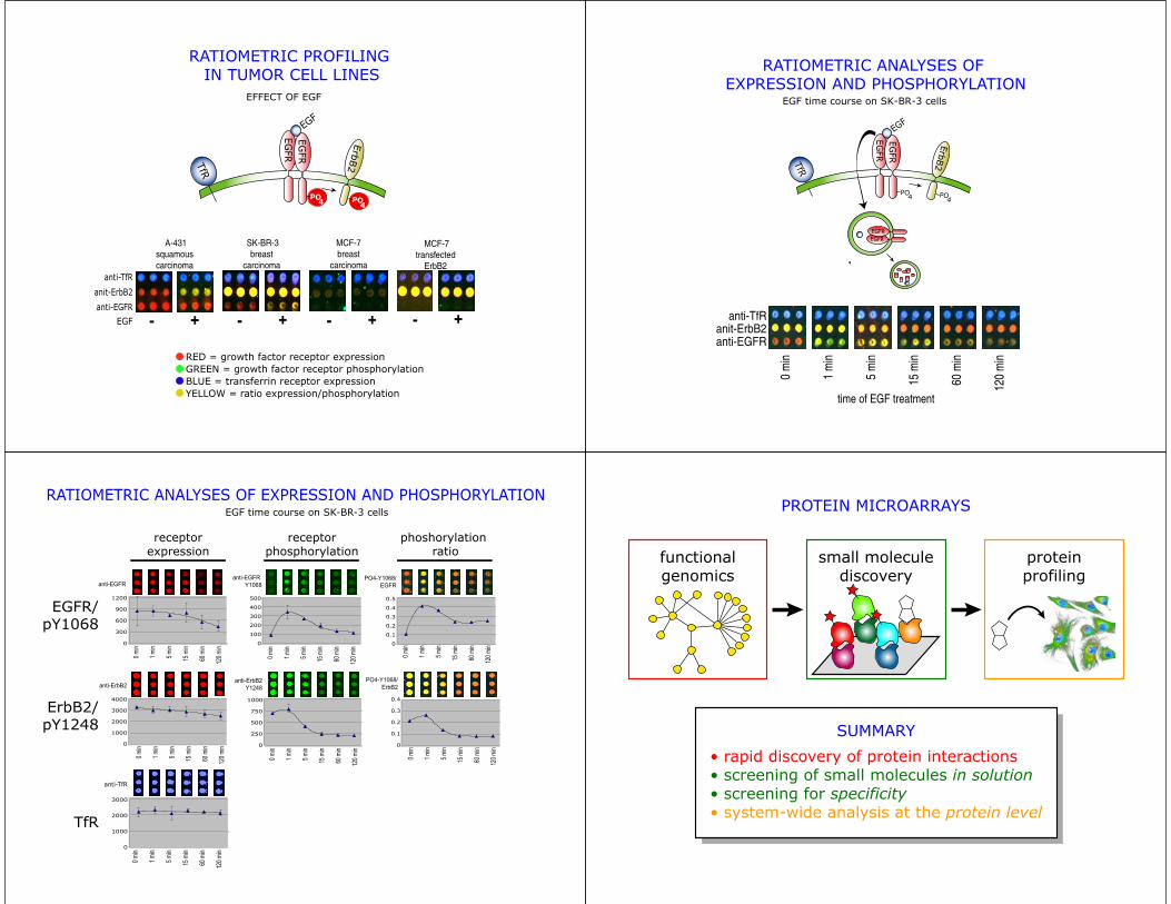
EFFECT OF EGF
RATIOMETRIC PROFILING IN TUMOR CELL LINES
RED = growth factor receptor expressionGREEN = growth factor receptor phosphorylationBLUE = transferrin receptor expressionYELLOW = ratio expression/phosphorylation
anti-EGFR
anit-ErbB2
anti-TfR
EGF
A-431squamouscarcinoma
+ -
SK-BR-3breast
carcinoma
+ -
MCF-7breast
carcinoma
+ -
MCF-7transfected
ErbB2
+ -
EGFR
EGFR
Erb
B2TfR
EGF
PO4 PO
4
RATIOMETRIC ANALYSES OF EXPRESSION AND PHOSPHORYLATION
EGF time course on SK-BR-3 cells
TfR
EGFREGFR
EGFR
EGFR
ErbB2
EGF
PO4 PO
4
time of EGF treatment
anti-EGFRanit-ErbB2
anti-TfR
0 m
in
1 m
in
5 m
in
15 m
in
60 m
in
120
min
0 min
1 min
5 min
15 m
in
60 m
in
120 m
in
0
100
200
300
400
500
anti-EGFRY1068
0
300
600
900
1200
anti-EGFR
0 min
1 min
5 min
15 m
in
60 m
in
120 m
in 0
0.1
0.2
0.3
0.4
0.5
0 min
1 min
5 min
15 m
in
60 m
in
120 m
in
PO4-Y1068/EGFR
anti-ErbB2Y1248
0
250
500
750
1000
0 min
1 min
5 min
15 m
in
60 m
in
120 m
in
0
1000
2000
3000
4000
anti-ErbB2
0 min
1 min
5 min
15 m
in
60 m
in
120 m
in
PO4-Y1068/ErbB2
0
0.1
0.2
0.3
0.4
0 min
1 min
5 min
15 m
in
60 m
in
120 m
inRATIOMETRIC ANALYSES OF EXPRESSION AND PHOSPHORYLATION
EGF time course on SK-BR-3 cells
EGFR/pY1068
ErbB2/pY1248
phoshorylationratio
anti-TfR
0
1000
2000
3000
0 min
1 min
5 min
15 m
in
60 m
in
120 m
in
TfR
receptorexpression
receptorphosphorylation functional
genomicsproteinprofiling
• rapid discovery of protein interactions• screening of small molecules in solution• screening for specificity• system-wide analysis at the protein level
SUMMARY
PROTEIN MICROARRAYS
small moleculediscovery
ACKNOWLEDGEMENTS
Antibody Microarrays
Prof. Peter Sorger (MIT)Dr. Ulrik Nielsen (MIT)Dr. Michael Cardone (MIT)
Dr. Raghida Bu-Khalid
Harvard Center for Genomics ResearchDARPA
Protein Microarrays
Prof. Peter Kim (Whitehead Institute)Dr. John Newman (Whitehead Institute)
Dr. Viara GrantcharovaLioudmila Zaslavskaia