Embed Size (px)
Citation preview

EXTENDED REPORT
Synovial features of patients with rheumatoidarthritis and psoriatic arthritis in clinical andultrasound remission differ under anti-TNF therapy:a clue to interpret different chances of relapse afterclinical remission?Stefano Alivernini,1 Barbara Tolusso,1 Luca Petricca,1 Laura Bui,2 Gabriele Di Sante,1
Giusy Peluso,1 Roberta Benvenuto,2 Anna Laura Fedele,1 Franco Federico,2
Gianfranco Ferraccioli,1 Elisa Gremese1
ABSTRACTObjective To define the synovial characteristics ofpatients with rheumatoid arthritis (RA) and psoriaticarthritis (PsA) in clinical and ultrasound remissionachieved by combination therapy with methotrexate(MTX) and tumour necrosis factor (TNF) blockers.Methods Patients with RA in remission (n=25)(disease activity score (DAS)<1.6 for at least 6 months),patients with RA in low disease activity (LDA) (n=10)(1.6<DAS<2.4 for at least 6 months) and patients withPsA in remission (n=18) (DAS<1.6 and Psoriasis AreaSeverity Index (PASI)=0 for at least 6 months) achievedby MTX+anti-TNF (adalimumab 40 mg or etanercept50 mg) with power Doppler (PDUS)-negative synovialhypertrophy underwent synovial tissue biopsy. Patientswith RA with high/moderate disease naïve to treatment(n=50) were included as a comparison group.Immunostaining for cluster designation (CD)68, CD21,CD20, CD3, CD31 and collagen was performed.Results PDUS-negative patients with RA in remissionshowed lower histological scores for synovial CD68+,CD20+, CD3+ cells and CD31+ vessels and collagendeposition (p<0.05 for both lining and sublining)compared with PDUS-positive patients with RA with high/moderate disease. In addition, there was no significantdifference in terms of lining and sublining CD68+,CD20+, CD3+, CD31+ cells and collagen comparingPDUS-negative patients with RA in remission and in LDA,respectively. On the contrary, PDUS-negative patients withPsA in remission showed higher histological scores forsublining CD68+ (p=0.02) and CD3+ cells (p=0.04) aswell as CD31+ vessels (p<0.001) than PDUS-negativepatients with RA in remission.Conclusions PDUS-negative patients with RA inremission have comparable synovial histological featuresthan PDUS-negative patients with RA in LDA. However,patients with PsA in remission are characterised by ahigher degree of residual synovial inflammation thanpatients with RA in remission, despite PDUS negativityunder TNF inhibition.
INTRODUCTIONStable clinical remission is the most important goalin the treatment of chronic arthritides as rheumatoid
arthritis (RA) and psoriatic arthritis (PsA). However,despite apparent clinical remission, defined withcomposite indices, patients with RA can experiencejoint damage progression.1 Ultrasound assessment(US), through synovial membrane hypertrophy (SH)and power Doppler (PDUS) evaluation, identifiesresidual synovitis in more than 50% of patients withRA in remission on the basis of their disease activityscore (DAS).1 It has been shown that significantlyfewer PDUS-negative patients with RA in clinicalremission had a flare during the 12-month follow-upperiod, compared with patients with RAwith PDUSpositivity at the time of remission.2
The use of combined clinical and PDUS criteriaallows to identify patients with RA in remission,increasing the success rate of persistence of the goodclinical outcome after tumour necrosis factor (TNF)inhibitor discontinuation in patients with estab-lished disease.3 The presence of SH seems to be afrequent finding in patients with long-standing RAwith DAS-based remission, owing to the contribu-tion of significant long-standing disease to SH.However, data on the composition of residual syno-vitis in patients with RA derive mostly from histo-logical studies enrolling patients in remission withPDUS-positive synovitis,4 and no data were pro-duced on patients with PsA in stable clinical remis-sion with PDUS-residual SH after standardisedtreatments. Moreover, it is still unknown if PDUS-negative patients with RAwith long-standing diseasein clinical remission differ from PDUS-negativepatients with RA in low disease activity (LDA)reached with the same treatment regimen.Based on this, the aims of the study were (i) to
define the histological features of PDUS-negativesynovial tissue of patients with RA and PsA inclinical remission through anti-TNF treatment;(ii) to dissect the correlations between US featuresand inflammatory (CD68, CD21, CD20 andCD3), vascular (CD31) and fibrotic (collagen)synovial parameters and (iii) to assess the possiblehistological differences in terms of inflammatorysynovial cells comparing patients with RA inclinical remission and in LDA status withPDUS-negative SH.
1228 Alivernini S, et al. Ann Rheum Dis 2017;76:1228–1236. doi:10.1136/annrheumdis-2016-210424
Clinical and epidemiological research
To cite: Alivernini S, Tolusso B, Petricca L, et al. Ann Rheum Dis 2017;76:1228–1236.
Handling editor Tore K Kvien
► Additional material is published online only. To view please visit the journal online (http://dx.doi.org/10.1136/annrheumdis-2016-210424).
1Institute of Rheumatology, Fondazione Policlinico Universitario Agostino Gemelli, Catholic University of the Sacred Heart, Rome, Italy2Institute of Pathology, Fondazione Policlinico Universitario Agostino Gemelli, Catholic University of the Sacred Heart, Rome, Italy
Correspondence toProfessor Gianfranco Ferraccioli, Professor of Rheumatology, Institute of Rheumatology, Fondazione Policlinico Universitario A. Gemelli—Catholic University of the Sacred Heart, Via Giuseppe Moscati, 31, Rome 00168, Italy; [email protected]
Received 27 August 2016Revised 14 December 2016Accepted 17 December 2016Published Online First 24 January 2017
on 14 February 2019 by guest. P
rotected by copyright.http://ard.bm
j.com/
Ann R
heum D
is: first published as 10.1136/annrheumdis-2016-210424 on 24 January 2017. D
ownloaded from

PATIENTS AND METHODSPatients recruitmentPatients with RA (n=25) fulfilling the American College ofRheumatology 2010 revised criteria for RA5 in stable clinicalremission (DAS<1.6 for at least 6 months) or stable LDA(n=10) (1.6<DAS<2.4 for at least 6 months), and patientswith PsA (n=18)6 in stable clinical remission (DAS<1.6 andPASI=0 for at least 6 months) and stable minimal disease activ-ity (MDA),7 were enrolled in the study. All patients were undertreatment with stable dose of methotrexate in association withTNF-α inhibitor (adalimumab 40 mg/14 days or etanercept50 mg/week, respectively). At study entry, each patient wasdefined as in US remission with negative PDUS despite the pres-ence of SH according to the published protocol.3 A comparisongroup of 50 patients with RA naive to any Disease ModifyingAnti-Rheumatic Drugs (DMARDs) treatment, with high/moder-ate disease activity with PDUS-positive SH, was included. Thestudy protocol was approved by the local ethical committee,and all subjects provided signed informed consent.
Synovial tissue biopsy and immunohistochemistry for CD68,CD21, CD20, CD3, CD31 and collagenAll enrolled patients underwent ultrasound-guided synovialtissue biopsy of the knee following the already published proto-col.8 Once collected, synovial tissue specimens were fixed in10% neutral-buffered formalin and embedded in paraffin forhistology. Briefly, paraffin-embedded synovial tissue (ST) speci-mens were sectioned at 3–4 μm. First, sections were stained forH&E as follows: sections were deparaffinised in xylene andrehydrated in a graded ethanol series. Then they were stained inhaematoxylin and counterstained in eosin/phloxine. Finally, sec-tions were dehydrated, cleared in xylene and mounted with BioMount (Bio-Optica). Other sections were stained for CD68mouse antihuman monoclonal antibody (clone 514H12) or
CD21 mouse antihuman monoclonal antibody (clone 2G9) orCD20 mouse antihuman monoclonal antibody (clone L26)or CD3 mouse antihuman monoclonal antibody (clone LN10)or CD31 mouse antihuman monoclonal antibody (clone 1A10)(all from Leica Biosystem, Newcastle, UK) by immunostainerBOND MAX III (Leica).9
ST slides were stained for collagen using the MassonTrichrome Goldner with light green (Bio-Optica; 04-011802) asfollows: slides were washed in distilled water; then six drops ofWeigert’s iron haematoxylin, provided by the manufacturer,were put on the sections and left to act 10 min. Withoutwashing, the slides were drained, and 10 drops of picric acidalcoholic stable solution were added on the sections. Then, theslides were quickly (3–4 s) washed in distilled water, and 10drops of ponceau acid fuchsin according to Masson were addedon the sections and left to act 4 min. Then, the slides werewashed in distilled water, and 10 drops of phosphomolybdicacid solution were added on the sections and left to act 10 min.Without washing, the slides were drained, and 10 drops ofLight Green Solution, according to Goldner, were added andleft to act 5 min. Then, the slides were washed in distilled waterand rapidly dehydrated through ascending alcohols. Reactionwas done for 1 min in absolute alcohol. Finally, the slides werecleared in xylene and mounted.
Slides were examined by two independent evaluators using alight microscope (Leica DM 2000), and all tissues were evalu-ated using a numerical score based on the number of CD68+,CD21+, CD3+, CD20+ and CD31+ cells in the lining and sub-lining areas of the section (three different fields in each section),with a score of 0 indicating no positive cells; 1 indicating<10% positive cells; 2 indicating 10%–50% positive cells; and3 indicating >50% positive cells.10 The inter-rater agreementcoefficient was assessed for each single immunohistochemistry(IHC) marker (see online supplementary table S1).
Table 1 Demographic, clinical and immunological parameters of enrolled patients with RA and PsA
Remission RAcohort (n=25)
LDA RA cohort(n=10)
High/moderate RAcohort (n=50)
Remission PsAcohort (n=18) p Value* p Value** p Value*** p Value ^
Age, years (mean±SD) 57.20±14.95 52.00±16.38 54.36±15.33 56.67±16.66 0.45 0.69 0.37 0.80
Gender, female (%) 21 (84.0) 6 (60.0) 40 (80.00) 11 (61.1) 0.68 0.17 0.13 0.08
Disease duration, years (mean±SD) 9.70±2.81 9.52±3.03 2.19±2.82 8.50±3.01 <0.001 <0.001 0.06 0.89
Treatment duration, years (mean±SD) 7.01±3.41 6.98±2.75 – 6.83±2.95 – – 0.98 0.63
AB positivity, n (%) 17 (68.0) 5 (50.0) 28 (56.0) 0 (0.0) 0.31 0.72 0.27 <0.001
DAS (mean±SD) 1.09±0.40 1.90±0.31 3.52±1.10 1.10±0.53 <0.001 <0.001 <0.001 0.87
DAS28 (mean±SD) 2.17±0.82 3.18±0.48 5.00±1.24 2.16±0.86 <0.001 <0.001 <0.001 0.51
GH (mean±SD) 32.00±12.73 37.88±27.90 55.80±23.56 33.44±31.93 0.001 0.02 0.37 0.89
TJC28 (mean±SD) 0.04±0.20 1.20±1.23 7.14±6.21 0.39±0.61 <0.001 <0.001 0.01 0.10
TJC44 (mean±SD) 0.16±0.47 1.60±1.95 8.66±8.84 0.39±0.61 <0.001 <0.001 0.02 0.11
SJC28 (mean±SD) 0.12±0.33 1.00±1.05 7.04±6.03 0.22±0.43 <0.001 <0.001 0.02 0.38
SJC44 (mean±SD) 0.16±0.37 1.00±1.05 8.68±8.72 0.22±0.43 <0.001 <0.001 0.02 0.61
ESR, mm/first hour (mean±SD) 17.00±16.10 16.20±16.28 54.65±31.05 14.17±13.62 <0.001 <0.001 0.90 0.62
CRP, mg/L (mean±SD) 2.14±2.00 2.58±2.13 21.43±21.54 2.77±3.20 <0.001 0.002 0.53 0.96
BMI (mean±SD) 25.78±4.11 29.54±8.19 26.55±5.65 26.64±4.25 0.65 0.19 0.15 0.42
Treatment regimen
MTX dose, mg/week 14.42±3.56 14.38±4.96 – 14.79±3.45 – – 0.97 0.97
Etanercept 50 mg/week 13 (52.0) 6 (60.0) – 10 (55.6) – – 0.67 0.82
Adalimumab 40 mg/2 weeks 12 (48.0) 4 (40.0) – 8 (44.4) – – 0.67 0.82
p*: Patients with RA in remission versus patients with RA with high/moderate disease; p**: Patients with RA in LDA versus patients with RA with high/moderate disease; p***: Patientswith RA in remission versus patients with RA in LDA; p^: Patients with RA in remission versus patients with PsA in remission; Bold: p<0.05.AB, autoantibody; BMI, body mass index; CRP, C reactive protein; DAS, disease activity score; ESR, erythrocyte sedimentation rate; GH, global health; LDA, low disease activity; MTX,methotrexate; PsA, psoriatic arthritis; RA, rheumatoid arthritis; SJC, swollen joint count; TJC, tender joint count.
1229Alivernini S, et al. Ann Rheum Dis 2017;76:1228–1236. doi:10.1136/annrheumdis-2016-210424
Clinical and epidemiological research on 14 F
ebruary 2019 by guest. Protected by copyright.
http://ard.bmj.com
/A
nn Rheum
Dis: first published as 10.1136/annrheum
dis-2016-210424 on 24 January 2017. Dow
nloaded from

Statistical analysisStatistical analysis was performed using SPSS V. 20.0 (SPSS.Chicago, Illinois, USA) and Prism software (GraphPad, SanDiego, California, USA). Categorical and quantitative variableswere described as frequencies, percentage and mean±SD. Dataon demographic and clinical features were compared betweenpatients by the non-parametric Mann-Whitney U test or χ2 test,as appropriate. Spearman’s rank correlation test was used forcorrelation in all analyses. A value of p<0.05 was consideredstatistically significant.
RESULTSDemographic, clinical and immunological characteristicsof enrolled patientsDemographic, clinical and immunological characteristics of theenrolled patients with RA are summarised in table 1.PDUS-negative patients with RA in remission and in LDA didnot differ for SH thickness in all the assessed joints (table 2)and showed significantly lower DAS and DAS28 values(p<0.001 and p<0.001, respectively), global health (GH)assessment (p<0.001), tender joint count over 44 and 28 joints(p<0.001 and p<0.001, respectively) and swollen joint countover 44 and 28 joints (p<0.001 and p<0.001, respectively)
compared with patients with RA with high/moderate diseaseactivity. Patients with RA in clinical remission did not differfrom patients with RA in LDA in terms of age, gender, diseaseduration and treatment duration (table 1). Using a stricter cri-teria to define stable clinical remission (Simplified DiseaseActivity Index (SDAI)≤3.3 for at least 6 months), 14/25 (56.0%)patients with RA were considered to be in SDAI remission inour cohort despite DAS-based definition. Analysing the differentparameters used for SDAI calculation, only the patient globalassessment of disease activity (p<0.001) was the item limitingthe achievement of SDAI remission in our PDUS-negative RAcohort (see online supplementary table S2).
Histological features of synovial tissue do not differ inPDUS-negative patients with RA in remission compared withPDUS-negative patients with RA in LDA under TNF blockadeAll included patients underwent synovial tissue biopsy of theknee, which was assessed for the presence of resident CD68+,CD21+, CD20+ and CD3+ cells. In figure 1A–C, exampleimages of immunostaining for CD68+, CD21+, CD20+ andCD3+ cells in synovial tissue from patients with RA with high/moderate disease, in LDA and in clinical remission, respectivelyare shown. In particular, PDUS-negative patients with RA in
Table 2 Parameters at US assessment of patients with RA in remission, patients with RA in LDA and patients with PsA in remission at studyentry
Remission RAcohort (n=25)
LDA RA cohort(n=10)
Remission PsAcohort (n=18) p Value* p Value**
Wrist
RC SH, mm (mean±SD) 1.53±0.88 1.54±1.06 1.31±0.61 0.98 0.37
RC SH score (mean±SD) 0.36±0.64 0.50±0.70 0.22±0.55 0.57 0.46
IC SH, mm (mean±SD) 2.39±1.14 2.75±1.33 2.74±0.54 0.43 0.23
IC SH score (mean±SD) 0.76±0.78 1.10±0.99 0.62±0.41 0.29 0.49
MCP-PIP (dorsal view)
II MCP SH, mm (mean±SD) 0.61±0.49 0.65±0.77 0.43±0.35 0.85 0.19
II MCP SH score (mean±SD) 0.50±0.51 0.67±0.50 0.51±0.48 0.44 0.95
III MCP SH, mm (mean±SD) 0.68±0.64 0.69±0.94 0.55±0.38 0.97 0.45
III MCP SH score (mean±SD) 0.75±0.53 0.78±0.67 0.54±0.52 0.89 0.20
II PIP SH, mm (mean±SD) 0.78±0.42 0.74±0.27 0.60±0.30 0.78 0.13
II PIP SH score (mean±SD) 0.38±0.67 0.10±0.20 0.34±0.36 0.21 0.45
III PIP SH, mm (mean±SD) 0.83±0.32 0.75±0.29 0.57±0.26 0.50 0.10
III PIP SH score (mean±SD) 0.22±0.43 0.25±0.46 0.23±0.44 0.62 0.98
MCP-PIP (volar view)
II MCP SH, mm (mean±SD) 0.45±0.53 0.84±0.71 0.47±0.54 0.10 0.91
II MCP SH score (mean±SD) 0.41±0.59 0.78±0.64 0.46±0.66 0.11 0.88
III MCP SH, mm (mean±SD) 0.43±0.47 0.20±0.21 0.39±0.43 0.15 0.91
III MCP SH score (mean±SD) 0.38±0.59 0.25±0.46 0.23±0.44 0.53 0.60
II PIP SH, mm (mean±SD) 0.79±0.54 0.51±0.45 0.63±0.36 0.16 0.54
II PIP SH score (mean±SD) 0.33±0.64 0.23±0.35 0.21±0.47 0.64 0.80
III PIP SH, mm (mean±SD) 0.76±0.37 0.61±0.53 0.57±0.38 0.35 0.61
III PIP SH score (mean±SD) 0.38±0.49 0.33±0.50 0.27±0.43 0.91 0.56
Knee
knee SH, mm (mean±SD) 6.50±2.91 8.01±2.59 5.50±2.41 0.16 0.34
MTP
II MTP SH, mm (mean±SD) 0.73±0.43 0.60±0.29 0.88±0.71 0.39 0.82
V MTP SH, mm (mean±SD) 0.30±0.34 0.30±0.36 0.37±0.44 0.99 0.87
p*: Patients with RA in remission versus patients with RA in LDA; p**: Patients with RA in remission versus patients with PsA in remission.IC, intercarpal joint; LDA, low disease activity; MCP, metacarpal–phalangeal joint; MTP, metatarsal–phalangeal joint; PIP, proximal interphalangeal joint; PsA, psoriatic arthritis; RA,rheumatoid arthritis; RC, radiocarpal joint; SH, synovial hypertrophy; US, ultrasound assessment.
1230 Alivernini S, et al. Ann Rheum Dis 2017;76:1228–1236. doi:10.1136/annrheumdis-2016-210424
Clinical and epidemiological research on 14 F
ebruary 2019 by guest. Protected by copyright.
http://ard.bmj.com
/A
nn Rheum
Dis: first published as 10.1136/annrheum
dis-2016-210424 on 24 January 2017. Dow
nloaded from

clinical remission showed lower histological scores for CD68+
cells (p<0.001 for both lining and sublining), CD20+ cells(p<0.001 for lining and p=0.02 for sublining, respectively) andCD3+ cells (p=0.002 for lining and p=0.003 for sublining)compared with patients with RA with high/moderate diseaseactivity. PDUS-negative patients with RA in LDA showed lowerhistological scores for lining (p=0.03) and sublining (p=0.01)CD68+ cells, lining (p=0.01) and sublining (p=0.05) CD20+
cells (p=0.05) and lining (p=0.04) and sublining (p=0.05)CD3+ cells compared with patients with RA with high/moderatedisease activity (figure 1D–I). In addition, follicular structureswere found in 8.0% of patients with RA in clinical remission(p<0.001), 10.0% of patients with RA in LDA (p=0.01) com-pared with 56.0% of patients with RA with high/moderate
disease activity. None of the synovial follicular structures foundin PDUS-negative patients with RA in clinical remission andLDA was positive for CD21+ cells compared with 71.4% ofCD21+ synovial follicles in patients with RAwith high/moderatedisease activity (p≤0.001) (figure 1A–C).
Moreover, PDUS-negative patients with RA in clinicalremission did not differ from PDUS-negative patients with RAin LDA in terms of histological scores for CD68+ cells(p=0.39 and p=0.28), CD20+ cells (p=0.49 and p=0.65)and CD3+ cells (p=0.92 and p=0.29), respectively, in thelining and sublining areas (figure 1D–I). These findings wereconfirmed dividing PDUS-negative patients with RA in re-mission using the SDAI cut-off (see online supplementarytable S2).
Figure 1 (A–I) IHC staining for CD68/CD21 and CD3/CD20 on ST of patients with rheumatoid arthritis (RA) in disease remission, in low diseaseactivity (LDA) and in high/moderate disease. (A) Example photos of CD68 (red)/CD21 (brown) (Aa, b) and CD3 (red)/CD20 (brown) (Ac,d) staining ofST biopsies from patients with RA with high/moderate disease activity naïve to any DMARDs treatment (magnification 20×). (B) Example photos ofCD68 (red)/CD21 (brown) (Ba, b) and CD3 (red)/CD20 (brown) (Bc,d) staining of ST biopsies from patients with RA in LDA status (magnification 20×).(C) Example photos of CD68 (red)/CD21 (brown) (Ca, b) and CD3 (red)/CD20 (brown) (Cc,d) staining of ST biopsies from patients with RA in clinicalremission (magnification 20×); ultrasound assessment (US) picture with power doppler (PD) scale of the knee used for ST biopsy is shown next tothe corresponding patient. (D) Lining IHC score for CD68+ cells; high/moderate versus LDA patients with RA, *p=0.03; high/moderate versusremission patients with RA, **p<0.001; LDA versus remission patients with RA, p=0.39. (E) Sublining IHC score for CD68+ cells; high/moderateversus LDA patients with RA, **p=0.01; high/moderate versus remission patients with RA, **p<0.001; LDA versus remission patients with RA,p=0.28. (F) Lining IHC score for CD20+ cells; high/moderate versus LDA patients RA, **p=0.01; high/moderate versus remission patients with RA,**p<0.001; LDA versus remission patients with RA, p=0.49. (G) Sublining IHC score for CD20+ cells; high/moderate versus LDA patients with RA,*p=0.05; high/moderate versus remission patients with RA, *p=0.02; LDA versus remission patients with RA, p=0.65. (H) Lining IHC score for CD3+
cells; high/moderate versus LDA patients with RA, *p=0.04; high/moderate versus remission patients with RA, **p=0.002; LDA versus remissionpatients with RA, p=0.92. (I) Sublining IHC score for CD3+ cells; high/moderate versus LDA patients with RA, *p=0.05; high/moderate versusremission patients with RA, **p=0.003; LDA versus remission patients with RA, p=0.29.
1231Alivernini S, et al. Ann Rheum Dis 2017;76:1228–1236. doi:10.1136/annrheumdis-2016-210424
Clinical and epidemiological research on 14 F
ebruary 2019 by guest. Protected by copyright.
http://ard.bmj.com
/A
nn Rheum
Dis: first published as 10.1136/annrheum
dis-2016-210424 on 24 January 2017. Dow
nloaded from
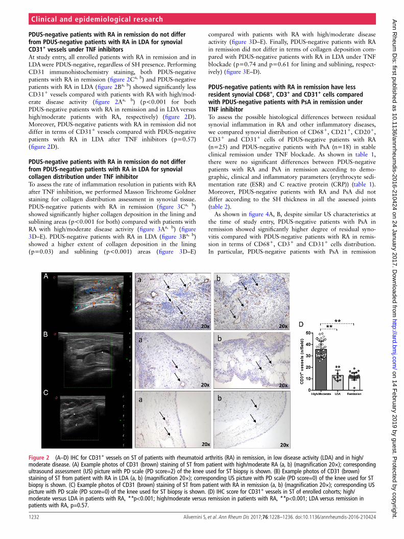
PDUS-negative patients with RA in remission do not differfrom PDUS-negative patients with RA in LDA for synovialCD31+ vessels under TNF inhibitorsAt study entry, all enrolled patients with RA in remission and inLDAwere PDUS-negative, regardless of SH presence. PerformingCD31 immunohistochemistry staining, both PDUS-negativepatients with RA in remission (figure 2Ca, b) and PDUS-negativepatients with RA in LDA (figure 2Ba, b) showed significantly lessCD31+ vessels compared with patients with RA with high/mod-erate disease activity (figure 2Aa, b) (p<0.001 for bothPDUS-negative patients with RA in remission and in LDA versushigh/moderate patients with RA, respectively) (figure 2D).Moreover, PDUS-negative patients with RA in remission did notdiffer in terms of CD31+ vessels compared with PDUS-negativepatients with RA in LDA after TNF inhibitors (p=0.57)(figure 2D).
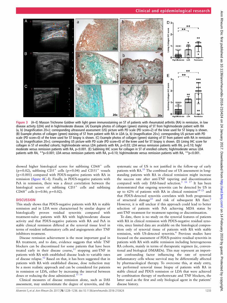
PDUS-negative patients with RA in remission do not differfrom PDUS-negative patients with RA in LDA for synovialcollagen distribution under TNF inhibitorTo assess the rate of inflammation resolution in patients with RAafter TNF inhibition, we performed Masson Trichrome Goldnerstaining for collagen distribution assessment in synovial tissue.PDUS-negative patients with RA in remission (figure 3Ca, b)showed significantly higher collagen deposition in the lining andsublining areas (p<0.001 for both) compared with patients withRA with high/moderate disease activity (figure 3Aa, b) (figure3D–E). PDUS-negative patients with RA in LDA (figure 3Ba, b)showed a higher extent of collagen deposition in the lining(p=0.03) and sublining (p<0.001) areas (figure 3D–E)
compared with patients with RA with high/moderate diseaseactivity (figure 3D–E). Finally, PDUS-negative patients with RAin remission did not differ in terms of collagen deposition com-pared with PDUS-negative patients with RA in LDA under TNFblockade (p=0.74 and p=0.61 for lining and sublining, respect-ively) (figure 3E–D).
PDUS-negative patients with RA in remission have lessresident synovial CD68+, CD3+ and CD31+ cells comparedwith PDUS-negative patients with PsA in remission underTNF inhibitorTo assess the possible histological differences between residualsynovial inflammation in RA and other inflammatory diseases,we compared synovial distribution of CD68+, CD21+, CD20+,CD3+ and CD31+ cells of PDUS-negative patients with RA(n=25) and PDUS-negative patients with PsA (n=18) in stableclinical remission under TNF blockade. As shown in table 1,there were no significant differences between PDUS-negativepatients with RA and PsA in remission according to demo-graphic, clinical and inflammatory parameters (erythrocyte sedi-mentation rate (ESR) and C reactive protein (CRP)) (table 1).Moreover, PDUS-negative patients with RA and PsA did notdiffer according to the SH thickness in all the assessed joints(table 2).
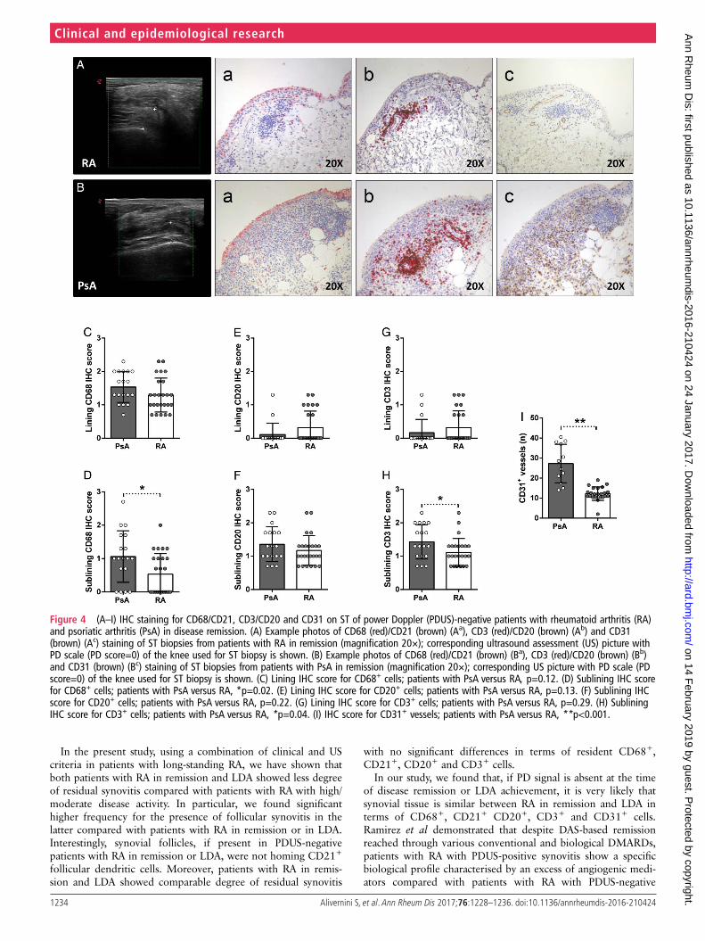
As shown in figure 4A, B, despite similar US characteristics atthe time of study entry, PDUS-negative patients with PsA inremission showed significantly higher degree of residual syno-vitis compared with PDUS-negative patients with RA in remis-sion in terms of CD68+, CD3+ and CD31+ cells distribution.In particular, PDUS-negative patients with PsA in remission
Figure 2 (A–D) IHC for CD31+ vessels on ST of patients with rheumatoid arthritis (RA) in remission, in low disease activity (LDA) and in high/moderate disease. (A) Example photos of CD31 (brown) staining of ST from patient with high/moderate RA (a, b) (magnification 20×); correspondingultrasound assessment (US) picture with PD scale (PD score=2) of the knee used for ST biopsy is shown. (B) Example photos of CD31 (brown)staining of ST from patient with RA in LDA (a, b) (magnification 20×); corresponding US picture with PD scale (PD score=0) of the knee used for STbiopsy is shown. (C) Example photos of CD31 (brown) staining of ST from patient with RA in remission (a, b) (magnification 20×); corresponding USpicture with PD scale (PD score=0) of the knee used for ST biopsy is shown. (D) IHC score for CD31+ vessels in ST of enrolled cohorts; high/moderate versus LDA in patients with RA, **p<0.001; high/moderate versus remission in patients with RA, **p<0.001; LDA versus remission inpatients with RA, p=0.57.
1232 Alivernini S, et al. Ann Rheum Dis 2017;76:1228–1236. doi:10.1136/annrheumdis-2016-210424
Clinical and epidemiological research on 14 F
ebruary 2019 by guest. Protected by copyright.
http://ard.bmj.com
/A
nn Rheum
Dis: first published as 10.1136/annrheum
dis-2016-210424 on 24 January 2017. Dow
nloaded from
showed higher histological scores for sublining CD68+ cells(p=0.02), sublining CD3+ cells (p=0.04) and CD31+ vessels(p<0.001) compared with PDUS-negative patients with RA inremission (figure 4C–I). Finally, in PDUS-negative patients withPsA in remission, there was a direct correlation between thehistological scores of sublining CD3+ cells and subliningCD68+ cells (r=0.86; p=0.02).
DISCUSSIONThis study shows that PDUS-negative patients with RA in stableremission and in LDA were characterised by similar degree ofhistologically proven residual synovitis compared withtreatment-naïve patients with RA with high/moderate diseaseactivity and that PDUS-negative patients with RA and PsA instable clinical remission differed at the synovial tissue level interms of resident inflammatory cells and angiogenesis after TNFinhibitors treatment.
Disease remission achievement is the most important goal inRA treatment, and to date, evidence suggests that while TNFblockers can be discontinued for some patients that have beentreated early in their disease course,11 stopping anti-TNF inpatients with RA with established disease leads to variable ratesof disease relapse.12 Based on that, it has been suggested that inpatients with RA with established disease, dose reduction maybe a more realistic approach and can be considered for patientsin remission or LDA, either by increasing the interval betweendoses or reducing the dose administered.13 14
Clinical measures of disease remission alone, such as DASassessment, may underestimate the degree of synovitis, and the
systematic use of US is not justified in the follow-up of earlypatients with RA.15 The combined use of US assessment in long-standing patients with RA in clinical remission might increasethe success rate after anti-TNF tapering and discontinuationcompared with only DAS-based selection.3 16 17 It has beendemonstrated that ongoing synovitis can be detected by US inup to 62% of patients with RA in clinical remission18–21 andthat PDUS-detected synovitis correlates with both progressionof structural damage22 and risk of subsequent RA flare.2
However, it is still unclear if this approach could lead to betterselection of patients with PsA achieving MDA status byanti-TNF treatment for treatment tapering or discontinuation.
To date, there is no study on the synovial features of patientswith RA in clinical remission with PDUS-negative residual syno-vitis, since limited data are available on the histological compos-ition only of synovial tissue of patients with RA with stableremission, with US-detected synovitis.4 Previous studies havefocused on the assessment of PDUS-positive residual synovitis inpatients with RA with stable remission including heterogeneousRA cohorts, mainly in terms of therapeutic regimen (ie, conven-tional and biological DMARDs). This may represent an import-ant confounding factor influencing the rate of synovialinflammatory cells whose survival may be differentially affectedby pharmacological therapy. To overcome this, at study entry,we performed synovial tissue biopsy of patients with RA instable clinical and PDUS remission or LDA that were achievedby combination therapy of methotrexate and TNF blockers, thelatter used as the first and only biological agent in the patients’disease history.
Figure 3 (A–E) Masson Trichrome Goldner with light green immunostaining on ST of patients with rheumatoid arthritis (RA) in remission, in lowdisease activity (LDA) and in high/moderate disease. (A) Example photos of collagen (green) staining of ST from high/moderate patient with RA(a, b) (magnification 20×); corresponding ultrasound assessment (US) picture with PD scale (PD score=2) of the knee used for ST biopsy is shown.(B) Example photos of collagen (green) staining of ST from patient with RA in LDA (a, b) (magnification 20×); corresponding US picture with PDscale (PD score=0) of the knee used for ST biopsy is shown. (C) Example photos of collagen (green) staining of ST from patient with RA in remission(a, b) (magnification 20×); corresponding US picture with PD scale (PD score=0) of the knee used for ST biopsy is shown. (D) Lining IHC score forcollagen in ST of enrolled cohorts; high/moderate versus LDA patients with RA, p=0.03; LDA versus remission patients with RA, p=0.10; high/moderate versus remission patients with RA, p<0.001. (E) Sublining IHC score for collagen in ST of enrolled cohorts; high/moderate versus LDApatients with RA, **p<0.001; LDA versus remission patients with RA, p=0.10; high/moderate versus remission patients with RA, **p<0.001.
1233Alivernini S, et al. Ann Rheum Dis 2017;76:1228–1236. doi:10.1136/annrheumdis-2016-210424
Clinical and epidemiological research on 14 F
ebruary 2019 by guest. Protected by copyright.
http://ard.bmj.com
/A
nn Rheum
Dis: first published as 10.1136/annrheum
dis-2016-210424 on 24 January 2017. Dow
nloaded from
In the present study, using a combination of clinical and UScriteria in patients with long-standing RA, we have shown thatboth patients with RA in remission and LDA showed less degreeof residual synovitis compared with patients with RA with high/moderate disease activity. In particular, we found significanthigher frequency for the presence of follicular synovitis in thelatter compared with patients with RA in remission or in LDA.Interestingly, synovial follicles, if present in PDUS-negativepatients with RA in remission or LDA, were not homing CD21+
follicular dendritic cells. Moreover, patients with RA in remis-sion and LDA showed comparable degree of residual synovitis
with no significant differences in terms of resident CD68+,CD21+, CD20+ and CD3+ cells.
In our study, we found that, if PD signal is absent at the timeof disease remission or LDA achievement, it is very likely thatsynovial tissue is similar between RA in remission and LDA interms of CD68+, CD21+ CD20+, CD3+ and CD31+ cells.Ramirez et al demonstrated that despite DAS-based remissionreached through various conventional and biological DMARDs,patients with RA with PDUS-positive synovitis show a specificbiological profile characterised by an excess of angiogenic medi-ators compared with patients with RA with PDUS-negative
Figure 4 (A–I) IHC staining for CD68/CD21, CD3/CD20 and CD31 on ST of power Doppler (PDUS)-negative patients with rheumatoid arthritis (RA)and psoriatic arthritis (PsA) in disease remission. (A) Example photos of CD68 (red)/CD21 (brown) (Aa), CD3 (red)/CD20 (brown) (Ab) and CD31(brown) (Ac) staining of ST biopsies from patients with RA in remission (magnification 20×); corresponding ultrasound assessment (US) picture withPD scale (PD score=0) of the knee used for ST biopsy is shown. (B) Example photos of CD68 (red)/CD21 (brown) (Ba), CD3 (red)/CD20 (brown) (Bb)and CD31 (brown) (Bc) staining of ST biopsies from patients with PsA in remission (magnification 20×); corresponding US picture with PD scale (PDscore=0) of the knee used for ST biopsy is shown. (C) Lining IHC score for CD68+ cells; patients with PsA versus RA, p=0.12. (D) Sublining IHC scorefor CD68+ cells; patients with PsA versus RA, *p=0.02. (E) Lining IHC score for CD20+ cells; patients with PsA versus RA, p=0.13. (F) Sublining IHCscore for CD20+ cells; patients with PsA versus RA, p=0.22. (G) Lining IHC score for CD3+ cells; patients with PsA versus RA, p=0.29. (H) SubliningIHC score for CD3+ cells; patients with PsA versus RA, *p=0.04. (I) IHC score for CD31+ vessels; patients with PsA versus RA, **p<0.001.
1234 Alivernini S, et al. Ann Rheum Dis 2017;76:1228–1236. doi:10.1136/annrheumdis-2016-210424
Clinical and epidemiological research on 14 F
ebruary 2019 by guest. Protected by copyright.
http://ard.bmj.com
/A
nn Rheum
Dis: first published as 10.1136/annrheum
dis-2016-210424 on 24 January 2017. Dow
nloaded from
remission,20 suggesting that the remaining synovial vascularity atlocal joints may increase the risk of structural deterioration,despite the anti-inflammatory therapy effect.23 24 These findingsconfirm that remission in RA requires an improvement ofoverall disease activity and disappearance of local synovial vas-cularity detected by PDUS.
In addition, in our study, we reported for the first time thehistological features of synovial tissue from PDUS-negativepatients with PsA in stable remission (defined using anRA-specific composite index as DAS) and MDA reached underanti-TNF treatment. So far, formal criteria for remission ofpatients with PsA have not been defined, whereas a state ofMDA has been proposed.7 Araujo et al25 recently demonstratedthat patients with PsA in stable clinical remission (defined ashaving documented absence of clinical symptoms related toarthritis, dactylitis, enthesitis and axial disease and minimal skindisease with PASI<1) have a high chance of disease relapse aftertreatment discontinuation, suggesting that patients with PsAmay be characterised by residual active synovitis. Moreover, theauthors suggested that the affected joints in patients with PsAmay repeatedly and effectively home inflammatory cells leadingto a memory function of the tissue, as the recurrence of thedisease usually occurs at the joints which were previouslyaffected by the disease. In our study, we performed synovialtissue biopsy of the knee which was affected during the diseasecourse, showing that patients with PsA in remission are charac-terised by a higher degree of residual synovitis in terms ofCD68+, CD3+ and CD31+ cells, mainly in the sublining, com-pared with patients with RA in clinical remission despite stablePDUS negativity, providing a biological support to the high rateof disease relapse of patients with PsA after treatment discon-tinuation.25 However, the analysis of the distribution of otherresident inflammatory cells, such as mast cells, demonstrated notto be affected by TNF inhibition at synovial tissue level in spon-dylarthritis,26 could provide additional information on the cellu-lar composition of residual synovitis of patients with PsA inMDA under TNF blockage.
In conclusion, the results of our study may have relevantimplications to better identify patients with RA in stable clinicalremission that could more safely undergo treatment reductionor discontinuation using combined clinical and US selection cri-teria. Moreover, since patients with RA with long-standingdisease reaching LDA status with PDUS-negative tissue havecomparable synovial characteristics than patients with RA inremission, tapering anti-TNF agent could be a reasonable choicefor clinicians in such patients. However, to definitely confirmthe prognostic power of residual inflammatory cells in the syn-ovial tissue of patients with RA or PsA in remission in foreseeingdisease relapse, prospective studies performing treatment taper-ing or discontinuation, based on the combination of clinical, USand histological selection criteria, are needed.
Contributors SA, GF and EG gave substantial contributions to study conceptionand design; SA, BT, LP, LB, GDS, RB, GP, ALF and FF gave substantial contributionsto acquisition of data; SA, BT GF and EG gave substantial contributions to analysisand interpretation of data; SA, BT, LP, LB, GDS, RB, GP, ALF, FF, GF and EG draftedthe article and revised it critically for important intellectual content; SA, BT, LP, LB,GDS, RB, GP, ALF, FF, GF and EG gave final approval for the version of the article tobe published.
Competing interests None declared.
Patient consent Obtained.
Ethics approval Fondazione Policlinico Universitario A. Gemelli—CatholicUniversity of the Sacred Heart Ethical Committee.
Provenance and peer review Not commissioned; externally peer reviewed.
Open Access This is an Open Access article distributed in accordance with theCreative Commons Attribution Non Commercial (CC BY-NC 4.0) license, whichpermits others to distribute, remix, adapt, build upon this work non-commercially,and license their derivative works on different terms, provided the original work isproperly cited and the use is non-commercial. See: http://creativecommons.org/licenses/by-nc/4.0/
REFERENCES1 Yoshimi R, Hama M, Takase K, et al. Ultrasonography is a potent tool for the
prediction of progressive joint destruction during clinical remission of rheumatoidarthritis. Mod Rheum 2013;23:456–65.
2 Peluso G, Michelutti A, Bosello SL, et al. Clinical and ultrasonographic remissiondetermines different chances of relapse in early and long standing rheumatoidarthritis. Ann Rheum Dis 2011;70:172–5.
3 Alivernini S, Peluso G, Fedele AL, et al. Tapering and discontinuation of TNF-αblockers without disease relapse using ultrasonography as a tool to identify patientswith rheumatoid arthritis in clinical and histological remission. Arthritis Res Ther2016;18:39.
4 Ramírez J, Celis R, Usategui A, et al. Immunopathologic characterization ofultrasound-defined synovitis in rheumatoid arthritis patients in clinical remission.Arthritis Res Ther 2016;18:74.
5 Aletaha D, Neogi T, Silman AJ, et al. 2010 rheumatoid arthritis classificationcriteria: an American College of Rheumatology/European League AgainstRheumatism collaborative initiative. Ann Rheum Dis 2010;69:1580–8.
6 Taylor W, Gladman D, Helliwell P, et al. Classification criteria for psoriatic arthritis:development of new criteria from a large international study. Arthritis Rheum2006;54:2665–73.
7 Coates LC, Fransen J, Helliwell PS. Defining minimal disease activity in psoriaticarthritis: a proposed objective target for treatment. Ann Rheum Dis2010;69:48–53.
8 Van de Sande MG, Gerlag DM, Lodde BM, et al. Evaluating anti-rheumatictreatments using synovial biopsy: a recommendation for standardization to be usedin clinical trials. Ann Rheum Dis 2011;70:423–7.
9 Bancroft JD, Stevens A. Theory and practice of histological techniques. 4th edn.New York: Churchill Livingstone, 1996.
10 Kurowska-Stolarska M, Alivernini S, Ballantine LE, et al. MicroRNA-155 as aproinflammatory regulator in clinical and experimental arthritis. Proc Natl Acad SciUSA 2011;108:11193–8.
11 Quinn MA, Conaghan PG, O’Connor PJ, et al. Very early treatment with infliximabin addition to methotrexate in early, poor-prognosis rheumatoid arthritis reducesmagnetic resonance imaging evidence of synovitis and damage, with sustainedbenefit after infliximab withdrawal: results from a twelve-month randomized,double-blind, placebo-controlled trial. Arthritis Rheum 2005;52:27–35.
12 Smolen JS, Nash P, Durez P, et al. Maintenance, reduction, or withdrawal ofetanercept after treatment with etanercept and methotrexate in patients withmoderate rheumatoid arthritis (PRESERVE): a randomised controlled trial. Lancet2013;381:918–29.
13 Smolen JS, Landewé R, Breedveld FC, et al. EULAR recommendations for themanagement of rheumatoid arthritis with synthetic and biological disease-modifyingantirheumatic drugs: 2013 update. Ann Rheum Dis 2014;73:492–509.
14 Rudolf MD, Deighton C, Bosworth A, et al. Rheumatoid arthritis: national clinicalguideline for management and treatment in adults. NICE Clinical Guidelines.London: Royal College of Physicians, 2009. http://www.nice.org.uk/nicemedia/pdf/CG79FullGuideline.pdf
15 Haavardsholm EA, Aga AB, Olsen IC, et al. Ultrasound in management ofrheumatoid arthritis: ARCTIC randomised controlled strategy trial. BMJ 2016;354:i4205.
16 Colebatch AN, Edwards CJ, Østergaard M, et al. EULAR recommendations for theuse of imaging of the joints in the clinical management of rheumatoid arthritis.Ann Rheum Dis 2013;72:804–14.
17 Navarro-Millán I, Sattui SE, Curtis JR. Systematic review of tumor necrosis factorinhibitor discontinuation studies in rheumatoid arthritis. Clin Ther2013;35:1850–61.
18 Saleem B, Brown AK, Keen H, et al. Should imaging be a component of rheumatoidarthritis remission criteria? A comparison between traditional and modifiedcomposite remission scores and imaging assessments. Ann Rheum Dis2011;70:792–8.
19 Ozgocmen S, Ozdemir H, Kiris A, et al. Clinical evaluation and power Dopplersonography in rheumatoid arthritis: evidence for ongoing synovial inflammation inclinical remission. South Med J 2008;101:240–5.
20 Brown AK, Quinn MA, Karim Z, et al. Presence of significant synovitis in rheumatoidarthritis patients with disease-modifying antirheumatic drug–induced clinicalremission: evidence from an imaging study may explain structural progression.Arthritis Rheum 2006;54:3761–73.
21 Ramírez J, Ruíz-Esquide V, Pomés I, et al. Patients with rheumatoid arthritis inclinical remission and ultrasound-defined active synovitis exhibit higher disease
1235Alivernini S, et al. Ann Rheum Dis 2017;76:1228–1236. doi:10.1136/annrheumdis-2016-210424
Clinical and epidemiological research on 14 F
ebruary 2019 by guest. Protected by copyright.
http://ard.bmj.com
/A
nn Rheum
Dis: first published as 10.1136/annrheum
dis-2016-210424 on 24 January 2017. Dow
nloaded from
activity and increased serum levels of angiogenic biomarkers. Arthritis Res Ther2014;16:R5.
22 Dougados M, Devauchelle-Pensec V, Ferlet JF, et al. The ability of synovitis topredict structural damage in rheumatoid arthritis: a comparative study betweenclinical examination and ultrasound. Ann Rheum Dis 2013;72:665–71.
23 Fukae J, Kon Y, Henmi M, et al. Change of synovial vascularity in a singlefinger joint assessed by power Doppler sonography correlated withradiographic change in rheumatoid arthritis: comparative study of a novelquantitative score with a semiquantitative score. Arthritis Care Res2010;62:657–63.
24 Fukae J, Isobe M, Kitano A, et al. Radiographic prognosis of finger joint damagepredicted by early alteration in synovial vascularity in patients with rheumatoidarthritis: potential utility of power Doppler sonography in clinical practice. ArthritisCare Res 2011;63:1247–53.
25 Araujo EG, Finzel S, Englbrecht M, et al. High incidence of disease recurrence afterdiscontinuation of disease-modifying antirheumatic drug treatment in patients withpsoriatic arthritis in remission. Ann Rheum Dis 2015;74:655–60.
26 Noordenbos T, Yeremenko N, Gofita I, et al. Interleukin-17-positive mast cellscontribute to synovial inflammation in spondylarthritis. Arthritis Rheum2012;64:99–109.
1236 Alivernini S, et al. Ann Rheum Dis 2017;76:1228–1236. doi:10.1136/annrheumdis-2016-210424
Clinical and epidemiological research on 14 F
ebruary 2019 by guest. Protected by copyright.
http://ard.bmj.com
/A
nn Rheum
Dis: first published as 10.1136/annrheum
dis-2016-210424 on 24 January 2017. Dow
nloaded from