Embed Size (px)
Citation preview
©FUNPEC-RP www.funpecrp.com.brGenetics and Molecular Research 12 (4): 5640-5650 (2013)
Expression pattern of the zona pellucida 3 (ZP3) gene during ovarian development and the location of ZP3 protein in oocytes in a natural, wild triploid crucian carp mutant, Carassius auratus var. Pingxiangnensis
J.W. Shi1, J.Q. Sheng1, K. Peng1, J.H. Wang1, W.J. Yi1, H.J. Wu2, Q. Gu1 andY.J. Hong1,3
1College of Life Sciences and Food Engineering, Nanchang University, Nanchang, China2Institute of Fisheries Science of Pingxiang, Pingxiang, China3Institute of Life Sciences, Nanchang University, Nanchang, China
Corresponding author: Y. HongE-mail: [email protected]
Genet. Mol. Res. 12 (4): 5640-5650 (2013)Received November 22, 2012Accepted August 10, 2013Published November 18, 2013DOI http://dx.doi.org/10.4238/2013.November.18.13
ABSTRACT. Carassius auratus var. Pingxiangnensis (designated CaP), distributed in the Pingxiang region of Jiangxi Province, China, is a natural, wild triploid crucian carp mutant that has two reproductive development modes: gynogenesis and bisexual reproduction. Little information is available about the expression pattern of the zona pellucida 3 (ZP3) gene during ovarian development and the location of the ZP3 protein in oocytes of this fish. In this study, we obtained the full-length cDNA of ZP3 (CaP_ZP3). CaP_ZP3 contains an open reading frame of 1305 bp that encodes 435 amino acid residues. Real-time polymerase chain reaction (PCR) was used to determine the CaP_ZP3 mRNA expression levels in the ovary at different stages of maturation. Results revealed high levels of CaP_ZP3 expression
5641
©FUNPEC-RP www.funpecrp.com.brGenetics and Molecular Research 12 (4): 5640-5650 (2013)
ZP3 in triploid Carassius auratus var. Pingxiangnensis
in 4- to 8-month-old ovaries (stage II-stage III), with a significant decline in 9- to 12-month-old ovaries (stages IV-stage V). The high levels of CaP_ZP3 transcripts during the early growth period suggest an important role for CaP_ZP3 in early oocyte development. In addition, a polyclonal antibody was prepared against CaP_ZP3, and the immunofluorescence localization was determined. CaP_ZP3 protein was detected close to the oocyte plasma membrane. The results also showed that no fluorescent signal was detected in stage I and II oocytes. CaP_ZP3 protein is primarily detected in stage III oocytes, and the protein accumulates as oocytes develop into stage IV oocytes. These results suggested that the transcription and translation of the CaP_ZP3 gene is asynchronous and that the transcription of the CaP_ZP3 protein occurs prior to its translation in this triploid fish.
Key words: Carassius auratus var. Pingxiangnensis; ZP3;Expression analysis; Real-time PCR; Immunofluorescence
INTRODUCTION
The zona pellucida (ZP) enclosing vertebrate oocytes is important in the processes of regulating the sperm-egg interaction, preventing polyspermy, and protecting the embryo during the successive stages of development (McLeskey et al., 1998). The ZP is composed of three kinds of glycoproteins encoded by multiple gene families including ZP1, ZP2, and ZP3 (Was-sarman, 1999, 2008). ZP proteins have a conservative homologous region known as the ZP domain (Bork and Sander, 1992). The ZP domain is thought to be involved in protein-protein interactions and is essential for ZP assembly (Qi et al., 2002). The ZP domain of ZP3 contains eight conserved Cys residues (Bork and Sander, 1992). Furthermore, ZP3 has been identified to regulate vertebrate fertilization (Barros et al., 1996; Snell and White, 1996), and it serves as a primary sperm receptor and induces the acrosome reaction in mice (Wassarman et al., 2004).
Oocyte growth and development are important issues in fish growth (Lubzens et al., 2010). Oocytes in all teleosts undergo the same basic pattern of growth: oogenesis, primary oocyte growth, cortical alveolus stage, vitellogenesis, maturation, and ovulation (Tyler and Sumpter, 1996). Mor-phological changes in developing oocytes have been described for several species of teleosts (Chen et al., 2010; Mohamed, 2010). Detailed information about the histological events accompanying ovarian development in Carassius auratus var. Pingxiangnensis can be found in reports from Hong (2005) and Wu et al. (2009), where 1- to 2-month ovaries (primordial germ cells), 3-month ovaries (stage I), 4- to 5-month ovaries (stage II), 6- to 8-month ovaries (stage III), nine- to 10-month ova-ries (stage IV), and 11- to 12-month ovaries (stage V) were studied and analyzed.
ZP3 cDNA molecules have already been characterized from some fishes, including Oryzias latipes (Murata et al., 1995), Sparus aurata (Modig et al., 2006), Danio rerio (Wang and Gong, 1999; Liu et al., 2006), Cyprinus carpio (Chang et al., 1996), C. auratus gibelio (Fan et al., 2001), Cynoglossus semilaevis (Sun et al., 2010), and Acipenser sinensis (Chuang-Ju et al., 2011). These studies have improved our understanding of the structural homology and tissue-specific expression of the ZP3 gene among fish. However, the expression profile of ZP3 mRNA during ovarian development and the cellular localization of ZP3 protein in oocytes have not been examined in the special triploid C. auratus var. Pingxiangnensis
5642
©FUNPEC-RP www.funpecrp.com.brGenetics and Molecular Research 12 (4): 5640-5650 (2013)
J.W. Shi et al.
(CaP). Understanding the molecular events of the ZP3 gene during oogenesis is a key step to determining the secret of “high-quality” eggs for aquaculture. In this study, the full-length cDNA of CaP_ZP3 was analyzed. In order to elucidate the CaP_ZP3 mRNA expression pattern during ovarian growth, we investigated the expression level of the CaP_ZP3 gene in 1- to 12-month-old ovaries. An immunofluorescence assay using a fluorescein isothiocyanate (FITC)-labeled antibody specific to CaP_ZP3 protein was carried out to further determine the cellular localization of ZP3 protein in oocytes in the mutant.
MATERIAL AND METHODS
RNA extraction and SMART cDNA synthesis
One- to 12-month-old triploid fish were collected from the Pingxiang Fisheries Re-search Institute of Jiangxi Province in China. CaP was identified to be triploid Carassius auratus by DNA content measurement and chromosome analysis (Hong et al., 2005). Arti-ficial propagation experiments indicated that CaP could reproduce by gynogenesis (Hong et al., 2005). Total RNA from the ovary was isolated using the SV Total RNA Isolation System (Promega, Madison, WI, USA). After the DNase treatment, total RNA (5 μg) extracted from the ovary was used to synthesize the 1st-strand cDNA using the SMARTTM cDNA Library Construction Kit (Clontech, Mountain View, CA, USA) (Zeng et al., 2012).
Cloning and sequencing
A pair of degenerate primers was designed according to the conserved region among the known ZP3 genes that were available from the GenBank database (AF180465, C. auratus; Z48974, C. auratus; L41636, C. auratus; Z48972, C. carpio; and L41638, C. carpio). A partial cDNA sequence that was homologous to ZP3 genes in other species was obtained, and some gene-specific primers were designed for rapid amplification of cDNA ends (RACE). The full-length CaP_ZP3 cDNA was amplified by 3'- and 5'-RACE (Clontech, Mountain View, CA, USA). The amplified cDNA was subcloned into the pMD19-T vector (Takara, Otsu, Japan) and sequenced.
Data and sequence analysis
All of the primers were designed by the Premier 5.0 and Oligo 6.0 software. The glycosylation sites and the putative signal peptide were predicted using YinOYang 1.2 and Signal P at http://www.cbs.dtu.dk/services/SignalP (Nielsen et al., 1997). Multiple alignments were performed with DNASTAR software. Phylogenetic analysis was performed using the MEGA4.1 software by bootstrap analysis.
Real-time polymerase chain reaction (PCR)
Prior to cDNA synthesis, DNase treatment with RQ1 RNase-free DNase (Promega) was carried out to eliminate the genomic DNA contamination following the manufacturer instructions. The RNA was reverse-transcribed using M-MLV Reverse Transcriptase (RT) and oligo(dT)18
5643
©FUNPEC-RP www.funpecrp.com.brGenetics and Molecular Research 12 (4): 5640-5650 (2013)
ZP3 in triploid Carassius auratus var. Pingxiangnensis
(Promega) following the manufacturer protocol. Samples were assayed in triplicate, and each experiment was repeated three times. The reaction mixture (25 μL) contained 1X SYBR® Premix EX TaqTM PCR Mix (Takara, Dalian, China), 1 μL template cDNA, and 0.2 μM each primer, with ddH2O added to reach 25 μL (Zeng et al., 2012). Quantitative real-time PCRs were performed in a 96-well microtiter plate (Applied Biosystems, California, America). The primer pairs, denoted CaP_ZP3.1F and CaP_ZP3.1R (Table 1), were designed to detect the differential expression pat-tern of CaP_ZP3 mRNA. Primers (CaP_β-actin-F and CaP_β-actin-R) for the detection of the ubiquitously expressed gene β-actin as a control gene are also given in Table 1.
Recombinant CaP_ZP3 protein expression
A pair of primers (CaP_ZP3F and CaP_ZP3R) (Table 1) were used to amplify a 300-bp cDNA fragment. The amplified fragment was initially cloned into the pGEM-T vector (Promega). The plasmid containing CaP_ZP3 was cut with NcoI and HindIII and subsequently subcloned into the pET-32α plasmid. Recombinant CaP_ZP3 protein expression was induced by isopropyl β-d-1-thiogalactopyranoside (IPTG) as an inducer in Escherichia coli BL21(DE3) cells (Mate et al., 2003). The recombinant CaP_ZP3 proteins were purified using nickel nitrilotriacetic acid (Ni-NTA) resin (Qiagen, GmbH, Hilden, Germany) as described previously (Kaul et al., 1997).
Preparation of polyclonal antibody and enzyme-linked immunosorbent assay (ELISA)
Primers Sequence (5'-3') Product size (bp)
Cap_ZP3.1F TGTTCCAGTACCGAGTG 171Cap_ZP3.1R CATCCTCCCAAAGTCAGACCCap_β-actin-F CACTGTGCCCATCTACGAG 200Cap_β-actin-R CCATCTCCTGCTCGAAGTCCap_ZP3F GAATTCCATATGCGCAGCCAAAGTCCAAGC 300Cap_ZP3R GGATCCTTAAAGCTTACTGCATTGAACAGCCAC
Table 1. Sequences of the primers used in this study.
Two New Zealand male white rabbits were injected subcutaneously at multiple sites using about 300 μg ZP3 fusion protein emulsified in complete Freund’s adjuvant (CFA). After 2 weeks, the rabbits were boosted intramuscularly with an equivalent amount of ZP3 fusion protein in incomplete Freund’s adjuvant (IFA). After 2 months, the blood was collected. The reactivity of anti-ZP3 protein antibodies was tested by ELISA (Li et al., 2008).
Immunofluorescence localization
The mature ovaries at different stages were embedded in optimal cutting temperature (OCT) (Reichert-Jung, Heidelberg Germany), and 10-μm-thick sections were obtained by the fro-zen microtomy method (Liu and Gui, 2005). The cryostat sections were rehydrated in phosphate-buffered saline (PBS) for 10 min and incubated for 1 h with 5% dry milk in PBS at room tem-perature. The sections were then incubated with the anti-ZP3 protein antiserum (1:100 dilution) overnight at 4°C. After washing with PBS, the sections were incubated for 1 h with a FITC-conju-gated secondary antibody, goat anti-rabbit IgG (Zhong Shan Jin Qiao, Beijing, China), and washed
5644
©FUNPEC-RP www.funpecrp.com.brGenetics and Molecular Research 12 (4): 5640-5650 (2013)
J.W. Shi et al.
with PBS. Finally, the sections were observed with a fluorescence microscope (Leica, Heidelberg, Germany). Control sections were treated with pre-immune serum as the primary antibody.
RESULTS
Cloning and sequence analysis of the full-length cDNA of CaP_ZP3
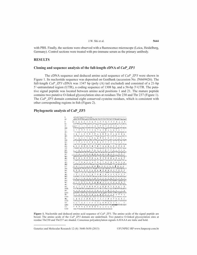
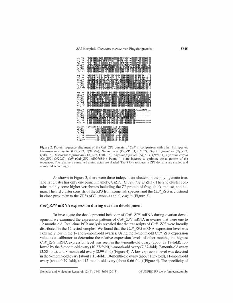
The cDNA sequence and deduced amino acid sequence of CaP_ZP3 were shown in Figure 1. Its nucleotide sequence was deposited on GenBank (accession No. JN669426). The full-length CaP_ZP3 cDNA was 1347 bp (poly (A) tail excluded) and consisted of a 21-bp 5’-untranslated region (UTR), a coding sequence of 1308 bp, and a 56-bp 3'-UTR. The puta-tive signal peptide was located between amino acid positions 1 and 21. The mature peptide contains two putative O-linked glycosylation sites at residues Thr 230 and Thr 237 (Figure 1). The CaP_ZP3 domain contained eight conserved cysteine residues, which is consistent with other corresponding regions in fish (Figure 2).
Phylogenetic analysis of CaP_ZP3
Figure 1. Nucleotide and deduced amino acid sequence of CaP_ZP3. The amino acids of the signal peptide are boxed. The amino acids of the CaP_ZP3 domain are underlined. Two putative O-linked glycosylation sites at residue Thr230 and Thr237 are shaded. Consensus polyadenylation signals AATAAA are italic and bold.
5645
©FUNPEC-RP www.funpecrp.com.brGenetics and Molecular Research 12 (4): 5640-5650 (2013)
ZP3 in triploid Carassius auratus var. Pingxiangnensis
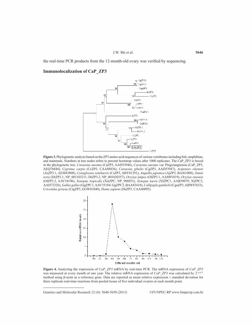
As shown in Figure 3, there were three independent clusters in the phylogenetic tree. The 1st cluster has only one branch, namely, CsZP3 (C. semilaevis ZP3). The 2nd cluster con-tains mainly some higher vertebrates including the ZP protein of frog, chick, mouse, and hu-man. The 3rd cluster consists of the ZP3 from some fish species, and the CaP_ZP3 is clustered in close proximity to the ZP3s of C. auratus and C. carpio (Figure 3).
CaP_ZP3 mRNA expression during ovarian development
To investigate the developmental behavior of CaP_ZP3 mRNA during ovarian devel-opment, we examined the expression patterns of CaP_ZP3 mRNA in ovaries that were one to 12 months old. Real-time PCR analysis revealed that the transcripts of CaP_ZP3 were broadly distributed in the 12 tested samples. We found that the CaP_ZP3 mRNA expression level was extremely low in the 1- and 2-month-old ovaries. Using the 3-month-old CaP_ZP3 expression value as a calibrator to determine the relative expression levels of other months, the highest CaP_ZP3 mRNA expression level was seen in the 4-month-old ovary (about 28.17-fold), fol-lowed by the 5-month-old ovary (10.27-fold), 6-month-old ovary (7.87-fold), 7-month-old ovary (3.88-fold), and 8-month-old ovary (2.99-fold) (Figure 4). A low expression level was detected in the 9-month-old ovary (about 1.13-fold), 10-month-old ovary (about 1.25-fold), 11-month-old ovary (about 0.79-fold), and 12-month-old ovary (about 0.66-fold) (Figure 4). The specificity of
Figure 2. Protein sequence alignment of the CaP_ZP3 domain of CaP in comparison with other fish species. Oncorhynchus mykiss (Om_ZP3, Q9I9M6), Danio rerio (Dr_ZP3, Q5TYP2), Oryzias javanicus (Oj_ZP3, Q5EC18), Tetraodon nigroviridis (Tn_ZP3, Q4RJR6), Anguilla japonica (Aj_ZP3, Q9YIB1), Cyprinus carpio (Cc_ZP3, Q92027), CaP (CaP_ZP3, AEQ76844). Points (---) are inserted to optimize the alignment of the sequences. The relatively conserved amino acids are shaded. The 8 Cys residues in ZP3 domains are shaded and numbered accordingly.
5646
©FUNPEC-RP www.funpecrp.com.brGenetics and Molecular Research 12 (4): 5640-5650 (2013)
J.W. Shi et al.
the real-time PCR products from the 12-month-old ovary was verified by sequencing.
Immunolocalization of CaP_ZP3
Figure 3. Phylogenetic analysis based on the ZP3 amino acid sequences of various vertebrates including fish, amphibian, and mammals. Numbers at tree nodes refers to percent bootstrap values after 1000 replicates. The CaP_ZP3 is boxed in the phylogenetic tree. Carassius auratus (CaZP3, AAD53946), Carassius auratus var. Pingxiangnensis (CaP_ZP3, AEQ76844), Cyprinus carpio (CcZP3, CAA88836), Carassius gibelio (CgZP3, AAD53947), Acipenser sinensis (AsZP3.1, AEM43808), Cynoglossus semilaevis (CsZP3, ABY81291), Anguilla japonica (AjZP3, BAJ61008), Danio rerio (DrZP3.1, NP_001103211; DrZP3.2, NP_001020357), Oryzias latipes (OlZP3.1, AAM91819), Oryzias sinensis (OlZP3.2, AAV34196), Xenopus tropicalis (XstZPC, NP_988853), Xenopus laevis (XlZPC1, AAB39079; XlZPC2, AAH72326), Gallus gallus (GgZPC1, AAV35184; GgZPC2, BAA83418), Callipepla gambelii (CgaZP3, ABW87653), Cricetulus griseus (CtgZP3, EGW01840), Homo sapiens (HsZP3, CAA40095).
Figure 4. Analyzing the expression of CaP_ZP3 mRNA by real-time PCR. The mRNA expression of CaP_ZP3 was measured at every month of one year. The relative mRNA expression of CaP_ZP3 was calculated by 2-△△CT method using β-actin as a reference gene. Data are reported as mean relative expression ± standard deviation for three replicate real-time reactions from pooled tissue of five individual ovaries at each month point.
5647
©FUNPEC-RP www.funpecrp.com.brGenetics and Molecular Research 12 (4): 5640-5650 (2013)
ZP3 in triploid Carassius auratus var. Pingxiangnensis
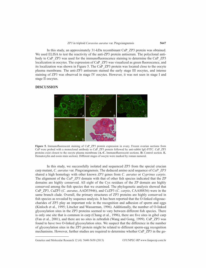
In this study, an approximately 31-kDa recombinant CaP_ZP3 protein was obtained. We used ELISA to test the reactivity of the anti-ZP3 protein antiserum. The polyclonal anti-body to CaP_ZP3 was used for the immunofluorescence staining to determine the CaP_ZP3 localization in oocytes. The expression of CaP_ZP3 was visualized as green fluorescence, and its localization was shown in Figure 5. The CaP_ZP3 protein was located close to the oocyte plasma membrane. The anti-ZP3 antiserum stained the early stage III oocytes, and intense staining of ZP3 was observed in stage IV oocytes. However, it was not seen in stage I and stage II oocytes.
DISCUSSION
Figure 5. Immunofluorescent staining of CaP_ZP3 protein expression in ovary. Frozen ovarian sections from CaP were probed with a monoclonal antibody to CaP_ZP3 protein followed by anti-rabbit IgG-FITC. CaP_ZP3 proteins exist closest to the oocyte plasma membrane (A.-C. Immunofluorescent sections. D. Control section. E. Hematoxylin and eosin stain section). Different stages of oocyte were marked by roman numeral.
In this study, we successfully isolated and sequenced ZP3 from the special crucian carp mutant, C. auratus var. Pingxiangnensis. The deduced amino acid sequence of CaP_ZP3 shared a high homology with other known ZP3 genes from C. auratus or Cyprinus carpio. The alignment of the CaP_ZP3 domain with that of other fish species indicated that the ZP domains are highly conserved. All eight of the Cys residues of the ZP domain are highly conserved among the fish species that we examined. The phylogenetic analysis showed that CaP_ZP3, CaZP3 (C. auratus, AAD53946), and CcZP3 (C. carpio, CAA88836) were in the same branch clade. Overall, the primary structures of ZP3 proteins are highly conserved in fish species as revealed by sequence analysis. It has been reported that the O-linked oligosac-charides of ZP3 play an important role in the recognition and adhesion of sperm and eggs (Kinloch et al., 1995; Litscher and Wassarman, 1996). Additionally, the number of O-linked glycosylation sites in the ZP3 proteins seemed to vary between different fish species. There is only one site that is common in carp (Chang et al., 1996), there are five sites in gibel carp (Fan et al., 2001), and there are no sites in zebrafish (Wang and Gong, 1999). CaP_ZP3 was found to have two O-linked glycosylation sites. We suspect that the difference in the number of glycosylation sites in the ZP3 protein might be related to different sperm-egg recognition mechanisms. However, further studies are required to determine whether CaP_ZP3 in the go-
5648
©FUNPEC-RP www.funpecrp.com.brGenetics and Molecular Research 12 (4): 5640-5650 (2013)
J.W. Shi et al.
nad is involved in controlling gynogenesis in this fish.As a major component of the ZP, the amount of CaP_ZP3 mRNA in oocytes is unusually
high (Chang et al., 1996). In mammals, ZP3 is indispensable for early ovarian development; in its absence, the zona matrix does not form and mice are infertile (Liu et al., 1996; Rankin et al., 1996). A major objective of this study was to determine the expression of CaP_ZP3 in ovaries of different developmental stages. We found that ovaries of all developmental stages contain ZP3 mRNA, and early stage ovaries have a higher expression level than the later stage ovaries. However, ZP3 mRNA is indeed present in the later ovary stages, although its expression level was much weaker than that of the early ovary stages. These facts suggest that ZP3 transcription begins in primitive ovary stages and persists in ovaries to the late developmental phases. In the ovaries that were 4-8 months old (stage II-stage III), the predominant presence and high expression of CaP_ZP3 suggests an important role for ZP3 during early ovarian development in this species. During the later period, when ovaries were 9-12 months old (stage IV-stage V), there is a dramatic decline in the abundance of CaP_ZP3 transcripts, and the level falls to less than 3% of the peak in mature eggs. Similar to CaP, ZP transcript levels in zebrafish are abundantly transcribed during oogenesis, where 10.3% of total transcripts expressed in the ovary code for proteins of the ZPA-ZPC families (Zeng and Gong, 2002). Consistent with this, Chang et al. (1996, 1997) used Northern blot analysis and found carp ZP2 and ZP3 mRNAs in all stages of oocytes, with decreasing levels from early to late stage. In mouse, the expression of all three ZP genes is precisely regulated during oogenesis and restricted to a 2-week growth phase, when they represent together approximately 1.5% of the total polyA+ RNA. However, in the later stages of oogenesis, their abundance declines, and each zona transcript is present in ovulated eggs at less than 5% of its maximal level (Roller et al., 1989; Epifano et al., 1995). These analogous expression patterns suggest that ZP3 genes are active during oogenesis, particularly during the early stages of oogenesis. The acellular vitelline envelope develops around the oocyte during the early phases of ovarian development in teleosts. During this period, the oocyte also accumulates RNA (known as maternal RNA) and completes the differentiation of its cellular and noncellular envelopes (Anderson, 1967; Wallace, 1985). In this study, high levels of ZP3 expression were observed during the early phases of ovarian development (stage II–stage III), which was consistent with results from earlier reports. During the later period of ovary development (9-12 months old; stage IV-stage V), when grown oocytes undergo meiotic maturation and become unfertilized eggs, the growth of the oocyte and formation of the ZP will be accomplished. Therefore, it is reasonable that the level of ZP3 mRNA falls quite dramatically and maintains a relatively low level in this period.
In this study, we generated antiserum against CaP_ZP3 protein that is specific for CaP_ZP3 protein in oocytes and carried out immunofluorescence assays further determine the localiza-tion of ZP3 protein in oocytes of this special mutant fish. The results showed that no staining or very faint staining was detected in the stage I and stage II oocytes with this antiserum. The posi-tive immunostaining signals were primarily detected in the stage III oocytes. Therefore, the trans-lation of CaP_ZP3 protein occurred in stage III oocytes. The high expression levels of CaP_ZP3 mRNA were observed in stage I and II oocytes and thereafter sharply diminished, in contrast to the observed levels of ZP3 protein. The results indicated that the transcription and translation of the ZP3 gene in this special triploid fish are asynchronous. Additionally, the translation of ZP3 protein was hysteretic and occurred in stage III oocytes. As the oocyte develops from stage III to stage IV, the ZP3 protein level increases, and the ZP3 content in oocytes increases as vitellogen-esis proceeds. ZP proteins appear to be highly conserved to serve as structural components of the
5649
©FUNPEC-RP www.funpecrp.com.brGenetics and Molecular Research 12 (4): 5640-5650 (2013)
ZP3 in triploid Carassius auratus var. Pingxiangnensis
eggshell in different species; their function in sperm recognition does not seem to be conserved (Hyllner et al., 2001). Sperm can penetrate the eggshell anywhere on mammalian eggs, and this is not the case in teleosts, in which sperm must enter through the micropyle (Griffin et al., 1996; Iwamatsu et al., 1997; Hyllner et al., 2001). In our study, the distribution of CaP_ZP3 in oocytes was not restricted to the micropylar region of the egg chorion, but it was also distributed to many sites of the ZP, as observed in mammals. Our results may provide some important clues to further understand whether CaP_ZP3 shares physiological functions with the mammalian homolog, as well as clues to the unknown diverse roles such as the regulation of gynogenesis and bisexual production in the natural triploid crucian carp mutant, C. auratus var. Pingxiangnensis.
Conflicts of interest
The authors declare that they have no conflict of interest.
ACKNOWLEDGMENTS
Research supported by the National Natural Science Foundation of China (#30660143, #31040082, and #31260282).
REFERENCESAnderson E (1967). The formation of the primary envelope during oocyte differentiation in teleosts. J. Cell Biol. 35:
193-212.Barros C, Crosby JA and Moreno RD (1996). Early steps of sperm-egg interactions during mammalian fertilization. Cell
Biol. Int. 20: 33-39.Bork P and Sander C (1992). A large domain common to sperm receptors (Zp2 and Zp3) and TGF-beta type III receptor.
FEBS Lett. 300: 237-240.Chang YS, Wang SC, Tsao CC and Huang FL (1996). Molecular cloning, structural analysis, and expression of carp ZP3
gene. Mol. Reprod. Dev. 44: 295-304.Chang YS, Hsu CC, Wang SC, Tsao CC, et al. (1997). Molecular cloning, structural analysis, and expression of carp ZP2
gene. Mol. Reprod. Dev. 46: 258-267.Chen KS, Crone PR and Hsu CC (2010). Reproductive biology of albacore Thunnus alalunga. J. Fish. Biol. 77: 119-136.Chuang-Ju L, Qi-Wei W, Xi-Hua C, Li Z, et al. (2011). Molecular characterization and expression pattern of three zona
pellucida 3 genes in the Chinese sturgeon, Acipenser sinensis. Fish Physiol. Biochem. 37: 471-484.Epifano O, Liang LF, Familari M, Moos MC, Jr., et al. (1995). Coordinate expression of the three zona pellucida genes
during mouse oogenesis. Development 121: 1947-1956.Fan LC, Yang ST and Gui JF (2001). Differential screening and characterization analysis of the egg envelope glycoprotein
ZP3 cDNAs between gynogenetic and gonochoristic crucian carp. Cell Res. 11: 17-27.Griffin FJ, Vines CA, Pillai MC, Yanagimachi R, et al. (1996). Sperm motility initiation factor is a minor component of
the Pacific herring egg chorion. Dev. growth & differ. 38: 193-202.Hong YJ, Yu ZJ, Zhou L and Gui J F (2005). A population of redtransparent, triploid Carassius auratus. J. Fish Biol. 67:
1139-1143.Hyllner SJ, Westerlund L, Olsson PE and Schopen A (2001). Cloning of rainbow trout egg envelope proteins: members of
a unique group of structural proteins. Biol. Reprod. 64: 805-811.Iwamatsu T, Yoshizaki N and Shibata Y (1997). Changes in the chorion and sperm entry into the micropyle during
fertilization in the teleostean fish, Oryzias latipes. Dev. Growth Differ. 39: 33-41.Kaul R, Afzalpurkar A and Gupta SK (1997). Expression of bonnet monkey (Macaca radiata) zona pellucida-3 (ZP3) in a
prokaryotic system and its immunogenicity. Mol. Reprod. Dev. 47: 140-147.Kinloch RA, Sakai Y and Wassarman PM (1995). Mapping the mouse ZP3 combining site for sperm by exon swapping
and site-directed mutagenesis. Proc. Natl. Acad. Sci. U. S. A. 92: 263-267.Li B, Shi HY and Wang MH (2008). An Enzyme-linked immunosorbent assay for the detection of bifenthrin. Chinese J.
5650
©FUNPEC-RP www.funpecrp.com.brGenetics and Molecular Research 12 (4): 5640-5650 (2013)
J.W. Shi et al.
Anal. Chemistry 36: 34-38.Litscher ES and Wassarman PM (1996). Characterization of mouse ZP3-derived glycopeptide, gp55, that exhibits sperm
receptor and acrosome reaction-inducing activity in vitro. Biochemistry 35: 3980-3985.Liu C, Litscher ES, Mortillo S, Sakai Y, et al. (1996). Targeted disruption of the mZP3 gene results in production of eggs
lacking a zona pellucida and infertility in female mice. Proc. Natl. Acad. Sci. U. S. A. 93: 5431-5436.Liu JX and Gui JF (2005). Expression pattern and developmental behaviour of cellular nucleic acid-binding protein
(CNBP) during folliculogenesis and oogenesis in fish. Gene 356: 181-192.Liu X, Wang H and Gong Z (2006). Tandem-repeated Zebrafish zp3 genes possess oocyte-specific promoters and are
insensitive to estrogen induction. Biol. Reprod. 74: 1016-1025.Lubzens E, Young G, Bobe J and Cerda J (2010). Oogenesis in teleosts: how eggs are formed. Gen. Comp. Endocrinol.
165: 367-389.Mate KE, Buist JM and Duckworth JA (2003). Expression in Escherichia coli and immunological characterization of three
zona pellucida proteins (ZP1, ZP2, and ZP3) from a marsupial, the brushtail possum (Trichosurus vulpecula). Mol. Reprod. Dev. 64: 136-143.
McLeskey SB, Dowds C, Carballada R, White RR, et al. (1998). Molecules involved in mammalian sperm-egg interaction. Int. Rev. Cytol. 177: 57-113.
Modig C, Modesto T, Canario A, Cerda J, et al. (2006). Molecular characterization and expression pattern of zona pellucida proteins in gilthead seabream (Sparus aurata). Biol. Reprod. 75: 717-725.
Mohamed A (2010). The reproductive biology and the histological and ultrastructural characteristics in ovaries of the female gadidae fish Merluccius merluccius from the Egyptian Mediterranean water. Afr. J. Biotechnol. 9: 2544-2559.
Murata K, Sasaki T, Yasumasu S, Iuchi I, et al. (1995). Cloning of cDNAs for the precursor protein of a low-molecular-weight subunit of the inner layer of the egg envelope (chorion) of the fish Oryzias latipes. Dev. Biol. 167: 9-17.
Nielsen H, Engelbrecht J, Brunak S and von Heijne G (1997). A neural network method for identification of prokaryotic and eukaryotic signal peptides and prediction of their cleavage sites. Int. J. Neural Syst. 8: 581-599.
Qi H, Williams Z and Wassarman PM (2002). Secretion and assembly of zona pellucida glycoproteins by growing mouse oocytes microinjected with epitope-tagged cDNAs for mZP2 and mZP3. Mol. Biol. Cell 13: 530-541.
Rankin T, Familari M, Lee E, Ginsberg A, et al. (1996). Mice homozygous for an insertional mutation in the Zp3 gene lack a zona pellucida and are infertile. Development 122: 2903-2910.
Roller RJ, Kinloch RA, Hiraoka BY, Li SS, et al. (1989). Gene expression during mammalian oogenesis and early embryogenesis: quantification of three messenger RNAs abundant in fully grown mouse oocytes. Development 106: 251-261.
Snell WJ and White JM (1996). The molecules of mammalian fertilization. Cell 85: 629-637.Sun Y, Yu H, Zhang Q, Qi J, et al. (2010). Molecular characterization and expression pattern of two zona pellucida genes
in half-smooth tongue sole (Cynoglossus semilaevis). Comparative biochemistry and physiology Part B. Biochem. Mol. Biol. 155: 316-321.
Tyler C and Sumpter J (1996). Oocyte growth and development in teleosts. Rev. Fish Biol. Fish. 6: 287-318.Wallace RA (1985). Vitellogenesis and Oocyte Growth in Non-Mammalian Vertebrates. In: Developmental Biology,
Oogenesis (Browder LW, ed.). Plenum Press, New York. 127-177.Wang H and Gong Z (1999). Characterization of two zebrafish cDNA clones encoding egg envelope proteins ZP2 and
ZP3. Biochim. Biophys. Acta 1446: 156-160.Wassarman PM (1999). Mammalian fertilization: molecular aspects of gamete adhesion, exocytosis, and fusion. Cell 96:
175-183.Wassarman PM, Jovine L and Litscher ES (2004). Mouse zona pellucida genes and glycoproteins. Cytogenet. Genome
Res. 105: 228-234.Wassarman PM (2008). Zona pellucida glycoproteins. J. Biol. Chem. 283: 24285-24289.Wu HF, Sheng JQ, Hong YJ, Wang JH, et al. (2009). Gonadal development in natural wildness triploid mutant Pingxiang
red-transparent crucian carp, Carassius auratus L. Acta. Hydrobiol. Sin. 6: 0-25.Zeng LG, Wang JH, Li YJ, Sheng JQ, et al. (2012). Molecular characteristics and expression of calmodulin cDNA from
the freshwater pearl mussel, Hyriopsis schlegelii. Genet. Mol. Res. 11: 42-52.Zeng S and Gong Z (2002). Expressed sequence tag analysis of expression profiles of zebrafish testis and ovary. Gene
294: 45-53.


![ZP3 Series アダプタ 適応パッド一覧ca01.smcworld.com/catalog/BEST-technical-data/pdf/6-4-p...ZP3-10B ZP3-13B ZP3-16B 適応パッド品番 アダプタ品番 ZP3A-T3-A5[質量:2.4g]](https://img.pdfslide.us/doc/110x75/5fca2fc152b0a9529d484818/zp3-series-ff-eoeffffeca01-zp3-10b-zp3-13b-zp3-16b-eoeffffc.jpg)

![arXiv:2006.00067v2 [cs.CV] 20 Jul 2020of zona pellucida segmentation, all these features are used for embryo selec tion [1,27,2,24]; we segment the zona pellucida both to improve the](https://img.pdfslide.us/doc/110x75/60af951b4e64854d4508408b/arxiv200600067v2-cscv-20-jul-2020-of-zona-pellucida-segmentation-all-these.jpg)