Embed Size (px)
Citation preview
EPS
AH*B†
R
aafiohfttbpCFtpA
btma(fieocbmgc
ONE
Biochemical and Biophysical Research Communications 258, 222–226 (1999)
Article ID bbrc.1999.0616, available online at http://www.idealibrary.com on
0CA
xpression of Saliva-Binding Epitopes of theorphyromonas gingivalis FimA Protein on theurface of Streptococcus gordonii
shu Sharma,*,1 Kiyonobu Honma,* Hakimuddin T. Sojar,* Dennis E. Hruby,†oward K. Kuramitsu,*,‡ and Robert J. Genco*,‡
Department of Oral Biology, School of Dental Medicine and ‡Department of Microbiology, School of Medicine andiomedical Sciences, State University of New York at Buffalo, Buffalo, New York 14214; andDepartment of Microbiology, Oregon State University, Corvallis, Oregon 97331
eceived March 22, 1999
induce protection against P. gingivalis associated alve-oFhtaic3asiobtCovdg
blipo
M
eigbgN(p5
Porphyromonas gingivalis, a gram-negative oral an-erobic bacterium, has been implicated in the onsetnd development of periodontitis. The P. gingivalismbriae which mediate bacterial adherence to hostral sites and induce host inflammatory responsesave been suggested as a potential antigen candidate.
or vaccine development. This study was undertakeno generate Streptococcus gordonii vectors expressinghe major subunit protein (FimA) of P. gingivalis fim-riae for testing as a potential live vaccine againsteriodontitis. We report here the expression of the-terminal saliva-binding epitopes of P. gingivalisimA on the surface of S. gordonii and demonstrate
hat domains containing free cysteine residues areoorly expressed on the surface of S. gordonii. © 1999
cademic Press
The nonpathogenic oral commensal, S. gordonii, haseen genetically modified to express heterologous an-igens for vaccine development (1, 2). One of the pri-ary aims of our laboratory is to develop a vaccine
gainst P. gingivalis associated periodontal diseaseperiodontitis). A major subunit protein of P. gingivalismbriae (FimA, fimbrillin) mediates bacterial adher-nce to the host oral sites via binding to saliva coatedral surfaces (3, 4), facilitates attachment to epithelialells (5), and directs interaction with other residentacteria (6) and induces tissue destructive host im-une responses (7–10). Immunization with purified P.
ingivalis fimbriae, fimbrillin or a synthetic peptideorresponding to a subdomain of the FimA protein can
1 To whom correspondence should be addressed at Department ofral Biology, 208 Foster Hall, 3435 Main Street, State University ofew York at Buffalo, Buffalo, NY 14214-3092. Fax: (716) 829-3942.-mail: [email protected].
222006-291X/99 $30.00opyright © 1999 by Academic Pressll rights of reproduction in any form reserved.
lar bone loss in rats (11–13). The peptide domains ofimA involved in bacterial adherence and induction ofost immune response have been mapped (14). Sincehe majority of the FimA domains involved in bacterialdherence to saliva coated oral surfaces and in thenduction of protective host immune responses are lo-alized in the C-terminal region from residues 226 to37 of FimA, this region is an attractive antigen for uses a vaccine (15, 16). Recently, we reported the expres-ion of portions of the N- (residue 55-145) and C- (res-dues 233-322) terminal regions of P. gingivalis FimAn the surface of S. gordonii using a strategy developedy Pozzi et al. where the FimA protein was targeted tohe S. gordonii cell surface as a fusion protein with the-terminal ‘anchor’ domain of the M6 molecule. Inrder for this strategy to be successful in developing aaccine against P. gingivalis, optimal expression ofesired FimA epitopes is required on the surface of S.ordonii.In this sudy, we report the expression of the saliva-
inding (PRP-1 and Statherin) domains and immuno-ogicaly protective epitope (226–246) of FimA localizedn the C-terminal region. We further show that theresence of cysteine residues in the C-terminal portionf FimA retards its surface-expression in S. gordonii.
ATERIALS AND METHODS
Bacterial strains, plasmids, media and growth conditions. Esch-richia coli strain DH5a (Gibco-BRL, Gaithersburg, MD) was grownn Luria-Bertani broth or agar (1.5%). S. gordonii GP251 (17) wasrown in Todd-Hewitt broth containing 0.2% yeast extract (THYroth) with or without 1.5% agar. For radiolabeling, the cells wererown in THY medium containing 5 mCi of [3H]-thymidine (DupontEN Research Products, Boston, MA) per ml at 37°C anaerobically
85% N2, 10% H2, 5% CO2). Where appropriate, antibiotics chloram-henicol (Cm) or erythromycin (Em) were added at a concentration ofmg/ml each. Plasmid pUC13Bg12.1 (18) was used as template for
PCR amplification of the P. gingivalis fimA gene. Insertion vectorpg
ifiastfvgoiKcsoafcbcm2pific(bpn
q(bbcadseocTt(aitFBpdwumm
bopNsw1
PBS/0.02% sodium azide. Following incubation, wells were washedwwtwctsm
R
pssmarpcgtCbCasto(w3sTwtTcfremccpubdaaMbgwtas
Vol. 258, No. 1, 1999 BIOCHEMICAL AND BIOPHYSICAL RESEARCH COMMUNICATIONS
SMB55 (17), a 5.73Kb E. coli plasmid that does not replicate in S.ordonii was maintained in E. coli strain DH5a.
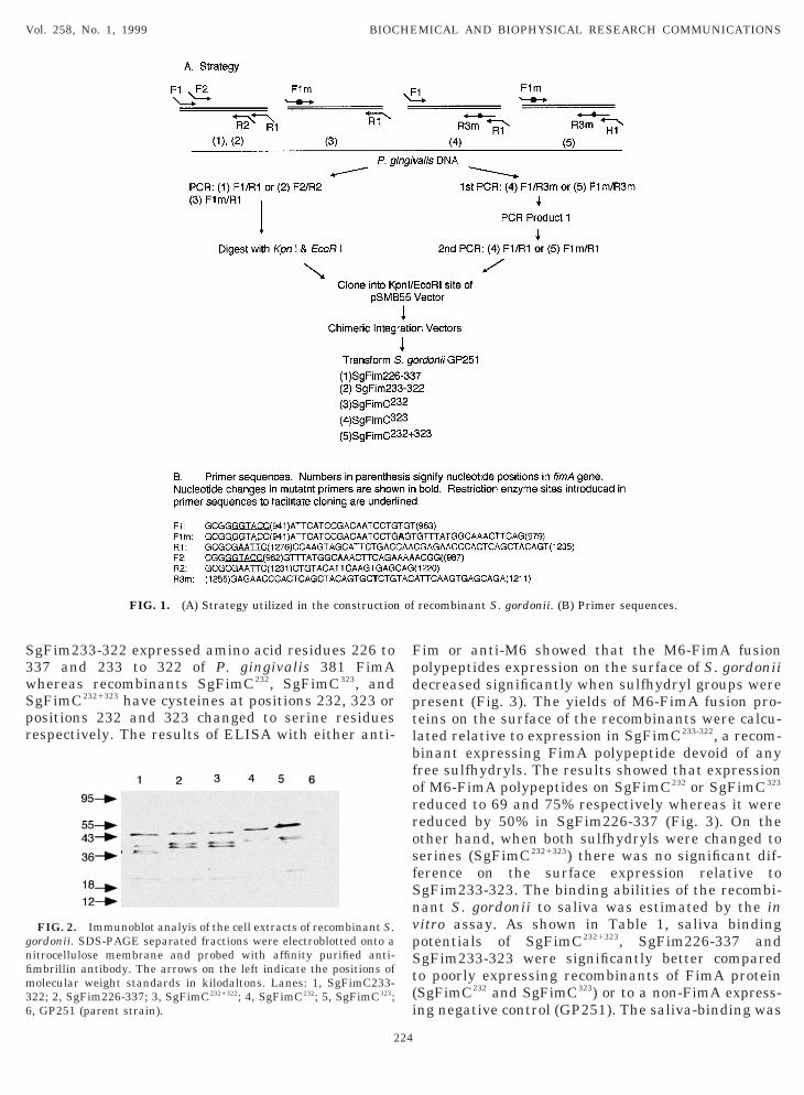
Construction of recombinant S. gordonii. DNA fragments encod-ng C-terminal portions (residues 226 to 337) of the P. gingivalismbrillin gene (fimA) were amplified by PCR utilizing pUC13Bg12.1s a template (18). Forward primers included the four-base ‘clamp’equence and a Kpn I restriction enzyme site at the 59-end whereashe reverse primers included a four base ‘clamp’ and a restriction siteor EcoR I to facilitate digestion and cloning into the integrationector. Insertion vector pSMB55 (1) was used to obtain translationalene fusions of the fimA gene with the M6 gene (emm6.1). Sequencef the primers used for amplifying wild type FimA encoding DNA orts mutants is shown in Fig. 1. The fimA gene was inserted into thepnI-EcoR I site of emm6.1 resident on pSMB55 and the resulting
himeric plasmid was used to transform S. gordonii GP251 as de-cribed previously (19). The transformants were selected by platingn THY-agar plates containing 5 mg/ml erythromycin. Followingnaerobic incubation at 37°C for 48-72 hours, colonies were analyzedor M6-FimA fusion protein expression by immunoblotting. Briefly,ultures (10 ml) of S. gordonii transformants were grown in THYroth to an optical density at 600 nm of 1.0. Cells were harvested byentrifugation and resuspended in 0.1 ml of protoplasting buffer (100M Tris-Cl, pH 8.0, 30% sucrose, 50 mM MgCl2, 5 mM dithiothreitol,
00 mg/ml lysozyme, 1 mM PMSF) and kept on ice for 1 h. Therotoplasts were centrifuged for 3 min at 16,000 x g and resuspendedn 100 ml of 50 mM Tris-Cl, pH 8.0. Thorough lysis was achieved byve cycles of quick freezing and thawing of the suspension. Unlysedells and gross debris were discarded by low speed centrifugation1,000 x g) for 15 min, whereas the supernatant containing mem-ranes and cytoplasm were subjected to sodium dodecyl sulfate-olyacrylamide gel electrophoresis (SDS-PAGE) followed by immu-oblot analysis (19).
Cell-surface ELISA. Cell-surface expression of fimbrilin wasuantified by a previously described whole cell ELISA technique19). Briefly, S. gordonii cells were grown as above in 15 ml THYroth culture to an absorbance of 1.0 at 600 nm. Cells were harvestedy centrifugation at 2,000 x g at 4°C and washed once with 25 ml ofold phophate-buffered saline containing 0.02% sodium azide (PBS-zide). The cells were resuspended in cold PBS-azide to an opticalensity at 590 nm of 1.0. The cells were diluted eightfold with 0.1 Modium bicarbonate buffer and 100 ml of diluted cells were added toach ELISA plate well (Dynatech Immulon II plate, Dynatech Lab-ratories). Plates were placed at room temperature for 2h to allowoating of wells with S. gordonii cells and stored overnight at 4°C.he wells were emptied by inverting the plates and then washedhree times with phosphate-buffered saline containing 0.05% Brij 35PBS-Brij). The unoccupied sites were blocked by 2% bovine serumlbubin in PBS (100 ml/well, PBS-BSA) for 1 h at 37°C. After block-ng, the wells were emptied and incubated for 1h at room tempera-ure with serially diluted primary antibody in PBS-BSA (50 ml/well).ollowing incubation, the wells were washed three times with PBS-rij and were incubated for 2 h at room temperature with alkalinehosphatase labeled goat anti-rabbit antibody (50 ml/well of 1:1000ilution in PBS-BSA; Bio-Rad Laboratories, Hercules, CA). Afterashing the wells three times with PBS-Brij, color was developedsing p-nitrophenyl phosphate (1 mg/ml) in 10% diethanolamine/1M MgCl2, pH 10.0. The plates were read at 405 nm using a Bio-Radicroplate reader.
Adherence assay. Adherence assays to determine the relativeinding abilities of recombinant S. gordonii to saliva were carriedut by a modification of a previuosly described assay (20). Microtiterlate wells were coated with human saliva (3) diluted 1:10 in 0.1 MaHCO3, pH 9.6 bufffer, for 2 h at 37°C (100 ml/well). Unoccupied
ites were blocked with 0.05% Tween-20 in PBS (PBST) and wellsere then incubated for 1 h at room temperature with 1 x 10⁵ to 1 x07 3H-labeled S. gordonii cells washed and suspended in 50 ml
223
ith PBST and bound-cells were dissociated by incubating each wellith 100 ml of 0.5 N NaOH/1% sodium dodecyl sulfate solution. Prior
o scintillation counting to measure bound radioactivity. samplesere neutralized by adding 100 ml of 0.5 N HCl. The percentage of
ell binding of each of the recombinant at sub-saturating concentra-ion was calculated and Tukey–Kramer multiple comparison tests ofignificance were performed to determine statistical differences inean binding.
ESULTS AND DISCUSSION
The C-terminal region of the P. gingivalis FimArotein (residues 226-337) contains domains respon-ible for bacterial adherenece to saliva-coated oralurfaces and the immunologically active peptide do-ain (peptide 226-246) that confers protection
gainst P. gingivalis associated alveolar bone loss inats. We reported earlier (19) that full length FimArotein or its polypeptide domains containing freeysteine residues express poorly on the surface of S.ordonii. The aim of the present study was thereforeo construct recombinant S. gordonii expressing the-terminal region of FimA and to study the contri-ution of free sulfhydryls on surface expression. The-terminal region contains cysteines at positions 232nd 333 respectively. To analyze the effect of freeulfhydryls on the surface expression of FimA pro-ein via the gram positive C-terminal anchor domainn S. gordonii, wild type and mutant C-terminal226 –337) polypeptdes of P. gingivalis fimbrillinith either one (residue 232 or 323) or both (232 and23) of the cysteines changed to serines were con-tructed by a PCR-based strategy depicted in Fig. 1.he fimA gene fragments were inserted in frameith the M6 gene residing in pSMB55 to obtain
ranslational fusions of the FimA and M6 proteins.he correct in-frame fusion and the nucleotidehanges in the cysteine to serine encoding fimA generagments were confirmed by DNA sequencing. Theecombinant plasmids were transformed into recipi-nt S. gordonii strain (GP251) and the transfor-ants selected on erythromycin selective plates were
haracterized by immunoblot analysis of the totalell extracts and cell-surface expression of M6-FimAolypeptides was quantified by cell-surface ELISAsing anti-fimbrillin and anti-M6 monoclonal anti-odies. The binding ability of the recombinants wasetermined by an in vitro binding assay. Immunoblotnalysis of cell extracts with anti-FimA (Fig. 2) andnti-M6 (data not shown) antibodies confirmed that6-FimA fusion proteins of expected size (45-kDa
and) were expressed in total cell extracts of the S.ordonii-FimA recombinants. Minor bands reactingith the anti-fimbrillin antibody likely represented
he products of the post translational processingnd/or the degradation products of the M6-FimA fu-ion protein. Recombinants SgFim226-337 and
S3wSpr
FpdptlbforrosfSnvpSt(i
gnfim36
Vol. 258, No. 1, 1999 BIOCHEMICAL AND BIOPHYSICAL RESEARCH COMMUNICATIONS
gFim233-322 expressed amino acid residues 226 to37 and 233 to 322 of P. gingivalis 381 FimAhereas recombinants SgFimC232, SgFimC323, andgFimC2321323 have cysteines at positions 232, 323 orositions 232 and 323 changed to serine residuesespectively. The results of ELISA with either anti-
FIG. 1. (A) Strategy utilized in the construction
FIG. 2. Immunoblot analyis of the cell extracts of recombinant S.ordonii. SDS-PAGE separated fractions were electroblotted onto aitrocellulose membrane and probed with affinity purified anti-mbrillin antibody. The arrows on the left indicate the positions ofolecular weight standards in kilodaltons. Lanes: 1, SgFimC233-
22; 2, SgFim226-337; 3, SgFimC2321322; 4, SgFimC232; 5, SgFimC323;, GP251 (parent strain).
224
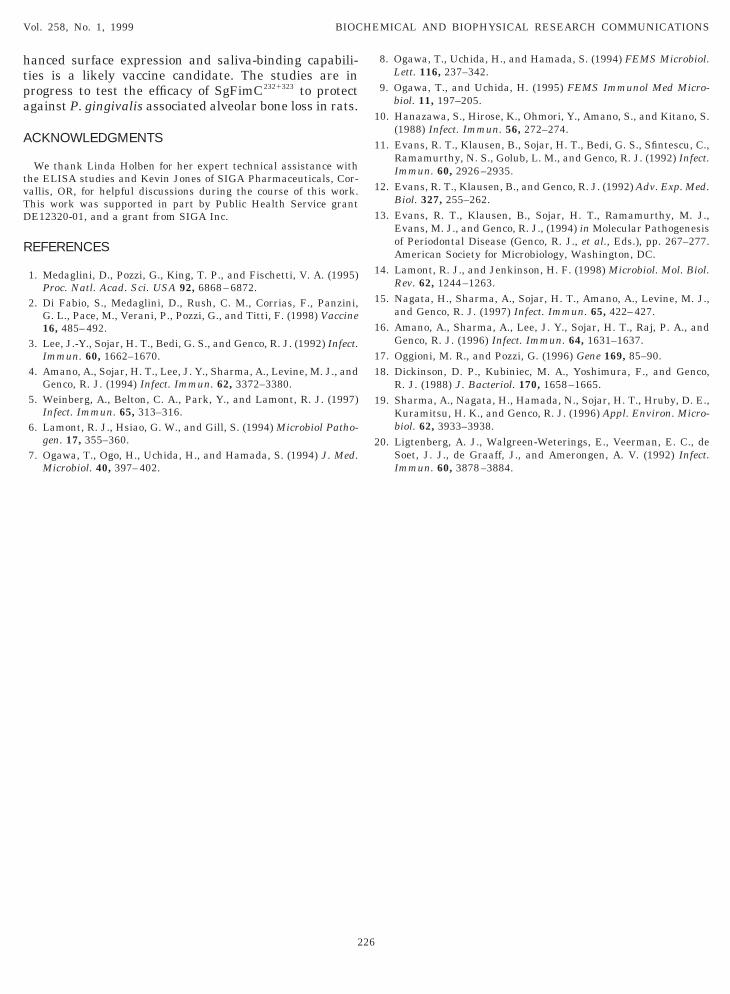
im or anti-M6 showed that the M6-FimA fusionolypeptides expression on the surface of S. gordoniiecreased significantly when sulfhydryl groups wereresent (Fig. 3). The yields of M6-FimA fusion pro-eins on the surface of the recombinants were calcu-ated relative to expression in SgFimC233-322, a recom-inant expressing FimA polypeptide devoid of anyree sulfhydryls. The results showed that expressionf M6-FimA polypeptides on SgFimC232 or SgFimC323
educed to 69 and 75% respectively whereas it wereeduced by 50% in SgFim226-337 (Fig. 3). On thether hand, when both sulfhydryls were changed toerines (SgFimC2321323) there was no significant dif-erence on the surface expression relative togFim233-323. The binding abilities of the recombi-ant S. gordonii to saliva was estimated by the initro assay. As shown in Table 1, saliva bindingotentials of SgFimC2321323, SgFim226-337 andgFim233-323 were significantly better comparedo poorly expressing recombinants of FimA proteinSgFimC232 and SgFimC323) or to a non-FimA express-ng negative control (GP251). The saliva-binding was
recombinant S. gordonii. (B) Primer sequences.
of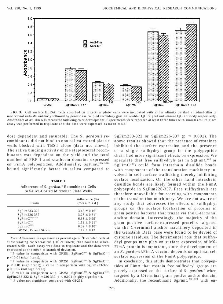
dcwTbnob
SaiocsSwvsdptoaggagvtcdFas
tptA
mAa
sce
p
pp
S
Vol. 258, No. 1, 1999 BIOCHEMICAL AND BIOPHYSICAL RESEARCH COMMUNICATIONS
ose dependent and saturable. The S. gordonii re-ombinants did not bind to non-saliva coated plasticells blocked with TBST alone (data not shown).he saliva binding activity of the steptococcal recom-inants was dependent on the yield and the totalumber of PRP-1 and statherin domains expressedn FimA polypeptides. Additionally, SgFimC2321323
ound significantly better to saliva compared to
FIG. 3. Cell surface ELISA. Cells absorbed on microtiter plateonoclonal anti-M6 antibody followed by peroxidase coupled secondbsorbance at 490 nm was measured following color development. Exssay was performed in triplicate and the data were expressed as m
TABLE 1
Adherence of S. gordonii Recombinant Cellsto Saliva-Coated Microtiter Plate Wells
StrainAdherence (%)(mean 6 s.d.)
SgFim233-322 3.45 6 0.16a
SgFim226-337 3.28 6 0.31b
SgFimC2321322 6.33 6 0.99c
SgFimC232 1.19 6 0.21ns
SgFimC323 0.82 6 0.18ns
GP251, Parent Strain 1.12 6 0.13
Note. Adherence is expressed as percentage of 3H-labeled cells atubsaturating concentrations (107 cells/well) that bound to saliva-oated wells. Each assay was done in triplicate and the data werexpressed as the mean 6 standard deviation.
a P value in comparison with GP251, SgFimC232 & SgFimC323,, 0.01 (significant).b P value in comparison with GP251, SgFimC232 & SgFimC323,, 0.01 (significant); P value in comparison with SgFim233-322,. 0.05 (not significant).c P value in comparison with GP251, SgFimC232 & SgFimC323,
gFim233-322 & SgFim226-337, p , 0.001 (highly significant).ns P value not significant compared with GP251.
225
gFim233-322 or SgFim226-337 (p # 0.001). Thebove results showed that the presence of cysteinesnhibited the surface expression and the presencef a single sulfhydryl group in the polypeptidehain had more significant effects on expression. Wepeculate that free sulfhydryls (as in SgFimC232 orgFimC323) could form interchain disulfide bondsith components of the translocation machinery in-olved in cell surface trafficking thereby inhibitingurface localization. On the other hand intrachainisulfide bonds are likely formed within the FimAolypeptde in SgFim226-337. Free sulfhydrayls areherefore unavailable for reacting with componentsf the translocation machinery. We are not aware ofny study that addresses the effects of sulfhydrylroups on the surface localization of proteins inram postive bacteria that traget via the C-terminalnchor domain. Interestingly, the majority of theram positive surface-expressed proteins targetedia the C-terminal anchor machinery deposited inhe GenBank Data base were found to be devoid ofysteine residues. The detrimental role that sulfhy-ryl groups may play on surface expression of M6-imA protein is important, since the development ofn S. gordonii:FimA vaccine depends on optimal cellurface expression of the FimA polypeptide.In conclusion, this study demonstrates that polypep-
ides of FimA that contain free sulfhydryl groups areoorely expressed on the surface of S. gordonii whenargeted by a C-terminal gram postive anchor domain.dditionally, the recombinant SgFimC2321323 with en-
ells were incubated with either affinity purified anti-fimbrillin orgoat anti-rabbit IgG or goat anti-mouse IgG antibody respectively.iments were repeated at least three times with simialr results. Each
6 s.d.
waryperean
hanced surface expression and saliva-binding capabili-tpa
A
tvTD
R
8. Ogawa, T., Uchida, H., and Hamada, S. (1994) FEMS Microbiol.
1
1
1
1
1
1
1
11
1
2
Vol. 258, No. 1, 1999 BIOCHEMICAL AND BIOPHYSICAL RESEARCH COMMUNICATIONS
ies is a likely vaccine candidate. The studies are inrogress to test the efficacy of SgFimC2321323 to protectgainst P. gingivalis associated alveolar bone loss in rats.
CKNOWLEDGMENTS
We thank Linda Holben for her expert technical assistance withhe ELISA studies and Kevin Jones of SIGA Pharmaceuticals, Cor-allis, OR, for helpful discussions during the course of this work.his work was supported in part by Public Health Service grantE12320-01, and a grant from SIGA Inc.
EFERENCES
1. Medaglini, D., Pozzi, G., King, T. P., and Fischetti, V. A. (1995)Proc. Natl. Acad. Sci. USA 92, 6868–6872.
2. Di Fabio, S., Medaglini, D., Rush, C. M., Corrias, F., Panzini,G. L., Pace, M., Verani, P., Pozzi, G., and Titti, F. (1998) Vaccine16, 485–492.
3. Lee, J.-Y., Sojar, H. T., Bedi, G. S., and Genco, R. J. (1992) Infect.Immun. 60, 1662–1670.
4. Amano, A., Sojar, H. T., Lee, J. Y., Sharma, A., Levine, M. J., andGenco, R. J. (1994) Infect. Immun. 62, 3372–3380.
5. Weinberg, A., Belton, C. A., Park, Y., and Lamont, R. J. (1997)Infect. Immun. 65, 313–316.
6. Lamont, R. J., Hsiao, G. W., and Gill, S. (1994) Microbiol Patho-gen. 17, 355–360.
7. Ogawa, T., Ogo, H., Uchida, H., and Hamada, S. (1994) J. Med.Microbiol. 40, 397–402.
226
Lett. 116, 237–342.9. Ogawa, T., and Uchida, H. (1995) FEMS Immunol Med Micro-
biol. 11, 197–205.0. Hanazawa, S., Hirose, K., Ohmori, Y., Amano, S., and Kitano, S.
(1988) Infect. Immun. 56, 272–274.1. Evans, R. T., Klausen, B., Sojar, H. T., Bedi, G. S., Sfintescu, C.,
Ramamurthy, N. S., Golub, L. M., and Genco, R. J. (1992) Infect.Immun. 60, 2926–2935.
2. Evans, R. T., Klausen, B., and Genco, R. J. (1992) Adv. Exp. Med.Biol. 327, 255–262.
3. Evans, R. T., Klausen, B., Sojar, H. T., Ramamurthy, M. J.,Evans, M. J., and Genco, R. J., (1994) in Molecular Pathogenesisof Periodontal Disease (Genco, R. J., et al., Eds.), pp. 267–277.American Society for Microbiology, Washington, DC.
4. Lamont, R. J., and Jenkinson, H. F. (1998) Microbiol. Mol. Biol.Rev. 62, 1244–1263.
5. Nagata, H., Sharma, A., Sojar, H. T., Amano, A., Levine, M. J.,and Genco, R. J. (1997) Infect. Immun. 65, 422–427.
6. Amano, A., Sharma, A., Lee, J. Y., Sojar, H. T., Raj, P. A., andGenco, R. J. (1996) Infect. Immun. 64, 1631–1637.
7. Oggioni, M. R., and Pozzi, G. (1996) Gene 169, 85–90.8. Dickinson, D. P., Kubiniec, M. A., Yoshimura, F., and Genco,
R. J. (1988) J. Bacteriol. 170, 1658–1665.9. Sharma, A., Nagata, H., Hamada, N., Sojar, H. T., Hruby, D. E.,
Kuramitsu, H. K., and Genco, R. J. (1996) Appl. Environ. Micro-biol. 62, 3933–3938.
0. Ligtenberg, A. J., Walgreen-Weterings, E., Veerman, E. C., deSoet, J. J., de Graaff, J., and Amerongen, A. V. (1992) Infect.Immun. 60, 3878–3884.