Embed Size (px)
Citation preview

121
1 University of Missouri at Kansas City, Pharmaceutical Sciences, Kansas City, Missouri.
2 University of Missouri at Kansas City, Medical School, Kansas City, Missouri.
*Current address: Howard University, School of Pharmacy, Washington, DC.
JOURNAL OF OCULAR PHARMACOLOGY AND THERAPEUTICSVolume 25, Number 2, 2009© Mary Ann Liebert, Inc.DOI: 10.1089/jop.2008.0084
Expression of Multidrug Resistance Associated Protein 5 (MRP5) on Cornea and Its Role in Drug Efflux
Pradeep K. Karla,1,* Tim L. Quinn,2 Betty L. Herndon,2 Priscilla Thomas,2 Dhananjay Pal,1 and Ashim Mitra1
Abstract
Purpose: The purpose of this manuscript is to investigate the presence of nucleoside/nucleotide effl ux trans-porter in cornea and to evaluate the role in ocular drug effl ux.Methods: RT-PCR, immunoprecipitation followed by Western blot analysis and immunostaining were employed to establish molecular presence of multidrug resistance associated protein 5 (MRP5) on cornea. Corneal effl uxby MRP5 was studied with bis(POM)-PMEA and acyclovir using rabbit and human corneal epithelial cells along with MRP5 over expressing cells (MDCKII-MRP5). Ex vivo studies using excised rabbit cornea and in vivo ocular microdialysis in male New Zealand white rabbits were used to further evaluate the role of MRP5 in conferring ocular drug resistance.Results: RT-PCR confi rms the expression of MRP5 in both rabbit and human corneal epithelial cells along with MDCKII-MRP5 cells. Immunoprecipitation followed by Western blot analysis using a rat (M511–54) monoclonal antibody that reacts with human epitope confi rms the expression of MRP5 protein in human corneal epithelial cells and MDCKII-MRP5 cells. Immunostaining performed on human cornea indicates the localization of this effl ux pump on both epithelium and endothelium. Effl ux studies reveal that depletion of ATP decreased PMEA effl ux signifi cantly. MRP5 inhibitors also diminished PMEA and acyclovir effl ux. However, depletion of gluta-thione did not alter effl ux. MDR1 and MRP2 did not contribute to PMEA effl ux. However, MRP2 is involved in acyclovir effl ux while MDR1 do not participate in this process. TLC/autoradiography suggested the conversion of bis(POM)-PMEA to PMEA in rabbit and human corneal epithelial cells. Two well known antiglaucoma drugs, bimatoprost and latanoprost were rapidly effl uxed by MRP5. Ex vivo study on intact rabbit corneas demonstrated accumulation of PMEA in cornea in the presence of ATP-depleting medium. In vivo ocular pharmacokinetics also revealed a signifi cant increase in maximum aqueous humor concentration (Cmax) and area under the aque-ous humor time curve (AUC) of acyclovir in the presence of MK-571, a specifi c MRP inhibitor.Conclusions: Taken together immunolocalization on human cornea, in vitro effl ux in human, rabbit corneal and MRP5 over expressing cells, ex vivo and in vivo studies in intact rabbit cornea suggest that MRP5 on cornea can signifi cantly lower the permeability of antiviral and glaucoma drugs. These fi ndings may be valuable in devel-oping formulation strategies to optimize ocular bioavailability of topically administered ocular agents.
Introduction
Cornea is considered a major barrier for topical ocular
drug delivery. Lipoidal nature of corneal epithelium
and tight junctions of epithelial cells are primarily respon-
sible for poor permeation.1,2 However, Pgp an effl ux pump
primarily responsible for emergence of drug resistance to a
wide spectrum of drugs was reported to be expressed in rab-
bit1 and human2 corneal epithelial cells. Also, various Pgp
inhibitors caused signifi cant alteration in pharmacokinetics
leading to enhanced anterior chamber bioavailability.3 Both
Pgp and multidrug resistance associated proteins (MRP)
belong to ATP-binding cassette (ABC) transporter family of
effl ux pumps. Recently, expression of organic anion trans-
porter effl ux pumps MRP1 and MRP2 were identifi ed for the
fi rst time in rabbit and human corneal epithelial cells from
our laboratory. MRP2 was indeed shown to confer signifi cant
04_JOP-2008_0084.indd 121 4/4/2009 2:37:35 AM

KARLA ET AL.122
purchased from Riken cell bank (Ibaraki, Japan). Protocol for
cell culture was provided by the cell bank. Mycoplasma-free
SV40-HCEC were grown at 37°C, humidifi ed 5% CO2/95%
air atmosphere/95% humidity in a culture medium contain-
ing 50% of Dulbecco’s modifi ed Eagle’s medium (DMEM)
and 50% of Ham’s nutrient mixture F-12 (from Gibco, Paisley,
UK) supplemented with 15% (v/v) fetal bovine serum (FBS,
from Atlanta biologics), antibiotic, gentamycin (30 μg/mL)
and 0.5 % dimethyl sulfoxide (DMSO). Cells were harvested
with trypsin-EDTA.
Statens Seruminstitut rabbit corneal epithelial cells (SIRC). SIRCs were plated in a T-75 culture fl ask and MEM con-
taining Sodium bicarbonate (3.8 mg/mL), HEPES (4.8 mg/
mL), Lactalbumin (1.76 mg/mL), Penicillin (0.1 mg/mL),
Streptomycin Sulfate (0.125 mg/mL), Fetal Bovine Serum
(10%), and MEM Nonessential amino acids (10%) were used.
Medium was changed twice a week, and cells were sub-
cultured every 5–7 days as 70%–80% confl uence is reached
(subculture ratio, 1:5).
Primary culture of rabbit corneal epithelial cells (rPCECs). Rabbit corneal epithelial cells were isolated and cultured as
per the protocol established in our laboratory.2 Cells were
grown using the same medium composition mentioned
earlier for SIRC. The medium was changed twice a week,
and the cells were subcultured every 7–10 days (subculture
ratio, 1:5).
MDCKII-Wt, MDCKII-MDR1, MDCKII-MRP2, and MDCKII-MRP5 cells. MDCKII-Wt and MDCKII cells overex-
pressing human MDR1, MRP2, and MRP5 genes at protein
level were obtained as a gift from Dr. Piet Borst (Netherlands
Cancer Institute, Amsterdam). Cells were maintained in
DMEM supplemented with 10% calf serum, 100 IU/mL pen-
icillin, 100 μg/mL streptomycin, and 20 mmol/L HEPES, pH
7.4. Cells were plated at a density of 100,000/cm2 in 12-well
tissue culture–treated plastic dishes. MDCKII cells were
incubated at 37°C in humidifi ed atmosphere of 5%CO2/95%
humidity and were allowed to grow for 5–8 days.
Reverse transcriptase polymerase chain
reaction (RT-PCR)
Human, rabbit corneal epithelial cells and MRP5 over
expressing cells (SV40-HCEC, rPCECs, SIRC, and MDCKII-
MRP5) were grown for mRNA extraction. Cells (3–5 million)
from a confl uent fl ask were taken, and 800 μL of Tri-reagent
LS (Molecular Research Center, Inc., Cincinnati, OH) was
added. The mixture was homogenized and transferred to
eppendorf tubes. RNA was extracted by the phenol-CHCl3-
isopropranolol method, purifi ed, and dissolved in 20 μL of
RNase-DNase-free water. RT-PCR was performed based on
established protocol with modifi cations using ~1 μg of total
RNA.18 MRP5 primers were designed from human MRP5
cDNA (Genbank Accession No: AB209454; http://www.ncbi.
nlm.nih.gov/Genbank; made available in the public domain
by the National Center for Biotechnology Information
[NCBI], Bethesda, MD). The forward and reverse primers
designed for MRP5 were 5’-ACCCGTTGTTGCCATCTTAG-3’
and 5’-GCTTTGACCCAGGCATACAT-3’, respectively. These
primers correspond to the nucleotide position 879–1106.
RT-PCR was performed with a kit (SuperScript III First-
Strand Synthesis System for RT-PCR; Invitrogen, Carlsbad,
CA). Conditions for reverse transcription were denaturation
resistance to drug disposition across cornea into the ante-
rior chamber.4,5 Substrate specifi city for the MRPs appears
to vary. Tissue distribution also varies, however the exact
function of the proteins in the cornea remains to be estab-
lished.6 MRP4 and MRP5, two prominent members of MRP
family are known to effl ux the nucleotides. MRP5 appears to
be localized on both the apical side of brain microvascular
endothelial cells and on the basolateral side of gut and liver
leading to drug resistance.7,8 Effl ux of PMEA by MRP5 in the
microglia has been demonstrated.9 MRP5-mediated drug
resistance is currently being investigated.6,10
Nucleoside analog such as acyclovir is indicated in the
treatment of severe corneal keratitis and stromal keratitis
which is one of the leading causes of blindness in the United
States.11 However, a very high dose of acyclovir needs to be
administered orally because of poor aqueous solubility and
low corneal permeability12 that renders acyclovir ineffective
via the topical route.13,14 So far, no evidence of MDR mediated
resistance related to topical acyclovir has been reported.
MRP2, a major effl ux pump identifi ed on intact rabbit cor-
nea was not shown to effl ux the nucleotide molecules like
PMEA.15 Prostaglandin analogs like bimatoprost and latano-
prost are currently employed extensively in the treatment of
glaucoma. Bimatoprost is available in the market as 0.03%
ophthalmic solution and is manufactured under the trade
name Lumigan by Allergan Inc. (Irvine, CA). Latanoprost is
manufactured under the trade name Xalatan by Pfi zer Inc.
(New York) and is composed of 50 μg/mL of latanoprost
along with 0.02% benzalkonium chloride. Both drugs are
administered as eye drops. Since MRP5-mediated effl ux of
prostaglandin analogs (PGE1 and PGA1) was reported ear-
lier16 it is important to investigate the role of MRP5 on the
ocular permeation of drugs like bimatoprost, latanoprost
along with PMEA and acyclovir. PMEA was the fi rst drug
of choice as it was proven to be a strong substrate of MRP5
and also used in the MRP5 characterization studies.9 In the
present study, transport of MRP5 was examined in both rab-
bit and human corneal epithelial cells. We have shown that
MRP5 is expressed at gene and protein level in human cor-
neal cells and is functionally active. We have also shown the
localization of MRP5 in human cornea and demonstrated
the functional activity with ex vivo and in vivo studies in rab-
bit cornea. Thus this article provides the fi rst evidence for
the corneal expression of MRP5-generating rapid effl ux of
antiviral and glaucoma drugs.
Materials and Methods
3H-bis(POM)PMEA, 3H-PMEA, and 3H-acyclovir were
obtained from Moravek Biochemicals and Radiochemicals
(Mercury Lane Brea, CA). Bimatoprost and latanoprost
were purchased from Cayman chemical (Ann Arbor, MI).
Indomethacin and sodium azide were obtained from Sigma
Chemical Co. (St. Louis, MO). Sulfi npyrazone was procured
from Alexis Biochemical (San Diego, CA). D,L-Buthionine
(S,R)-Sulfoximine (BSO) and 2-Deoxy-d-glucose were acquired
from Acros Organics (Morris Plains, NJ). MK-571 was received
from Biomol International, L.P. (Plymouth Meeting, PA).
Cell culture
Transfected human corneal epithelial cells (SV40-HCEC). SV40-immortalized human corneal epithelial cell line17 was
04_JOP-2008_0084.indd 122 4/4/2009 2:37:36 AM

ROLE OF MRP5 IN CORNEAL DRUG EFFLUX 123
monoclonal antibody (1:100 dilution). Tissues were rinsed
three times 15 min each with PBST at 37°C. Then the sections
were treated with biotin conjugated donkey anti-rat sec-
ondary antibody (1:2500) (BioFX Laboratories, Inc., Owings
Mills, MD) followed by horseradish peroxidase–conjugated
streptavidin (1:2000) for 20 min. This procedure was fol-
lowed by PBST wash, as previously mentioned. Finally, the
sections were treated with 3,3’-Diaminobenzidine (DAB) for
5 min and counterstained with harris hematoxylin.
Transport of nucleotides across cells
To determine functional activity of MRP5 in corneal epi-
thelial cells, transport studies were performed with PMEA
and acyclovir. Transport studies were performed based
on the protocol established by Schuetz et al., 1999 with
minor modifi cations. Monolayers of MDCKII-MRP5, SV40-
HCEC, rPCEC, and SIRC cultured in 12-well tissue culture
plates (Fisher Scientifi c, PA) were employed. As reported
previously, addition of 10-mM sodium azide and 10-mM
2-deoxy-d-glucose to cell monolayer’s reduced intracellu-
lar ATP by >90% in the fi rst 5 min of incubation but did
not affect cell viability.9 The principle behind this study
involves preloading drug in to cells by deactivating the
effl ux transporter (ATP-depleting conditions), washing and
then reactivate the transporter with a medium containing
glucose. Transport kinetics of MRP5 was observed with and
without inhibitors.
Cells were incubated for 60 min with 3H-bis (POM)
PMEA (1uCi) and 3H-acyclovir (1uCi) in the presence of
10-mM sodium azide and 2-deoxy-d-glucose. Following 1 h
incubation period under normal and ATP-depleting condi-
tions cell viability was determined by trypan blue exclusion
test. Cells were examined for viability (unstained) and non-
viability/cytotoxicity (stained). Hemocytometer (Hausser
scientifi c, Horsham, PA) was used to count the cells. After
incubation drug solution was removed from the wells and
cells were washed with 0.16 M NaCl and subsequently
incubated for 60 min with Hank’s buffered saline solution
(HBSS) with glucose, pH-7.2 and HBSS with glucose incor-
porating the inhibitors. Preparation of HBSS with inhibitors
is described in the next section. The supernatant was col-
lected and radioactivity was measured with Beckmann scin-
tillation counter (Beckman Coulter, Inc., CA). The amount of
radioactivity in the supernatant represents amount of drug
effl uxed. Radioactivity was converted to drug concentration
based on specifi c activity of the compound.
Preparation of inhibitor solutions
Indomethacin (10 mg), bimatoprost (1 mg), and latano-
prost (1 mg) were dissolved in ethanol (1 mL) to prepare a
stock solution and the solution was aliquoted in HBSS to
achieve the desired concentrations. Sulfi npyrazone (10 mg)
was dissolved in chloroform (1 mL) and aliquoted with HBSS.
Organic solvents were employed at ≤0.05% which was same
in control. MK-571 was freely soluble in water. Cytotoxicity
was measured with CytoTox-ONE Homogeneous Membrane
Integrity Assay (Promega, San Luis Obispo, CA) on all the
cell lines employed. No cytotoxicity was noticed even at
the highest concentrations of inhibitors employed. (data not
shown)
of the template RNA for 10 min at 70°C and reverse tran-
scription for 60 min at 42°C. Conditions for PCR amplifi ca-
tion were denaturation for 1 min at 94°C, annealing for 1 min
at 58.5°C and extension for 1 min at 72°C for 35 cycles, and
a fi nal extension for 10 min at 72°C. RT-PCR products were
analyzed by electrophoresis on 2% agarose gel. Sequencing
analysis was performed by Agencourt Biosciences (Beverly,
MA) using Quicklane sequencing technology.
Immunoprecipitation—Western blot
SV40-HCEC and MDCKII-MRP5 cells grown on T-75
(75 cm2 growth area) were washed twice with PBS and
harvested with a cell scraper into 5 mL of PBS. Mem-PER
Eukaryotic Membrane Protein Extraction Reagent Kit
(Pierce Biotechnology, Inc., Rockford, IL) was used to sep-
arate membrane fraction from cytoplasmic fraction. The
membrane fraction and supernatant were separated and
stored at −80°C until further use. Protein content was
determined with Bradford method (Bio-Rad protein esti-
mation kit; Bio-Rad, Hercules, CA). Immunoprecipitation
followed by Western blot was performed utilizing Seize
Classic (G) Immunoprecipitation Kit (Pierce Biotechnology,
Inc., Rockford, IL). Twenty micrograms of protein (100 μL
lysate) were immunoprecipitated overnight at 4°C with the
ABCAM (#M511–54) rat monoclonal antibody for MRP5
(Abcam Inc, Cambridge, MA). Protein G-sepharose beads
containing antigen–antibody complexes were collected by
centrifugation, washed three times with immunoprecip-
itation buffer, resuspended in lane marker sample buffer
(0.3 M Tris–HCl, 5% SDS, 50% glycerol, lane marker track-
ing dye; pH-6.8) (Pierce Biotechnology, Inc., Rockford, IL)
and boiled for 5 min. All immunoprecipitated samples and
molecular weight protein markers were separated by SDS-
PAGE (8% Tris-glycine gels for 2 h at 120 V). Protein was
transferred onto nitrocellulose membranes and stored for 2
h at 250 mA on ice. Protein transfer effi ciency was checked
by staining the nitrocellulose membranes in 0.2% ponceau
S (in 3% wt/vol trichloroacetic acid and 3% wt/vol sulfos-
alicylic acid) for 10 min. A portion of the blot containing
MRP5 samples was incubated in freshly prepared block-
ing buffer (3% wt/vol BSA and 5% nonfat dry milk in Tris-
buffered saline) for 1 h at room temperature. The blot was
then treated with MRP5 antibody (1:50) overnight at 4°C.
After fi ve 10-min washes with TBST (Tris-buffered saline
+ 0.1% Tween 20), the membrane was treated with sec-
ondary antibody in TBST (1:1500 anti-rat IgG-horseradish
peroxidase [HRP]) for 1 h. The blots were fi nally washed
three times (15 min each) with TBST. Opti-4CN Western
blot amplifi cation kit (Bio-Rad, Herculus, CA) was used to
develop the blots.
Immunostaining of human cornea
Human corneas were obtained from Heartland Lion’s
Eye Bank (Kansas City, MO). The tissues were fi xed in 3.7%
formalin and embedded in paraffi n. These sections were
dissected at 5 microns and deparafi nized using xylene and
ethanol. Antigen retrieval was performed with citrate buffer
(pH-6.0) with 0.05% Tween 20 in a 95°C steam bath for 30
min. Sections were rinsed in PBST (phosphate-buffered
saline + 0.05% Tween 20) for 5 min on a shaker and then
incubated overnight at 4 °C with ABCAM (#M511–54) rat
04_JOP-2008_0084.indd 123 4/4/2009 2:37:36 AM

KARLA ET AL.124
Residual radioactivity from the entire plate is determined
and added to the fi nal value to minimize error. Results were
expressed as percentage of total intracellular radioactivity
recovered (mean ± SD).
In vivo anterior segment microdialysis
Probe implantation. The role of MRP on absorption kinetics
of acyclovir was assessed by an in vivo topical well infusion
model coupled with aqueous humor microdialysis technique
established in our laboratory.3,20 Absorption kinetics of acy-
clovir across the cornea were measured alone and in the pres-
ence of a potent MRP inhibitor, MK-571. Rabbits were given
an anesthetic of Ketamine HCl (35 mg/kg) and xylazine (3.5
mg/kg) by intramuscular injection. A diagrammatic repre-
sentation of ocular microdialysis by a topical well infusion
model is shown in Figure 1. Isotonic phosphate buffer saline
solution (IPBS – pH, 7.2) was used for the studies.
IPBS was added to fi ll needles (25 G1). The posterior end
of needle was then closed with a plastic cap to prevent the
loss of liquid, including the loss of aqueous humor during
surgery. The needle was surgically inserted into the anterior
side of cornea and carefully penetrated through the aqueous
humor and withdrawn from the posterior side of cornea.
The sample-collecting end of the microdialysis probe
was inserted into the needle and the needle was withdrawn
slowly to ensure placement of the dialyzing membrane probe
in the aqueous humor. Extreme care is taken to ensure that
there is no damage to the other tissues of the eye (Fig. 2A).
The other end of the probe was connected to the syringe
needle (3 mL) fi lled with IPBS which was further connected
to a CMA/100 microdialysis pump (Bioanalytical Inc., West
Lafayette, IN) which controls the rate of infusion of IPBS
through the probe. The fl ow rate was set at 3.0 μL/min based
on standard protocol established in our laboratory. Heart
rate and respiratory rate are carefully monitored every
30 min to ensure the animal safety during surgery. Probe
implantation was followed by a 2 h stabilization period of
the anesthetized animal during which aqueous humor and
intra ocular pressure returned to normal.3,20
Evaluation of active nucleotide transport on freshly
excised cornea
New Zealand male albino rabbits (n = 6) were eutha-
nized, Corneas were immediately dissected and trans-
ferred to HBSS, pH 7.2. Within approximately 1–2 min,
the tissues were successfully mounted on Side-by-Side
diffusion cells (type-VSC-1, Crown Glass Company Inc.,
Somerville, NJ). Side-bi-Side chamber has a donor (apical)
and a receiver compartment (basolateral) with the cornea
mounted in between. The chamber is maintained at 34°C
which is the temperature of cornea exposed to external
environment. The donor and receiver compartments were
continuously stirred with magnetic console stirrer (Crown
Glass Company Inc., Somerville, NJ) to minimize the aque-
ous boundary layer and to simulate in vivo dynamic condi-
tions. The chambers were fi lled with 3 mL of HBSS with
glucose, pH-7.2 (normal conditions) and with10-mM NaN3
+ 10-mM 2-Deoxy-d-Glucose (ATP-depleting conditions)
for 30 min. Control chambers were fi lled with HBSS con-
taining glucose in both apical and basolateral sides. Test
chambers are loaded with ATP-depleting medium on apical
side and HBSS with glucose on basolateral side. This condi-
tion simulates actual in vivo environment, where apical side
(corneal surface exposed to external environment) is mod-
ulated with ATP-depleting agents. After the specifi c expo-
sure period, corneas were dismounted, thoroughly washed
to remove any residual radioactivity and placed in lysis
solution (1% tritonex + 1N NaOH solution) overnight. Later,
cornea was thoroughly ground using tissue grind pestle
(Fischer scientifi c, USA) and resuspended in scintillation
cocktail (ScintiSafe30) (Fischer scientifi c, USA) to determine
accumulated radioactivity. Pestle was thoroughly swiped
with whatmann fi lter paper and radioactivity determined
was added to the initial radioactivity to ensure no superfi -
cial loss due to grinding.
Intracellular conversion of bis(POM)PMEA to PMEA
Intracellular conversion of 3H-bis(POM)PMEA to 3H-PMEA
within the human and rabbit corneal cells was confi rmed
using thin layer chromatography (TLC) by the method
adopted from Dallas et al., 2004 with modifi cations. Cells
were incubated for 1 h (37°C) under ATP-depleting con-
ditions with 3H-bis(POM)PMEA (0.5 mCi/mL) and then
washed twice with 0.16 M NaCl to remove any residual drug.
Cells were then incubated in lysis solution overnight. Five
microliter of cell lysate, 5 μL donor (3H-bis(POM)PMEA),
and 1 μL controls (3H-bis(POM)PMEA and 3H-PMEA)
were spotted on a 250-μm thick silica-gel-coated TLC plate
(Sigma-Aldrich, St. Louis, MO). Plates were air dried for 1
h and chromatographic separation of 3H-bis(POM)PMEA
and 3H-PMEA was performed using the solvent system
isopropanol:ammonia:water (7.5:1:1.5 v/v/v). The plates were
subsequently air dried for 2 h and sprayed with beta emitter
enhancer spray (EN3HANCE) (PerkinElmer, Waltham, MA).
TLC plates were exposed to X-ray fi lm and stored at −20°C
for 36 h. Care was taken to protect the fi lms from light. The
fi lms were developed and the bands obtained were matched
to the respective position of the compounds on TLC plates.
The areas corresponding to bis(POM)PMEA and PMEA were
scrapped and radioactivity was determined by Beckman
scintillation counter (Beckman Coulter, Inc., Fullerton, CA).
Wt
PMEA **
0
2
4
6
Cum
. A
mt. E
fflu
x (
Pm
ole
s/m
g p
rote
in)
8
10
12
14
MDR1 MRP2 MRP5
FIG. 1. Effl ux of active drug PMEA from MDCKII-wt,
MDR1, MRP2, and MRP5 cells preloaded with prodrug Bis
(POM)-PMEA. HBSS with glucose was used to measure the
relative effl ux of transporters. Statistical signifi cance was
tested by two-factor ANOVA. **P ≤ 0.01 (n = 5).
04_JOP-2008_0084.indd 124 4/4/2009 2:37:36 AM

ROLE OF MRP5 IN CORNEAL DRUG EFFLUX 125
the animal study. Ten microcurie per milliliter is employed
for the in vivo studies, and a 5% of the concentration, that is,
0.5 μCi/mL was employed for the recovery studies. IPBS per-
fused the probe at 3.0 μL/min and the dialyzing membrane
was placed in a solution containing 0.5 μCi/mL of the drug.
As indicated by Dey and colleagues, 2003; recovery is con-
sidered as the ratio of the concentration of drug obtained in
the sample to the concentration present in the solution sur-
rounding the probe.21 The recovery of the drug by microdi-
alysis probe is designated by equation 1.
Recovery = Cs/Cm (Eq. 1)
Cs is the concentration in the sample and Cm represents
concentration in the medium. Concentration of the drug in
aqueous humor (CAH) is calculated based on concentrations
obtained in the dialysate sample (Cdl) from equation 2.
CAH = Cdl/Recovery (Eq. 2)
Percent recovery was between 12% and 18% for both acyclo-
vir and erythromycin. Recovery was performed after the in vivo study and MK-571 and GF120918 employed did not alter
the recovery. This confi rms that the recovery was uniform
throughout the duration of study.
Microdialysis
After 2 h, colibri retractors obtained from Spectrum (Stow,
OH) were employed to retract the eye lids to provide good
exposure of the cornea for the experiment. A topical well
was adhered on top of the cornea and after 40 min, 150 μL
HBSS containing 14C-erythromycin (10 μCi/mL) alone or in
combination with 100 μM MK-571 was added to the well.
The wells are monitored for any leakage of drug solution
and samples are collected from anterior end of the probe
every 20 min. The experiment is designed for 2 h of absorp-
tion phase and the drug solution is removed along with the
wells after 120 min. The surface of the cornea is washed
and aspirated with distilled water to remove any superfi cial
drug solution. Samples are collected from the probe for a 6
h postinfusion period at which time the animals are eutha-
nized following the approved protocol.
In vitro probe calibration
Ocular bioavailability on topical administration is ~5%
(ranges from 1% to 10%) for majority of the drugs.1 Probe
recovery is performed on all the probes used before and after
Cornea
Probe Outlet
Aqueous Humor
Topical Well with Drug
A
Probe Inlet
Injector with IPBS
Microdialysis Pump
Vitreous Humor
B
8 mm
14 mm
4 mm
Rabbit CorneaAqueous Humor
Peripheral Tissue
K0
K10
KPT
XPT
VPT
KAH
XAH,VAH
16 mm
Topical Glass Well
FIG. 2. (A) Schematic representation of probe insertion in the anterior segment of the eye. As shown in the fi gure, dialyzing
membrane of the probe was placed in the aqueous humor; probe inlet connected to an IPBS injector connected to a microdi-
alysis pump, which perfuses IPBS at 3 μL/min. Sample was collected at probe outlet. Topical glass well where drug solution is
loaded and placed on the cornea with a surgical adhesive as shown. (B) Schematic representation of aqueous humor kinetics
of single-dose constant infusion of drug across the cornea. Diagrams drawn based on inspiration from references 2.
04_JOP-2008_0084.indd 125 4/4/2009 2:37:36 AM

KARLA ET AL.126
Data treatment
Data analysis was performed with pharmacokinetic soft-
ware WinNonlin, v5.0 (Pharsight, CA). Noncompartmental
analysis of drug concentrations–time profi les in the aqueous
humor was performed according to a noncompartmental
model with a constant infusion rate. All the pharmacokinetic
parameters were obtained from the analysis. Absorption
rate constant (ka) was calculated from equation 6.
Results
MRP5 mRNA and protein expression in corneal
epithelial cells
A single specifi c band at ~228 bp corresponding to MRP5
was observed from RT-PCR conducted on rPCEC, SIRC, and
positive control (MDCKII-MRP5). Unique band at ~294 bp
for GAPDH (internal control) was also observed (Fig. 3A).
RT-PCR on SV40-HCEC also yielded a similar result, that is,
a band at ~228 bp. GAPDH band (~294 bp) served as an inter-
nal control (Fig. 3B). Sequencing, alignment in Clustal W
Mathematical data treatment
The mathematical treatment was adapted based on the
protocol established by Dey et al., 2004 in our laboratory.
As the HBSS containing the radioactive drug is loaded in
the topical well, it enters the aqueous humor via cornea by
passive diffusion. Drug in aqueous humor is distributed to
surrounding tissues and the distribution can be reversible.
Concentrations of the drug in aqueous humor and surround-
ing tissues (peripheral) are denoted by XAH and XPT. Rate of
infusion across the cornea is represented by K0. First order
rate constant for distribution of drug present in the aqueous
humor to surrounding tissues and surrounding tissues to
the aqueous humor are represented by kAH and kPT. As the
drug is eliminated through aqueous outfl ow, apparent elim-
ination rate constant, which is the fi rst order, is denoted by
K10. The absorption, distribution, and elimination are repre-
sented as a scheme in Figure 2B.
Designating Y, as aqueous humor and number of sur-
rounding tissues as X, and X ≥ Y, equation 3 can be repre-
sented as follows.
dXdt
k X k X k XAHAH PT PT
i
y
AH� � � ���
0
1
10kAHi
x
∑∑1
(Eq. 3)
Elimination is considered minimal from the surrounding
tissues and is mainly by aqueous out fl ow pathway. The rela-
tionship between rate constant of infusion and concentra-
tion of drug in reservoir (CR) is represented as equation 4. VR
is the reservoir volume and ka is the fi rst order rate constant
for drug absorption.
k0 = ka CR VR (Eq. 4)
Equation 4 can be further represented as k0 = ka XR.
Equation 3 can be further represented as equation 5.
dX Vdt
k X k X k XAH AHAH PT PT
i
y
AH� � � ���
0
1
10kAHi
x
∑∑1
(Eq. 5)
Concentration of drug in the reservoir is very high com-
pared to the concentration in the aqueous humor or tissue
compartments. Hence eliminating the insignifi cant terms,
equation 6 can be obtained.
dCdt
K C VV
AH R R
AH
a� (Eq. 6)
Corneal absorption rate constant is calculated based on
equation 7 that is derived from equation 6.
K
dCdt
V
C Va
AH
IAH
R R
=
⎛⎝⎜
⎞⎠⎟
( ) (Eq. 7)
Taken together these equations will allow determination of
absorption, distribution, and elimination rate constant (ocu-
lar pharmacokinetics) with high accuracy and reliability.
Analytical procedure
Beckman scintillation counter (Model LS 6500; Beckman
Instruments Inc., Fullerton, CA) was employed to measure
radioactivity with anterior segment microdialysis samples
containing 3H-acyclovir.
1500
600 500 400
300
200
100
600 500 400
300
200
100
GAPDH
(294 bp)
A
B
MDCKII-
MRP5
(228 bp)
rPCEC
(228 bp)
SIRC
(228 bp)
FIG. 3. (A) RT-PCR confi rming the presence of MRP5 in
rPCEC, SIRC, MDCKII-MRP5 (positive control), and GAPDH
(internal control). A specifi c band at ~228 bp corresponding
to MRP5 can be observed in all cell lines. A band at 294 bp
represents internal control. (B) RT-PCR confi rming the pres-
ence of MRP5 in SV40-HCEC (228 bp) and GAPDH (294 bp).
04_JOP-2008_0084.indd 126 4/4/2009 2:37:37 AM
ROLE OF MRP5 IN CORNEAL DRUG EFFLUX 127
Efflux studies in SIRC, rPCEC, and SV40-HCEC
Similar to MDCKII-MRP5, sulfi npyrazone (1 mM and
2.5 mM), Indomethacin (100 μM, 500 μM, and 1000 μM) and
MK-571 (50 μM) signifi cantly inhibited the effl ux of PMEA
and comparison with the Genbank sequences indicated that
MRP5 from corneal cells is ~95% identical to human MRP5
sequence (Pubmed data base). Western blot analysis with
rat monoclonal MRP5 antibodies which react with human
epitope detected bands of expected size (~200 kDa) in SV40-
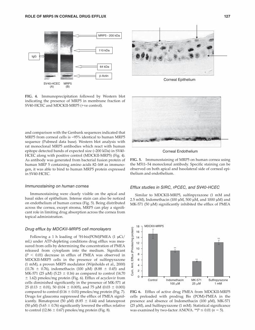
HCEC along with positive control (MDCKII-MRP5) (Fig. 4).
As antibody was generated from bacterial fusion protein of
human MRP 5 containing amino acids 82–168 as immuno-
gen, it was able to bind to human MRP5 protein expressed
in SV40-HCEC.
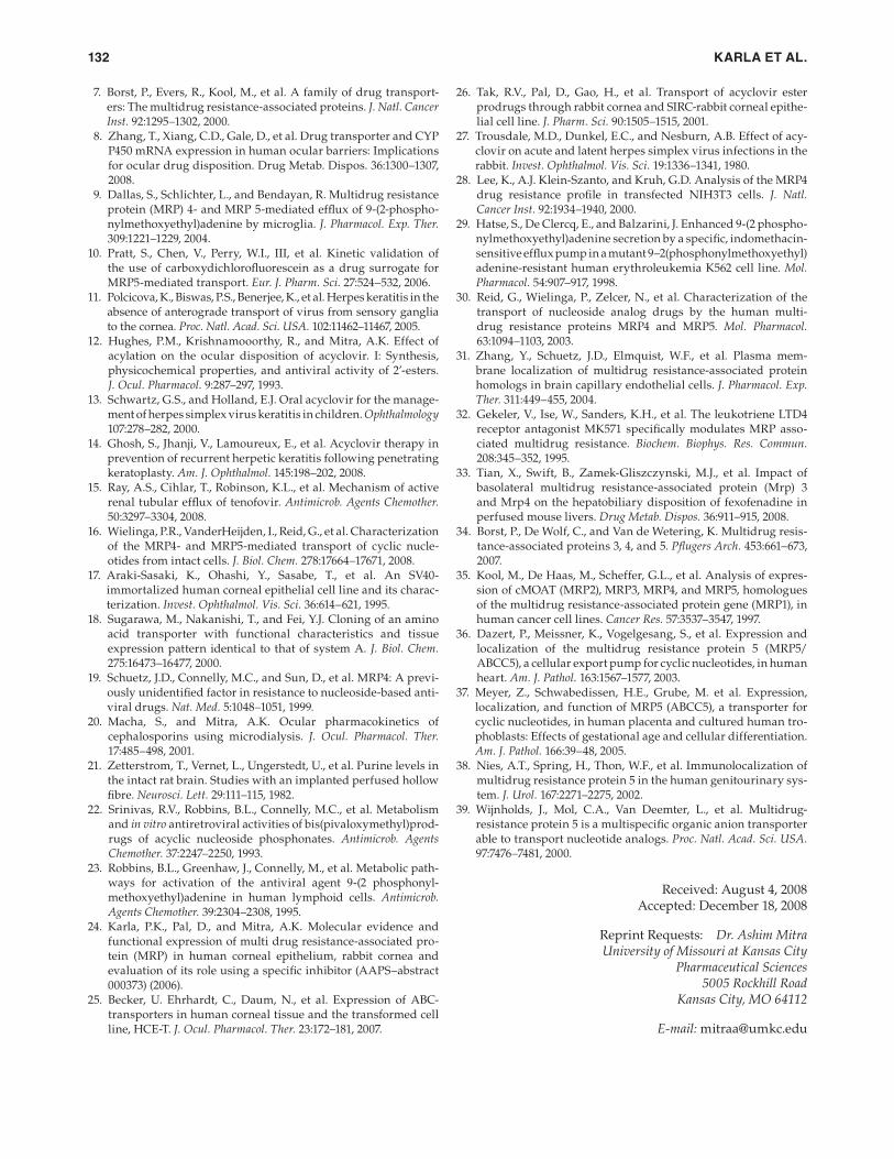
Immunostaining on human cornea
Immunostaining were clearly visible on the apical and
basal sides of epithelium. Intense stain can also be noticed
on endothelium of human cornea (Fig. 5). Being distributed
across the cornea, except stroma, MRP5 can play a signifi -
cant role in limiting drug absorption across the cornea from
topical administration.
Drug efflux by MDCKII-MRP5 cell monolayers
Following a 1 h loading of 3H-bis(POM)PMEA (1 μCi/
mL) under ATP-depleting conditions drug effl ux was mea-
sured from cells by determining the concentration of PMEA
released from cytoplasm into the medium. Signifi cant
(P < 0.01) decrease in effl ux of PMEA was observed in
MDCKII-MRP5 cells in the presence of sulfi npyrazone
(1 mM), a proven MRP5 modulator (Wijnholds et al., 2000)
(11.76 ± 0.76), indomethacin (100 μM) (8.88 ± 0.45) and
MK-571 (25 μM) (5.23 ± 0.16) as compared to control (14.70
± 1.62) pmoles/mg protein (Fig. 6). Effl ux of acyclovir from
cells diminished signifi cantly in the presence of MK-571 at
25 (0.13 ± 0.01), 50 (0.04 ± 0.005), and 75 μM (0.03 ± 0.001)
compared to control (0.16 ± 0.01) pmoles/mg protein (Fig. 7).
Drugs for glaucoma suppressed the effl ux of PMEA signif-
icantly. Bimatoprost (50 μM) (8.85 ± 0.44) and latanoprost
(50 μM) (5.65 ± 0.76) signifi cantly lowered the effl ux relative
to control (12.86 ± 0.67) pmoles/mg protein (Fig. 8).
IgG
SV40-HCEC MRP5
MRP5 - 200 kDa
110 kDa
64 kDa
β-Actin
(B) (A)
FIG. 4. Immunoprecipitation followed by Western blot
indicating the presence of MRP5 in membrane fraction of
SV40-HCEC and MDCKII-MRP5 (+ve control).
Corneal Epithelium
Corneal Endothelium
FIG. 5. Immunostaining of MRP5 on human cornea using
the M511–54 monoclonal antibody. Specifi c staining can be
observed on both apical and basolateral side of corneal epi-
thelium and endothelium.
MDCKII-MRP5
Cum
. A
mt. E
fflu
x (
Pm
ole
s/m
g p
rote
in)
**
Control Indomethacin
100 μM
MK-571
25 μM
Sulfinpyrazone
1 mM
18
16
14
12
10
8
6
4
2
0
**
**
FIG 6. Effl ux of active drug PMEA from MDCKII-MRP5
cells preloaded with prodrug Bis (POM)-PMEA in the
presence and absence of Indomethacin (100 μM), MK-571
(25 μM), and Sulfi npyrazone (1 mM). Statistical signifi cance
was examined by two-factor ANOVA. **P ≤ 0.01 (n = 5).
04_JOP-2008_0084.indd 127 4/4/2009 2:37:38 AM

KARLA ET AL.128
Role of glutathione and ATP
Treatment with 50-μM BSO for 24 h has been reported
to deplete glutathione levels by >90% (Dallas et al., 2004).
BSO treatment did not show signifi cant decrease in PMEA
effl ux in the cell lines employed (Table 1). Depletion of ATP
by 10-mM sodium azide and 10-mM 2-deoxy-d-glucose
resulted in signifi cant decrease in PMEA effl ux compared
to control (Fig. 10). Effl ux was low at all time points (30, 60,
and 90 min) compared to control in rPCEC, SIRC, and SV40-
HCEC. In rPCEC effl ux under ATP-depleting conditions at
30 min was (0.35 ± 0.11) versus control (1.39 ± 0.21), at 60
min (0.50 ± 0.03) versus control (2.54 ± 0.32), at 90 min (1.23
± 0.19) versus control (3.24 ± 0.26) pmoles/mg protein. In
SIRC, the effl ux at 30 min under ATP-depleting conditions
was (0.28 ± 0.17) versus control (1.23 ± 0.12), at 60 min (0.40
± 0.15) versus control (2.27 ± 0.04), at 90 min (1.05 ± 0.18)
versus control (3.12 ± 0.021) pmoles/mg protein. Similarly,
in SV40-HCEC, effl ux under ATP-depleting conditions was
at 30 min (0.21 ± 0.10) versus control (0.96 ± 0.11), at 60 min
(0.60 ± 0.02) versus control (1.78 ± 0.21), and at 90 min was
(0.98 ± 0.23) versus control (2.46 ± 0.12) pmoles/mg protein.
Figure 10 clearly illustrates that ATP depletion suppressed
the effl ux signifi cantly at all time points.
Active nucleotide efflux pump on freshly
excised cornea
Cumulative amount of PMEA accumulated in freshly
excised rabbit cornea under ATP-depleting conditions was
signifi cantly higher (8.25 ± 0.16) compared to normal con-
ditions (HBSS with glucose) (4.32 ± 0.86) pmoles/cornea
(Fig. 11).
Conversion of 3H-bis(POM)PMEA to PMEA human
and rabbit corneal epithelial cells
Lipophilic prodrug bis(POM)PMEA is metabolized
in the cell cytoplasm to PMEA which is a substrate for
MRP5. This mechanism is verifi ed in both human and rab-
bit corneal epithelial cells. X-ray fi lm developed after TLC
and autoradiography revealed representative bands for
bis(POM)PMEA and PMEA recovered from cell lysates
of SV40-HCEC (lane 5), SIRC (lane 6), and MDCKII-MRP5
(lane 3) (Fig. 12). Control bands for bis(POM)PMEA (lane 1),
PMEA (lane 2), and donor band for bis(POM)PMEA (lane
3) were also detected. At zero time point, no conversion of
bis(POM)PMEA was observed as demonstrated in lane 3.
Also, no intracellular radioactivity was detected. After 1 h
incubation with 3H-bis(POM)PMEA under ATP-depleting
conditions, ~93% of the radioactivity in the supernatant
is identifi ed as bis(POM)PMEA and ~5% corresponds to
PMEA. However intracellularly in MDCKII-MRP5, SV40-
HCEC, and SIRC, ~80% of the radioactivity is associated
with PMEA and ~15% with bis(POM)PMEA (lanes 4, 5, 6).
Remaining ~5% of intracellular radioactivity may repre-
sent mono(POM)PMEA (intermediate formed during the
conversion of bis(POM)PMEA to PMEA) and several PMEA
phosphorylated metabolites (i.e., PMEA monophosphate
and PMEA diphosphate).9 Similar result was obtained with
rPCEC (data now shown). Lower concentrations of these
metabolites were detected after bis(POM)PMEA adminis-
tration in human lymphoid cell lines.22,23 As demonstrated
(Table 1). PMEA effl ux was very low in the presence of
strong MRP inhibitor MK-571. Effl ux of acyclovir was signif-
icantly lower with MK-571 in rPCEC at 25 (0.16 ± 0.006), 50
(0.12 ± 0.01), and 75 μM (0.10 ± 0.02) relative to control (0.16
± 0.01); SIRC, 25 (0.20 ± 0.004), 50 (0.17 ± 0.01), and 75 μM
(0.16 ± 0.009) relative to control (0.25 ± 0.004); SV40-HCEC, 25
(0.13 ± 0.007), 50 (0.12 ± 0.005), and 75 μM (0.08 ± 0.007) com-
pared to control (0.16 ± 0.005) pmoles/mg protein (Fig. 7).
Glaucoma-treating drugs also caused a marked lowering in
PMEA effl ux signifi cantly in rPCEC, that is, bimatoprost (50
μM) (1.21 ± 0.20) and latanoprost (50 μM) (0.68 ± 0.13) rela-
tive to control (2.63 ± 0.23); SIRC, bimatoprost (50 μM) (0.98
± 0.08) and latanoprost (50 μM) (0.64 ± 0.01) compared to
control (2.20 ± 0.14); SV40-HCEC, bimatoprost (50 μM) (0.74
± 0.07) and latanoprost (50 μM) (0.70 ± 0.12) compared to
control (1.81 ± 0.51) pmoles/mg protein (Fig. 9).
MK571 (�M)
Cum
. A
mt.
Eff
lux (
Pm
ole
s/
mg p
rote
in)
MRP5
ACV
**
****
**
**
****
**
**
**
0
0.05
0.1
0.15
0.2
0.25
0.3
C 25 50 75 C 25 50 75 C 25 50 75 C 25 50 75
rPCEC SIRC SV40-HCEC
FIG. 7. Effl ux of acyclovir was measured from MDCKII-
MRP5, rPCEC, SIRC, and SV40-HCEC. Monolayers of cells
were incubated with 3H-acyclovir under ATP-depleting con-
ditions (10-mM sodium azide; 10-mM 2-deoxy-d-glucose) for
1 h at 37°C. HBSS with glucose was added, with and without
25, 50, 75 μM MK-571, and cumulative amount of acyclovir
effl ux was determined by measuring the radioactivity in the
supernatant. Statistical signifi cance was measured by two-
factor ANOVA. **P ≤ 0.01 (n = 5).
Control
MRP5
**
**
0
2
4
6
8
10
12
14
Cum
. A
mt. E
fflu
x (
Pm
ole
s/ m
g p
rote
in)
16
Bimatoprost
50 �M
Latanoprost
50 �M
FIG. 8. Effl ux of PMEA from MDCKII-MRP5 cells pre-
loaded with Bis (POM)-PMEA under ATP-depleting condi-
tions (10-mM sodium azide; 10-mM 2-deoxy-d-glucose) for
1 h at 37°C. HBSS with glucose was added, with and with-
out bimatoprost, latanoprost, and effl ux was determined.
Statistical signifi cance was determined by two-factor
ANOVA. **P ≤ 0.01 (n = 5).
04_JOP-2008_0084.indd 128 4/4/2009 2:37:39 AM

ROLE OF MRP5 IN CORNEAL DRUG EFFLUX 129
various drugs has not been explored. Also, there are con-
tradicting reports on the expression of ABC transporters on
human cornea.25 Dey and colleagues, 2003 and Karla and
colleagues, 2007 confi rmed the expression of Pgp, MRP2,
and MRP1 on human corneal cells. Presence of MRP3 on
corneal cells was also reported from our laboratory.24 Becker
et al., 2007 contradicted the expression of Pgp and MRP2
on human cornea, but indicated the expression of MRP2
in human corneal epithelial cells. However, a recent study
by Zhang et al., 2008 on quantitative expression of ABC
transporters revealed that intact human cornea expresses
MRP1>MRP3>MDR1=MRP2. This study confi rms our ear-
lier reports on molecular expression of ABC transporters in
cornea. Relative role of individual transporters on ocular
drug disposition is yet to be established.
Our results demonstrate for the fi rst time the presence
and localization of MRP5 on human cornea. Effl ux data
on human and rabbit corneal epithelial cells demonstrate
rapid effl ux of acyclovir and PMEA. As shown in Figure 8,
bimatoprost and latanoprost, the two widely used antiglau-
coma drugs currently on the market are found to be excel-
lent substrates for MRP5. Figure 9 shows the inhibition of
PMEA effl ux in rabbit and human corneal epithelial cells
by both bimatoprost and latanoprost. With later compound
marketed under the trade name Xalatan by Pfi zer appears to
by TLC/autoradiography, conversion of inactive prodrug
Bis(POM)PMEA to active MRP5 substrate PMEA occurs in
the cell cytoplasm of human and rabbit corneal epithelial
cells causing active drug effl ux.
In vivo ocular absorption of 3H-acyclovir in the
presence of MRP inhibitor (MK571)
Pharmacokinetic analysis revealed a signifi cant elevation
in the absorption of acyclovir across the cornea in presence
of 100 μM MK-571 (Table 2). Cmax, AUC0-α, and ka increased
~2 fold with the inhibition of MRP. Even the Clast, the last
measured concentration in aqueous humor is signifi cantly
higher (0.15 ± 0.01) than control (0.05 ± 0.02) μg/mL. Highest
aqueous humor concentration was observed at 140 min and
drug concentrations in aqueous humor were superior at all
time points when MK-571 was employed (Fig. 13). Based on
the in vitro substrate specifi city data (Fig. 14), predominantly
MRP5 and to a certain extent MRP2 appear to play a critical
role in acyclovir effl ux.
Discussion
Expression of ATP-binding cassette transporters on
human cornea and their role on transport processes of
Cont
rPCEC
**
**
**
**** **
SIRC SV40-HCEC
Cum
. A
mt. E
fflu
x (
Pm
ole
s/m
g p
rote
in)
0
0.5
1
1.5
2
2.5
3
3.5
Bim Lat Cont Bim Lat Cont Bim Lat
FIG. 9. Effl ux of PMEA from rPCEC, SIRC, and SV40-HCEC
preloaded with Bis (POM)-PMEA under ATP-depleting con-
ditions (10 mM sodium azide; 10 mM 2-deoxy-d-glucose) for
1 h at 37°C. HBSS with glucose was added, with and with-
out bimatoprost, latanoprost, and effl ux was determined.
Statistical signifi cance was examined by two-factor ANOVA.
**P ≤ 0.01 (n = 5).
Table 1. Efflux of Active Drug PMEA From SIRC, rPCEC, and SV40-HCEC Preloaded With Prodrug Bis
(POM)-PMEA in the Presence and Absence of Indomethacin (100 μM, 500 μM, 1000 μM), Sulfinpyrazone
(1 mM and 2.5 mM), Glutathione Depleting Agent BSO (50 μM), and Under ATP-Depleting
Conditions (10-mM Sodium Azide; 10-mM 2-Deoxy-d-Glucose) Was Shown
Sulfi npyrazone (mM) Indomethacin (mM)BSO
50 μM Cont. 1.0 2.5 Cont. 0.1 0.5 1.0
SIRC 2.27 ± 0.04 1.51 ± 0.21 1.24 ± 0.10 2.36 ± 0.11 1.53 ± 0.06 0.90 ± 0.04 0.82 ± 0.16 2.18 ± 0.08
rPCEC 2.54 ± 0.32 1.12 ± 0.04 0.42 ± 0.10 1.97 ± 0.19 2.02 ± 0.34 1.23 ± 0.16 0.80 ± 0.23 2.38 ± 0.14
SV40-HCEC 1.78 ± 0.21 0.98 ± 0.14 0.67 ± 0.08 1.65 ± 0.19 1.34 ± 0.07 1.36 ± 0.20 1.22 ± 0.05 1.87 ± 0.21
Statistical signifi cance was tested by two-factor ANOVA. P ≤ 0.01.
3.5
3
2.5
2
Cum
. A
mt. E
fflu
x (
Pm
ole
s/m
g p
rote
in)
1.5
1
0.5
0
Con 30 60 90 30 60 90 30 60 90
Con
Con
Con
Con
Con
Con
Con
Con
****
**
**
SIRC SV40-HCECrPCEC
**
**
**
**
**
FIG. 10. Time-dependant effl ux of active drug PMEA from
SIRC, rPCEC, and SV40-HCEC preloaded with prodrug
Bis (POM)-PMEA under ATP-depleting conditions (10 mM
sodium azide; 10 mM 2-deoxy-d-glucose). ATP depletion
suppressed PMEA effl ux signifi cantly at all time points.
Statistical signifi cance was examined by two-factor ANOVA.
**P ≤ 0.01 (n = 5).
04_JOP-2008_0084.indd 129 4/4/2009 2:37:39 AM

KARLA ET AL.130
However, it is found to have a low ocular bioavailability when
administered topically.26 A 3% acyclovir ointment applied 5
times a day did not treat herpes keratitis.27 Effl ux-mediated
resistance of acyclovir absorption in the cornea is a quiet
unexpected result. In vitro effl ux studies and in vivo microdi-
alysis results reveal that MRP5 along with MRP2 can play
a vital role in imparting drug resistance to ocular acyclovir
absorption. As shown in Figure 14, both MRP5 and MRP2
can effl ux the acyclovir but acyclovir seems to be a better
substrate for MRP5 than MRP2. MDR1 did not play a role in
acyclovir effl ux. Results from Figure 7, indicates strong sup-
pression in acyclovir effl ux in MRP5 over expressing cells
with higher concentration of MK-571 (50 and 75 μM). Dose-
dependant suppression in acyclovir effl ux was also noticed
in rPCEC, SIRC, and SV40-HCEC. It can be concluded that
both MRP5 and MRP2 (MRP5 > MRP2) can play a critical
role in conferring resistance to the absorption of acyclovir
across cornea. MK-571 was employed in the in vivo studies
as it is the only specifi c MRP inhibitor currently available
in the market. It was found to not interact with other effl ux
pumps, such as Pgp.32 As MRP is a relatively new class of
be a better substrate for MRP5 than bimatoprost marketed
as Lumigan by Allergan. This is evident from the results in
all three cell lines, that is, MDCKII-MRP5, rPCEC, and SIRC.
However, in SV40-HCEC bimatoprost and latanoprost were
equally effective in lowering the effl ux of PMEA. Effl ux data
in Table 1 shows that PMEA effl ux is more predominant in
rPCEC and SIRC as compared to SV40-HCEC. This result
indicates relatively higher expression levels of MRP5 in
rPCEC and SIRC when relative to SV40-HCEC. Latanoprost
at 50 μM was suffi cient to saturate the MRP5 effl ux in SV40-
HCEC. However, PMEA effl ux decreased by >50% in rPCEC,
SIRC, and SV40-HCEC. Glaucoma drug formulations with
added effl ux pump inhibitors might provide better clin-
ical outcome. However, an in vivo analysis on the effi cacy
of transporter in modulating drug absorption of these com-
pounds and a comprehensive clinical analysis on cytotox-
icity along with inhibitors should be studied in humans. In vivo pharmacokinetic parameters in male New Zealand rab-
bits provide exciting results with a ~2 fold increase in Cmax
and AUC0-α of acyclovir in the presence of MK-571. Acyclovir
can be employed for the treatment of ocular herpes keratitis.
Donor
1 2 3 4 5 6
Bis(POM)PMEA-Control
PMEA-Control
MRP5 HCEC SIRC
FIG. 12. TLC separation of PMEA and bis(POM)PMEA
shown by autoradiography. MDCKII-MRP5, SV40-HCEC,
and SIRC were loaded with 3H-bis(POM)PMEA and radio-
activity present in the cellular extracts was quantifi ed. Lane 1, bis(POM)PMEA standard; Lane 2, PMEA standard; Lane 3,
donor, intracellular conversion of bis(POM)PMEA to PMEA
in 60 min; Lane 4, MRP5; Lane 5, SV40-HCEC; Lane 6, SIRC.
Table 2. Pharmacokinetic Parameters After a Single-
Dose Infusion of 3H-Acyclovir (10 mci/ml)
in the Absence and Presence of MK-571 (50 mM).
Drug and/or Inhibitor AcyclovirAcyclovir +
100 μM MK571
AUC0–α (μg.min/mL) 215.43 ± 35.20 466.28 ± 25.35
Cmax (μg/mL) 1.19 ± 0.15 2.64 ± 0.16
Ka 7.28 × 10⁻⁴ 13.31 × 10⁻⁴
K10 × 103 (min−1) 5.37 ± 1.87 8.02 ± 1.42
As evident from the data, Cmax and AUC0-α values of acyclovir
were signifi cantly elevated in the presence of MK-571 relative
to control. Corneal absorption rate constant (ka) signifi cantly
increased in the presence of MK-571 leading to higher Cmax and
AUC0-α values relative to control. Data is expressed as mean ± S.D.
(n = 4).
0
0
0.5
1
1.5
2
2.5
3
3.5
100 200 300
Time (min)
Control
MK571
Dru
g in a
quoes h
um
or
(nano g
ram
s/m
L)
400 500
FIG. 13. Scatter plot representing the amount of 3H-acy-
clovir present in the aqueous humor in the presence and
absence of MK-571. Amount of acyclovir was signifi cantly
higher in the presence of MK-571 compared to control.
Statistical signifi cance was tested by two-factor ANOVA.
**P ≤ 0.01 and data is expressed as mean ± S.D. (n = 4).
Control
0
2
4
6
8
10
ATP depleted
**
Cu
m.
Am
t. A
ccu
mu
late
d
(Pm
ole
s/c
orn
ea
)
FIG. 11. Cumulative amount of PMEA accumulated in
freshly excised rabbit cornea mounted on a side-by-side dif-
fusion chamber under normal conditions and under ATP-
depleting conditions at 34°C. Statistical signifi cance was
tested by two-factor ANOVA. **P ≤ 0.01.
04_JOP-2008_0084.indd 130 4/4/2009 2:37:40 AM

ROLE OF MRP5 IN CORNEAL DRUG EFFLUX 131
25-μM BSO did not alter the transport of PMEA by MRP5.
Also, Wielinga et al., 2003 indicated that GSH depletion did
not alter the effl ux of cGMP/cAMP and cyclic nucleotides by
MRP5. These results are consistent with earlier reports about
the role of glutathione in MRP5-mediated effl ux. Various cell
lines were found to express MRP5, and in vivo, MRP5 has
been detected in the majority of tissues of the body except
the eye.31,34,35. Human urinary-genital tract smooth muscle,
epithelial cells of blood vessels, uterus and urethra, syncy-
tiotrophoblasts and fetal cells of placenta, astrocytes and
pyramidal neurons of brain, cadiomyocytes, endothelial
cells, and smooth muscle cells of heart have all been found
to abundantly express MRP5.34–39
For the fi rst time, we have demonstrated functional and
molecular expression of a nucleotide effl ux transporter
(MRP5) in human and rabbit cornea. Also, for the fi rst time,
we have demonstrated that acyclovir, a widely used antivi-
ral agent and glaucoma drugs (bimatoprost and latanoprost)
are excellent substrates for MRP5 and are effl uxed by cor-
neal epithelial cells. Overall distribution and accumulation
of various classes of drugs in anterior segment of eye might
depend on their relative substrate specifi city for the effl ux
pumps. Incorporation of one or a combination of inhibitors
in ophthalmic formulation can be an effective strategy to
improve ocular bioavailability. In light of new effl ux trans-
porters like MRP5, extensive research needs to be performed
to delineate relative roles of these transporters in ocular
drug disposition.
Acknowledgments
We thank Dr. Piet Borst for contributing MRP5-
transfected cell line for this project. This work was pre-
sented in a preliminary format to NIH, AAPS, ARVO, and
National Eye Foundation. The work was supported by NIH
05R01EY009171–14 and Sara Morrison grant from UMKC
School of Medicine.
Disclosure Statement
The authors report no confl ict of interest.
References
1. Kawazu, K., Nakamura, M., and Ota, A. Characterization of
cyclosporin A transport in cultured rabbit corneal epithelial
cells: P-glycoprotein transport activity and binding to cyclophi-
lin. Invest. Ophthalmol. Vis. Sci. 40:1738–1744, 1999.
2. Dey, S., Patel, J., Anand, B.S., et al. Molecular evidence and func-
tional expression of P-glycoprotein (MDR1) in human and rab-
bit cornea and corneal epithelial cell lines. Invest. Ophthalmol. Vis. Sci. 44:2909–2918, 2003.
3. Dey, S., Gunda, S., and Mitra, A.K. Pharmacokinetics of eryth-
romycin in rabbit corneas after single-dose infusion: Role of
P-glycoprotein as a barrier to in vivo ocular drug absorption. J. Pharmacol. Exp. Ther. 311:246–255, 2004.
4. Karla, P.K., Pal, D., Quinn, T., et al. Molecular evidence and
functional expression of a novel drug effl ux pump (ABCC2) in
human corneal epithelium and rabbit cornea and its role in ocu-
lar drug effl ux. Int. J. Pharm. 84:53–60, 2006.
5. Karla, P.K., Pal, D., and Mitra, A.K. Molecular evidence and func-
tional expression of multidrug resistance associated protein (MRP)
in rabbit corneal epithelial cells. Exp. Eye. Res. 84:53–60, 2007.
6. Bosrt, P., Dewolf, C., and Van De Wetering, K. Multidrug resistance
associated proteins 3, 4, and 5. Eur. J. Physiol. 453:661–673, 2007.
effl ux pump, there is no specifi c inhibitor available for indi-
vidual members of MRP family. Employing the cells which
over express the specifi c MRP transporters or using the
gene knockout mice are the methods suggested in the liter-
ature.32,33 Hence, we procured the MRP overexpressing cells
(MDCKII-MRP1, MDCKII-MRP2, and MDCKII-MRP5) from
Dr. Piet Borst (Netherlands cancer institute, Amsterdam,
Netherlands). MDCKII-MRP5 cells along with other MRP-
transfected cells were used to delineate the role of various
MRPs in ocular drug resistance.
The probe recovery was below ~10% with PMEA and
hence it was not employed in in vivo pharmacokinetics stud-
ies. However, to measure the role of MRP5 on resistance to
PMEA, we performed transport studies on freshly excised
rabbit cornea using side-by-side diffusion cells. The study
indicated a ~2-fold increase in corneal accumulation of
PMEA with ATP-depleting medium. Reid G et al. indicated
that PMEA is not a substrate for ABC transporters, Pgp (or)
breast cancer resistance protein (BCRP). Also MRP1–3 was
found not to play a role in PMEA effl ux.9,28 As demonstrated
in Figure 1, only MRP5 played a signifi cant role in the PMEA
effl ux. Neither MRP2 nor MDR1 participated in the effl ux of
PMEA.
Bis(POM)-PMEA enters cells rapidly by passive diffu-
sion.29 Rapid uptake of bis(POM)-PMEA by different cell
types and its fast conversion to PMEA which is an excel-
lent substrate for nucleotide effl ux transporter makes it an
attractive candidate to study effl ux mechanism. Following
bis(POM)-PMEA loading in cells overexpressing MRP5, 98%
of prodrug is converted to PMEA.7,30 TLC/autoradiography
data in Figure 12 proved the conversion of bis(POM)-PMEA
to PMEA in both human and rabbit corneal epithelial cells.
This fi nding further confi rms the expression of a nucleotide
effl ux transporter in cornea. MRP5 belongs to the class of
ATP-dependant transporters and Figure 10 clearly depicts
time-dependant suppression in PMEA effl ux with the use of
ATP-depleting medium. Effl ux was low at all the time points
(30, 60, 90 min) relative to control. As indicated from data in
Table 1, 50-μM BSO did not alter the effl ux of PMEA. Our
results agree with Dallas et al., 2004 who demonstrated that
Wt
ACVC
um
. A
mt. E
fflu
x (
Pm
ole
s/ m
g p
rote
in)
0
0.05
0.1
0.15
0.2
MDR1 MRP2
*
**
MRP5
FIG. 14. Effl ux of drug was measured from MDCKII-wt,
MDR1, MRP2, and MRP5 cells preloaded with 3H-acyclovir
under ATP-depleting conditions (10 mM sodium azide;
10 mM 2-deoxy-d-glucose) for 1 h at 37°C. HBSS with glu-
cose was added and comparative effl ux of transporters was
determined. Statistical signifi cance was examined by two-
factor ANOVA. **P ≤ 0.01 (n = 5).
04_JOP-2008_0084.indd 131 4/4/2009 2:37:41 AM
KARLA ET AL.132
7. Borst, P., Evers, R., Kool, M., et al. A family of drug transport-
ers: The multidrug resistance-associated proteins. J. Natl. Cancer Inst. 92:1295–1302, 2000.
8. Zhang, T., Xiang, C.D., Gale, D., et al. Drug transporter and CYP
P450 mRNA expression in human ocular barriers: Implications
for ocular drug disposition. Drug Metab. Dispos. 36:1300–1307,
2008.
9. Dallas, S., Schlichter, L., and Bendayan, R. Multidrug resistance
protein (MRP) 4- and MRP 5-mediated effl ux of 9-(2-phospho-
nylmethoxyethyl)adenine by microglia. J. Pharmacol. Exp. Ther. 309:1221–1229, 2004.
10. Pratt, S., Chen, V., Perry, W.I., III, et al. Kinetic validation of
the use of carboxydichlorofl uorescein as a drug surrogate for
MRP5-mediated transport. Eur. J. Pharm. Sci. 27:524–532, 2006.
11. Polcicova, K., Biswas, P.S., Benerjee, K., et al. Herpes keratitis in the
absence of anterograde transport of virus from sensory ganglia
to the cornea. Proc. Natl. Acad. Sci. USA. 102:11462–11467, 2005.
12. Hughes, P.M., Krishnamooorthy, R., and Mitra, A.K. Effect of
acylation on the ocular disposition of acyclovir. I: Synthesis,
physicochemical properties, and antiviral activity of 2’-esters.
J. Ocul. Pharmacol. 9:287–297, 1993.
13. Schwartz, G.S., and Holland, E.J. Oral acyclovir for the manage-
ment of herpes simplex virus keratitis in children. Ophthalmology
107:278–282, 2000.
14. Ghosh, S., Jhanji, V., Lamoureux, E., et al. Acyclovir therapy in
prevention of recurrent herpetic keratitis following penetrating
keratoplasty. Am. J. Ophthalmol. 145:198–202, 2008.
15. Ray, A.S., Cihlar, T., Robinson, K.L., et al. Mechanism of active
renal tubular effl ux of tenofovir. Antimicrob. Agents Chemother. 50:3297–3304, 2008.
16. Wielinga, P.R., VanderHeijden, I., Reid, G., et al. Characterization
of the MRP4- and MRP5-mediated transport of cyclic nucle-
otides from intact cells. J. Biol. Chem. 278:17664–17671, 2008.
17. Araki-Sasaki, K., Ohashi, Y., Sasabe, T., et al. An SV40-
immortalized human corneal epithelial cell line and its charac-
terization. Invest. Ophthalmol. Vis. Sci. 36:614–621, 1995.
18. Sugarawa, M., Nakanishi, T., and Fei, Y.J. Cloning of an amino
acid transporter with functional characteristics and tissue
expression pattern identical to that of system A. J. Biol. Chem. 275:16473–16477, 2000.
19. Schuetz, J.D., Connelly, M.C., and Sun, D., et al. MRP4: A previ-
ously unidentifi ed factor in resistance to nucleoside-based anti-
viral drugs. Nat. Med. 5:1048–1051, 1999.
20. Macha, S., and Mitra, A.K. Ocular pharmacokinetics of
cephalosporins using microdialysis. J. Ocul. Pharmacol. Ther. 17:485–498, 2001.
21. Zetterstrom, T., Vernet, L., Ungerstedt, U., et al. Purine levels in
the intact rat brain. Studies with an implanted perfused hollow
fi bre. Neurosci. Lett. 29:111–115, 1982.
22. Srinivas, R.V., Robbins, B.L., Connelly, M.C., et al. Metabolism
and in vitro antiretroviral activities of bis(pivaloxymethyl)prod-
rugs of acyclic nucleoside phosphonates. Antimicrob. Agents Chemother. 37:2247–2250, 1993.
23. Robbins, B.L., Greenhaw, J., Connelly, M., et al. Metabolic path-
ways for activation of the antiviral agent 9-(2 phosphonyl-
methoxyethyl)adenine in human lymphoid cells. Antimicrob. Agents Chemother. 39:2304–2308, 1995.
24. Karla, P.K., Pal, D., and Mitra, A.K. Molecular evidence and
functional expression of multi drug resistance-associated pro-
tein (MRP) in human corneal epithelium, rabbit cornea and
evaluation of its role using a specifi c inhibitor (AAPS–abstract
000373) (2006).
25. Becker, U. Ehrhardt, C., Daum, N., et al. Expression of ABC-
transporters in human corneal tissue and the transformed cell
line, HCE-T. J. Ocul. Pharmacol. Ther. 23:172–181, 2007.
26. Tak, R.V., Pal, D., Gao, H., et al. Transport of acyclovir ester
prodrugs through rabbit cornea and SIRC-rabbit corneal epithe-
lial cell line. J. Pharm. Sci. 90:1505–1515, 2001.
27. Trousdale, M.D., Dunkel, E.C., and Nesburn, A.B. Effect of acy-
clovir on acute and latent herpes simplex virus infections in the
rabbit. Invest. Ophthalmol. Vis. Sci. 19:1336–1341, 1980.
28. Lee, K., A.J. Klein-Szanto, and Kruh, G.D. Analysis of the MRP4
drug resistance profi le in transfected NIH3T3 cells. J. Natl. Cancer Inst. 92:1934–1940, 2000.
29. Hatse, S., De Clercq, E., and Balzarini, J. Enhanced 9-(2 phospho-
nylmethoxyethyl)adenine secretion by a specifi c, indomethacin-
sensitive effl ux pump in a mutant 9–2(phosphonylmethoxyethyl)
adenine-resistant human erythroleukemia K562 cell line. Mol. Pharmacol. 54:907–917, 1998.
30. Reid, G., Wielinga, P., Zelcer, N., et al. Characterization of the
transport of nucleoside analog drugs by the human multi-
drug resistance proteins MRP4 and MRP5. Mol. Pharmacol. 63:1094–1103, 2003.
31. Zhang, Y., Schuetz, J.D., Elmquist, W.F., et al. Plasma mem-
brane localization of multidrug resistance-associated protein
homologs in brain capillary endothelial cells. J. Pharmacol. Exp. Ther. 311:449–455, 2004.
32. Gekeler, V., Ise, W., Sanders, K.H., et al. The leukotriene LTD4
receptor antagonist MK571 specifi cally modulates MRP asso-
ciated multidrug resistance. Biochem. Biophys. Res. Commun. 208:345–352, 1995.
33. Tian, X., Swift, B., Zamek-Gliszczynski, M.J., et al. Impact of
basolateral multidrug resistance-associated protein (Mrp) 3
and Mrp4 on the hepatobiliary disposition of fexofenadine in
perfused mouse livers. Drug Metab. Dispos. 36:911–915, 2008.
34. Borst, P., De Wolf, C., and Van de Wetering, K. Multidrug resis-
tance-associated proteins 3, 4, and 5. Pfl ugers Arch. 453:661–673,
2007.
35. Kool, M., De Haas, M., Scheffer, G.L., et al. Analysis of expres-
sion of cMOAT (MRP2), MRP3, MRP4, and MRP5, homologues
of the multidrug resistance-associated protein gene (MRP1), in
human cancer cell lines. Cancer Res. 57:3537–3547, 1997.
36. Dazert, P., Meissner, K., Vogelgesang, S., et al. Expression and
localization of the multidrug resistance protein 5 (MRP5/
ABCC5), a cellular export pump for cyclic nucleotides, in human
heart. Am. J. Pathol. 163:1567–1577, 2003.
37. Meyer, Z., Schwabedissen, H.E., Grube, M. et al. Expression,
localization, and function of MRP5 (ABCC5), a transporter for
cyclic nucleotides, in human placenta and cultured human tro-
phoblasts: Effects of gestational age and cellular differentiation.
Am. J. Pathol. 166:39–48, 2005.
38. Nies, A.T., Spring, H., Thon, W.F., et al. Immunolocalization of
multidrug resistance protein 5 in the human genitourinary sys-
tem. J. Urol. 167:2271–2275, 2002.
39. Wijnholds, J., Mol, C.A., Van Deemter, L., et al. Multidrug-
resistance protein 5 is a multispecifi c organic anion transporter
able to transport nucleotide analogs. Proc. Natl. Acad. Sci. USA. 97:7476–7481, 2000.
Received: August 4, 2008
Accepted: December 18, 2008
Reprint Requests: Dr. Ashim MitraUniversity of Missouri at Kansas City
Pharmaceutical Sciences5005 Rockhill Road
Kansas City, MO 64112
E-mail: [email protected]
04_JOP-2008_0084.indd 132 4/4/2009 2:37:41 AM


![COMMENTARY Antibiotic Efflux Pumps · the drug efflux pumps in eucaryotic cells ( [7]; drug efflux transporters are classically energized by ATP). The second-ary active transporters,](https://img.pdfslide.us/doc/110x75/6132c0d4dfd10f4dd73aa6ef/commentary-antibiotic-efflux-pumps-the-drug-efflux-pumps-in-eucaryotic-cells-7.jpg)