Embed Size (px)
Citation preview
Expression of hormone receptors and markers for
metastatic potential in relation to tumor associated
macrophages in breast cancer
Version 1
Author: Anna Ramberg, bachelor of medicine
Supervisor: Ann Erlandsson, lecturer
Karlstad University, Karlstad, Sweden
Örebro University
School of Medical Sciences
Degree project, 30 ECTS
January 2016
2
Abstract
Introduction Hormone receptor status influences prognosis in breast cancer. Tumor
associated macrophages are part of the tumor microenvironment and are correlated with
hormone receptor negativity and poor survival, including increased risk of metastasis.
Estrogen receptor β is a novel estrogen receptor with a putative anti-proliferative effect and is
associated with favorable clinical outcome.
Aim To examine pre- and post-treatment associations between macrophage infiltration and
expression of estrogen receptor α (ERα), estrogen receptor β (ERβ), progesterone receptor
(PR), human epithelial growth factor receptor-2 (HER-2), Ki-67, matrix metallopeptidase 9
(MMP-9) and urokinase-type plasminogen activator receptor (uPAR) in breast cancer patients
and in cultured T47D breast cancer cell line.
Methods Macrophage infiltration (CD68 and CD163) and expression of ERα, ERβ, PR,
HER-2, Ki-67, MMP-9 and uPAR were evaluated by immunohistochemistry in 19 pre-
treatment breast tumor biopsies and 16 post-treatment breast surgical specimens. Furthermore,
T47D breast cancer cell line was cultured and treated with media from M1 and M2
macrophages, respectively. Quantitative real-time PCR was performed to evaluate expression
of ERα, ERβ, PR and HER-2 mRNA in T47D.
Results We found that 75.0% of tumor biopsies with dense infiltration of CD163+
macrophages expressed high levels of ERβ. Low or negative expression of ERα and PR were
seen in 36.8% and 42.1% of these tumor biopsies, respectively. Expression of MMP-9 was
lower in surgical specimens compared to tumor biopsies (18.8% vs. 57.9%) and expression of
uPAR was higher in surgical specimens (37.5% vs. 26.3%). In T47D breast cancer cell line,
ERα and PR mRNA was significantly downregulated with M1 conditioned media, while ERβ
showed a significant up-regulation.
Conclusions Dense infiltration of CD68+ and CD163+ macrophages in breast tumor biopsies
are related to high expression of ERβ and negative expression of ERα and PR and this was
accompanied in T47D breast cancer cell line. Estrogen receptor β might be a prognostic
marker in patients with estrogen receptor α negativity and triple negative breast cancer.
Keywords: Breast cancer, Estrogen receptor alpha, Estrogen receptor beta, Progesterone
receptor, HER-2, Tumor associated macrophages, Breast cancer cell line
Abbreviations: CM, conditioned media; CXCL9, chemokine (C-X-C motif) ligand 9; ER,
estrogen receptor; HER-2, human epithelial growth factor receptor-2; IFN-γ, interferon-
3
gamma; IL, interleukin; LPS, lipopolysaccharide; MMP-9, matrix metallopeptidase 9; PR,
progesterone receptor; TAM, tumor associated macrophage; TNBC, triple negative breast
cancer; uPAR, urokinase-type plasminogen activator receptor
Introduction
Breast cancer is the most common cancer among women worldwide and the fifth cause of
death from cancer overall [1]. Hormone receptor status, including estrogen receptor (ER)
negativity and progesterone receptor (PR) negativity, human epithelial growth factor receptor-
2 (HER-2) positivity and fast proliferation (Ki67 positivity) are well recognized parameters to
negatively influence the prognosis [2,3].
Malignant tumors consist of cancer cells and the tumor microenvironment, including
extracellular matrix, endothelial cells, fibroblasts and leukocytes [4]. Tumor associated
macrophages (TAMs) constitutes 5-40% of the tumor mass in solid tumors [5]. Two subsets
of macrophages exist, pro-inflammatory M1 macrophages and anti-inflammatory M2
macrophages [4,6]. Classically activated M1 macrophages are stimulated by interferon
gamma (IFN-γ) and lipopolysaccharide (LPS) and secrete pro-inflammatory cytokines [5].
They stimulate helper T cells to destroy pathogens and have tumoricidal capabilities [4]. The
alternatively activated M2 macrophages on the other hand arises from interleukin-4 (IL-4) and
interleukin-13 (IL-13) and are involved in tissue repair and have pre tumor functions. Tumor
cells secrete chemotactic factors leading to recruitment of macrophages to the tumor
microenvironment. In the tumor microenvironment the macrophages shift polarization state
from M1 phenotype to M2 phenotype, thus TAMs resembles the M2 phenotype [5,6]. CD68
is a pan-macrophage marker staining for both M1 and M2 macrophages and are more
frequently used compared with CD163, which is regarded as a marker for M2 macrophages
[4]. TAMs enhances tumor initiation, progression and metastasis by various factors (e.g.
tumor invasion, angiogenesis and intravasation), as is well-established hallmarks of cancer
[6,7]. TAMs produces growth promoting and sustaining cytokines, including epithelial growth
factor, vascular endothelial growth factor and matrix metalloproteinases (MMPs) [2].
Activation of cell-signaling urokinase receptor (uPAR) initiates MMPs to degrade
components of extracellular matrix contributing to tumor cell invasion and metastasis [8,9].
Macrophages, in particular CD68+, are of prognostic value comprising survival rates in
breast cancer. Dense infiltration of CD68+ macrophages in tumor stroma but not tumor nest
are correlated with poor overall survival and breast cancer specific survival [4,10,11]. TAM
infiltration is correlated with higher tumor grade and larger tumor size. CD163+ cancer is
4
more common in histologically advanced cancers and this might be an explanation for earlier
distant metastasis in patients with expression of CD163 [4,12]. Furthermore, M2 macrophage
number is correlated with Ki67 positivity, ER-negativity, PR-negativity, HER-2-positivity
and triple negative breast cancer [4,5,10,13]. However, also myeloid-derived suppressor cells
can express CD163, which is known to enhance tumor progression [4,5].
Estrogen is a growth promoting hormone in mammary glands and is associated with
increased risk for breast cancer. The effect is mediated through two nuclear receptors:
estrogen receptor alpha (ERα) and estrogen receptor beta (ERβ). The latter was discovered for
about 15 years ago and is not currently used in routine diagnostics. Unlike ERα, ERβ has a
putative anti-proliferative effect probably by negative actions on ERα, but this remains to be
established. ERβ, with multiple isoforms, is the most abundant of the ER in normal mammary
glands, but decline with increased tumor growth [14,15], and consequently, 65% of breast
tumors have shown to be ERβ negative [16]. ERα has a limited expression to epithelial cells
while ERβ, despite epithelial cells, is expressed in fibroblasts, adipocytes, endothelial cells
and macrophages [14]. Furthermore, ERβ can be expressed in the nucleus and cytoplasm of
tumor cells [16]. Expression of ERβ is associated with favorable clinical outcome and
clinicopathological features, including small tumor size, low tumor grade and lymph node
negativity [15,17,18]. About 70% of ERα positive and 5-10% of ERα negative breast cancer
responds to tamoxifen, a selective ER modulator [15,19]. This can be a result of an ERα-
independent mechanism of action of tamoxifen via ERβ. Subsequently, ERβ is a predictive
marker for responsiveness to tamoxifen in ERα negative tumors and these patients do benefit
from treatment with tamoxifen [19]. ERβ has been shown to be expressed in 33% of ERα
positive tumors and 25% of triple negative breast cancers (TNBC) and activation of ERβ have
resulted in an inhibitory effect on cell proliferation in TNBC. Furthermore, 24-44% of ERα
negative tumors are ERβ positive [16].
Aim
The aim of the present study was to examine pre- and post-treatment associations between the
extent of macrophage infiltration (CD68 and CD163) and expression of ERα, ERβ, PR, HER-
2, Ki-67, MMP-9 and uPAR in patients with primary breast carcinoma. A second aim was to
evaluate the effect of medium from M1 and M2 macrophages on mRNA expression of ERα,
ERβ, PR and HER-2 in cultured T47D breast cancer cell line.
5
Materials and methods
Human samples
The breast cancer specimens analyzed in this retrospective study consists of patients
diagnosed with primary breast carcinoma at Karlstad Hospital (Karlstad, Sweden) between
2009 and 2012. De-identified, archival material including 19 formalin-fixed, paraffin-
embedded pre-treatment tumor biopsies and 16 post-treatment surgical specimens was
obtained from biobank at Department of Pathology and Cytology, Karlstad Hospital
(Karlstad, Sweden).
Immunohistochemical analysis
Sections (4-µm) of tissue blocks from breast cancer specimens were mounted onto IHC
microscope glass slides (Dako, Glostrup, Denmark). Sections were deparaffinised followed by
antigen retrieval using PT-link (Dako) following the manufacturers protocol. Primary
antibodies included: monoclonal Estrogen receptor α (clone EP1, ready-to-use), monoclonal
Estrogen receptor β1 (clone PPG5/10, 1:40 dilution), monoclonal Progesterone receptor
(clone PgR 636, ready-to-use), polyclonal HercepTest™ HER2, monoclonal CD68 antibody
(clone Kp1, ready-to-use), monoclonal Ki-67 (clone MIB1), polyclonal MMP-9 (1:50
dilution), monoclonal uPAR (clone R4, 1:50 dilution) (all from Dako) and monoclonal
CD163 antibody (clone 10D6, 1:200 dilution, Novocastra, Leica Microsystems, Newcastle,
United Kingdom). Immunohistochemical staining was standardized performed with
horseradish peroxidase and 3,3’-diaminobenzidine in Autostainer Link 48 with EnVision
visualization system (both from Dako) according to the manufacturer’s instructions. Estrogen
receptor β1 was additionally incubated for 15 minutes with EnVision FLEX/mouse linker
(Dako). Following immunohistochemical staining, slides were counterstained with Mayer’s
haematoxylin, dehydrated and mounted using Tissue-Tek coverslipping film (Sakura Finetek,
Torrence CA, USA). Appropriate controls for ERα, PR and ERβ1 was benign human cervix
tissue (for ERβ1 also breast carcinoma), for HER-2 breast carcinoma and for CD68, CD163,
Ki-67, MMP-9 and uPAR tonsil tissue. For pathological assessments of immunostainings, a
Leica DMD108 light microscope was used. The immunostainings were reviewed by a breast
cancer pathologist (Anja Solterbeck, Department of Clinical Pathology and Cytology,
Karlstad hospital, Karlstad, Sweden) in a blinded fashion. ERα and PR positivity was
determined using standard procedures (i.e. percentage of positive breast carcinoma cells) and
HER-2 from 0 to 3+. The CD68 and CD163 staining was scored as percentage of positive
macrophage like cells in tumor nest, categorized into 1 (1-10 %), 2 (10-30 %) and 3 (30-60
6
%), and into low (1), moderate (2) and high (3) expression levels (Figure 1). The
immunoreactivity of ERβ was determined as a sum of the extent and intensity scores as
previously described [16]. A total sum of 0-2 was denoted as ERβ-negative/low (1), 3-5 as
ERβ-moderate (2) and 6-7 as ERβ-high (3) (Figure 2). Ki-67 expression in the nucleus was
denoted as percentage of positive cells, categorized into low (<20%) and high (≥20%) Ki-67
status. MMP-9 and uPAR was scored as percentage and intensity of staining of positive tumor
like cells, ranging from 0 (absent/negative) up to 2 (dense/positive).
Cell culture
The human ductal breast epithelial cancer cell line T47D, obtained from American Type
Culture Collection (Manassas, VA, USA), were cultured in RPMI medium supplemented with
10 % FCS, 2 mM L-glutamine, 100 U/ml of penicillin and 100 µg/ml of streptomycin at 37°C
in a 5 % CO2 atmosphere. Cells were seeded at 25 000 cells/cm2 onto cell culture plates
(Greiner Bio-One, Frickenhausen, Germany) and allowed to adhere for 48 hours, and
thereafter treated with M1 and M2 conditioned media (CM) for another 48 hours.
Isolation of human monocytes
Human monocyte-derived macrophages was generated as previously described [20]. Briefly,
about 45 ml buffy coat from anonymous healthy blood donors were obtained from the
division of Clinical Immunology and Transfusion Medicine, Uppsala University Hospital
(Uppsala, Sweden) and was diluted with an equal volume PBS containing 3 mM EDTA
(Sigma-Aldrich, St. Louis, MO, USA) and gradient centrifuged with Ficoll Paques PLUS (GE
Healthcare, Little Chalfont, UK). The separated monocyte band was collected and cells were
washed by repeated centrifugations. After 6 days of culture, in RPMI+20 % FCS with 20
ng/ml macrophage colony-stimulating factor (M-CSF) (R&D Systems, Minneapolis, MN,
USA), macrophages were generated.
Macrophage differentiation
To obtain M1 and M2 macrophages, 100 ng/ml LPS (Sigma-Aldrich) and 20 ng/ml IFN-γ
(R&D Systems) were added to generate the M1 phenotype and 20 ng/ml IL-4 and 20 ng/ml
IL-13 (both from R&D Systems) for the M2 phenotype. M0 macrophages were cultured in
RPMI+5 % FCS with no further additions. Following 48 hours of culture, the differentiated
macrophages were washed twice with PBS and cultured in RPMI with 5 % FCS for another
48 hours. Thereafter the CM from M1 and M2 macrophages, without LPS/IFN-γ or IL-4/IL-
13, respectively, was collected and centrifuged to remove cellular debris and stored in -20°C.
7
Quantification of mRNA by real-time PCR
For quantitative real-time PCR (qPCR), total RNA was isolated from cultured T47D cells
treated as previously described using RNeasy Plus Mini kit (Qiagen, Hilden, Germany)
according to the manufacturer’s instructions. Absorbance of the isolated RNA, to quantify
mRNA, was measured at 260 nm and 280 nm using NanoQuant plate and M200 Pro plate
reader (Tecan, Männedorf, Switzerland). cDNA was synthesized from 0,2 µg total RNA using
High Capacity cDNA Reverse Transcription Kit (Applied Biosystems, Foster City, CA, USA)
with a total reaction volume of 20 μl according to the manufacturer’s instructions. qPCR to
evaluate expression of ERα, ERβ, PR and HER-2 mRNA in T47D was performed using
StepOnePlus with Power SYBR-Green Master Mix (both from Applied Biosystems) in a total
volume of 25 μl containing 4 μl of cDNA (1:5 dilution) and 200 nM of each primer.
Gene expression analysis
Primers used to amplify ERα were 5´-GGGAAGTATGGCTATGGAATCTG-3´and 5´-
TGGCTGGACACATATAGTCGTT-3´ (NCBI Accession No. NM_000125.3), ERβ 5´-
TCCATCGCCAGTTATCACATCT-3´ and 5´-CTGGACCAGTAACAGGGCTG-3´ (NCBI
Accession No. NM_001437.2), PR 5´- ACCCGCCCTATCTCAACTACC-3´and 5´-
AGGACACCATAATGACAGCCT-3´ (NCBI Accession No. NM_000926.4) and HER-2 5´-
TGTGACTGCCTGTCCCTACAA-3´and 5´- CCAGACCATAGCACACTCGG-3´ (NCBI
Accession No. NM_001005862.2). For the reference gene POL2RF, primer sequences were
5´- ATGTCAGACAACGAGGACAATTT-3´and 5´-TTCGGCATTCTCCAAGTCATC-3´
(NCBI Accession No. NM_001301129.1). LinRegPCR software was utilized to calculate the
efficiency of the primers [21]. Agarose gel- electrophoresis was used to validate the size of
the amplified PCR products. Fold changes were calculated using the comparative CT method
(∆∆CT method) [22].
Immunocytochemistry
About 200 000 T47D cells treated as previously described were detached by trypsinization
and centrifuged at 300×g for 10 min. Thereafter, cells were resuspended in PBS and spun
onto positively charged microscopic glass slides (Thermo Scientific, Waltham, MA, USA)
before dehydration and fixation in formalin. Immunohistochemical staining was performed
using monoclonal Estrogen receptor α (clone EP1, ready-to-use), monoclonal Estrogen
receptor (clone PPG5/10, dilution 1:40) and monoclonal Progesterone receptor (clone PgR
636, ready-to-use) (all from Dako) as previously described.
8
Statistics
For the gene expression data, fold changes were calculated using the comparative CT method
(∆∆CT method) [22]. For quantitative real-time PCR, a paired Student´s t-test was utilized. P-
values <.05 were considered to be statistically significant.
Study approval
The ethics committee in Uppsala (Uppsala, Sweden) approved the current study (permission
2014/498).
Results
Infiltration of TAMs and their association with expression of hormone receptors and HER-2
H&E-stained histological sections from pre-treatment breast carcinoma tumor biopsies (n=19)
and post-treatment surgical specimens (n=16) were evaluated to assess tumor areas. Out of 19
patients with primary breast cancer, expression of CD68 and CD163 in tumor biopsies was
determined to be low in 5 (26.3%), moderate in 6 (31.6%) and high in 8 (42.1%) samples.
This is in contrast to expression of CD68 and CD163 in surgical specimens, with high
expression of CD163 in 9 (56.3%) samples compared with 4 (25.0%) samples with high
expression of CD68. Among tumor biopsies with dense infiltration of CD163+ macrophages,
6 (75.0%) samples had high levels of ERβ. Low or negative expression of ERα and PR were
seen in 7 (36.8%) and 8 (42.1%) of these tumor biopsies, respectively. Expression of CD163
was higher than expression of CD68 in 7 (43.8%) surgical specimens. High levels of ERβ
were seen in 9 (47.3%) samples pre-treatment compared to 6 (37.5%) samples post-treatment.
None of surgical specimens was determined to be ERβ-negative/low. ERα-negativity, PR-
negativity and absence of HER-2 expression (i.e. TNBC) were seen in 3 samples, and two of
them had high levels of ERβ. In tumor biopsies, high levels of ERβ were seen in 5 (62.5%)
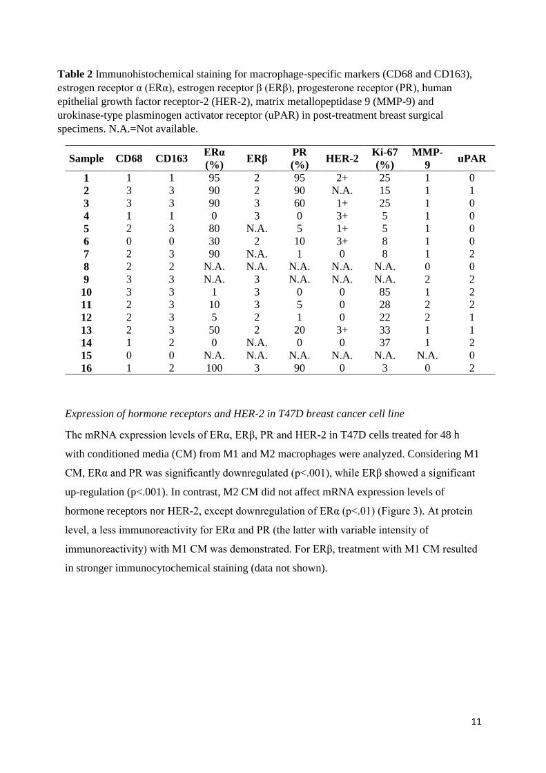
samples with negative or low expression of ERα. The results are listed in Table 1 and Table 2.
Data presented as not available (N.A.) stands for tumor regression following treatment and,
fibrosis formation.
9
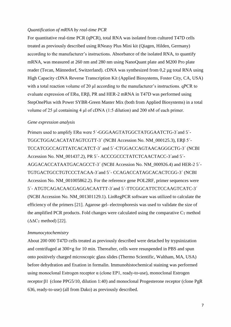
Figure 1 Immunohistochemical staining for macrophage-specific markers (CD68 and CD163)
in breast carcinoma. Representative images demonstrating low (1), moderate (2) and high (3)
expression of CD68 and CD163. Magnification×400.
Low Moderate High
CD
68
CD
163
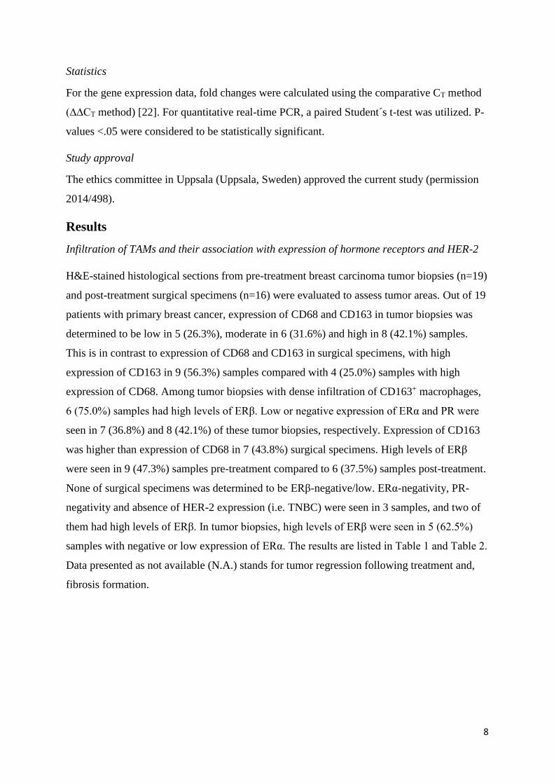
Figure 2 Immunohistochemical staining for estrogen receptor β (ERβ) in breast carcinoma.
Representative images demonstrating negative/low (1), moderate (2) and high (3) expression
of nuclear ERβ. ERβ score 1 A, ERβ score 2 B and ERβ score 3 C. Magnification×400.
A B C
10
Associations between TAMs and expression of Ki-67, MMP-9 and uPAR
High Ki-67 proliferation index (≥20%) was seen in 5 (62.5%) samples with dense infiltration
of CD68+and CD163+ macrophages in tumor biopsies and in 5 (55.6%) surgical specimens.
High expression of MMP-9 was seen in 11 (57.9%) tumor biopsies and in 3 (18.8%) surgical
specimens. This is in contrast to expression of uPAR, with high expression of uPAR in 5
(26.3%) tumor biopsies and 6 (37.5%) surgical specimens. Among tumor biopsies with high
expression of uPAR, 80% also expressed high levels of CD68 and CD163. In surgical
specimens the corresponding figure was 66.7%, but CD163 was more often expressed
compared to CD68 in samples with high expression of uPAR (Table 1 and Table 2).
Sample CD68 CD163 ERα
(%) ERβ
PR
(%) HER-2
Ki-67
(%)
MMP-
9 uPAR
1 3 3 90 3 0 3+ 15 2 2
2 1 1 100 3 100 2+ 0 2 0
3 2 2 75 2 75 1+ 25 2 0
4 3 3 0 3 0 3+ 50 2 1
5 2 2 0 1 0 3+ 25 1 0
6 1 1 80 3 5 1+ 5 1 0
7 2 2 30 2 10 3+ 8 1 1
8 1 1 80 2 90 0 5 1 0
9 1 2 90 3 1 0 8 2 0
10 3 3 0 3 0 0 70 2 2
11 3 3 1 3 1 2+ 50 2 2
12 3 3 0 3 0 3+ 25 2 1
13 3 3 1 N.A. 0 0 85 1 1
14 2 1 40 2 0 2+ 10 2 1
15 2 2 60 2 70 1+ 45 1 2
16 3 3 1 3 4 1+ 15 2 1
17 3 3 0 2 0 2+ 17 2 2
18 1 1 80 N.A. 0 3+ 40 N.A. N.A.
19 2 2 90 1 100 0 15 1 1
Table 1 Immunohistochemical staining for macrophage-specific markers (CD68 and
CD163), estrogen receptor α (ERα), estrogen receptor β (ERβ), progesterone receptor (PR),
human epithelial growth factor receptor-2 (HER-2), matrix metallopeptidase 9 (MMP-9) and
urokinase-type plasminogen activator receptor (uPAR) in pre-treatment breast tumor
biopsies. N.A.=Not available.
11
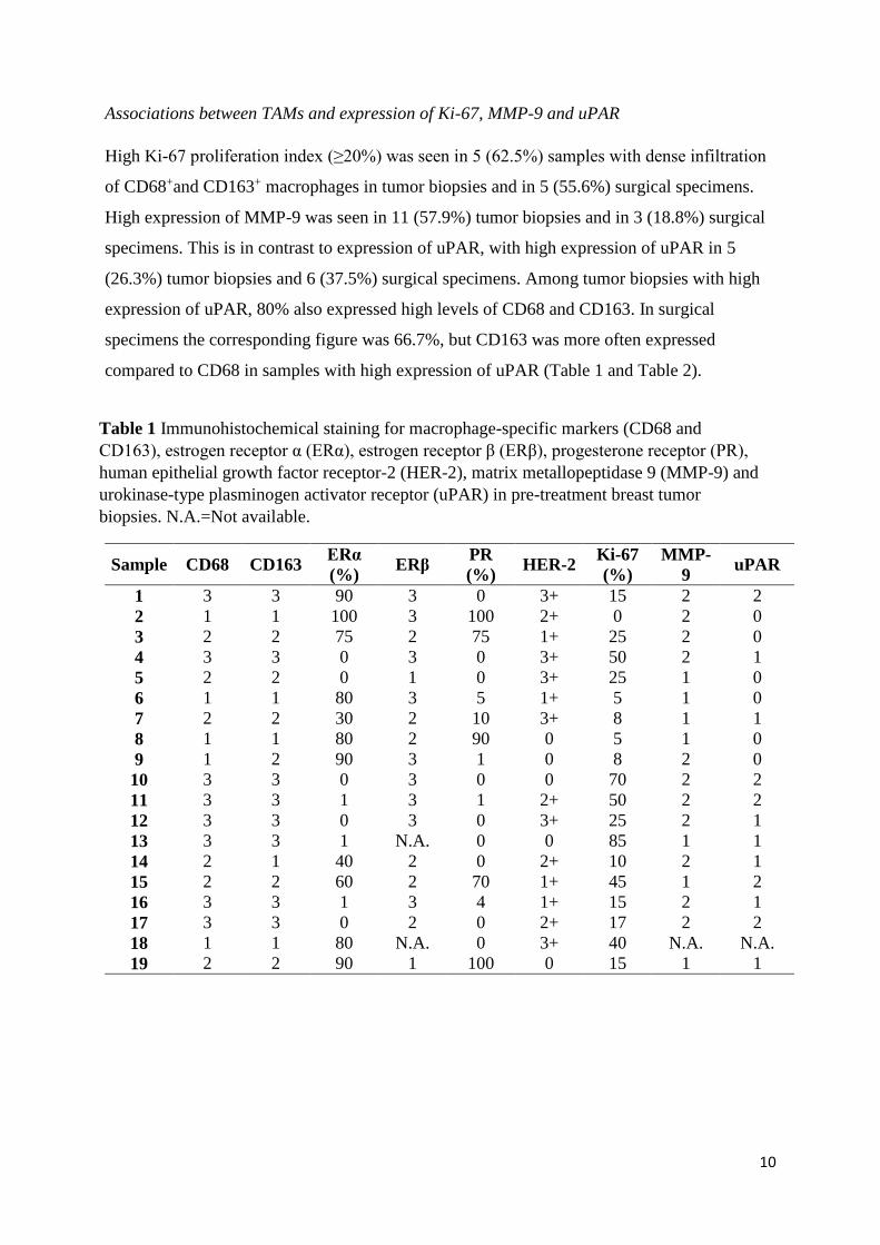
Expression of hormone receptors and HER-2 in T47D breast cancer cell line
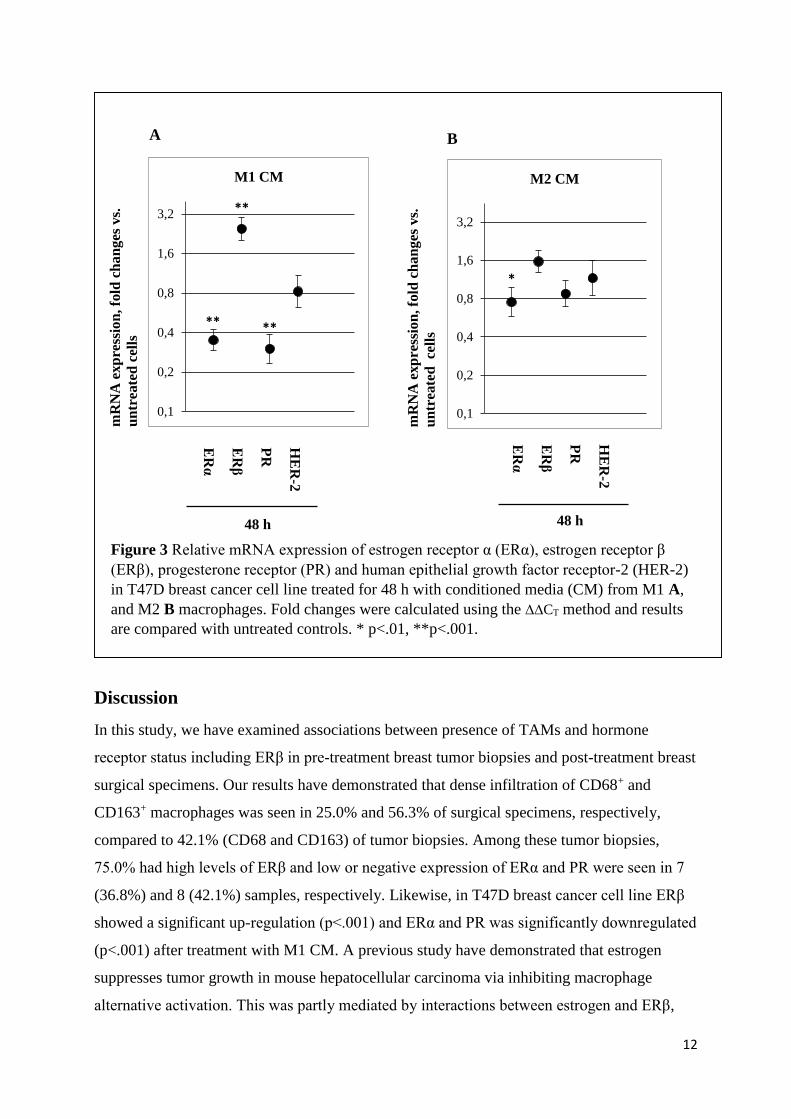
The mRNA expression levels of ERα, ERβ, PR and HER-2 in T47D cells treated for 48 h
with conditioned media (CM) from M1 and M2 macrophages were analyzed. Considering M1
CM, ERα and PR was significantly downregulated (p<.001), while ERβ showed a significant
up-regulation (p<.001). In contrast, M2 CM did not affect mRNA expression levels of
hormone receptors nor HER-2, except downregulation of ERα (p<.01) (Figure 3). At protein
level, a less immunoreactivity for ERα and PR (the latter with variable intensity of
immunoreactivity) with M1 CM was demonstrated. For ERβ, treatment with M1 CM resulted
in stronger immunocytochemical staining (data not shown).
Sample CD68 CD163 ERα
(%) ERβ
PR
(%) HER-2
Ki-67
(%)
MMP-
9 uPAR
1 1 1 95 2 95 2+ 25 1 0
2 3 3 90 2 90 N.A. 15 1 1
3 3 3 90 3 60 1+ 25 1 0
4 1 1 0 3 0 3+ 5 1 0
5 2 3 80 N.A. 5 1+ 5 1 0
6 0 0 30 2 10 3+ 8 1 0
7 2 3 90 N.A. 1 0 8 1 2
8 2 2 N.A. N.A. N.A. N.A. N.A. 0 0
9 3 3 N.A. 3 N.A. N.A. N.A. 2 2
10 3 3 1 3 0 0 85 1 2
11 2 3 10 3 5 0 28 2 2
12 2 3 5 2 1 0 22 2 1
13 2 3 50 2 20 3+ 33 1 1
14 1 2 0 N.A. 0 0 37 1 2
15 0 0 N.A. N.A. N.A. N.A. N.A. N.A. 0
16 1 2 100 3 90 0 3 0 2
Table 2 Immunohistochemical staining for macrophage-specific markers (CD68 and CD163),
estrogen receptor α (ERα), estrogen receptor β (ERβ), progesterone receptor (PR), human
epithelial growth factor receptor-2 (HER-2), matrix metallopeptidase 9 (MMP-9) and
urokinase-type plasminogen activator receptor (uPAR) in post-treatment breast surgical
specimens. N.A.=Not available.
12
0,1
0,2
0,4
0,8
1,6
3,2
M2 CM
Discussion
In this study, we have examined associations between presence of TAMs and hormone
receptor status including ERβ in pre-treatment breast tumor biopsies and post-treatment breast
surgical specimens. Our results have demonstrated that dense infiltration of CD68+ and
CD163+ macrophages was seen in 25.0% and 56.3% of surgical specimens, respectively,
compared to 42.1% (CD68 and CD163) of tumor biopsies. Among these tumor biopsies,
75.0% had high levels of ERβ and low or negative expression of ERα and PR were seen in 7
(36.8%) and 8 (42.1%) samples, respectively. Likewise, in T47D breast cancer cell line ERβ
showed a significant up-regulation (p<.001) and ERα and PR was significantly downregulated
(p<.001) after treatment with M1 CM. A previous study have demonstrated that estrogen
suppresses tumor growth in mouse hepatocellular carcinoma via inhibiting macrophage
alternative activation. This was partly mediated by interactions between estrogen and ERβ,
A B
mR
NA
exp
ress
ion
, fo
ld c
ha
ng
es v
s.
un
trea
ted
cel
ls
mR
NA
exp
ress
ion
, fo
ld c
ha
ng
es v
s.
un
trea
ted
ce
lls
HE
R-2
PR
ER
β
ER
α
HE
R-2
PR
ER
β
ER
α
Figure 3 Relative mRNA expression of estrogen receptor α (ERα), estrogen receptor β
(ERβ), progesterone receptor (PR) and human epithelial growth factor receptor-2 (HER-2)
in T47D breast cancer cell line treated for 48 h with conditioned media (CM) from M1 A,
and M2 B macrophages. Fold changes were calculated using the ∆∆CT method and results
are compared with untreated controls. * p<.01, **p<.001.
48 h
48 h
0,1
0,2
0,4
0,8
1,6
3,2
M1 CM
**
**
**
*
13
leading to inhibition of polarization of macrophages toward M2. Consequently, administration
of estrogen to males may suppress tumor growth in this male-predominant cancer [23].
The fact that our in vitro study was limited to only one breast cancer cell line might be a
limitation of the present study as it is equal to a single breast cancer case, but we aimed to
confirm whether hormone receptors was downregulated with M1 and/or M2 CM. M2 CM did
not affect mRNA expression levels to the same extent as M1 CM. Not yet published data
indicates an up-regulation of a number of target genes in M1 macrophages, including
interleukin-8 (IL-8) and chemokine (C-X-C motif) ligand 9 (CXCL9). A strength of the
present study, on the other hand, is human monocyte-derived macrophages differentiated into
M1 and M2 phenotypes.
Expression of CD163 was higher than expression of CD68 in 7 (43.8%) surgical
specimens. Since CD68 is widely used as a pan-macrophage marker for TAMs, expression of
CD68 should be higher than expression of CD163. CD163 is a scavenger receptor expressed
by macrophages and neoplasms with monocytic differentiation, but not cancer cells.
However, a study with 127 patients with primary breast cancer have demonstrated breast
cancer cell expression of CD163 up to 48% [12]. Moreover, also myeloid-derived suppressor
cells can express CD163, which is known to enhance tumor progression [4,5]. TAMs may
build hybrids with tumor cells by fusion, and these are prone to metastasize [12]. Taken
together, further investigation with double staining of CD68 and CD163, and ERα and CD163
to identify possible CD163+ tumor cells is required.
High levels of ERβ were seen in 9 (47.3%) samples pre-treatment compared to 6 (37.5%)
samples post-treatment. Although not fully understood, ERβ has a putative anti-proliferative
effect and as a result, expression of ERβ should be higher in surgical specimens compared to
tumor biopsies. However, 5 (55.6%) tumor biopsies with high levels of ERβ had low Ki-67
proliferation index (<20%) and, vice versa, 3 (30.0%) samples with high Ki-67 proliferation
index was determined to express low or moderate levels of ERβ.
Gruvberger-Saal et al. reported that ERβ is an independent prognostic marker for response
to tamoxifen in ERα-negative breast cancer, hence, patients diagnosed with ERα-negative but
ERβ-positive breast cancer may benefit from treatment with tamoxifen [19]. In our study,
high levels of ERβ were seen in 5 (62.5%) tumor biopsies with negative or low expression of
ERα. A previous study reported 24-44% of ERα negative tumors to be ERβ positive [16].
This discrepancy can be due to small sample size in the present study. Triple negative breast
14
cancer was seen in three cases, and among these, two of them had high levels of ERβ which
might be a prognostic marker for response to treatment in these patients.
A limitation of the present study is that no ethical approval was required and, subsequently,
clinicopathological features (e.g. age, tumor size, treatment regimen etc.) are not available. In
general, patients with primary breast carcinoma receives chemotherapy, radiation therapy
and/or hormone therapy [24]. Response to treatment with tumor regression were seen in 5
surgical specimens.
High Ki-67 proliferation index (≥20%) was seen in 62.5% of samples with dense
infiltration of CD68+and CD163+ macrophages in tumor biopsies and in 55.6% of surgical
specimens. These results are consistent with previous reports [4,5]. Expression of MMP-9 was
lower in surgical specimens compared to tumor biopsies (18.8% vs. 57.9%) but, in contrast,
expression of uPAR was higher in surgical specimens compared to tumor biopsies (37.5% vs.
26.3%). This might be unexpected since activation of uPAR initiates MMPs to degrade
components of extracellular matrix [8]. However, elevated levels of uPAR in surgical
specimens can be a result of treatment which in turn re-gives normal function of uPAR.
Two anti-ERβ antibodies of mono- and polyclonal origin were evaluated, PPG5/10 and
PA1-311, respectively. Four different ERβ isoforms exist and PPG5/10 is regarded as ERβ1-
specific, while PA1-311 are not [15,16,25]. In our study, PA1-311 gave a non-specific
staining and, therefore, PPG5/10 was used which has proven to be highly sensitive and
specific [16]. Moreover, PPG5/10 are useful in paraffin-embedded breast tissue [25], as was
used in the present study. Although PPG5/10 is a well-validated antibody, there are
difficulties in pathological assessments because anti-ERβ antibodies has not been considered
to be of diagnostic value so far and, therefore, are not used in routine diagnostics. In addition,
antibody concentrations and scoring systems differs which further complicates the
assessments.
The present study are also limited by the small number of patients included which does not
permit statistical analysis, but these are preliminary results. During winter 2015/16 pre-
treatment tumor biopsies with adjacent post-treatment surgical specimens from 151 breast
cancer patients obtained from Karolinska Institutet, (Stockholm, Sweden) will be evaluated.
The study has been approved and data about neoadjuvant chemotherapy and other
clinicopathological features are available. Immunohistochemical staining will possibly be
performed for tumor-infiltrating lymphocytes (TILs) and double staining of CD68 and
15
CD163. Data about prognostic factors including ERα, PR, HER-2 and Ki-67 are available in
database.
Conclusion
Dense infiltration of CD68+ and CD163+ macrophages in breast tumor biopsies are related to
high expression of ERβ and negative expression of ERα and PR. In cultured T47D breast
cancer cell line, ERβ showed a significant up-regulation whereas ERα and PR was
significantly downregulated after treatment with M1 conditioned media. High expression of
ERβ were seen in 62.5% of tumor biopsies with negative expression of ERα and, thus, ERβ
might be a prognostic marker for treatment in these patients. Expression of MMP-9 was lower
in breast surgical specimens compared to tumor biopsies and expression of uPAR was higher
in surgical specimens. This study suggests further investigation with double staining of CD68
and CD163, and ERα and CD163 since expression of CD163 was higher than expression of
CD68 in 43.8% of surgical specimens.
Acknowledgements
I would like to thank Ann Erlandsson, Karlstad University (Karlstad, Sweden) for expertise
and reading of the manuscript. I would also like to thank Margareta Ericsson, Department of
Pathology and Cytology (Karlstad, Sweden) for performance of immunohistochemical
stainings and Anja Solterbeck, Department of Pathology and Cytology (Karlstad, Sweden) for
evaluation of immunostainings. Finally, I would like to thank Therése Lindsten, Department
of Pathology and Cytology (Karlstad, Sweden) for sharing of data and support.
References
1. Ferlay J, Soerjomataram I, Ervik M, Dikshit R, Eser S, Mathers C, Rebelo M, Parkin DM,
Forman D, Bray, F. World Health Organization. GLOBOCAN 2012: Estimated Cancer
Incidence, Prevalence and Mortality Worldwide in 2012. Available at:
http://globocan.iarc.fr/Pages/fact_sheets_cancer.aspx. Accessed 2015-10-09.
2. Matsumoto H, Koo SL, Dent R, Tan PH, Iqbal J. Role of inflammatory infiltrates in triple
negative breast cancer. J Clin Pathol 2015 Jul;68(7):506-510.
3. Mohammed ZM, Going JJ, Edwards J, Elsberger B, Doughty JC, McMillan DC. The
relationship between components of tumour inflammatory cell infiltrate and
clinicopathological factors and survival in patients with primary operable invasive ductal
breast cancer. Br J Cancer 2012 Aug 21;107(5):864-873.
4. Medrek C, Ponten F, Jirstrom K, Leandersson K. The presence of tumor associated
macrophages in tumor stroma as a prognostic marker for breast cancer patients. BMC
Cancer 2012 Jul 23;12:306-2407-12-306.
16
5. Sousa S, Brion R, Lintunen M, Kronqvist P, Sandholm J, Monkkonen J, et al. Human
breast cancer cells educate macrophages toward the M2 activation status. Breast Cancer
Res 2015 Aug 5;17(1):101-015-0621-0.
6. Ramanathan S, Jagannathan N. Tumor associated macrophage: a review on the phenotypes,
traits and functions. Iran J Cancer Prev 2014 Winter;7(1):1-8.
7. Hanahan D, Weinberg RA. Hallmarks of cancer: the next generation. Cell 2011 Mar
4;144(5):646-674.
8. Noh H, Hong S, Huang S. Role of urokinase receptor in tumor progression and
development. Theranostics 2013 Jun 25;3(7):487-495.
9. Hu J, Jo M, Eastman BM, Gilder AS, Bui JD, Gonias SL. uPAR induces expression of
transforming growth factor beta and interleukin-4 in cancer cells to promote tumor-
permissive conditioning of macrophages. Am J Pathol 2014 Dec;184(12):3384-3393.
10. Mahmoud SM, Lee AH, Paish EC, Macmillan RD, Ellis IO, Green AR. Tumour-
infiltrating macrophages and clinical outcome in breast cancer. J Clin Pathol 2012
Feb;65(2):159-163.
11. Yang J, Li X, Liu X, Liu Y. The role of tumor-associated macrophages in breast
carcinoma invasion and metastasis. Int J Clin Exp Pathol 2015 Jun 1;8(6):6656-6664.
12. Shabo I, Stal O, Olsson H, Dore S, Svanvik J. Breast cancer expression of CD163, a
macrophage scavenger receptor, is related to early distant recurrence and reduced patient
survival. Int J Cancer 2008 Aug 15;123(4):780-786.
13. Gwak JM, Jang MH, Kim DI, Seo AN, Park SY. Prognostic value of tumor-associated
macrophages according to histologic locations and hormone receptor status in breast
cancer. PLoS One 2015 Apr 17;10(4):e0125728.
14. Omoto Y, Iwase H. Clinical significance of estrogen receptor beta in breast and prostate
cancer from biological aspects. Cancer Sci 2015 Apr;106(4):337-343.
15. Haldosen LA, Zhao C, Dahlman-Wright K. Estrogen receptor beta in breast cancer. Mol
Cell Endocrinol 2014 Jan 25;382(1):665-672.
16. Reese JM, Suman VJ, Subramaniam M, Wu X, Negron V, Gingery A, et al. ERbeta1:
characterization, prognosis, and evaluation of treatment strategies in ERalpha-positive
and -negative breast cancer. BMC Cancer 2014 Oct 7;14:749-2407-14-749.
17. Huang B, Warner M, Gustafsson JA. Estrogen receptors in breast carcinogenesis and
endocrine therapy. Mol Cell Endocrinol 2014 Nov 26.
18. Fuqua SA, Schiff R, Parra I, Moore JT, Mohsin SK, Osborne CK, et al. Estrogen receptor
beta protein in human breast cancer: correlation with clinical tumor parameters. Cancer
Res 2003 May 15;63(10):2434-2439.
17
19. Gruvberger-Saal SK, Bendahl PO, Saal LH, Laakso M, Hegardt C, Eden P, et al. Estrogen
receptor beta expression is associated with tamoxifen response in ERalpha-negative
breast carcinoma. Clin Cancer Res 2007 Apr 1;13(7):1987-1994.
20. Engstrom A, Erlandsson A, Delbro D, Wijkander J. Conditioned media from macrophages
of M1, but not M2 phenotype, inhibit the proliferation of the colon cancer cell lines HT-
29 and CACO-2. Int J Oncol 2014 Feb;44(2):385-392.
21. Ruijter JM, Ramakers C, Hoogaars WM, Karlen Y, Bakker O, van den Hoff MJ, et al.
Amplification efficiency: linking baseline and bias in the analysis of quantitative PCR
data. Nucleic Acids Res 2009 Apr;37(6):e45.
22. Applied Biosystems. Guide to Performing Relative Quantitation of Gene Expression
Using Real-Time Quantitative PCR. 2008; Available at:
http://www3.appliedbiosystems.com/cms/groups/mcb_support/documents/generaldocum
ents/cms_042380.pdf. Accessed 2015-12-10.
23. Yang W, Lu Y, Xu Y, Xu L, Zheng W, Wu Y, et al. Estrogen represses hepatocellular
carcinoma (HCC) growth via inhibiting alternative activation of tumor-associated
macrophages (TAMs). J Biol Chem 2012 Nov 23;287(48):40140-40149.
24. Regionala cancercentrum i samverkan. Bröstcancer Nationellt vårdprogram. 2014;
Available at:
http://www.cancercentrum.se/globalassets/cancerdiagnoser/brost/vardprogram/natvp_bro
stcancer_2014-11-11_final.pdf. Accessed 2015-12-08.
25. Skliris GP, Parkes AT, Limer JL, Burdall SE, Carder PJ, Speirs V. Evaluation of seven
oestrogen receptor beta antibodies for immunohistochemistry, western blotting, and flow
cytometry in human breast tissue. J Pathol 2002 Jun;197(2):155-162.
18
Letter of intent
January 06, 2016
Corresponding author:
Anna Ramberg
School of Medical Sciences
Örebro University, Sweden
Dear Editor,
On behalf of the authors, we hereby submit a manuscript titled Expression of hormone
receptors and markers for metastatic potential in relation to tumor associated macrophages
in breast cancer for publication in X, authored by Anna Ramberg and Ann Erlandsson.
Hormone receptor status influences prognosis in breast cancer. Tumor associated
macrophages are part of the tumor microenvironment and is correlated with poor survival and
hormone receptor negativity. Estrogen receptor β is a novel estrogen receptor with a putative
anti-proliferative effect and is associated with favorable clinical outcome and tamoxifen
responsiveness.
This study examines pre- and post-treatment associations between macrophage infiltration
(CD68 and CD163) and expression of estrogen receptor α, estrogen receptor β, progesterone
receptor, human epithelial growth factor receptor-2, proliferation marker Ki-67, matrix
metallopeptidase 9 and urokinase receptor in 19 breast cancer patients and in cultured T47D
breast cancer cell line. We found that dense infiltration of CD68+ and CD163+ macrophages
in tumor biopsies are related to high expression of estrogen receptor β and negative
expression of estrogen receptor α and progesterone receptor. This was accompanied in T47D
breast cancer cell line.
We suggest that estrogen receptor β might be a prognostic marker in patients with estrogen
receptor α negativity and triple negative breast cancer.
The manuscript is original and has not been published elsewhere or previously. There are
no conflicts of interest.
We look forward to your review.
Best wishes,
Anna Ramberg
19
Ny potentiell behandlingsmarkör för bröstcancerpatienter
Pressmeddelande 2016-01-06
Bröstcancer är den vanligaste cancerformen hos kvinnor över hela världen.
Framtidsutsikten beror bland annat på om cancercellerna har mottagare (receptorer)
dit hormonet östrogen kan binda. Generellt sett är det svårare att behandla patienter
vars cancerceller saknar dessa mottagare. Vi har visat att en viss typ av
immunförsvarscell, så kallade makrofager, minskar mängden av den vanliga
östrogenreceptorn men ökar mängden av en ny östrogenreceptor. Detta skulle kunna
hjälpa de bröstcancerpatienter som har den nyare typen av östrogenreceptor.
Syftet med studien som genomfördes via avdelningen för Klinisk patologi på Centralsjukhuset
i Karlstad och Karlstads universitet var att ta reda på om det finns något samband mellan
makrofager och hormonreceptorer hos bröstcancerpatienter. Efter att ha studerat material från
19 stycken bröstcancerpatienter visar resultaten att i material där det finns mycket makrofager
ser man färre hormonreceptorer. Däremot ser man en ökning av en ny östrogenreceptor, som
skulle kunna användas som behandlingsmarkör för patienter med en vanligtvis mycket
svårbehandlad typ av bröstcancer, så kallad trippelnegativ bröstcancer. Förutom att använda
oss av material från bröstcancerpatienter har även bröstcancerceller och makrofager odlats
fram. Makrofagerna har sedan tillsatts till cancercellerna för att se vad som händer med dess
receptorer. Vi ser samma resultat i de odlade bröstcancercellerna som vi behandlat med
makrofager som hos materialet från bröstcancerpatienterna, det vill säga att nivåerna av
hormonreceptorer sjunker när man tillsätter makrofager. I framtiden kan det få betydelse att
undersöka om bröstcancerpatienter även har den ”nya” typen av receptor, eftersom de då kan
dra nytta av behandling som även riktar sig mot denna receptor.
20
Ethics
The current study was approved by the ethics committee in Uppsala (Uppsala, Sweden),
however, the application was not tested. According to the law of ethical approval, ethical
approval was not required since no participants were affected or sensitive personal data was
handled. Moreover, since the material is de-identified there is no ethical doubts regarding
traceability and, hence, personal data is unavailable. Unlike decoded material, de-identified
material refers to data that cannot be identifiable retrospectively and is handled by persons not
directly linked to the study. We hope that breast cancer patients will benefit from the results
of this study and we cannot imagine any risks. Samples are taken from all breast cancer
patients, for diagnostic purposes and to determine responsiveness to treatment, and no further
samples have been taken for the present study. The immunostainings were evaluated at
Department of Clinical Pathology and Cytology (Karlstad, Sweden) and microscope glass
slides was not brought out of this area. There was no defined inclusion criteria, but de-
identified archival material was obtained from biobank. Consequently, data regarding age and
treatment regimen etc. is not available. Moreover, pre-treatment tumor biopsies cannot be
coded with adjacent surgical specimens. These are preliminary results and during winter
2015/16 tumor biopsies with adjacent surgical specimens from 151 breast cancer patients
obtained from Karolinska Institutet (Stockholm, Sweden) will be evaluated. The study has
been approved and, therefore, data about age and other clinicopathological features are
provided for analysis. The patients have given their written informed consent to use the breast
tissues for research purposes. The buffy coat used in the present study to generate human
macrophages were obtained from anonymous healthy blood donors, and can be considered as
de-identified material. Finally, the company where the T47D breast cancer cell line is
obtained from are adhered to the highest ethical standards and ensures integrity of their
products.