Embed Size (px)
Citation preview

Comprehensive Summaries of Uppsala Dissertationsfrom the Faculty of Pharmacy 297
Expression of Genes Encoding forDrug Metabolism in the
Small Intestine
BY
MONICA LINDELL
ACTA UNIVERSITATIS UPSALIENSISUPPSALA 2003


7
Genes Encoding for Drug Metabolism in Small Intestine
INTRODUCTION
It is common that drugs and other foreign substances (xenobiotics) are undergoing metabolism before they are eliminated from the body. The routes by which drugs may be metabolised are many and varied. Drug metabolism is normally divided into two phases; phase I and phase II. Phase I enzymes introduce a functional group, such as –OH, into the substrate. In general, they render the compound less lipophilic, but additionally serve to exposes reactive sites, or to add functionally reactive sites to which polar groups may be conjugated. Phase II enzymes then use this functional group as a handle for conjugation with such moieties as glucuronic acid, sulfate, and glutathiol. The end result is a hydrophilic product that can be more easily eliminated from the body [1, 2]. Cytochrome P450 (CYP) probably comprises more than 95% of all phase I enzymes. Phase II enzymes include a group of diverse enzymes, among them the UDP-glucuronosyltransferases (UGT) [2].
The liver is the organ with the highest capacity for both phase I and phase II reactions, but it is not the only organ involved. Drug metabolism has been observed in different organs such as the gastrointestinal tract (GI tract), lung, white blood cells, brain, kidney, and placenta [3]. While the liver has been thought to be the major organ involved in drug metabolism, the small intestine is also capable of performing many of the same metabolic reactions on orally ingested drugs, or potentially toxic compounds ingested with the food [4 - 6]. But the metabolic activities of both phase I and phase II enzymes are considerably much lower in the whole small intestine compared to the liver [7].
The focus of this thesis is on the variation of expression of both CYP and UGT genes in the small intestine of both humans and rats; in particular variation between the two species, individual variation in expression in human populations, and how the expression is affected by endogenous and/or exogenous factors.
It is important to clarify the differences between the human and the rat expression, and also the differences between the expression of rat small intestine and liver. Rat hepatic tissue has been used extensive by drug metabolism studies. Also in humans the most common used organ in metabolic studies has been the liver, but for orally used drugs the small intestine is the first organ that is exposed for a drug. It is therefore important to elucidate how much the small intestine is contributing in the first-pass drug metabolism. The humans are also exposed to a lot of different exogenous factors, such as other drugs, xenobiotics from the diet and environmental pollutants, factors that may inhibit and/or induce drug metabolism.

8
Monica Lindell
The small intestine
The small intestine forms the largest metabolically active external surface of the human digestive system [8]. In humans, the small intestine is about 5 to 6 meter and is divided into three parts: duodenum, jejunum, and ileum [9, 10]. These regions are not anatomically distinct, although there are differences in their absorptive and secretory capabilities. The duodenum is the shortest, widest, and least mobile section. In humans it measures 20 to 30 cm in length and 3 to 5 cm in diameter. The rest of the small intestine is about 5 m long; the proximal two-fifths is referred to as the jejunum and the distal three-fifths is called the ileum [11]. The primary function of the small intestine is to absorb nutrients and water. This is achieved by mixing the passing foodstuff with digestive enzymes to increase its contact with the absorptive cells of the mucosa. Approximately 6 to 12 litres of partially digested foodstuffs, water, and secretions are delivered daily to the small intestine. Of this, only 10 to 20% are passed on to the colon, because most nutrients, electrolytes, and water are absorbed as they are transported through the small intestine [9, 10]. In addition to this fundamental role, a secondary function of the small intestine arises from the fact that it is also a major route of entry into the body for many xenobiotics including pharmaceutical drugs.
Blood and lymph vesse
Cells shed from the tip
crypt
Differentiated enterocytes
Direction of cellmovement
Figure 1. Structure of a single intestinal villius tip. The intestinal mucosa is continuously renewed by new cells that originate from the crypt (modifi ed from Hole and Koos.)
Throughout its length, the inner wall of the small intestine has velvet like appearance. This is due to the presence of innumerable tiny projections of the mucous membrane called intestinal villi. These project into the lumen of the

9
Genes Encoding for Drug Metabolism in Small Intestine
alimentary canal containing the intestinal contents. The villi increase the surface of the intestinal lining and play an important role in the absorption of digestive products. Each villi consist of a layer of simple columnar epithelium and a core of connective tissue containing blood capillaries, a lymphatic capillary, and nerve fibres (Fig. 1). At their free surface, the epithelial cells possess many fine extensions, called microvilli, that create a brush like border and greatly increase the surface area of the intestinal cells (Fig. 2) [12]. The epithelial cells at the tip of the villi are functionally mature and non-dividing cells, whereas the crypt cells are immature and evolving. The crypt cells continue to mature as they ascend toward the villi and are extruded at its tip (Fig. 1).
Folds Villi Enerocytes with microvillae
Figure 2. The morphology and surface of the small intestine.
The enterocytes are important for the absorption and metabolism of nutrients and drugs [11]. The enzyme activity is greatest in the villius tips and decreases progressively towards the crypts. The highest concentration of metabolising enzymes in the GI tract is in the upper small intestine: the duodenum and the jejunum followed by the ileum and colon [13]. Epithelial cells of the intestinal mucosa have a programmed life span. They are formed by cell division within the crypts and undergo differentiation of cell function for two to five days as they migrate towards the apical tip. Immunohistochemical analysis of mucosal cross-sections indicates that drug-metabolising enzymes are expressed only after cell transformation, and thus, are found exclusively in mature enterocytes [14].
Although the small intestine is regarded as an absorptive organ in the uptake of orally administered drugs, it also has the ability to metabolise drugs by numerous pathways involving both phase I and phase II reactions [5, 15-17]. It has been shown that the metabolism in the enterocytes are minimal compared to the metabolism in the liver. This is based on the relative rates of metabolism in intestinal microsomes compared to hepatic microsomes. This observation may, however, in part be explained by the localisation of the drug metabolising enzymes (e.g. CYPs), in the villius tip, which makes up a relatively small portion of the total intestinal mucosa [18]. Despite significant lower expression of CYP enzymes, the intestine contributes considerably to first-pass metabolism of some substances.

10
Monica Lindell
Drug metabolism and Transport
There are essentially four mechanisms (Fig. 3), by which an orally ingested drug may cross the epithelial layer:
1)3)
4)
2)
Extracellular
Intracellular
Figure 3. Different transport mechanisms for drugs across the intestinal epithelium; 1) passive transcellular diffusion; 2) paracellular transport; 3) carrier-mediated transport and 4) endocytosis (modifi ed from Barthe et al 1999).
(1) Passive transcellular diffusion, a rout most likely used by lipophilic drugs with low molecular weight [19].
(2) Paracellular transport. Only small hydrophilic molecules cross the tight-junctions and pass between the cells. Compounds using this paracellular route should not be metabolised by the intracellular enzymes [20].
(3) Carrier-mediated transport. This mechanism involves specific interactions between the molecule and the transporter or carrier and is a saturable mechanism, utilised by hydrophilic molecules. Briefly, two types of carrier-mediated transport exist: (I) active transport that requires metabolic energy (ATP) and that can act against a concentration gradient and (II) transport that are driven by a co-transport or a counter transport of various ions. Drug absorption has generally been assumed to occur by passive processes. Transporters in the intestine have been shown to facilitate transport of some drugs possessing structural similarities to natural occurring substances [21].
(4) Endocytosis. This is a constitutive process for the uptake of macromolecules that requires metabolic energy and is generally a slow uptake mechanism. [19].

11
Genes Encoding for Drug Metabolism in Small Intestine
Phase I metabolism
Cytochrom P450 (CYP)
The Cytochrom P450 super family (CYP) is an enzyme group that is critical for oxidation reactions that affect the biological activities of drugs, environmental chemicals and endogenous compounds and that plays a predominant role in the phase I metabolism of xenobiotics.
The CYP enzymes are classified into families and subfamilies based on their amino acid sequence similarity. The root symbol ‘CYP’, denoting Cytochrome P450 and an Arabic number designating the family (e.g. CYP3). Members of the same family are more than 40% identical with respect to their amino acid sequences. If the sequences are more than 55% identical, the enzymes belong to the same subfamily, indicated by an additional letter (e.g. CYP3A). Finally, each individual enzyme is represented by an Arabic numeral (e.g. CYP3A4) [22]. An updated listing of CYP enzymes can be accessed at http://drnelson.utmem.edu/CytochromeP450.html.
Only three CYP gene families are currently identified to be responsible for drug metabolism in both humans and rats i.e. CYP1, CYP2, and CYP3 [23].
CYP1 have two subfamilies CYP1A (i.e. CYP1A1 and 1A2) and CYP1B. CYP1A1 is mainly found extrahepatically and CYP1A2 is a hepatic enzyme [24]. Both are induced by polycyclic aromatic hydrocarbons (PAH), found for example in cigarette smoke and charbroiled meat [24-26]. CYP1A2 is responsible for metabolism of several drugs such as caffeine and paracetamol [24].
CYP2 is the largest family of human CYPs identified to date, with up to seven different subfamilies [22]. In the small intestine, mRNA from only three CYP2 subfamilies has been reported: CYP2C, CYP2D6, and CYP2E1 [27-29] whereas the human liver also expresses CYP2A6 and CYP2B6 [30]. All CYP2s are involved in the metabolism of numerous clinically important drugs and endogenous compounds [30]. There are also differences between CYP2 gene family members in terms of response to inducers. For example, CYP2Bs are phenobarbital induced and CYP2E1 is induced by ethanol [31]. Some members of the CYP2C and CYP2D subfamilies appear to be constitutively expressed [32].
CYP3A is the primary CYP subfamily in humans; responsible for metabolism of more than 50% of administered drugs. It is an enzyme family that has shown to be highly conserved, in all mammals examined, including humans [33,34]. To date four members of the CYP3A subfamily are expressed in humans; CYP3A3, CYP3A4, CYP3A5 and CYP3A7 [35]. Studies have shown that CYP3A4 and/or CYP3A3 proteins are the major forms in human liver, jejunum, colon, and pancreas, whereas CYP3A5 is the major protein in the stomach [35]. The subfamily has a broad substrate specificity, where the substrates are structurally diverse including clinically important drugs, covering a wide therapeutic range [23]. The CYP3As has shown to be induced by a multitude of pharmaceutical drugs [32, 36-40] as well as environmental toxins [41, 42]. It therefore appears that CYP3A enzymes may

12
Monica Lindell
play a role in some forms of diseases mediated by environmental factors, in addition to playing a role in pharmacokinetics of a vast array of clinically used drugs.
Phase II metabolism
Phase II metabolism is a synthetic or conjugation step that is usually (with the exception of glucuronidation) a nonmicrosomal process occurring in the cytosol [43]. The human intestinal tissues have been reported to contain relatively high activity of glucuronosyltransferase, together with sulfotransferase, and glutathione-S-transferase as the main enzymes in phase II conjugation [44].
UDP-glucuronosyltransferases (UGT)
Glucuronidation is a central process in which many xenobiotics and endobiotics are converted to more hydrophilic compounds, which are readily eliminated from the body. Glucuronidation reactions are catalysed by UDP-glucuronosyltransferases (UGT) that are located on the luminal side of the endoplasmatic reticulum and in the nuclear envelope of hepatocytes and cells in other organs including the intestine [17, 45-47]. The range of target substances is large and spans divergent chemical classes, dietary constituents, environmental pollutants, therapeutic drugs and numerous endogenous substrates [48, 49].
Based on evolutionary divergence of the proteins, at least two UGT subfamilies have been described: UGT1 and UGT2 [48]. In naming the UGTs one uses a similar system as for the CYP enzymes, i.e. using UGT as a root symbol, representing UDP-glucuronosyltransferase, followed by an Arabic number denoting the family having ≥45% amino acid identity within a family (e.g. UGT2), and a letter designating the subfamily (e.g. UGT2B). Mammalian sequences within the same protein subfamily are approximately 60% or more identical. Finally, an Arabic number represents an individual gene within the subfamily (e.g. UGT2B15) [2, 48]. An updated listing of the UGT enzymes can be found at: http://som.flinders.edu.au/FUSA/ClinPharm/UGT/
Members of the UGT1 gene complex are encoded by at least 12 unique first exons located at the 5’ end of the locus. They are then selectivity spliced with four constant exons (exon 2-5) to form unique RNA transcripts with varied 5’ ends and identical 3’ ends (Fig. 4) [50]. Members of the UGT1 family conjugate mainly bilirubin, bile acids, phenols and steroid hormones [51, 52]. The gene products of the UGT2 subfamily are, on the other hand, transcribed from unique genes [53, 54]. UGT2B enzymes catalyse the glucuronidation of bile- and fatty acids, and in particular, steroid hormones [2, 51, 52]. Currently, 15 human UGT cDNAs have been identified, eight UGT1A proteins encoded by the UGT1A locus (and three pseudogenes) and seven proteins encoded by UGT2 genes [49].

13
Genes Encoding for Drug Metabolism in Small Intestine
13p11p12p 8 13467910 5p 2p
UGT1A10
UGT1A6
UGT1A4
UGT1A3
UGT1A1
198872 bp
5432
Exon 1 Exons
Figure 4 The organisation of the UGT1A1 locus (modifi ed from Tukey and Strassburg 2001 [55]).
In the small intestine the UGT1A proteins have been localised to the epithelial cell layer and the UGT staining of the villi is homogenous. The gene expression of UGT1A is uniform along the length of the human small intestine [8].
Transport through intestinal epithelium
P-glycoprotein (Pgp)
P-glycoprotein (Pgp or ABCB1 [Human Gene Nomenclature Committée: http://www.gene.ucl.ac.uk/nomenclature] ) is a member of the ABC-transporter family (ATP binding cassette), a group of proteins that is a participant in our defence against foreign substances. Pgp was first discovered in cancer cells resistant to several chemotheraputic agents. Where the agents were pumped out of the cell [56].
The Pgp:s is encoded by the multidrug resistance genes (MDR). In humans, two MDR genes have been found; MDR1 (ABCB1) that encodes a drug transporting Pgp that confers drug resistance, and MDR2 (ABCB4 ) that encodes an enzyme that is specific for phosphatidylcholine translocation in cells. In rodents, there are three mdr genes: mdr1a (mdr1) and mdr1b (mdr3) conferring multidrug resistance, whereas mdr2 is responsible for transport of cell phospholipids [23].
The human MDR1 mRNA encodes 1280 amino acids and the gene product is a 135 to 180 kDa membrane transporter, the variation in molecular mass may be linked to differences in the glycosylation of Pgp [57]. Sequence analysis indicates the presence of 12 transmembrane domains in two homologous halves each containing six regions and a putative ATP-binding site (Fig. 5). The transmembrane segments are thought to form a channel through which the drug is extruded. An ATP-binding site is thought to be involved and ATP hydrolysis required for ‘pumping’ out drugs against a concentration gradient [58].

14
Monica Lindell
ATP- binding domain
Drug efflux
Figure 5 P-glycoprotein structure through the plasma membrane. The transport protein has 12 transmembrane domains in two homologous halves with ATP binding sites on the intracellular side of the molecule (modifi cation from www.nature.com/nre/journl/v2/images/nrc823-f3.gif).
Pgp may play a significant role in drug absorption and disposition in both animals and humans. Pgp is present in normal epithelial cells from liver, kidney, GI-tract among other tissues [59, 60]. The localisation suggests that Pgp functionally can protect the body against toxic xenobiotics by excreting these compounds into bile, urine, and the intestinal lumen [7]. In the GI-tract, the distribution of Pgp is not uniform along the length of the intestine or along the villi within a cross section of the mucosa. The mRNA levels of Pgp increase progressively from the stomach to the colon with low levels in the stomach, intermediate in the jejunum and high levels in colon [61, 62]. However, more studies of the regional distribution of this protein transporter along the gastrointestinal tract are needed. It has been speculated that, since Pgp is located on the epithelium of intestinal cells, it may act as a counter-transport pump that transports drugs back into the intestinal lumen as they begin to be absorbed across the luminal plasma membrane.
CYP3A4 and Pgp
Both CYP3A4 and Pgp function to protect the body from toxic accumulation of hydrophobic xenobiotics via metabolism and excretion. They are both co-localised in the small bowel (being present in villius tip and not in the crypt cells). Moreover, data obtained in rat and man suggest that CYP3A4 are localised primarily at the apex of the enterocytes, just below the brush border containing Pgp [35, 38, 63].
Studies have found that there is an overlap between substrates for CYP3A4 and Pgp [21, 64, 65]. Overlap has also been found among inhibitors [66-68] and inducers of the two proteins [69-72]. A hypothesis has been put forward that CYP3A and Pgp

15
Genes Encoding for Drug Metabolism in Small Intestine
together form efficient machinery for the elimination of drugs from cells. The drug is entering the enterocyte and is either metabolised by CYP3A or effluxed by Pgp. The drug may then enter the cell again. This recirculation of the drug may allow a greater access to a limited amount of CYP3A, and a greater proportion of the drug will be metabolised [6] (Fig. 6). However, some other evidence suggests that the expression of CYP3A4 and Pgp is independently and non-coordinatedly regulated [64, 73, 74].
Pgp
CYP3A
Drug
Metabolite
Drug
Metabolite
Metabolite
Basolateral side
Epical side
Figure 6. A model of the role of P-glycoprotein and CYP3A4 in the intestinal absorption and gut wall metabolism of drugs (modifi cation from Hunter and Hirst [6]).
Drug metabolising enzymes in the small intestine
The small intestine is the first line of defence against orally ingested xenobiotics including therapeutically used drugs. It has been established that the small intestine has drug metabolising enzymes located in the enterocytes. The duodenum is the intestinal segment that has the highest expression levels of CYP genes and the enzymes are located in the villius tip only [28] The UGT genes are expressed throughout the whole intestine and in both the villius tip and in the crypt [8]. The activity of efflux proteins, Pgp, has been shown to be region dependent [75], whereas the regional aspect of metabolite extrusion has not yet been reported.
Several studies have found high amounts of mainly the CYP3A4, CYP2C, and the CYP2D6 in the human small intestine [28, 29, 38, 76]. And in the rat the prominent forms are CYP1A1, CYP2B1 and CYP3A1 [28, 77, 78].

16
Monica Lindell
The major UGT form in the human small intestine is the UGT1A10, but many other forms have also been reported [8, 55]. The role of UGTs in intestinal tissues is not currently known, however, there are several lines of evidence to support a role for UGTs as a part of the detoxification mechanisms; large amounts of intestinal UGT mRNA and protein, as well as the ability to glucuronidate a wide variety of substances that are found in the GI tract [79-81].
Regulation
In accordance with the multiplicity of the CYP and UGT genes, is the considerable diversity in the mechanisms of their regulation. Not surprisingly, the most common and best understood means of regulation is transcriptional. Post-transcriptional mechanisms include both mRNA and protein stabilisation that may be mediated through transacting regulators or through changes in the phosphorylation status of the enzyme (Fig. 7). Moreover, many CYPs and UGTs are subject to tissue-specific expression, with resulting differences in isoform composition and activities in various tissues [8, 82].
AAAAA
Genetranscripton Processing
mRNAstabilisation Translation
Enzymestabilisation
Figure 7. Different mechanisms of regulation of gene expression (modifi ed from Porter and Coon [82]).
Studies on transcriptional regulation have mainly involved the CYP gene families and the regulation is now characterised for several isoforms. The regulation of both the UGT and MDR genes is still poorly understood, but preliminary results indicate that both groups of enzymes use similar receptor-dependent mechanisms as CYP genes [83-85].
Transcriptional regulation
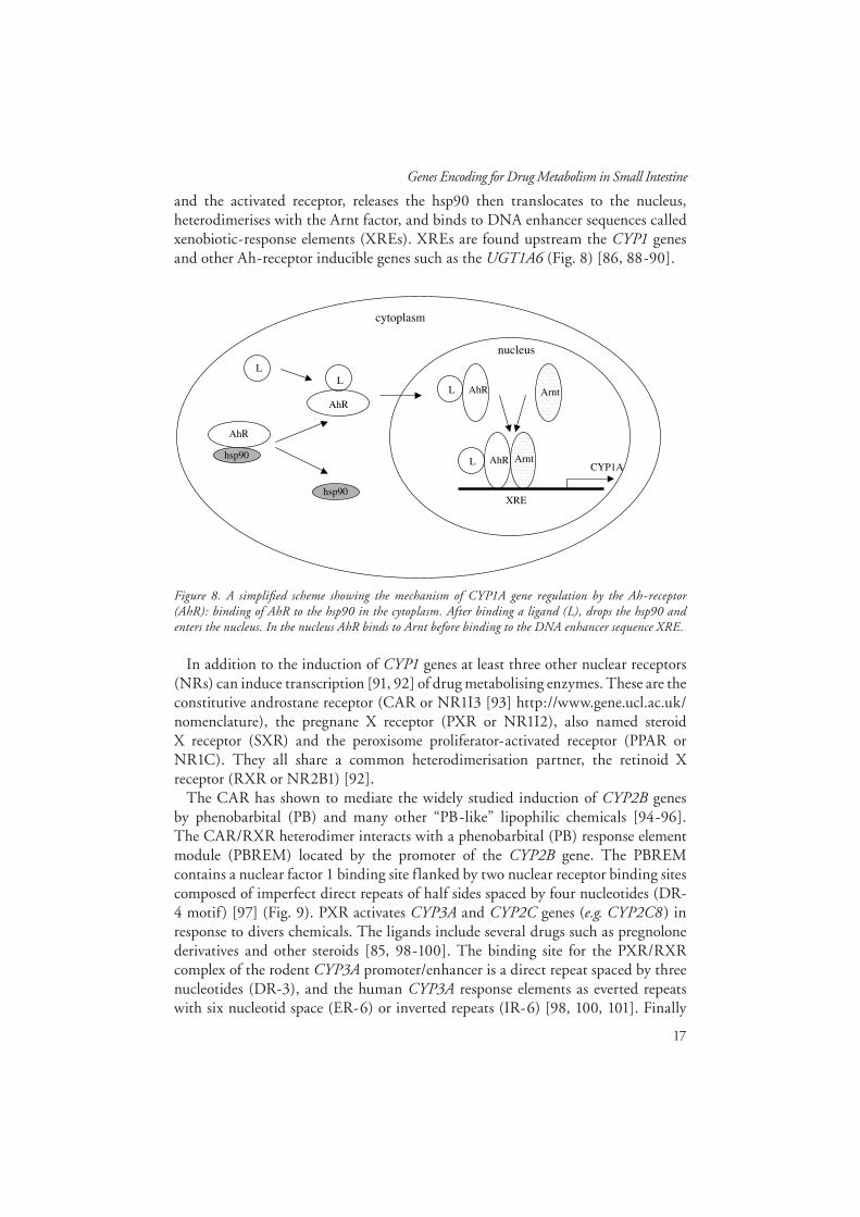
Many genes belonging to the CYP1-3, UGT1-2 and MDR families can be transcriptionally activated by foreign chemicals that will induce the gene expression through different receptor-dependent mechanisms. The most studied is the Aryl hydrocarbon receptor (Ah-receptor). This is a transcription factor that stimulates the expression of CYP1 and UGT1A6 genes [86, 87]. Without a ligand, the Ah-receptor is merged with hsp90, a chaperon protein, and present in the cytosol. Binding an aromatic hydrocarbon ligand in the cytosol activates the Ah-receptor
17
Genes Encoding for Drug Metabolism in Small Intestine
AhR
AhR
AhR
AhR
hsp90
hsp90
LL
L
L Arnt
ArntCYP1A
XRE
cytoplasm
nucleus
Figure 8. A simplifi ed scheme showing the mechanism of CYP1A gene regulation by the Ah-receptor (AhR): binding of AhR to the hsp90 in the cytoplasm. After binding a ligand (L), drops the hsp90 and enters the nucleus. In the nucleus AhR binds to Arnt before binding to the DNA enhancer sequence XRE.
In addition to the induction of CYP1 genes at least three other nuclear receptors (NRs) can induce transcription [91, 92] of drug metabolising enzymes. These are the constitutive androstane receptor (CAR or NR1I3 [93] http://www.gene.ucl.ac.uk/nomenclature), the pregnane X receptor (PXR or NR1I2), also named steroid X receptor (SXR) and the peroxisome proliferator-activated receptor (PPAR or NR1C). They all share a common heterodimerisation partner, the retinoid X receptor (RXR or NR2B1) [92].
The CAR has shown to mediate the widely studied induction of CYP2B genes by phenobarbital (PB) and many other “PB-like” lipophilic chemicals [94-96]. The CAR/RXR heterodimer interacts with a phenobarbital (PB) response element module (PBREM) located by the promoter of the CYP2B gene. The PBREM contains a nuclear factor 1 binding site flanked by two nuclear receptor binding sites composed of imperfect direct repeats of half sides spaced by four nucleotides (DR-4 motif) [97] (Fig. 9). PXR activates CYP3A and CYP2C genes (e.g. CYP2C8 ) in response to divers chemicals. The ligands include several drugs such as pregnolone derivatives and other steroids [85, 98-100]. The binding site for the PXR/RXR complex of the rodent CYP3A promoter/enhancer is a direct repeat spaced by three nucleotides (DR-3), and the human CYP3A response elements as everted repeats with six nucleotid space (ER-6) or inverted repeats (IR-6) [98, 100, 101]. Finally
and the activated receptor, releases the hsp90 then translocates to the nucleus, heterodimerises with the Arnt factor, and binds to DNA enhancer sequences called xenobiotic-response elements (XREs). XREs are found upstream the CYP1 genes and other Ah-receptor inducible genes such as the UGT1A6 (Fig. 8) [86, 88-90].

18
Monica Lindell
X,Y,Z
CAR
PXR
PPAR
CAR
PXR
PPAR
RXR
RXR
RXR
CYP2B
CYP3A
CYP4A
DR4
DR3
DR1
(AGGTCA - NX)2
ER6
cytoplasm
nucleus
X
Y
Z
PPARα, which mediates induction of fatty acid hydroxylases of the CYP4 family by many acidic chemicals, classified as “non-genotoxic” carcinogens and peroxisome proliferators [102, 103]. The CYP4 family could be a candidate for metabolism of fatty acid like drugs. The PPARα/RXR complex binds responsive elements (PPRE) in the promoter of target genes. A PPRE is a six nucleotide direct repeat separated by one nucleotide (DR-1) [104, 105].
Figure 9. A simplifi ed scheme of the role of nuclear receptors CAR, PXR, and PPAR in drug metabolism. Various drugs (X,Y,Z) are ligands to the receptors. These, drug activated the nuclear receptors form heterodimers with RXR and bind then to their respective enhancer elements on the promoter and activate transcription.
Post-transcriptional regulation
Post-transcriptional regulation mechanisms include both mRNA and protein stabilisation as well as stimulated protein degradation. These processes can result in an increase or decrease of enzyme expression [82].
A good example is CYP2E1 that is regulated by several post-transcriptional mechanisms. Both diabetes and fasting increases the expression by stabilising the mRNA and by an increase in gene transcription, respectively [106-108]. Insulin, on the other hand, decreases the mRNA levels of the CYP2E1 due to RNA-protein interaction mediating the destabilisation of the mRNA [109, 110].
Other CYP enzymes are both transcriptionally and post-transcriptionally regulated. The CYP1A2 mRNA levels are induced by PAH, a ligand to the Ah-receptor, but can also be post-transcriptionally regulated by enhancing the stability and the intracellular processing of the mRNA precursor [111-113].
The murine Cyp2a5 is also both transcriptionally and post-transcriptionally regulated [114-116]; transcriptionally by the transcription factor DBP (DNA binding protein) [117], whereas the post-transcriptional regulation is mediated

19
Genes Encoding for Drug Metabolism in Small Intestine
by stabilisation of the mRNA by binding of hnRNP1A (heterogeneous nuclear ribonucleo protein) to the 3’UTR (untranslated region) of the CYP2A5 mRNA [118, 119].
Regulation of CYP and UGT genes in the small intestine
As the gene regulation of drug metabolism is mainly studied in the liver, the different mechanisms presented apply basically for this organ only. However, it is established that also several intestinal CYPs are induced by xenobiotics [120-122]. The relevant NRs have also been found in the small intestine, but their role in the small intestinal gene regulation is not clear. [85, 123].
Artemisinin - an antimalarial drug
Malaria is an infectious disease that affects millions of people in the world. The high resistance to commonly used drugs has focused attention on the artemisinin group of compounds, now used in first-line treatment in several regions in Southeast Asia. Artemisinin (Fig. 10) is isolated from Artemisia annua L.; a plant with a long history of medical use against malaria in China [124, 125].
Figure 10. The molecular structure of antimalariae artemisinin (molecular weight 282 Da)
Despite the widespread use of artemisinin, information on its pharmacokinetics and metabolism is limited. Studies using rat liver microsomes indicate that artemisinin metabolism is primarily mediated by CYP2B6 with a minor contribution of CYP2A6 [126]. It has also been demonstrated that after multiple doses of artemisinin, the elimination of omeprazole is enhanced due to an increased capacity of CYP2C19 [127]. Artemisinin also exhibits an autoinduction of its elimination and this phenomenon is possibly due to an induction of CYP2B6 [128].
The evidence so far suggest that artemisinin induction may be similar to that of phenobarbital or other CAR activation compounds, however this has never been demonstrated, and, based on artemisinins structure this is not obvious (Fig.10).

20
Monica Lindell
OBJECTIVES OF THE PRESENT INVESTIGATION
Two overall questions have been addressed in this thesis:
A) How is the expression of drug metabolising CYPs, Pgp and UGTs in the small intestine? For that purpose we performed the following studies:
1. To investigate the mRNA expression of several major drug metabolising CYPs and Pgp in the small intestine and the liver of rats.
2. To analyse the expression of eight CYPs and Pgp in human small intestine; interindividual variability and regulation by internal and external factors.
3. Expression of seven UGTs in the human small intestine; interindividual variability and regulation, by endogenous and exogenous factors.
B) What are the mechanisms of induction of drug metabolising CYP enzymes by artemisinin?

21
Genes Encoding for Drug Metabolism in Small Intestine
EXPERIMENTAL PROCEDURES
Enterocytes from three segments of the rat small intestines, (i.e. duodenum, jejunum, and ileum) were collected and total RNA from both the enterocytes and the liver were purified. The human biopsies, from duodenum, were collected at Uppsala University Hospital, Sweden, in collaboration with Prof. L. Påhlman at the Gastro-intestinal ward. The site were the biopsies were taken showed no visible infection or inflammation. Total RNA was purified within two hours from that the biopsies were taken.
To study gene expression RT-PCR (Reverse transcriptace PCR) technique was chosen allowing us to detect very small amounts of mRNA. This is of importance when using human intestinal biopsies as source of mRNA. Biopsies do not give a large amount of mRNA, and therefore a sensitive method is necessary.
In the study on the human CYP genes the mRNA expression levels was compared to the expression of Pgp that was set on 100%. Pgp is found in many different tissues in high amounts. In the study on UGT expression the comparison was done to β-actin, an enzyme that is constitutively expressed in several tissues and therefore serves as a good internal standard for a technique called DRT-PCR (Duplex reverse transcriptase PCR). The mRNA expression of β-actin was first standardised so that the band intensity was equal for the different UGT amplifications.
The PCR amplification was visualised on an agarose gel stained with ethidiumbromide which visualise the bands under UV-light. The gel was photographed, and the photo was scanned and the band intensity was determined using the ‘NIH Image (1.62)’ program; a public domain image-processing and analysis program for Apple Macintosh (http://rsb.info.nih.gov/nih-image/). Details of the used methods are given in the respective papers.
The densitometric data were further statistically analysed to see if any correlation could be found between the expressions of the different CYP forms and UGT forms. We also investigated if any of the endogenous or exogenous factors were affecting the mRNA expression. A detailed description of the statistical analysis is given in papers II and III.
For the artemisinin studies three different techniques were used: In vivo experiments where male DBA/2J mice were given artemisinin for three days. After that the animal were killed and the livers removed and microsomes were prepared. The activity of CYP2A5, CYP2B10, and CYP1A2 was determined by measuring the coumarin 7-hydroxylation, pentoxyresorufin O-dealkylase and methoxyresorufin O-dealkylase activity, respectively.
Primary mouse hepatocytes were prepared and treated with three different concentrations of artemisinin and exposed for the drug for 16 – 40 hours. The cells

22
Monica Lindell
were then harvested and RNA was isolated and analysed with a Northern blot. The RNA was hybridised with radiolabeled cDNA from CYP2A5, CYP2B10, and CYP1A2.
Based on the result from these experiments we wanted to investigate whether nuclear receptor CAR was responsible for the induction of CYP2B10. HEK293 cells were transfected with a CAR; alternatively PXR, expression vectors and reporter gene, and the cells were exposed to artemisinin. The cells were lysed and the CAR-dependent luciferase and β-galactosidase activities were determined. The PXR vector responding reporter gene was used to see if artemisinin exhibited any receptor specificity and because PXR may cross-regulate CYP2B genes [129]. Further details are given in paper IV.

23
Genes Encoding for Drug Metabolism in Small Intestine
RESULTS AND DISCUSSION
mRNA expression of some important drug metabolising CYP genes and Pgp in the rat small intestine, (paper I)
More than 40 forms of CYP enzymes have been described in the rat liver [130]. By using different techniques such as the PCR and immunoblots, it has been established that several of these CYP forms are also expressed in the rat small intestine. Mainly the CYP1A1, CYP2B1, CYP2C, and CYP3A1 have been detected [28, 63, 77, 131]. By using both immuno-, and Northern blot analysis, it has been determined that the total CYP content of the small intestine is much lower than in the liver [131].
In this study we examined the mRNA expression levels of nine known genes encoding for drug metabolising CYP enzymes and Pgp in the three regions (duodenum, jejunum, and ileum) of the rat small intestine and compared the expression with that in the rat liver. We used Sprague-Dawley rats, one of the most used experimental models in studies of drug kinetics.
The intestinal expression of the different CYP genes and Pgp differed remarkably from the liver. The liver expression was strong for all tested CYP forms except the CYP1A1, which did not show any expression. Four of the tested CYPs, the CYP2B1, CYP2C6, CYP2C11, and CYP2D1 together with Pgp showed higher mRNA expression in the small intestine compared to the liver. On the contrary, CYP2A3, CYP3A1, CYP1A2, and CYP2E1 showed low or no expression in the small intestine. The CYP1A1 that was not expressed in the liver had a very strong expression in the small intestine. Earlier studies have found that the CYP1A1 is a strictly extrahepatic enzyme [111].
Studies on hepatic induction of the CYP1A1 have indicated that glucocorticoid receptors, in addition to the Ah receptor, may play a role in the CYP1A1 induction [132]. However, this receptor does not participate in the intestinal induction despite its presence in the enterocytes [133, 134].
One of the questions addressed was whether or not a gender difference existed in the mRNA levels, however, we could not find any such difference. All tested CYP forms and Pgp were expressed in both males and female at equal levels. There are earlier reports on sex-differences of CYP expression in the rat liver [63, 135, 136]. Yoshioka et al [135] found that there was a difference in protein expression between the genders in the rat liver, but when they performed a RNA blot analysis both genders displayed a hybridisation band of the same intensity. These and our results indicate therefore that mRNA could be transcribed in both sexes but that the maturation of the protein could to be an important step in sex-differentiation of some of the CYP enzymes. It is also possible that sex-dependent regulation is tissue specific. This has been seen in studies comparing the liver and the kidney of male and female mice [137]. In liver microsomes the expression of CYP2A4 is higher in

24
Monica Lindell
female mice compared to male, but the opposite is the case for the kidney [137]. It is therefore possible that CYP enzymes may show one gender pattern in the liver and a reversed or no gender difference in other tissues. In the case of Pgp one study has shown that the basal protein level was approximately 40% higher in female than in male rat liver [138].
Only CYP2A3 showed variation in mRNA expression along the length of the small intestine, the ileum seemed to lack expression whereas the other two sections had some. Previous studies have shown differential expression of CYPs such as the CYP1A1 and CYP2B1 between the segments of the intestine [28, 63]. When studying the localisation of Pgp, activity was found along the entire length of the small intestine, but with a higher activity in the ileum than in the duodenum [75]. In neither of these studies the mRNA expression was determined and are therefore not directly comparable to our study. However other studies have shown a correlation in mRNA expression and the enzyme activity in human liver for CYP1A1, CYP1A2, CYP3A4, CYP2D6, and CYP2B6 [139]. Whether or not similar correlation exists in the intestine is unclear.
A conclusion from this investigation is that the mRNA expression of both CYPs and Pgp is different in the rat small intestine as compared to the liver. Results from metabolic studies in the liver can therefore not be assumed to be the same in the GI-tract.
Interindividual variation of CYPs and Pgp in the human duodenum (paper II)
To study the CYP and Pgp expression in the human small intestine, biopsies from the duodenum were used. This choice was based on two criteria; (I) earlier studies have shown that the CYP content and activity are generally higher in the duodenum as compared to jejunum and ileum [28, 140]. (II) it is easier to get duodenal sample from patients going through gastroscopic examinations than from the other two sections of the human small intestine, which need more invasive operations.
The mRNA expression was studied using RT-PCR and the analysis was done by comparing expressions of CYP mRNA to the expressions of Pgp mRNA. Pgp is encoded by the MDR1 gene that is supposed to be expressed in the human small intestine at high levels [60].
An extensive interindividual variation was found in the expression of the eight CYP genes and the Pgp. Only two CYP forms, the CYP2C9 and CYP3A4, in addition to Pgp were found in all 51 subjects. The other forms were expressed at 82% (CYP1A1), 88% (CYP1A2) and 72% (CYP2A6). The frequency for the remaining three CYP forms was approximately 95%. In addition the level of expression varied extensively between individuals based on densitometric determination. In earlier studies, CYP3A4 was found in high amounts in the human liver and small intestine [28, 35]. Our study shows that the intestinal expression can vary dramatically between individuals which has not been reported before [28, 131, 141, 142].

25
Genes Encoding for Drug Metabolism in Small Intestine
CYP2C9 has been reported to be expressed in the human small intestine but only at low levels [27, 28]. In our study the CYP2C9 mRNA was expressed at a high average level and with the smallest interindividual variation among the CYP genes indicating that the CYP2Cs could be one of the major forms in the human intestine.
The expression of the different CYP forms and the Pgp were compared to each other with a help of a densitometric analysis. The results show that the correlation was in general low. Only three combinations exhibit a r> 0.5, with a high significance (p< 0,01) CYP2D6/CYP1A2, CYP2E1/CYP1A2, and CYP2E1/CYP2D6 (Table 1). This was not expected since these three genes are known to be differently regulated, at least in the liver [82, 143, 144]; CYP1A2 is regulated by the Ah-receptor having substances like PAH as substrates. But the enzyme is also posttranscriptionally regulated by RNA stabilisation [88, 111-113].
Table 1. CYPs with the best correlation coeffi cient (r >5) based on the densitometric analysis.
The CYP2D6 is involved in metabolism of a diversity of drugs [145, 6] and earlier studies indicated that CYP2D6 is not induced by xenobiotics. The co-ordinated transcriptional regulation of the enzyme in both extrahepatic and hepatic tissue should be the same, with no tissue specificity [147].
The CYP2E1 is showing a complex regulation pattern with several post-transcriptional regulatory mechanisms, for example different physiological conditions such as diabetes; that increases the expression by mRNA stabilisation or fasting, where the activation is transcriptionally activated [106-108].
The reason for this high correlation in the expression of the three CYP genes is not known but could be due to some transacting factors working in a tissue specific manner.
It has been speculated that Pgp and CYP3A4 may act synergistically in the small intestine as a major defence against xenobiotics [6]. A suggestion that is based on the fact that they have a broad overlapping substrate specificity [21, 64, 65]. Based on the rather poor correlation of their mRNA expression (r= 0.38) in our study, this does not seem to be the case. Rather, the results indicate that there must be differences in the regulation of these two genes. Both CYP3A4 and Pgp are known to be regulated by the nuclear receptor PXR (NR1I2) [83-85, 99-101, 148], but a differences in the protein-DNA interaction have been discovered: in CYP3A4 a XREM (xenobiotic-responsive enhancer module) has a DR3 or ER6 motif that mediate the induction whereas in Pgp it is a DR4 motif [149]. A second difference is that the PXR dependent induction of CYP3A4 is achieved in co-operation with
CYP1A2 CYP2D6
CYP2D6 0.74 p < 0.001
CYP2E1 0.55 p < 0.001 0.62 p < 0.01

26
Monica Lindell
a promoter element, which is missing in the MDR1 gene [83]. This could be the reason for their different regulation. Alternatively tissue specific factors may be involved explaining the variation in their expression.
The study does not support the hypothesis of a strict co-regulation of Pgp and CYP3A4 in the small intestine [65].
When comparing the expression of the different CYP forms to the demographic data we could see that CYP2A6 was affected by gender with a lower expression in females compared to male. CYP2E1 was affected by ethnic background with a higher expression among North Europeans compared to individuals from the Middle East. Among the external factors the intake of both acetylsalicylicacid (ASA) and omeprazole (Omep) increased the mRNA of some CYPs: ASA induced both CYP2A6 and CYP2E1, whereas omeprazole upregulated CYP3A4 (Table 2).
Table 2. Correlation between different CYPs and internal and external factors, based on densitometric analysis.
This study showed an extensive interindividual variation in the expression of drug metabolising CYP genes in the human small intestine. Accordingly it seems possible that any of the forms included in the study could be a major form in some individuals. The mRNA expression is also affected by the usage of common drugs/xenobiotics such as acetylsalicylic acid, omeprazole and smoking. The possibility that this large interindividual variation may lead to a varied bioavalibility of orally used drugs and complicate optimal drug therapy, should be carefully examined.
Expression of UGTs in the human duodenum (paper III)
The mRNA expression of seven UGT forms was analysed by DRT-PCR and densitometric quantification by comparing with the expression of β-actin set at 100% for each sample.
Interindividual variation in the expression of the different UGT forms was considerable. Only UGT1A1 was expressed in all 45 individuals. The UGT1A10 and UGT2B7 were found in 44/45 of the subjects. These three forms were also expressed at the highest average level based on the densitometric analysis. The lowest frequency was found for UGT2B15; present in 53% of the individuals which also was expressed at the lowest average levels when compared to the other forms (Table 3). Earlier studies have shown activity in most of the studied UGTs found
CYP2A6 CYP2E1 CYP3A4
Sex P = 0.0087
Ethnic background P= 0.0193
ASA P= 0.0118 P= 0.0235
Omep P= 0.0299

27
Genes Encoding for Drug Metabolism in Small Intestine
in the small intestine. [8, 150]. However, in these studies too few individuals were recruited so that reliable conclusions can been drawn from them. In particular on the interindividual variability.
Table 3. A summery of the densitometric analysis on the seven different UDP-glucuronosyltransferases is given. The mean values are based on the densitometric analysis relative units.
Table 4. UGTs with the best correlation coeffi cient (r >5) based on the densitometric analysis.
%UGT1A1 %UGT1A3 %UGT1A4 %UGT1A6 %UGT1A10 %UGT2B7 %UGT2B15
mean 265 60 63 41 131 288 46
CV 40% 64% 49% 48% 64% 73% 61%
No normal
distribution
No normal
distribution.
No normal
distribution
No normal
distribution
UGT1A1 UGT1A3 UGT1A10
UGT1A1
UGT1A3
UGT1A10 0.55 P< 0.001 0.52 P< 0.001
UGT2B7 0.61 P< 0.001 0.58 P< 0.001
UGT2B15 0.64 P< 0.01
The expression levels of the studied UGTs were compared to each other, several of which seemed to correlate well, with a r> 0.5 and a high significance: UGT1A1/UGT1A10, UGT1A1/UGT2B7, UGT1A3/UGT1A10 UGT1A3/UGT2B15, and UGT1A10/UGT2B7 (Table 4). This rather high correlation could indicate that some common regulation mechanisms are involved. On the other hand the scores (r= 0.5 – 0.6), also suggest some differences in the regulation. The overlapping substrate specificity of some of the UGT forms [8, 55, 89, 151] would speak for a common regulation. Both UGT1 and UGT2 genes are known to exhibit tissue specific regulation; for example, UGT1A10 is only found in extrahepatic tissues such as the small intestine and the biliary tract [55, 152]. Little is known about the regulation of human UGTs, as most of the studies so far have used either hepatocytes or transfected cells It has been established however, that the UGT1A1 expression is regulated by the CAR receptor [153], and the UGT2B7 expression is PXR mediated. What factors mediate the UGT2B15 expression is not yet determined but at least the PXR seems not to be involved [52]. The only studies on regulation that has been done on extrahepatic tissues is on UGT1A6 showing a CAR dependent expression in Caco 2-cells [86].

28
Monica Lindell
None of the endogenous factors included in this study: age, sex, and ethnic background affected the expression of the UGTs. Earlier reports have shown sex differences in drug metabolism by UGTs, however these studies focused on in vivo glucuronidation by measuring plasma levels or the urine excretion after an oral dose of the different test drugs [154-157]. Therefore, these studies are not comparable to the present as they measure the systemic activity of in vivo.
Only UGT1A4 was significantly increased by the intake of omeprazole and by smoking (Table 5). To our knowledge this is the first report on omeprazole impact on any UGT form.
Table 5. Relationship between UGT1A4 and external factors.
Which UGT enzyme(s) are involved in nicotine metabolism is not yet known [58]. However, it has been shown that smoking upregulates glucuronidation reactions catalysed by UGT1A6 [87, 159]; but no reports has shown affects on UGT1A4.
This study revealed a large interindividual variation in the mRNA expression of seven UGT forms of the human intestine. The major forms seem to be UGT1A1, UGT1A10 and UGT2B7; they are expressed in most individuals and also at a high average levels.
The expression of these three forms also appeared to correlate with each other. It could therefore be that these three forms together form a major glucuronidation machinery of the intestine. A possibility that should be examined in detail.
The expression of one form; the UGT1A4 is affected by smoking and by usage of omeprazole. This could be of importance for drug-drug interactions at least for some individuals.
Induction of drug metabolism by Artemisinin (paper IV)
Artemisinin is used orally in multidrug treatment of malaria. It has been suggested to induce drug metabolism and therefore be involved in drug-drug interactions.
Earlier experiments, by us and others [128], have indicated that artemisinin can affect CYP1A2, CYP2A, and CYP2B in rodents. Indirect evidence also indicated that artemisinin may act like phenobarbital and other CAR activating compounds in the induction of drug metabolism, but this has never been confirmed. In humans the drug is primarily metabolised by CYP2B6 with some contributions by CYP3A4 and CYP2A6 [127]. In this paper we characterised the inductive properties of artemisinin on CYPs by using three different techniques: in vivo, primary hepatocytes in culture and recombinant luc-reproter genes with a CAR or PXR driven promoters
UGT1A4 Omeprazole p = 0.003
Smoking p = 0.029

29
Genes Encoding for Drug Metabolism in Small Intestine
In vivo experiments (Table 6) showed that the drug increased the activity of CYP2B10 and to a lesser extend that of the CYP2A5 of the mouse liver. Hardly any effect was seen on CYP1A2 activity.
Table 6. Effect of artemisinin on the three CYP catalysed enzyme activities of mouse liver microsmes. Activities are given as pmol/min x mg protein. Fold of increased activity is given in parenthesis.
In cultured primary hepatocytes a strong effect was seen on the mRNA expression of CYP2B10 and a weak effect on CYP2A5 upon exposure to artemisinin. The CYP1A2 mRNA expression was not affected. (Fig. 11).
CYP dependant activity Control Artemisinin treated
CYP2B10 52 + 5 390 + 42 P≥ 0.001
(7.5)
CYP2A5 135 + 15 365 + 30 P≥ 0.05
(2.7)
CYP1A2 480 + 60 510 + 70
(1.06)
Artemisinin, µM 0 5 10
CYP2B10
CYP2A5
CYP1A2
Figure 11. Northern blot analysis of total RNA from mouse primary hepatocytes exposed to artemisinin at three different concentrations (from paper IV).
Based on the results it appeared that artemisinin mainly upregulates the CYP2B enzymes at a pretranslational level.
HEK293 cells were transfected with mouse and human CMX-GAL4-CAR or CMX-GAL4-PXR constructs and treated with artemisinin. The drug showed a dose dependent increase in both mouse and human CAR-mediated expression, with a slightly higher activation with the mouse construct. Neither mouse nor human

30
Monica Lindell
PXR driven promoters were affected by artemisinin. (Fig. 12). This results provide evidence that artemisinin acts as an inducer via the CAR receptor, thus confirming the earlier indirect evidence.
Figure 12. Effects of artemisinin on the expression of CAR or PXR driven luciferase recombinants in HEK293 cells (paper IV).
The study shows that artemisinin induces hepatic CYP2B, at a pretransitional level by a mechanism involving the nuclear receptor CAR. The weaker effect, seen on the CYP2A5 expression, may involve a different regulation mechanism as compared to the CYP2B where artemisinin toxicity may play a role.
31
Genes Encoding for Drug Metabolism in Small Intestine
GENERAL SUMMARY
This work shows extensive interindividual variations in the intestinal expression of several CYP and UGT genes encoding for drug metabolising enzymes in humans. Each individual appears to have his or her unique CYP and UGT profile or fingerprint. Due to effects of external factors, the profile may vary over time.
It was shown that the genes expressed at the highest levels in a human population are the CYP2C, CYP2D6, and CYP3A4 and the UGT1A1, UGT1A10, and UGT2B7. Due to the extensive variability, any of the genes included in the study may have a major role in intestinal drug metabolism for a given individual.
Expression levels of the different genes were determined by analysing concentrations of corresponding mRNAs in the human specimen. It was not established how well the mRNA levels reflect the corresponding protein or enzyme activity levels. However, the results should help designing functional studies in search for the key catalysts of drug metabolism in the human intestine.
Some common external factors such as smoking, and clinical drugs omeprazole and acetylsalicylic acid affected the expression of some of the CYP and UGT genes. This may lead to drug-drug interactions affecting drug absorption and metabolism in the intestine of exposed individuals; a possibility that should be studied in more detail.
The study also established that artemisinin, a widely used antimalarial, affects the expression of drug metabolising enzymes mainly via the CAR receptor with minor effects possibly due to its toxicity. This result should help predicting and avoiding drug-drug interaction in therapies where artemisinin is used.

32
Monica Lindell
ACKNOWLEDGEMENTS
Many people at the Department of Pharmaceutical Bioscience, Division of Biochemistry, Faculty of Pharmacy, Uppsala University have contributed in different ways for the conclusion of this thesis. In particular I would like to express my sincere gratitude to:
My supervisors Prof. Hans Lennernäs and Prof. Matti Lang for once accepting me at the department and for all the support and encouragement through all those years
My former room mates Dr Zufan Araya and Dr Fardin Hoseinpour for all our nice discussions about everything and nothing.
Present members at the division: Malin Söderberg, Kyle Christian, Wanjin Tang, Angela Lannerbro, Dr. Kerstin Lundell, Dr. Tina Glisovic, Dr Maria Norlin, Dr Ronnie Hansson, Prof. Kjell Wikvall, Dr Francoise Raffalli-Mathieu and former members: Marianne Åström, Britt-Marie Johansson and Kerstin Rönnqvist it is all your effort that the environmen in the lab and in the ‘fika’ room is a place were the discussion easy can jump from this years ice-cream to the seven deadly sins.
The technical duo: Hans Darberg and Magnus Jansson, with out your help not many machines and computer would work as splendid as they do.
The staff and patients at the Gasto-intestinal ward at the University Hospital here in Uppsala with out your effort this thesis would never have been done.
To my friends out side the department who helped me to keep a distance to science.
Etha, Anne-Sofie, and Karin for our, nearly manic, trips to garden centre to expand my collection in pelargoniums and to your contribution to the now overgrown miniature ‘garden’ I have. Here I also have to put in Mark and Anders who in some strange way has been able to coop with this obsession of our.
Ulrika, Cristina, Lasse, Elise and Graham, with whom I have, more than once taken a ‘Rocket to the Moon’ for the ‘Search of Angels’ but found that ‘Place under the sun were hearts of olden glory grow young’. I hope there will be more opportunities for us to go ‘Back to the Stamping Ground’.

33
Genes Encoding for Drug Metabolism in Small Intestine
And finally my family: Lars Erik, Reija, Björn and Nils, you have all been a part in this work and also to change my conception on were it is possible to drive or not.
And my parents, Marianne and Gunnar for their encouragement and support.
Monica 15/9

34
Monica Lindell
REFERENCES
1. Woodhouse K.W., and James O.F.W.; Hepatic Drug Metabolism and Ageing (1990) Br. Medical Bulletin 46: 22-35
2. Burchell B., Nebert D.W., Nelson D.R., Bock K.W., Iyanagi T., Jansen P.L.M., Lancet D., Mulder G.J., Chowdhury J.R., Siest G., Tephly T.R., Mackenzie P.I.; The UDP Glucuronosyltransferase Gene Superfamily: Suggested Nomenclature Based on Evolutionary Divergence. (1991) DNA Cell Biol. 10: 487-494
3. Gibson G.G., and Skett P.; Introduction to Drug Metabolism 2nd Edition (1994) Blackie Academic & Professional, London, UK
4. Watkins P.B.; The Barrier Function of CYP3A4 and P-glycoprotein in the Small Bowel. (1997) Adv. Drug Delivery Reveiw 27: 161-170
5. Ilett K.F., Tee L.B.G., Reeves P.T., and Minchin R.F.; Metabolism of Drugs and Other Xenobiotics in the Gut Lumen and Wall. (1990) Pharmac. The. 46: 67-93
6. Hunter J., and Hirst B.H.; Intestinal Secretion of Drugs. The Role of P-glycoprotein and Related Drug Effl ux Systems in Limiting Oral Drug Absorption. (1997) Adv. Drug Delivery Reviews 25: 129-157
7. Lin J.H., Chiba M., and Baillie T.A.; Is the Role of the Small Intestine in First-pass Metabolism Overemphasized? Pharmacol. Reveiw (1999) 51: 135-157
8. Strassburg C.P., Kneip S., Topp J., Obermayer-Straub P., Barut A., Tukey R.H., and Manns M.P.; Polymorphic Gene Regulation and Interindividual Variation of UDP-glucuronosyltransferase Activity in Human Small Intestine. J. Biol. Chem. (2000) 275: 36164-36171
9. Weisbrodt N.W.; Motility of the small intestine. (1987) Physiology of the Gastrointestinal Tract, 2nd Edition. 631-663 Leonard R. Johnson ed., Raven Press, New York; USA
10. Guyton A.C.; Transport and Mixing of Food in the Alimentary Tract. . (1991) Textbook of Medical Physiology 8thEdition 698-708 W.B. Saunders, Philadelphia, USA
11. Thomson A.B.R.; Relationship Between Physiology and Clinical Disorder of the Small Intestine. (1988) Principles and Practice of Gastroenterology and Hepatology 281-289 Elsevier, Amsterdam
12. Hole J.W., and Koos K.A.; The Digestive System. (1991) Human Anatomy 384-387, 405-410 Wm.C. Brown Publishers, Dubuque IA, USA
13. Doherty M.M., and Charman W.N.; The Mucosa of the Small Intestine. How Clinically Relevant as an Organ of Drug Metabolism? (2002) Clin. Pharmacokinet 41: 235-253

35
Genes Encoding for Drug Metabolism in Small Intestine
14. Thummel K.E., Kunze K.L., and Shen D.D.; Enzyme-catalyzed Processes of First-pass Hepatic and Intestinal Drug Extraction. (1997) Adv. Drug Delivery Reveiws 27: 99-127
15. Caldwell J., and Varwell Marsh M.; Metabolism of Drugs by the Gastrointestinal Tract. (1982) Clinical Pharmacology and Therapeutics: Presystemic Drug Elimination 29-42 George D.F., Shand D.E., and Rernwick A.G. eds. Butterworths, London
16. Renwick A.G., and George C.F.; Metabolism of Xenobiotics in Gastrointestinal Tract. (1989) Intermediary Xenobiotic Metabolism in Animals: Methology, Mechanisms and Signifi cance 13-40 Taylor and Francis, London, UK
17. Krishna D.R., and Klotz U.; Extrahepatic Metabolism of Drugs in Humans. (1994) Clin. Pharmacokinetic Concepts 26: 144-160
18. Hebert M.F.; Contributions of Hepatic and Intestinal Metabolism and P-glycoprotein to Cyclosporine and Tacrolimus Oral Drug Delivery. (1997) Adv. Drug Delivery Reviews 27: 201-214
19. Barthe L., Woodley J., and Houin G.; Gastrointestinal Absorption of Drugs: Methods and Studies. (1999) Fundam. Clin. Pharmacol. 13: 134-168
20. Lennernäs H.; Does Fluid Flow Across the Intestinal Mucosa Affect Quantitative Oral Drug Absorption? Is It Time for a Reevaluation? (1995) Pharm. Res. 12: 1573-1582
21. Benet L.Z., Wu C-Y., Hebert M.F., and Wacher V.J.; Intestinal Drug Metabolism and Antitransport Processes: A Potential Paradigm shift in Oral Drug Delivery. (1996) J. Controlled Release 39: 139-143
22. Nelson D.R., Koymans L., Kamataki T., Stegeman J.J., Feyereisen R., Waxman D.J., Waterman M.R., Gotoh O., Coon M.J., Estabrook R.W., Gunsalus I.C., and Nebert D.W.; P450 Superfamily: Update on New Sequences, Gene Mapping, Accession Numbers and Nomenclature. (1996) Pharmacogenetics 6: 1-42
23. Zhang Y., and Benet L.Z.; The Gut as a Barrier to Drug Absorption. Combined Role of Cytochrome P450 3A and P-glycoprotein. (2001) Clin. Pharmacokinet 40: 159-168
24. Zevin S. and Benowitz N.L.; Drug Interactions with Tobacco Smoking: An Update. (1999) Clin. Pharmacokinet. 36: 425-438
25. Koide A., Fuwa K., Furukawa F., Hirose M., Nishikawa A., and Mori Y.; Effect of Cigarette Smoke on the Mutagenic Activation of Environmental Carcinogens by Rodent Liver. (1999) Mutation Research 428: 165-176
26. Fontana R.J., Lown K.S., Pain M.F., Fortlage L., Santella R.M., Felton J.S., Knize M.G., Greenberg A., and Watkins P.B.; Effects of a Chargrilled Meat Diet on Expression of CYP3A, CYP1A, and P-glycoprotein Levels in Healthy Volunteers. (1999) Gastroenterology 117: 89-98

36
Monica Lindell
27. Zhang Q-Y., Dunbar D., Ostrowska A., Zeisloft S., Yang J., and Kaminsky L.S.; Characterization of Human Small Intestinal Cytochromes P-450. (1999) Drug Metab. Disp. 27: 804-809
28. deWaziers I., Cugnenc P.H., Yang C.S., Leroux J-P., and Beaune P.H.; Cytochrome P450 Isoenzymes, Epoxide Hydrolase and Gluthathione Transferases in Rat and Human Hepatic and Extrahepatic Tissues. (1990) J. Pharmacol. Exp. Ther. 253: 387-394
29. Madani S., Paine M.F., Lewis L., Thummel K.E., and Shen D.D.; Comparison of CYP2D6 Content and Metoprolol Oxidation Between Microsomes Isolated from Human Livers and Small Intestines. (1999) Pharm. Rev. 16: 1199-1205
30. Rendic S., and Di Carlo F.J.; Human Cytochrome P450 Enzymes: A Status Report Summarizing their Reactions, Substrates, Inducers, and Inhibitors. (1997) Drug Metab. Reviews 29: 413-580
31. Wrighton S.A., Thomas P.E., Molowa D.T., Haniu, M., Shively J.E., Maines S.L., Watkins P.B., Parker G., Mendez-Picon G., Levin W., and Guzelian P.S.; Characterization of Ethanol-inducible Human Liver N- Nitrosdimethylamine Demethylase. (1986) Biochemistry 25: 6731-6735
32. Watkins P.B.; Role of Cytochromes P450 in Drug Metabolism and Hepatotoxicity. (1990) Sem. Liver Dis. 10: 235-250
33. Wrighton S.A., Schuetz E.G., Watkins P.B., Maurel P., Barwick J., Bailey B.S., Hartle H.T., Young B., and Gunzelian P.; Demonstration in Multiple Species of Inducible Hepatic Cytochromes P-450 and Their mRNAs Related to the Glucocorticoid-inducible Cytochrome P-450 in the Rat. (1985) Mol. Pharmacol .28: 312-321
34. Watkins P.B., Murray S.A., Winkelman L.G., Heuman D.M., Wrighton S.A., and Guzelian P.S.; Erythromycin Breath Test as an Assay of Glucocorticoid-inducible Liver Cytochromes P-450. (1989) J. Clin. Invest. 83: 688-697
35. Kolars J.C., Lown K.S., Schmiedlin-Ren P., Ghosh M., Fang C., Wrighton S.A., Merion R.M., and Watkins P.B.; CYP3A Gene Expression in Human Gut Epithelium. (1994) Pharmacogenetics 4: 247-259
36. Kronbach T., Fischer V., and Meyer U.A.; Cyclosporine Metabolism in Human Liver: Identifi cation of a Cytochrome P-450III Gene Family as the Major Cyclosporine-metabolizing Enzyme Explains Interactions of Cyclosporine with Other Drugs. (1988) Clin. Pharmacol. Ther. 43: 630-635
37. Combalbert J, Fabre I., Fabre G., Dalet I., Derancourt J., Cano J.P., and Maurel P.; Metabolism of Cyclosporine A. IV. Purifi cation and Identifi cation of the Rifampicin-inducible Human Liver Cytochrome P-450 (Cyclosporine A Oxidase) as a Product of P450IIIA Gene Subfamily. (1989) Drug Metab. Dispos. 17: 197-207
38. Kolars J.C., Schmiedlin-Ren P., Schuetz J.D., Fang C., and Watkins P.B.; Identifi cation of Rifampin-inducible P450IIIA4 (CYP3A4) in Human Small Bowel Enterocytes (1992) J. Clin. Invest. 90: 1871-1878

37
Genes Encoding for Drug Metabolism in Small Intestine
39. Gonzalez F.J., Schmid B.J., Umeno M., McBride O.W., Hardwick J.P., Meyer U.A., Gelboin H.V., and Idle J.R.; Human P450PCN1: Sequence, Chromosome Localization, and Direct Evidence Through cDNA Expression That P450PCN1 is Nifedipine Oxidase. (1988) DNA 7: 79-86
40. Guengerich F.P., Martin M.V., Beaune P.H., Kremers P., Wolff T., and Waxman D.J.; Characterization of Rat and Human Liver Microsomal Cytochrome P-450 Forms Involved in Nifedipine Oxidation, a Prototype for Genetic Polymorphism in Oxidative Drug Metabolism (1986) J. Biol. Chem. 261: 5051-5060
41. Shimada T., and Guengerich F.P.; Evidence for Cytochrome P-450NF
, the Nifedipine Oxidase, Being the Principal Enzyme Involved in the Bioactivation of Afl atoxins in Human Liver. (1989) Proc. Natl. Acad. Sci. USA 86: 462-465
42. van Ommen B., Hendriks W., Bessems J.G.M., Geesink G., Müller F., and van Bladeren P.J.; The Relation Between the Oxidative Biotransformation of Hexachlorobezene and its Porphyrinogetic Activity (1989) Toxicol. Appl. Pharmacol. 100: 517-528
43. Durnas C., Loi C-M., and Cusack B.J.; Hepatic Drug Metabolism and Aging. (1990) Clin. Pharmacokinet. 19: 359-389
44. Prueksaritanont, T., Gorham L.M., Hochman J.H., Tran L.O., and Vyas K.P.; Comparative Studies of Drug-metabolizing Enzymes in Dog, Monkey, and Human Small Intestines, and in Caco-2 Cells. (1996) Drug Metab. Disp. 24: 634-642
45. Pacifi ci G.M., Franchi M., Bencini C., Repetti F., Di Lascio N., and Muraro G.B.; Tissue Distribution of Drug-metabolizing Enzymes in Humans. (1988) Xenobiotica 18: 849-856
46. Cappiello M., Giuliani L., and Pacifi ci G.M.; Distribution of UDP-glucuronosyltransferase and its Endogenous Substrate Uridine 5’-diphosphoglucuronic Acid in Human Tissues. (1991) Eur. J. Clin. Pharmacol. 41: 345-350
47. Cheng Z., Radominska-Pandya A., and Tephly T.R.; Studies on the Substrate Specifi city of Human Intestinal UDP-glucuronosyltransferases 1A8 and 1A10. (1999) Drug Metab. Disp. 27: 1165-1170
48. Mackenzie P.I., Owens I.S., Burchell B., Bock K.W., Bairoch A., Bélanger A., Fournel-Gigleux S., Green M., Hum D.W., Iyanagi T., Lancet D., Louisot P., Magdalou J., Chowdhury J.R., Ritter J.K., Schachter H., Tephly T.R., Tipton K.F., and Nebert D.W.; The UDP-glycosyltransferase Gene Superfamily: Recomended Nomenclature Update Based on Evolutionary Divergence. (1997) Pharmacogenetics 7: 255-269
49. Tukey R.H., and Strassburg C.P.; Human UDP-glucuronosyltransferases: Metabolism, Expression, and Disease. (2000) Annu. Rev. Pharmacol. Toxicol. 40: 581-616

38
Monica Lindell
50. Ritter J.K., Chen F., Sheen Y.Y., Tran H.M., Kimura S., Yeatman M.T., and Owens I.S.; A Novel Complex Locus UGT1 Encodes Human Bilirubin, Phenol, and Other UDP-glucuronosyltransferase Isozymes with Identical Carboxyl Termini. (1992) J. Biol. Chem. 267: 3257-3261
51. Radominska-Pandya A., Czernik P.J., and Little J.M.; Structural and Functional Studies of UDP-glucuronosyltransferases. (1999) Drug Metab. Reviews 31: 817-899
52. Turgeon D., Carrier J-S., Lévesque É, Beatty B.G., Bélanger A., and Hum D.W.; Isolation and Characterization of the Human UGT2B15 Gene, Localized Within a Cluster of UGT2B Genes and Pseudogenes on Chromosome 4. (2000) J. Mol. Biol. 295: 489-504
53. Mackenzie P.I., and Rodbourn L.; Organization of the Rat UDP-glucuronosyltransferase, UDPGTr-2, Gene and Characterization of Its Promoter. (1990) J. Biol. Chem. 265: 11328-11332
54. Haque S.J., Petersen D.D., Nebert D.W., and Mackenzie P.I.; Isolaion, Sequence, and Developmental Expression of Rat UGT2B2: The Gene Encoding a Constitutive UDP Glucuronosyltransferase That Metabolizes Etiocholanolone and Androsterone. (1991) DNA Cell Biol. 10: 515-524
55. Tukey R.H., and Strassburg C.P.; Genetic Multiplicity of the Human UDP-glucuronosyltransferases and Regulation in the Gastrointestinal Tract. (2001) Mol. Pharmacol. 59: 405-414
56. Gottesman M.M., and Pastan I.; Biochemistry of Multidrug Resistance Mediated by the Multidrug Transporter. (1993) Annu. Rev. Biochem. 62: 385-427
57. Fardel O., Lecureur V., Daval S., Corlu A., and Guillouzo A.; Up-regulation of P-glycoprotein Expression in Rat Liver Cells by Acute Doxorubicin Treatment. (1997) Eur. J. Biochem. 246: 186-192
58. Borst P.; Genetic Mechanisms of Drug Resistance. A Review. (1991) Reviews in Oncology 4: 87-105
59. Thiebaut F., Tsuruo T., Hamada H., Gottesman M.M., Pastan I., and Willingham M.C.; Cellular Localization of the Multidrug-resistance Gene Product P-glycoprotein in Normal Human Tissues. (1987) Proc. Natl. Acad. Sci. USA 84: 7735-7738
60. Cordon-Cardo C., O’Brien J.P., Boccia J., Casals D., Bertino J.R., and Melamed M.R.; Expression of the Multidrug Resistance Gene Product (P-glycoprotein) in Human Normal and Tumor Tissues. (1990) J. Histochem. Cytochem. 38: 1277-1287
61. Fojo A.T., Ueda K., Slamon D.J., Poplack D.G., Gottesman M.M. and Pastan I.; Expression of a Multidrug-resistance Gene in Human Tumors and Tissues. (1987) Proc. Natl. Acad. Sci. USA 84: 265-269

39
Genes Encoding for Drug Metabolism in Small Intestine
62. Fricker G., Drewe J., Huwyler J., Gutmann H., and Berlinger C.; Relevance of P-glycoprotein for the Enteral Absorption of Cyclosporin A: in vitro – in vivo Correlation (1996) Br. J. Pharmacol. 118: 1841-1847
63. Kolars J.C., Schmiedlin-Ren P., Dobbins III W.O., Schuetz J., Wrighton S.A., and Watkins P.B.; Heterogeneity of Cytochrome P450IIIA Expression in Rat Gut Epithelia. (1992) Gastroenterology 102: 1186-1198
64. Schuetz, E.G., Furuya K.N., and Schuetz J.D.; Interindividual Variation in Expression of P-glycoprotein in Normal Human Liver and Secondary Hepatic Neoplasms. (1995) J. Pharmaco. Exp. Ther. 275: 1011-1018
65. Wacher V.J., Wu C-Y., and Benet L.Z.; Overlapping Substrate Specifi cities and Tissue Distribution of Cytochrome P450 3A and P-glycoprotein Implications for Drug Delivery and Activity in Cancer Chemotherapy. (1995) Mol. Carcinog. 13: 129-134
66. Hofsli E., and Nissen-Meyer J.; Reversal of Drug Resistance by Erythromycin: Erythromycin Increases the Accumulation of Actinomycin D and Doxorubicin in Multidrug-Resistant Cells. (1989) Int. J. Cancer 44: 149-154
67. Gupta S., Kim J., and Gollapudi S.; Reversal of Daunorubicin Resistance in P388/ADR Cells by Itraconazol. (1991) J. Clin. Invest. 87: 1467-1469
68. Siegsmund M.J., Cardarelli C., Aksentijevich I., Sugimoto Y., Pastan I., and Gottesman M.M.; Ketoconazole Effectively Reverses Multidrug Resistance in Highly Resistant KB Cells. (1994) J. Urology 151: 485-491
69. Schuetz E.G., Beck W.T., and Schuetz J.D.; Modulators and Substrates of P-glycoprotein and Cytochrome P4503A Coordinately Up-regulate these Proteins in Human Colon Carcinoma Cells. (1996) Mol. Pharmacol. 49: 311-318
70. Pichard L., Fabre I., Daujat M., Domergue J., Joyeux H., and Maurel P.; Effect of Corticosteroids on the Expression of Cytochromes P450 and on Cyclosporin A Oxidase Activity in Primary Cultures of Human Hepatocytes. (1992) Mol. Pharmacol. 41: 1047-1055
71. Fardel O., Lecureur V., and Guillouzo A.; Regulation by Dexamethasone of P-glycoprotein Expression in Cultured Rat Hepatocytes. (1993) FEBS 327: 189-193
72. Zhao J-Y., Ikeguchi M., Eckersberg T., and Kui M.T.; Modulation of Multidrug Resistence Gene Expression by Dexamethasone in Cultured Hepatoma Cells. (1993) Endocrinology 133: 521-528
73. Lown K.S., Mayo R.R., Leichtman A.B., Hsiao H-L., Turgeon D.K., Schmiedlin-Ren P., Brown M.B., Guo W., Rossi S.J., Benet L.Z., and Watkins P.B.; Role of Intestinal P-glycoprotein (mdr1) in Interpatient Variation in the Oral Bioavailability of Cyclosorine. (1997) Clin. PharmacoL. Ther. 62: 248-260
74. Vickers A.E.M., Alegert M., Meyer E., Smiley S., and Guertler J.; Hydroxyethyl Cyclosporine A Induces and Decreases P4503A and P-glycoprotein Levels in Rat Liver. (1996) Xenobiotica 26: 27-39

40
Monica Lindell
75. Makhey V.D., Guo A., Norris D.A., Hu P., Yan J., and Sinko P.J.; Characterization of the Regional Intestinal Kinetics of Drug Effl ux in Rat and Human Intestine and in Caco-2 Cells. (1998) Pharm. Res. 15: 1160-1167
76. Obach R.S., Zhang Q-Y., Dunbar D., and Kaminsky L.S.; Metabolic Characterization of the Major Human Small Intestinal Cytochrome P450s. (2001) Drug Metab. Disp. 29: 347-352
77. Zhang Q-Y., Wikoff J., Dunbar D., and Kaminsky L.; Characterization of Rat Small Intestinal Cytochrome P450 Composition and Inducibility. (1996) Drug Metab. Disp. 24: 322-328
78. Kaminsky L.S., and Fasco M.J.; Small Intestinal Cytochromes P450. (1992) Crit. Review Toxicol. 21: 407-422
79. Münzel P.A., Bookjans G., Mehner G., Lehmköster T., and Bock K.W.; Tissue-specifi c 2,3,7,8-Tetrachlorodibenzo-p-dioxin-Inducible Expression of Human UDP-glucuronosyltransferase UGT1A6. (1996) Arch. Biochem. Biophys. 335: 205-210
80. Mojarrabi B., Butler R., and Mackenzie P.I.; cDNA Cloning and Characterization of the Human UDP-glucuronosyltransferase, UGT1A3. (1996) Biochem. Biophys. Res. Commun. 225: 785-790
81. Mojarrabi B., and Mackenzie P.I.; The Humna UDP Glucuronosyltransferase, UGT1A10, Glucuronidates Mycophenolic Acid. (1997) Biochem. Biophys. Res. Commun. 238: 775-778
82. Porter T.D., and Coon M.J.; Cytochrome P-450: Multiplicity of Isoforms, Substrates, and Catalytic and Regulatory Mechanisms. (1991) J. Biol. Chem. 266: 13469-13472
83. Geick A., Eichelbaum M., and Burk O.; Nuclear Receptor Respone Elements Mediate Induction of Intestinal MDR1 by Rifampin. (2001) J. Biol. Chem. 276: 14581-14587
84. Kast H.R., Goodwin B., Tarr P.T., Jones S.A., Anisfeld A.M., Stoltz C.M., Tontonoz P., Kliewer S., Willson T.M., and Edwards P.A.; Regulation of Multidrug Resistance-associated Protein 2 (ABCC2) by the Nuclear Receptors Pregnane X Receptor, Farnesoid X-activated Receptor, and Constitutive Androstane Receptor. (2002) J. Biol. Chem. 277: 2908-2915
85. Synold T.W., Dussault I., and Forman B.M.; The Orphan Nuclear Receptor SXR Coordinately Regulates Drug Metabolism and Effl ux. (2001) Nature Medicine 7: 584-590
86. Münzel P.A., Lehmköster T., Brück M., Ritter J.K., and Bock K.W.; Aryl Hydrocarbon Receptor-inducible or Constitutive Expression of Human UDP Glucuronosyltransferase UGT1A6. (1998) Arch. Biochem. Biophys. 350: 72-78
87. Whitlock J.P.; Induction of Cytochrome P4501A1. (1999) Annu. Rev. Pharmacol. Toxicol. 39: 103-125

41
Genes Encoding for Drug Metabolism in Small Intestine
88. Wilhelmsson A., Cuthill S., Denis M., Wikström A-C., Gustafsson J-Å., and Poellinger L.; The Specifi c DNA Binding Activity of the Dioxin Receptor is Modulated by the 90 kd Heat Shoch Protein. (1990) EMBO J 9: 69-76
89. Lin J.H., and Wong B.K.; Complexities of Glucuronidation Affecting In vitro – In vivo Extrapolation. (2002) Curr. Drug Metab. 3: 623-646
90. Bock K.W., Gschaidmeier H., Heel H., Lehmköster T., Münzel P.A., and Bock-Hennig B.S.; Functions and Transcriptional Regulation of PAH-inducible Human UDP-glucuronosyltransferases. (1999) Drug Metab. Review 31: 411-422
91. Meier C.A.; Regulation of Gene Expression by Nuclear Hormone Receptors. (1997) J. Receptor Signal Transduction Research 17: 319-335
92. Mangelsdorf D.J., and Evans R.M.; The RXR Heterodimers and Orphan Receptors. (1995) Cell 83: 841-850
93. Nuclear Receptors Nomenclature Committee; A Unifi ed Nomenclature System for the Nuclear Receptor Superfamily. (1999) Cell 97: 161-163
94. Waxman D.J., and Azaroff L.; Phenobarbital Induction of Cytochrome P-450 Gene Expression. (1992) Biochem J. 281: 577-592
95. Honkakoski P., Zelko I., Sueyoshi T., and Negishi M.; The Nuclear Orphan Receptor CAR-retinoid X Receptor Heterodimer Activates the Phenobarbital-responsive Enhnacer Module of the CYP2B Gene (1998) Mol. Cell. Biol. 18: 5652-5658
96. Sueyoshi T., Kawamoto T., Zelko I., Honkakoski P., and Negishi M.; The Repressed Nuclear Receptor CAR Responds to Phenobarbital in Activating the Human CYP2B6 Gene (1999) J. Biol. Chem. 274: 6043-6046
97. Quattrochi L.C., and Guzelian P.S.; CYP3A Regulation: From Pharmacology to Nuclear Receptors. (2001) Drug Metab. Disp. 29: 615-622
98. Kliewer S.A., Moore J.T., Wade L., Staudinger J.L., Watson M.A., Jones S.A. McKee D.D., Oliver B.B., Willson T.M., Zetterström R.H., Perlmann T., and Lehmann J.M.; An Orphan Nuclear Receptor Activated by Pregnanes Defi nes a Novel Steroid Signaling Pathway. (1998) Cell 92: 73-82
99. Blumberg B., Sabbagh W., Juguilon H., Bolado J., van Meter C.M., Ong E.S., and Evans R.M.; SXR, a Novel Steroid and Xenobiotic-sensing Nuclear Receptor. (1998) Genes Dev. 12: 3195-3205
100. Bertilsson G., Heidrich J., Svensson K., Åsman M., Jendeberg L., Sydow-Bäckman M, Ohlsson R., Postlind H., Blomquist P., and Berkenstam A.; Identifi cation of a Human Nuclear Receptor Defi nes a New Signaling Pathway for CYP3A Induction. (1998) Proc. Natl. Acad. Sci. USA 95: 12208-12213
101. Lehmann J.M., McKee D.D., Watson M.A., Willson T.M., Moore J.T., and Kliewer S.A.; The Human Orphan Nuclear Receptor PXR is Activated by Compounds that Regulate CYP3A4 Gene Expression and Cause Drug Interaction. (1998) J. Clin. Invest. 102: 1016-1023

42
Monica Lindell
102. Issemann I., and Green S.; Activation of a Member of the Steroid Hormone Receptor Superfamily by Peroxisome Proliferators. (1990) Nature 347: 645-650
103. Gonzalez F.J., Peters J.M., and Cattley R.C.; Mechanism of Action of the Nongenotoxic Peroxisome Proliferators: Role of the Peroxisome Proliferator-activated Receptor α. (1998) J. Natl. Cancer Inst. 90: 1702-1709
104. Göttlicher M., Widmark E., Li Q., and Gustafsson J-Å.; Fatty Acids Activate a Chimera of the Colfi bric Acid-activated Receptor and the Glucocorticoid Receptor. (1992) Proc. Natl. Acad. Sci.USA. 89: 4653-4657
105. Keller H., Dreyer C., Medin J., Mahfoudi A., Ozato K., and Wahli W.; Fatty Acids and Retinoids Control Lipid Metabolism through Activation of Peroxisome Proliferator-activated Receptor-retinoid X Receptor Heterodimers. (1993) Proc. Natl. Acad. Sci. USA. 90: 2160-2164
106. Song B.J., Matsunaga T., Hardwick J.P., Park S.S., Veech R.L., Yang C.S., Gelboin H.V., and Gonzalez F.J.; Stabilization of Cytochrome P450j Messenger Ribonucleic Acid in the Diabetic Rat. (1987) Mol. Endo. 1: 542-547
107. Hong J., Pan J., Gonzalez F.J., Gelboin H.V., and Yang C.S.; The Induction of a Specifi c Form of Cytochrome P-450 (P-450j) by Fasting. (1987) Biochem. Biophys. Res. Commun. 142: 1077-1083
108. Johansson I., Lindros K.O., Eriksson H., and Ingelman-Sundberg M.; Transcriptional Control of CYP2E1 in the Perivenous Liver Region and During Starvation. (1990) Biochem. Biophys. Res. Comm. 173: 331-338
109. Peng H-M., and Coon M.J.; Regulation of Rabbit Cytochrome P450 2E1 Expression in HepG2 Cells by Insulin and Thyroid Hormone. (1998) Mol. Pharmacol. 54: 740-747
110. Moncion A., Truong N.T., Garrone A., Beaune P., Barouki R., and De Waziers I.; Identifi cation of a 16-nucleotide Sequence that Mediates Post-transcriptional Regulation of Rat CYP2E1 by Insulin. (2002) J. Biol. Chem. 277: 45904-45910
111. Kimura S., Gonzalez F.J., and Nebert D.W.; Tissue-specifi c Expression of the Mouse Dioxin-inducible P
1450 and P
3450 Genes: Differential Transcriptional Activation
and mRNA Stability in Liver and Extrahepatic Tissues. (1986) Mol. Cell Biol. 6: 1471-1477
112. Pasco D.S., Boyum K.W., Merchant S.N., Chalberg S.C., and Fagan J.B.; Transcriptional and Post-transcriptional Regulation of the Genes Encoding Cytochromes P-450c and P450-d in Vivo and in Primary Hepatocyte Cultures. (1988) J. Biol. Chem. 263: 8671-8676
113. Silver G., and Krauter K.S.; Expression of Cytochrome P-450c and P-450d mRNAs in Cultured Rat Hepatocytes. (1988) J. Biol. Chem. 263: 11802-11807
114. Salonpää P., Pelkonen O., Kojo A., Pasanen M., Negishi M., and Raunio H.; Cytochrome P4502A5 Expression and Inducibility by Phenobarbital is Modulated by cAMP in Mouse Primary Hepatocytes. (1994) Biochem. Biophys. Res. Commu. 205: 631-637

43
Genes Encoding for Drug Metabolism in Small Intestine
115. Aida K., and Negishi M.; Posttranscriptional Regulation of Coumarin 7-Hydroxylase Induction by Xenobiotics in Mouse Liver: mRNA Stabilization by Pyrazole. (1991) Biochemistry 30: 8041-8045
116. Lang M.A., Juvonen R., Järvinen P., Honkakoski P., and Raunio H.; Mouse Liver P450Coh: Genetic Regulation of the Pyrazole-inducible Enzyme and Comparison with Other P450 Isoenzymes. (1989) Arch. Biochem. Biophys. 1: 139-148
117. Lavery D.J., Lopez-Molina L., Margueron R., Fleury-Olela F., Conquet F., Schibler U., and Bonfi ls C.; Circadian Expression of the Steroid 15α-Hydroxylase (Cyp2a4) and Coumarin 7-Hydroxylase (Cyp2a5) Genes in Mouse Liver is Regulated by the PAR Leucine Zipper Transcription Factor DBP. (1999) Mol. Cell Biol. 19: 6488-6499
118. Raffalli-Mathieu F., Glisovic T., Ben-David Y., and Lang M.A.; Heterogeneous Nuclear Ribonucleoprotein A1 and Regulation of the Xenobiotic-inducible Gene Cyp2a5. (2002) Mol. Pharmacol. 61: 795-799
119. Glisovic T., Söderberg M., Christian K., Lang M., Raffalli-Mathieu F.; Interplay Between Transcriptional and Post-Transcriptional Regulation of Cyp2a5 Expression. (2003) Biochem. Pharmacol. 65: 1653-1661
120. Fromm M.F., Busse D., Kroemer H.K., and Eichelbaum M.; Differential Induction of Prehepatic and Hepatic Metabolism of Verapamil by Rifampin. (1996) Hepatology 24: 796-801
121. Cotreau M.M.; von Moltke L.L., Bienfeld M.C., and Greenblatt D.J.; Methodologies to Study the Induction of Rat Hepatic and Intestinal Cytochrome P450 3A at the mRNA, Protein, and Catalytic Activity Level. (2000) J. Pharmacol. Toxicol. Methods 43: 41-54
122. Paine M.F., Schmiedlin-Ren P., and Watkins P.B.; Cytochrome P-450 1A1 Expression in Human Small Bowel: Interindividual Variation and Inhibition by Ketoconazole. (1999) Drug Metab. Disp. 27: 360-364
123. Waxman D.J.; P450 Gene Induction by Structurally Diverse Xenochemicals: Central Role of Nuclear Receptors CAR, PXR, and PPAR. (1999) Arch. Biochem. Biophys. 369: 11-23
124. Ashton M., Hai T.N., Sy N.D., Huong D.X., Huong N.V., Niêu N.T., and Công L.D.; Artemisinin Pharmacokinetics in Time-dependent During Repeated Oral Administration in Healthy Male Adults. (1998) Drug Metab. Disp. 26: 25-27
125. Svensson U.S.H., Alin M.H., Karlsson M.O., Bergqvist Y., and Ashton M.; Population Pharmacokinetic and Pharmacodynamic Modeling of Artemisinin and Mefl oquine Enantiomers in Patients with Falcparium Malaria. (2002) Eur. J. Clin. Pharmacol. 58: 339-351
126. Svensson U.S.H., and Ashton M., Identifi cation of the Human Cytochrome P450 Enzymes Involved in the in vitro Metabolism of Artemisinin. (1999) Br. J. Clin. Pharmacol. 48: 528-535

44
Monica Lindell
127. Svensson U.S.H., Ashton M., Hai T.N., Bertilsson L., Huong D.X., Huong N.V., Niêu N.T., Sy N.D., Lykkesfeldt J., and Công L.D.; Artemisinin Induces Omeprazole Metabolism in Human Beings. (1998) Clin. Pharmacol. Ther. 64: 160-167
128. Simonsson U.S.H., Jansson B., Hai T.N., Huong D.X., Tybring G., and Ashton M.; Artemisinin Autoinduction is Caused by Involvment of Cytochrome P450 2B6 But Not 2C9. (2003) Clin. Pharmacol. Ther. 74: 32-43
129. Goodwin B., Redinbo M.R., and Kliewer S.A.; Regulation of CYP3A Gene Transcription by the Pregnane X Receptor. (2002) Annu. Rev. Pharmacol. Toxicol. 42: 1-23
130. Nelson D.R., Kamataki T., Waxman D.J., Guengerich F.P., Estabrook R.W., Feyereisen R., Gonzalez F.J., Coon M.J., Gunsalus I.C., Gotoh O., Okuda K., and Nebert D.W.; The P450 Superfamily: Update on New Sequences, Gene Mapping, Accession Numbers, Early Trivial Names of Enzymes, and Nomenclature. (1993) DNA Cell Biol. 12: 1-51
131. Watkins P.B., Wrighton S.A., Schuetz E.G., Molowa D.T., and Guzelian P.S.; Identifi cation of Glucocorticoid-inducible Cytochromes P-450 in the Intestinal Mucosa of Rats and Men. (1987) J. Clin. Invest. 80: 1029-1036
132. Kaminsky L.S., Fasco M.J., and Guengerich F.P.; Production and Application of Antibodies to Rat Liver Cytochrome P-450. (1981) Methods in Enzymology 74: 262-272
133. Lentze M.J., Colony P.C., and Trier J.S., Glucocorticoid Receptors in Isolated Intestinal Epithelial Cells in Rats. (1985) Am. J. Physiol. 249: G58-G65
134. Zhang Q-Y., Wikoff J., Dunbar D., Fasco M., and Kaminsky L. Regulation of Cytochrome P4501A1 Expression in Rat Small Intestine. (1997) Drug Metab. Disp. 25: 21-26
135. Yoshioka H., Morohashi K-I., Sogawa K., Miyata T., Kawajiri K., Hirose T., Inayama S., Fujii-Kuriyama Y., and Omura T.; Structual Analysis and Specifi c Expression of Microsomal Cytochrome P450(M-1) mRNA in Male Rat Livers. (1987) J. Biol. Chem. 262: 1706-1711
136. Mugford C.A., and Kedderis G.L.; Sex-dependent Metabolism of Xenobiotics. (1998) Drug Metab. Review 30: 441-498
137. Squires E.J. and Negishi M.; Reciprocal Regulation of Sex-dependent Expression of Testosterone 15α-Hydroxylase (P-450
15α) in Liver and Kidney of Male Mice by Androgen. (1988) J. Biol. Chem. 263: 4166-4171
138. Salphati L., and Benet L.Z.; Modulation of P-glycoprotein Expression by Cytochrome P450 3A Inducers in Male and Female Rat Livers. (1998) Biochem. Pharmacol. 55: 387-395
139. Rodríguez-Antona C., Donato M.T., Pareja E., Gómez-Lechón M-J., and Castell J.V.; Cytochrome P-450 mRNA Expression in Human Liver and Its Relationship with Enzyme Activity. (2001) Arch. Biochem. Biophys. 393: 308-315

45
Genes Encoding for Drug Metabolism in Small Intestine
140. Peters W.H., and Kremers P.G.; Cytochrome P-450 in the Intestinal Mucosa of Man. (1989) Biochem. Pharmacol. 38: 1535-1538
141. Shimada T., Yamazaki H., Mimura M., Inui Y., and Guengerich F.P.; Interindividual Variation in Human Liver Cytochrome P-450 Enzymes Involved in the Oxidation of Drugs, Carcinogens and Toxic Chemicals: Studies with Liver Microsomes of 30 Japanese and 30 Caucasians. (1994) J. Pharmacol. Exp. Therap. 270: 414-423
142. Paine M.F., Khalighi M., Fisher J.M., Shen D.D., Kunze K.L., Marsh C.L., Perkins J.D., and Thummel K.E.; Characterization of Interintestinal and Intraintestinal Variations in Human CYP3A-dependent Metabolism. (1997) J. Pharm. Exp. Ther. 283: 1552-1562
143. Ramana K.V., and Kohli K.K.; Gene Regulation of Cytochrome P450 – An Overview. (1998) Indian J. Exp. Biol. 36: 437-446
144. Pelkonen O., Rautio A., Raunio H., and Pasanen M.; CYP2A6: a Human Coumarin 7-hydroxylase. (2000) Toxicology 144: 139-147
145. Wrighton S.A., VenderBranden M., and Ring B.J.; The Human Drug Metabolizing Cytochromes P450. (1996) J. Pharmakinet. Biopharmaceutics 24: 461-478
146. Wong J.Y.Y., Seah E.S., and Lee E.J.D.; Pharmacogenetics: The Molecular Genetics of Cyp2D6 Dependent Drug Metabolism. (2000) Ann. Acad. Med. Singapore 29: 401-406
147. Carcillo J.A., Adedoyin A., Burckart G.J., Frye R.F., Venkataramanan R., Knoll C., Thummel K., Roskos L., Wilson J.W., Sereika S., Romkes M., Bebia Z., and Branch R.A.; Coordinated Intrahepatic and Extrahepatic Regulation of Cytochrome P4502D6 in Healthy Subjects and in Patients After Liver Transplantation. (2003) Clin. Pharmacol. Ther. 73: 456-467
148. Zhang H., LeCulyse E., Liu L., Hu M., Matoney L., Zhu W., and Yan B.; Rat Pregnane X Receptor: Molecular Cloning, Tissue Distribution, and Xenobiotic Regulation. (1999) Arch. Biochem. Biophys. 368: 14-22
149. Goodwin B., Hodgson E., and Liddle C.; The Orphan Human Pregnane X Receptor Mediates the Transcriptionel Activation of CYP3A4 by Rifampicin Through a Distal Enhancer Module. (1999) Mol. Pharmacol. 56: 1329-1339
150. Radominska-Pandya A., Little J.M., Pandya J.T., Tephly T.R., King C.D., Barone G.W., and Raufman J-P.; UDP-glucuronosyltransferases in Human Intestinal Mucosa. (1998) Biochim. Biophys. Acta. 1394: 199-208
151. Fisher M.B., Paine M.F., Strelevitz T.J., and Wrighton S.A.; The Role of Hepatic and Extrahepatic UDP-glucuronosyltransferases in Human Drug Metabolism. (2001) Drug Metab. Reviews 33: 273-297
152. Strassburg C.P., Oldhafer K., Manns M.P., and Tukey R.H.; Differential Expression of the UGT1A Locus in Human Liver, Biliary, and Gastric Tissue: Identifi cation of UGT1A7 and UGT1A10 Transcripts in Extrahepatic Tissue. (1997) Mol. Pharmacol. 52: 212-220

46
Monica Lindell
153. Sugatani J., Kojima H., Ueda A., Kakizaki S., Yoshinari K., Gong Q-H., Owens I.S., Negishi M., and Sueyoshi T.; The Phenobarbital Response Enhancer Module in the Human Bilirubin UDP-glucuronosyltransferase UGT1A1 Gene and Regulation by the Nuclear Receptor CAR. (2001) Hepatology 33: 1232-1238
154. Divoll M., Greenblatt D.J., Harmatz J.S., and Shader R.I.; Effect of Age and Gender on Disposition of Temazepam. (1981) J. Pharm. Sci. 70: 1104-1107
155. Greenblatt D.J., Divoll M., Harmatz J.S., and Shader R.I.; Oxazepam Kinetics: Effects of Age and Sex. (1980) J. Pharm. Exp. Ther. 215: 86-91
156. Macdonald J.I., Herman R.J., and Verbeeck R.K.; Sex-difference and the Effects of Smoking and Oral Contraceptive Steroids on the Kinetics of Difl unisal. (1990) Eur. J. Clin. Pharmacol. 38: 175-179
157. Liu H.F., Vincent-Viry M., Galteau M.M., Guéguen R., Magdalou J., Nicolas A., Leroy P., and Siest G.; Urinary Glucuronide Excretion of Fenofi bric and Clofi bric Acid Glucuronides in Man. Is it Polymorphic? (1991) Eur. J. Clin. Pharmacol. 41: 153-159
158. Tricker A.R.; Nicotine Metabolism, Human Drug Metabolism Polymorphisms, and Smoking Behaviour. (2003) Toxicology 183: 151-173
159. Bock K.W., Schrenk D., Forster A., Griese E-U., Mörike K., Brockmeier D., and Eichelbaum M.; The Infl uence of Environmental and Genetic Factors on CYP2D6, CYP1A2 and UDP-glucuronosyltransferases in Man Using Sparteine, Caffeine, and Paracetamol as Probes. (1994) Pharmacogenetics 4: 209-218

Acta Universitatis UpsaliensisComprehensive Summaries of Uppsala Dissertations
from the Faculty of PharmacyEditor: The Dean of the Faculty of Pharmacy
Distribution:Uppsala University Library
Box 510, SE-751 20 Uppsala, Swedenwww.uu.se, [email protected]
ISSN 0282-7484ISBN 91-554-5753-3
A doctoral dissertation from the Faculty of Pharmacy, Uppsala University,is usually a summary of a number of papers. A few copies of the completedissertation are kept at major Swedish research libraries, while the sum-mary alone is distributed internationally through the series Comprehen-sive Summaries of Uppsala Dissertations from the Faculty of Pharmacy.(Prior to July, 1985, the series was published under the title “Abstracts ofUppsala Dissertations from the Faculty of Pharmacy”.)
![Fingolimod Affects Transcription of Genes Encoding Enzymes ...Aβ [27]. Aβ peptides modulate the enzymes of sphingolipid metabolism and S1P receptors in cellular models; thus, Aβ’s](https://img.pdfslide.us/doc/110x75/60c8a68d47f86855c059212d/fingolimod-affects-transcription-of-genes-encoding-enzymes-a-27-a-peptides.jpg)


![Intimate bacterial–fungal interaction triggers ... · metabolism array (ASMA) [supporting information (SI) Fig. S1B]. The ASMA comprises genes encoding the central enzymes of each](https://img.pdfslide.us/doc/110x75/5e25dbd717932b029d04b002/intimate-bacterialafungal-interaction-triggers-metabolism-array-asma-supporting.jpg)