Embed Size (px)
Citation preview
Summary. The aim of the present study was tocharacterise the expression pattern of claudin-1, -3, -4, -5 and -7 tight junction proteins in canine normalcolorectum and in the low-grade, tubulopapillarycolorectal carcinoma in canines. Methods and results:The biopsy samples included 10 canine normalcolorectal tissues and 20 canine low grade colorectalcarcinomas (CLGCCs). The canine normal colorectalmucosa was negative for claudin-1. Claudin-1 wasdetected as a non-diffuse intense membrane labelling ofneoplastic epithelial cells in low grade colorectal cancerin canines. Fifty five per cent of all tumours showed aweak cytoplasmic pattern of staining for claudin-1protein. The normal colorectal mucosa showed diffusepunctate positivity for claudin-3. Claudin-3 was detectedas an intense lateral membrane labelling of tumour cellsin CLGCCs. Claudin-4 expression in surface and cryptepithelial cells of the intact colorectal mucosa in canineswas punctate. Claudin-4 molecule was detected as alateral membrane labelling of neoplastic cells inCLGCCs. The epithelium of the CLGCCs and the lowgrade colorectal carcinoma were negative for claudin-5.The surface and crypt epithlial cells of the canine normalcolorectal mucosa showed a diffuse lateral membranouspattern of staining for claudin-7. Claudin-7 moleculewas detected as an intense membrane labelling ofneoplastic cells in CLGCCs. Seventy per cent of alltumours showed weak cytoplasmic positivity forclaudin-7. Conclusion: Consequently, we hypothesizethat claudin-1 plays a role in the progression ofCLGCCs. Further functional studies are needed toclarify the biological role of the mislocalization of theclaudin-1 molecule from cell membrane to the
cytoplasm in CLGCCs. Lower claudin-4 expressionsuggests that reduced expression of claudin-4 moleculemay lead to cellular disorientation, detachment andinvasion of CLGCCs. Further functional studies areneeded to clarify the biological role of overexpressionand mislocalisation of claudin-7 in CLGCCs.
Key words: Canine low grade colorectal carcinoma,Immunohistochemistry, Claudin-1, -3, -4, -5, -7
Introduction
Human colorectal carcinoma (HCRC) is animportant cause of death by cancer worldwide and hasvariable geographical distribution. Virtually 98% of allcancers in the large intestine are adenocarcinomas(Jemal et al., 2002; Kinzler and Vogelstein, 2002). Theyusually arise in polyps and produce symptoms relativelyearly and at a stage generally curable by surgicalresection (Burgart, 2002). Major predisposing factors inthe etiopathogenesis of HCRC include the presence ofpre-malignant conditions, such as familial polyposissyndrome, inflammatory bowel disease and dietaryfactors. A diet rich in refined carbohydrate and low inunabsorbable vegetable fibre content and freshvegetables, the intake of red meat, and a decreasedintake of protective micronutrients, all increase fecaltransit time (Eaden et al., 2001; Jass, 2001; Wei et al.,2004; Kumar et al., 2005). The peak incidence forHCRC is between ages 60 and 79 years (Kumar et al.,2005). The histological grade of HCRCs is an importantprognostic variable and depends on the degree ofglandular differentiation and cellular polarity. High-grade or poorly differentiated HCRC’s are usually moreaggressive than their low-grade or well differentiatedcounterparts (Hermanek and Sobin, 1995).
In canines up to 60 percent of all intestinal tumours
Expression of claudin-1, -3, -4, -5 and -7 proteins in low grade colorectal carcinoma of caninesCs. Jakab1, M. Rusvai1, P. Gálfi3, Z. Szabó4, Á. Szabára1 and J. Kulka2
1Szent István University, Faculty of Veterinary Science, Department of Pathology and Forensic Veterinary Medicine, Budapest,
Hungary, 22nd Department of Pathology, Semmelweis University, Budapest, Hungary and 3Szent István University, Faculty of
Veterinary Science, Department of Pharmacology and Toxicology Budapest, Hungary and 4Szent István University, Faculty of
Veterinary Science, Department and Clinic of Internal Medicine
Histol Histopathol (2010) 25: 55-62
Offprint requests to: Csaba Jakab, Szent István University, Faculty ofVeterinary Medicine, Department of Pathology and Forensic VeterinaryMedicine, 1078 István utca 2., Budapest, Hungary. e-mail:[email protected]
http://www.hh.um.es
Histology andHistopathology
Cellular and Molecular Biology
are located in the colon and rectum (Holt and Lucke,1985). Colorectal carcinomas develop in older canine,with an avarage of 8-9 years, and male canines have ahigher incidence rate than females (Schaffer andSchieffer, 1968; Patnaik et al., 1977). Malignanttransformation of adenoma to carcinoma, observed inhumans, may be encountered in canines (Silverberg,1971). Canine colorectal carcinoma (CCRC) can spreadin the lymphatics to lymph nodes and producesperitoneal seeding (Brunnert et al., 1993) and distantmetastases (Stampley et al., 1987; Hampson et al.,1990). In the veterinary oncopathology the fourcategories of CCRC, namely colorectal adenocarcinoma,mucinous adenocarcinoma, signet ring cell carcinoma,and undifferentiated or solid carcinoma, can be dividedinto two mainly histological groups: (1) low gradeCCRC, well or moderately differentiated and (2) highgrade CCRC, undifferentiated or poorly differentiated(Head et al., 2002).
Adhesion between neighboring epithelial cells is acrucial and tightly controlled process. In epithelial cells,specialized structures, such as tight junctions, adherentjunctions and desmosomes are responsible for theestablishment of contacts between neighboring cells.Claudins are key components of epithelial andendothelial tight junctions, which act as a barrier toparacellular flux of water, solutes, and transmigration ofother cells. There are currently at least 24 knownmembers of the claudin family, which are expressed in atissue specific pattern. These transmembrane proteinsare connected to the actin cytoskeleton via a network ofproteins, such as zonula occludens-1 (ZO-1) (Furuse etal., 1999; Tsukita et al., 2001). In carcinogenesis, it hasbeen proposed that the loss of cell polarity is followedby an abnormal influx of different growth factors, whichgives rise to auto- and paracrine stimulation ofneoplastic epithelia (Mullin, 1997). The loss of claudinsand other tight junction proteins has been suggested toaccount for the loss of cell adhesion and to be animportant step in the development of tumour invasionand metastasis (Karen et al., 2005). Several previousstudies have examined the role of claudins in humancolorectal carcinogenesis (Miwa et al., 2001; DeOliveira et al., 2005; Dhawan et al., 2005; Resnick et al.,2005; Grone et al., 2007; Kinguasa et al., 2007;Nakayama et al., 2008; Oshima et al., 2008; Weber et al.,2008).
In our previous study we described that loss orreduction of expression of claudin-1, -2, -5 and -7 maylead to cellular disorientation, detachment and invasionin canine mammary neoplasia, and we deteceted a strongexpression of claudin-3 and -4 in carcinoma in situ andgrade I, II and III simple infiltrating carcinomas (Jakabet al., 2008d). In addition to our previous studies weinvestigated the microvessel density in canine mammarygland tumours by quantitative claudin-5 moleculeimmunohistochemistry (Jakab et al., 2008c), claudin-5protein expression and localization in the normal caninemammary gland (Jakab et al., 2008a) and neoplasticlesions of mammary gland (Jakab et al., 2008d).
The aim of the present study was to characterise theexpression pattern of claudin-1, -3, -4, -5 and -7 tightjunction proteins in canine normal colorectum and in thelow-grade, tubulopapillary colorectal carcinoma incanines. The hypothesis of the present study was thatsimilarly to mammary gland carcinomas (Jakab et al.,2008d), in the case of CLGCCs there is a change in theexpression pattern of the claudin proteins, which couldfacilitate the detachment and cellular disorientation andinvasiveness of these tumours. We also wanted toinvestigate whether colorectal epithelial cells andCLGCC cells express claudin-5 endothel-specificprotein, and whether it could be used for microvesseldensity assessment. To our knowledge, this is the firststudy that has examined claudin-1, -3, -4, -5 and -7molecule expression in canine normal colorectum andlow-grade colorectal cancer.
Materials and methods
Samples of canine tumours
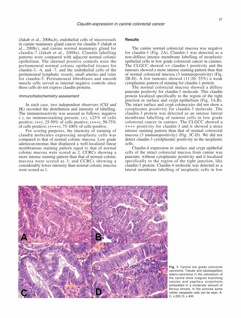
Tissue samples were collected between 2005 and2008 at Szent István University, Faculty of VeterinaryScience, Department of Pathology and ForensicVeterinary Medicine (Budapest, HU). The samplesincluded 10 canine normal colorectal tissue samples and20 canine low grade colorectal carcinoma (CLGCC).Samples were fixed in 8% neutral buffered formalin for24 hours at room temperature, dehydrated in a series ofethanol and xylene, and embedded in paraffin. The 3-4µ m thick sections were routinely stained withhematoxylin and eosin (HE). Each neoplastic case wasclassified by the pathologists (CSJ and JK) according tograde (Greene et al., 2002) (Fig. 1A-D).
Immunohistochemistry
Serial sections (3-4 µm) were initially dewaxed inxylene and graded ethanol. After treatment withappropriate antigen retrieval (Target Retrieval Soluton,DAKO, Glostrup, Denmark, pH 6; microwave - 800W -oven for 30 min), the sections were incubated withprimary antibodies against claudin-1 (rabbit polyclonal,diluted 1 in 100), claudin-3 and claudin -7 (both rabbitpolyclonal and diluted 1 in 80), claudin-4 and claudin-5(both mouse monoclonal and diluted 1 in 100) for 60min at room temperature. All primary antibodies werefrom Zymed Inc., San Francisco, CA, USA.Immunohistochemical labelling was performed using thestreptavidin-peroxidase procedure. Antigen-boundprimary antibody was detected using standard avidin-biotin immunoperoxidase complex (DAKO, LSAB2Kit). The chromogen substrate was 3, 3’-diaminobenzidine tetrahydrochloride (DAB substrate-chromogen, DAKO). Sections were counterstained withMayer’s hematoxylin.
Negative controls were performed by omission ofthe primary antibody and positive controls was caninenormal mammary gland for claudin-1, -3, -4 and -7
56
Claudin-expression in canine colorectal cancer
(Jakab et al., 2008a,b), endothelial cells of microvesselsin canine mammary gland cancer for claudin-5 (Jakab etal., 2008c), and canine normal mammary gland forclaudin-7 (Jakab et al., 2008a). Claudin labellingpatterns were compared with adjacent normal colonicepithelium. The internal positive controls were theperitumoural normal colonic epithelial tissues forclaudin-3, -4, and -7, and the endothelial cells of theperitumoural lymphatic vessels, small arteries and veinsfor claudin-5. Peritumoural fibroblasts and smoothmuscle cells served as internal negative controls sincethese cells do not express claudin proteins.
Immunohistochemistry assessment
In each case, two independent observers (CSJ andJK) recorded the distribution and intensity of labelling.The immunoreactivity was assesed as follows: negative(-), no immunostaining present; (+), <25% of cellspositive; (++), 25-50% of cells positive; (+++), 50-75%of cells positive; (++++), 75-100% of cells positive.
For scoring purposes, the intensity of staining ofclaudin molecules expressing neoplastic cells wascompared to that of normal colonic mucosa. Low gradeadenocarcinomas that displayed a well-localized linearmembranous staining pattern equal to that of normalcolonic mucosa were scored as 2. CCRCs showing amore intense staining pattern than that of normal colonicmucosa were scored as 3; and CCRCs showing aconsiderably lower intensity than normal colonic mucosawere scored as 1.
Results
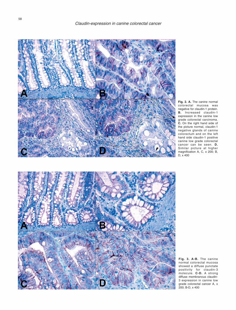
The canine normal colorectal mucosa was negativefor claudin-1 (Fig. 2A). Claudin-1 was detected as anon-diffuse intense membrane labelling of neoplasticepithelial cells in low grade colorectal cancer in canines.The CLGCC showed ++ claudin-1 positivity and thetumours showed a more intense staining pattern than thatof normal colorectal mucosa (3 immunopositivity) (Fig.2B-D). A few tumours showed (11:20; 55%) a weakcytoplasmic pattern of staining for claudin-1 protein.
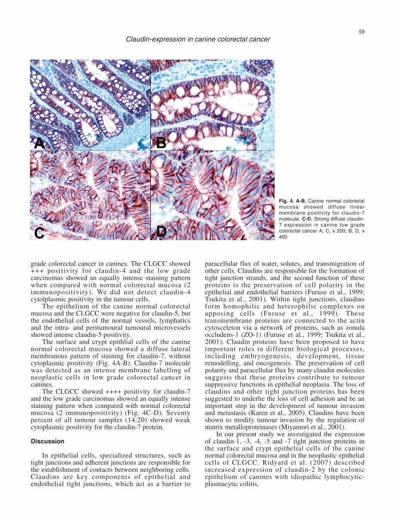
The normal colorectal mucosa showed a diffusepunctate positivity for claudin-3 molecule. This claudinprotein localized specifically to the region of the tightjunction in surface and crypt epithelium (Fig. 3A,B).The intact surface and crypt colonocytes did not show acytoplasmic positivity for claudin-3 molecule. Theclaudin-3 protein was detected as an intense lateralmembrane labelling of tumour cells in low gradecolorectal cancer in canines. The CLGCC showed a++++ positivity for claudin-3 and it showed a moreintense staining pattern than that of normal colorectalmucosa (3 immunopositivity) (Fig. 3C-D). We did notdetect claudin-3 cytolplasmic positivity in the neoplasticcells.
Claudin-4 expression in surface and crypt epithelialcells of the intact colorectal mucosa from canine waspunctate, without cytoplasmic positivity and it localizedspecifically to the region of the tight junction, likeclaudin-3 protein. Claudin-4 molecule was detected as alateral membrane labelling of neoplastic cells in low
57
Claudin-expression in canine colorectal cancer
Fig. 1. Canine low grade colorectalcarcinoma. Tubular and tubulopapillaryadeno-carcinoma in the colorectum ofthe canine show irregular branchingtubules and papil lary projectionsembedded in a moderate amount offibrous stroma. In the pictures somemitotic neoplastic cells can be seen. A-C, x 200; D, x 400
58
Claudin-expression in canine colorectal cancer
Fig. 2. A. The canine normalcolorectal mucosa wasnegative for claudin-1 protein.B. Increased claudin-1expression in the canine lowgrade colorectal carcinoma.C. On the right hand side ofthe picture normal, claudin-1negative glands of caninecolorectum and on the lefthand side claudin-1 positivecanine low grade colorectalcancer can be seen. D.Similar picture at highermagnification A, C, x 200; B,D, x 400
Fig. 3. A-B. The caninenormal colorectal mucosashowed a diffuse punctateposit ivity for claudin-3molecule. C-D. A strongdiffuse membranous claudin-3 expression in canine lowgrade colorectal cancer A, x200; B-D, x 400
grade colorectal cancer in canines. The CLGCC showed+++ positivity for claudin-4 and the low gradecarcinomas showed an equally intense staining patternwhen compared with normal colorectal mucosa (2immunopositivity). We did not detect claudin-4cytolplasmic positivity in the tumour cells.
The epithelium of the canine normal colorectalmucosa and the CLGCC were negative for claudin-5, butthe endothelial cells of the normal vessels, lymphaticsand the intra- and peritumoural tumoural microvesselsshowed intense claudin-5 positivity.
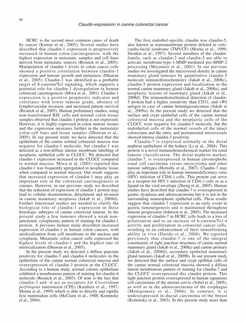
The surface and crypt epithlial cells of the caninenormal colorectal mucosa showed a diffuse lateralmembranous pattern of staining for claudin-7, withoutcytoplasmic positivity (Fig. 4A-B). Claudin-7 moleculewas detected as an intense membrane labelling ofneoplastic cells in low grade colorectal cancer incanines.
The CLGCC showed ++++ positivity for claudin-7and the low grade carcinomas showed an equally intensestaining pattern when compared with normal colorectalmucosa (2 immunopositivity) (Fig. 4C-D). Seventypercent of all tumour samples (14:20) showed weakcytoplasmic positivity for the claudin-7 protein.
Discussion
In epithelial cells, specialized structures, such astight junctions and adherent junctions are responsible forthe establishment of contacts between neighboring cells.Claudins are key components of epithelial andendothelial tight junctions, which act as a barrier to
paracellular flux of water, solutes, and transmigration ofother cells. Claudins are responsible for the formation oftight junction strands, and the second function of theseproteins is the preservation of cell polarity in theepithelial and endothelial barriers (Furuse et al., 1999;Tsukita et al., 2001). Within tight junctions, claudinsform homophilic and heterophilic complexes onapposing cells (Furuse et al., 1999). Thesetransmembrane proteins are connected to the actincytosceleton via a network of proteins, such as zonulaoccludens-1 (ZO-1) (Furuse et al., 1999; Tsukita et al.,2001). Claudin proteins have been proposed to haveimportant roles in different biological processes,including embryogenesis, development, tissueremodelling, and oncogenesis. The preservation of cellpolarity and paracellular flux by many claudin moleculessuggests that these proteins contribute to tumoursuppressive functions in epithelial neoplasia. The loss ofclaudins and other tight junction proteins has beensuggested to underlie the loss of cell adhesion and be animportant step in the development of tumour invasionand metastasis (Karen et al., 2005). Claudins have beenshown to modify tumour invasion by the regulation ofmatrix metalloproteinases (Miyamori et al., 2001).
In our present study we investigated the expressionof claudin-1, -3, -4, -5 and -7 tight junction proteins inthe surface and crypt epithelial cells of the caninenormal colorectal mucosa and in the neoplastic epithelialcells of CLGCC. Ridyard et al. (2007) describedincreased expression of claudin-2 by the colonicepithelium of canines with idiopathic lymphocytic-plasmacytic colitis.
59
Claudin-expression in canine colorectal cancer
Fig. 4. A-B. Canine normal colorectalmucosa showed diffuse l inearmembrane positivity for claudin-7molecule. C-D. Strong diffuse claudin-7 expression in canine low gradecolorectal cancer A, C, x 200; B, D, x400
HCRC is the second most common cause of deathby cancer (Kumar et al., 2005). Several studies havedescribed that claudin-1 expression is progressivelyincreased in human colon carcinogenesis, with thehighest expression in metastatic samples and cell linesderived from metastatic sources (Resnick et al., 2005).Manipulation of claudin-1 levels in colon cancer cellsshowed a positive correlation between claudin-1expression and tumour growth and metastasis (Dhawanet al., 2005). Claudin-1 was identified as a probabletarget of ß-catenin/Tcf signaling, which supports apotential role for claudin-1 dysregulation in humancolorectal carcinogenesis (Miwa et al., 2001). Claudin-1expression is a positive prognostic indicator andcorrelates with lower tumour grade, absence oflymphovascular invasion, and increased patient survival(Resnick et al., 2005). Human preliminary studies, usingnon-transformed RIE cells and normal colon tissuesamples observed that claudin-1 protein is not expressed.However, it is highly expressed in colon tumour tissuesand the expression increases further in the metastaticcolon cell lines and tissue samples (Dhawan et al.,2005). In our present study we have detected that theepithelium of the canine normal colorectal mucosa wasnegative for claudin-1 molecule, but claudin-1 wasdetected as a non-diffuse intense membrane labelling ofneoplastic epithelial cells in CLGCC. We detected thatclaudin-1 expression increased in the CLGCC comparedto normal mucosa. Miwa et al. (2001) reported thatclaudin-1 was frequently upregulated in neoplastic tissuewhen compared to normal mucosa. Our result suggeststhat increased expression of claudin-1 may play animportant role in the colorectal carcinogenesis incanines. However, in our previous study we describedthat the reduction of expression of claudin-1 protein maylead to cellular disorientation, detachment and invasionin canine mammary neoplasia (Jakab et al., 2008d).Further functional studies are needed to clarify thebiological role of claudin-1 expression in thesehistologic subtypes of canine colorectal tumour. In thepresent study a few tumours showed a weak non-junctional, cytoplasmic pattern of staining for claudin-1protein. A previous human study described increasedexpression of claudin-1 in human colon cancers, withmislocalization from cell membrane to the nucleus andcytoplasm. Metastatic colon cancer cells expressed thehighest levels of claudin-1 and the highest rate ofmislocalization (Dhawan et al., 2005).
In the present study we detected a diffuse punctatepositivity for claudin-3 and claudin-4 molecules in theepithelium of the canine normal colorectal mucosa andoverexpression of claudin-3 protein in the CLGCC.According to a human study, normal colonic epitheliumexhibited a membranous pattern of staining for claudin-4molecule (Resnick et al., 2005). Of note is the fact thatclaudin-3 and -4 act as receptors for Clostridiumperfringens enterotoxin (CPE) (Katahira et al., 1997;Morita et al., 1999) which is able to directly and rapidlylyse mammalian cells (McClane et al., 1988; Kominskyet al., 2004).
The first endothel-specific claudin was claudin-5,also known as transmembrane protein deleted in velo-cardio-facial syndrome (TMVCF) (Morita et al., 1999;Sirotkin et al., 1997). Several members of the claudin-family, such as claudin-2 and claudin-5 are able toactivate membrane-type-1-MMP-mediated pro-MMP-2processing (Miyamori et al., 2001). In our previousstudies we investigated the microvessel density in caninemammary gland tumours by quantitative claudin-5molecule immunohistochemistry (Jakab et al., 2008c),claudin-5 protein expression and localization in thenormal canine mammary gland (Jakab et al., 2008a), andneoplastic lesions of mammary gland (Jakab et al.,2008d). The immunohistochemical detection of claudin-5 protein had a higher sensitivity than CD31, and vWfantigen in case of canine hemangiosarcomas (Jakab etal., 2009a). In the present study we detected that thesurface and crypt epithelial cells of the canine normalcolorectal mucosa and the neoplastic cells of theCLGCC were negative for claudin-5 molecule, but theendothelial cells of the normal vessels of the intactcolorectum and the intra- and peritumoural microvesselsshowed intense claudin-5 positivity.
Claudin-7 is expressed normally in the distalnephron epithelium of the kidney (Li et al., 2004). Thisprotein is a novel immunohistochemical marker for renaltumour classification. The distal nephron marker,claudin-7, is overexpressed in human chromophoberenal cell carcinoma versus oncocytoma and othertumour subtypes (Hornsby et al., 2007). Claudin-7 mayplay an important role in human immunodeficiency virus(HIV) infection of CD4(-) cells. This protein can serveas a receptor for HIV-1 infection of CD4(-) cells or as aligand on the viral envelope (Zheng et al., 2005). Humanstudies have desrcibed that claudin-7 is overexpressed ingastric dysplasia and adenocarcinoma but it is not in thesurrounding nonneoplastic epithelial cells. These resultssuggest that claudin-7 expression is an early event ingastric tumourigenesis and is maintained throughouttumour progression (Johnson et al., 2005). The increasedexpression of claudin-7 in HCRC cells leads to a loss ofpolarization and to an increase of h-catenin/Tcf-4activity and proliferation in colorectal cancer cells,resulting in an enhancement of their tumorformingability in vivo (Darido et al., 2008). We reportedpreviously that claudin-7 is one of the integralconstituents of tight junction structures of canine normalmammary gland (Jakab et al., 2008a) and canine primary(Jakab et al., 2008d), secondary epithelial mammarygland tumours (Jakab et al., 2009b). In our present studywe detected that the surface and crypt epithlial cells ofthe canine normal colorectal mucosa showed a diffuselateral membranous pattern of staining for claudin-7 andthe CLGCC overexpressed this claudin protein. Thistight junction protein overexpressed in human squamouscell carcinomas of the uterine cervix (Sobel et al., 2005),as well as in the adenocarcinoma of the esophagus(Montgomery et al., 2006). In contrast, it isunderexpressed in ductal carcinoma of the breast(Kominsky et al., 2003). In this present study more than
60
Claudin-expression in canine colorectal cancer
50 % of the CLGCCs showed a lateral membrane andcytoplasmic positivity for claudin-7 molecule. Furtherfunctional studies are needed to clarify the biologicalrole of mislocalisation of claudin-7 from the cellmembrane to the cytoplasm in canine low gradecarcinoma.
In conclusion, the results of the present study haveshown that claudin-1 is not expressed in the epitheliumof canine normal colorectal mucosa, but increasedexpression of this protein is observed in CLGCCs.Consequently, we hypothesize that claudin-1 plays a rolein the progression of canine colorectal carcinoma.Further functional studies are needed to clarify thebiological role of the mislocalization of claudin-1molecule from the cell membrane to the cytoplasm inthese histologic subtypes of canine colorectal tumour.The canine intact colorectal epithelium showed diffusepunctate positivity for claudin-3 and claudin-4molecules, and the low grade colorectal canceroverexpressed the claudin-3 protein. The CLGCCsshowed a lower claudin-4 expression and this resultsuggests that reduced expression of claudin-4 moleculemay lead to cellular disorientation, detachment andinvasion of canine colorectal cancers. In the presentstudy we detected that the epithelial cells of the caninenormal colorectal mucosa and the neoplastic cells of theCLGCCs were negative for claudin-5 molecule, but theendothelial cells of the intact vessels, lymphatics andintratumoural, peritumoural microvessels showed strongclaudin-5 positivity. This can help during lightmicroscopic analysis in the detection of lymphovascularinvasion of the colorectal cancer cells (claudin-5 positivevessels, lymphpatics with claudin-5 negative tumouremboli), as well as in the case of canine mammary glandcancer intravasation (Jakab et al., 2008e). In addition,claudin-5 can be used for the intra-, and peritumouralmicrovessel density assessment of canine colorectalcancers. The epithelial cells of the canine normalcolorectal mucosa showed a diffuse lateral membranouspositivity for claudin-7, and the CLGCCs overexpressedthis claudin. More than 50% of canine low grade cancershowed a lateral membrane and cytoplasmic positivityfor claudin-7 molecule. Further functional studies areneeded to clarify the biological role of overexpressionand mislocalisation of claudin-7 in CLGCCs.
Acknowledgements. We would like to thank Renáta Pop and MagdolnaPekár for their assistance with the immunohistochemical reactions, andEdit Oláh for her kind help in locating the relevant references.
References
Brunnert S.R., Deel L.A., Herron A.J. and Altman N.H. (1993). Primarylinitis plastica (singet ring) carcinoma of the colon in a dog. J. Am.Anim. Hosp. Assoc. 29, 75-77.
Burgart L.J. (2002). Colorectal polyps and other precursor lesions. Needfor an expanded view. Gastroenterol. Clin. North. Am. 31, 959-970.
Darido C., Buchert M., Pannequin J., Bastide P., Zalzali H.,Mantamadiotis T., Bourgaux J.F., Garambois V., Jay P., Blache P.,
Joubert D. and Hollande F. (2008). Defective claudin-7 regulation byTcf-4 and Sox-9 disrupts the polarity and increases thetumorigenicity of colorectal cancer cells. Cancer. Res. 68, 4258-4268.
De Oliveira S.S., De Oliveira I.M., De Souza W. and Morgado-Díaz J.A.(2005). Claudin upregulation in human colorectal cancer. FEBS Lett.579, 6179-6185.
Dhawan P., Singh A.B., Deane N.G., No Y., Shiou S.R., Schmidt C.,Neff J., Washington M.K. and Beauchamp R.D. (2005). Claudin-1regulates cellular transformation and metastatic behavior in coloncancer. J. Clin. Invest.115, 1765-1776.
Eaden J.A., Abram K.R. and Mayberry J.F. (2001). The risk of colorectalcancer in ulcerative colitis: a meta-analysis. Gut 48, 526-535.
Furuse M., Sasaki H. and Tsukita S. (1999). Manner of interaction ofheterogeneous claudin species within and between tight junctionstrands. J. Cell Biol. 147, 891-903.
Greene F.L., Page D.L., Fleming I.D., Fritz A.G., Blach C.M., Haller D.G.and Morrow M. (2002). In: AJCC: Cancer Staging Handbook: Fromthe AJCC Cancer Staging Manual. 6th edn. Springer-Verlag. NewYork. pp 127-129.
Grone J., Weber B., Staub E., Heinze M., Klaman I., Pilarsky C.,Hermann K., Castanos-Velez E., Röpcke S., Mann B., Rosenthal A.and Buhr H.J.(2007). Differential expression of genes encoding tightjunction proteins in colorectal cancer: frequent dysregulation ofclaudin-1, -8 and -12. Int. J. Colorectal. Dis. 22, 651-659.
Hampson E.C.G.M., Wilkinson G.T., Sutton R.H. and Turner S.A.(1990). Cutaneous metastasis of a colonic carcinoma in a dog. J.Small Anim. Prac. 31, 155-158.
Head K.W., Else R.W. and Dubielzig R.R. (2002). Tumors of theintestines. In: Meuten D.J. (2002). Tumors in domestic animals.Fourth edition. Iowa State Press, 461-469.
Hermanek P. and Sobin L.H. (1995). Colorectal carcinoma. In:Prognostic factors in cancer. Hermanek P., Gospodarowicz M.K.and Hendson D.E. (eds). Springer-Verlag. New York. pp 64-79.
Holt P.E. and Lucke V.M. (1985). Rectal neoplasia in the dog: Aclinicopathological review of 31 cases. Vet. Rec. 116, 400-405.
Hornsby C.D., Cohen C., Amin M.B., Picken M.M., Lawson D., Yin-Goen Q. and Young A.N. (2007). Claudin-7 immunohistochemistryin renal tumors: a candidate marker for chromophobe renal cellcarcinoma identified by gene expression profiling. Arch. Pathol. Lab.Med. 131, 1541-1547.
Jakab Cs., Halász J., Szász A. M., Batmunkh E., Kiss A., Schaff Zs.,Rusvai M., Gálf i P. and Kulka J. (2008a). Expression andlocalisation of claudin-1, -2, -3, -4, -5, -7 and -10 proteins in thenormal canine mammary gland. Acta Vet. Hung. 56, 341-352.
Jakab Cs., Halász J., Kiss A., Szász A. M., Schaff Zs., Rusvai M. andKulka J. (2008b). Use of external positive controls in claudin-expression immunhistochemical examination. Magy. Állatorv. Lap.130, 433-438. (In Hungarian, with English abstract and figures).
Jakab Cs., Halász J., Szász A.M., Kiss A., Schaff Zs., Rusvai M.,Abonyi T.Zs. and Kulka J. (2008c). Evaulation of microvesseldensity (MVD) in canine mammary tumours by quantitative claudin-5molecule immunohistochemistry. Acta Vet. Hung. 56, 495-510.
Jakab Cs., Halász J., Szász A.M., Batmunkh E., Kiss A., Schaff Zs.,Rusvai M., Gálfi P. and Kulka J. (2008d). Expression of claudin-1, -2, -3, -4, -5, and -7 proteins in benign and malignant caninemammary gland epithelial tumors. J. Comp. Pathol. 139, 238-245.
Jakab Cs., Halász J., Kiss A., Schaff Zs., Szabára Á., Rusvai M.,Szatmári Sz. and Kulka J. (2008e). Examination of claudin-5 proteinexpression of the endothelial cells of lymph vessels in caninemammary glands and mammary gland carcinoma by
61
Claudin-expression in canine colorectal cancer
immunohistochemical methods. Magy. Állatorv. Lap. 130, 296-303.(In Hungarian, with English abstract and figures).
Jakab Cs., Halász J., Kiss A., Schaff Zs., Rusvai M., Gálfi P., AbonyiT.Zs. and Kulka J. (2009a). Claudin-5 protein is a new differentialmarker for histopathological differential diagnosis of caninehemangiosarcoma. Histol. Histopathol. 24, 801-813.
Jakab Cs., Szász A.M., Kiss A., Schaff Zs., Rusvai M., Szabára Á. andKulka J. (2009b). Immunohistochemical examintion of expressionpatterns of claudin molecules in metastases of solid carcinomasimplex from canines. Magy. Állatorv. Lap. 131, 33-41. (InHungarian, with English abstract and figures).
Jass J.R. (2001). Pathogenesis of colorectal cancer. Surg. Clin. N. Am.82, 891-894.
Jemal A., Thomas A., Murray T. and Thun M. (2002). Cancer statistics.CA Cancer J. Clin. 52, 23-47.
Johnson A.H., Frierson H.F., Zaika A., Powell S.M., Roche J., Crowe S.,Moskaluk C.A. and El-Rifai W. (2005). Expression of tight-junctionprotein claudin-7 is an early event in gastric tumorigenesis. Am. J.Pathol. 167, 577-584.
Karen S., Robert M. and Manfred K. (2005). Role of claudins intumorigenesis. Adv. Drug Del. Rev. 57, 919-928.
Katahira J., Inoue N., Horiguchi Y., Matsuda M. and Sugimoto, N.(1997). Molecular cloning and functional characterization of thereceptor for Clostridium perfringens enterotoxin. J. Cell Biol. 136,1239-1247.
Kinguasa T., Huo Q., Higasi D., Shibaguchi H., Kuroki M., Tnaka T.,Futami K., Yamashita Y., Hachimine K., Maekawa S., NabeshimaK., Iwasaki H. and Kuroki M. (2007). Selective up-regulation ofclaudin-1 and claudin-2 in colorectal cancer. Anticancer Res. 27,3729-3734.
Kinzler K.W. and Vogelstein B. (2002). Colorectal tumors. In: Thegenetic basis of human cancer. 2nd Edition. Kinzler K.W. andVogelstein B. (eds). McGraw-Hill. New York. pp 583-590.
Kominsky S.L., Argani P., Korz D., Evron E., Raman V., Garrett E., ReinA., Sauter G., Kallioniemi O.P. and Sukumar S. (2003). Loss of thetight junction protein claudin-7 correlates with histological grade inboth ductal carcinoma in situ and invasive ductal carcinoma of thebreast. Oncogene 22, 2021-2033.
Kominsky S.L., Vali M., Korz D, Gabig T.G., Weitzman S.A., Argani P.and Sukumar S. (2004). Clostridium perfringens enterotoxin elicitsrapid and specific cytolysis of breast carcinoma cells mediatedthrough tight junction proteins claudin 3 and 4. Am. J. Pathol. 164,1627-1634.
Kumar V., Abbas A.K. and Fausto N. (2005). Robbins and CotranPathologic basis of disease. 7th ed. Elsevier Saunders.Philadelphia. pp 864-865.
Li W.Y., Huey C.L. and Yu A.S. (2004). Expression of claudin-7 and -8along the mouse nephron. Am. J. Physiol. Renal. Physiol. 286,1063-1071.
McClane B.A., Hanna P.C. and Wnek A.P. (1988). Clostridiumperfringens enterotoxin. Microb. Pathogen. 4, 317-323.
Miyamori H., Takino T., Kobayashi Y., Tokai H., Itoh Y., Seiki M. andSato H. (2001). Claudin promotes activation of pro-matrixmetalloproteinase-2 mediated by membrane-type matrixmetalloproteinases. J. Biol. Chem., 276, 28204-28211.
Miwa N., Furuse M., Tsukita S., Niikawa N., Nakamura Y. and FurukawaY. (2001). Involvement of claudin-1 in the beta-catenin/Tcf signalingpathway and its frequent upregulation in human colorectal cancers.Oncol. Res. 12, 469-476.
Montgomery E., Mamelak A.J., Gibson M., Maitra A., Sheikh S., Amr
S.S., Yang S., Brock M., Forstiere A., Zhang S., Murphy K.M. andBerg K.D. (2006). Overexpression of claudin proteins in esophagealadenocarcinoma and its precusor lesions. Appl. Immunohistochem.Mol. Morphol. 14, 24-30.
Morita K., Furuse M., Fujimoto K. and Tsukita S. (1999). Claudinmultigene family encoding four-transmembrane domain proteincomponents of tight junction strands. Proc. Natl. Acad. Sci. USA 96,511-516.
Mullin J.M. (1997). Potential interplay between luminal growth factorsand increased tight junction permeability in epithelial carcinogenesis.J. Exp. Zool. 279, 484-489.
Nakayama F., Semba S., Usami Y., Chiba H., Sawada N. and YokozakiH. (2008). Hypermethylation-modulated downregulation of claudin-7expression promotes the progression of colorectal carcinoma.Pathobiology 75, 177-185.
Oshima T., Kunisaki C., Yoshihara K., Yamada R., Yamamoto N., SatoT., Makino H., Yamagishi S., Nagano Y., Fujii S., Shiozawa M.,Akaike M., Wada N., Rino Y., Masuda M., Tanaka K. and Imada T.(2008). Reduced expression of the claudin-7 gene correlates withvenous invasion and liver metastasis in colorectal cancer. Oncol.Rep. 19, 953-959.
Patnaik A.K., Hurvitz A.I. and Johnson G.F. (1977). Caninegastrointestinal neoplasms. Vet. Pathol. 14, 547-555.
Resnick M.B., Konkin T., Routhier J., Sabo E. and Pricolo V.E. (2005).Claudin-1 is a strong prognostic indicator in stage II colonic cancer:a tissue microarray study. Mod. Pathol. 18, 511-518.
Ridyard A.E., Brown J.K., Rhind S.M., Else R.W., Simpson J.W. andMiller H.R.P. (2007). Apical junction complex protein expression inthe canine colon: differential expression of claudin-2 in the colonicmucosa in dogs with idiopathic colitis. J. Histochem. Cytochem. 55,1049-1058.
Schaffer E. and Schieffer B. (1968). Incidence and types of canine rectalcarcinomas. J. Small Anim. Pract. 9, 491-595.
Silverberg S.G. (1971). Carcinoma arising in adenomatous polyps of therectum in a dog. Dis. Colon Rectum 14, 191-194.
Sirotkin H., Morrow B., Saint-Jore B., Puech A., Das Gupta R., PatanjaliS.R., Skoultchi A., Weissman S.M. and Kucherlapati R. (1997).Identification, characterization, and precise mapping of a humangene encoding a novel membrane-spanning protein from the 22q11region deleted in velo-cardiofacial syndrome. Genomics 42, 245-251.
Sobel G., Paska Cs., Szabó I., Kiss A., Kádár A. and Schaff Zs. (2005).Increased expression of claudins in cervical squamous intraepithelialneoplasia and invasive carcinoma. Human Pathol. 36, 162-169.
Stampley A.R., Swayne D.E. and Prasse K.W. (1987). Meningealcarcinomatosis secondary to a colonic signet-ring cell carcinoma in adog. J. Am. Anim. Hosp. Assoc. 23, 655-658.
Tsukita S., Furuse M. and Itoh M. (2001). Multifunctional strands in tightjunctions. Nat. Reviews Mol. Cell. Biol. 2, 285-293.
Weber C.R., Nalle S.C., Tretiakova M., Rubin D.T. and Turner J.R.(2008). Claudin-1 and claudin-2 expression is elevated ininflammatory bowel disease and may contribute to early neoplastictransformation. Lab. Invest. 88, 1110-1120.
Wei E.K. Giovannucci E., Wu K., Rosner B., Fuchs C.S., Willett W.C.and Colditz G.A. (2004). Comparison of risk factors for colon andrectal cancer. Int. J. Cancer 108, 433-442.
Zheng J., Xie Y., Campbel R., Song J., Massachi S., Razi M., Chiu R.,Berenson J., Yang O.O., Chen I.S. and Pang S. (2005). Involvementof claudin-7 in HIV infection of CD4(-) cells. Retrovirol. 2, 79-92.
Accepted July 20, 2009
62
Claudin-expression in canine colorectal cancer