Embed Size (px)
Citation preview
Proc. Natl. Acad. Sci. USA 83 (1986)
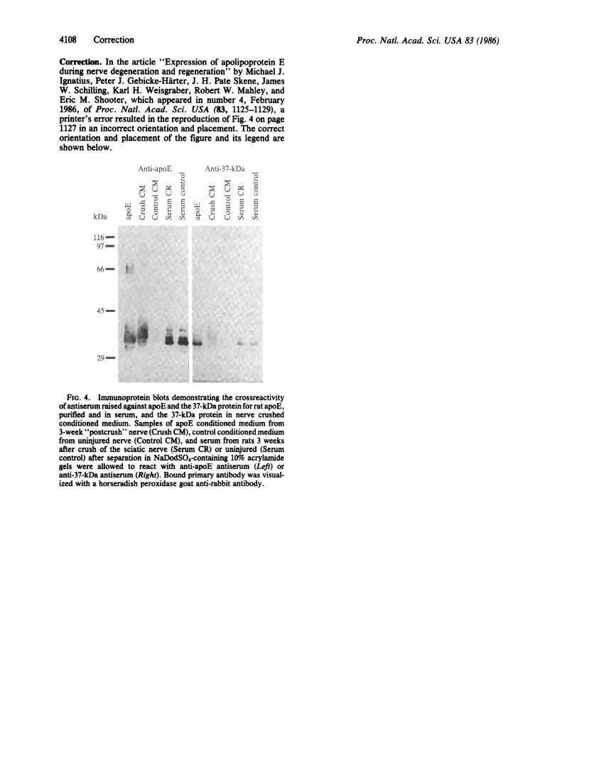
Correction. In the article "Expression of apolipoprotein Eduring nerve degeneration and regeneration" by Michael J.Ignatius, Peter J. Gebicke-Hirter, J. H. Pate Skene, JamesW. Schilling, Karl H. Weisgraber, Robert W. Mahley, andEric M. Shooter, which appeared in number 4, February1986, of Proc. Nadl. Acad. Sci. USA (83, 1125-1129), aprinter's error resulted in the reproduction of Fig. 4 on page1127 in an incorrect orientation and placement. The correctorientation and placement of the figure and its legend areshown below.
Anti-apoE Anti-37-kDa _
U _ Uto _)
Xa;C 5 L1 2 = =
kDa U U c co Uv u en
116-97-
66- t
45-
*:=
29-
FIG. 4. Immunoprotein blots demonstrating the crossreactivityofantiserum raised against apoE and the 37-kDa protein for rat apoE,purified and in serum, and the 37-kDa protein in nerve crushedconditioned medium. Samples of apoE conditioned medium from3-week "postcrush" nerve (Crush CM), control conditioned mediumfrom uninjured nerve (Control CM), and serum from rats 3 weeksafter crush of the sciatic nerve (Serum CR) or uninjured (Serumcontrol) after separation in NaDodSO4-containing 10%o acrylamidegels were allowed to react with anti-apoE antiserum (Left) oranti-37-kDa antiserum (Right). Bound primary antibody was visual-ized with a horseradish peroxidase goat anti-rabbit antibody.
4108 Correction
Proc. Natl. Acad. Sci. USAVol. 83, pp. 1125-1129, February 1986Neurobiology
Expression of apolipoprotein E during nerve degenerationand regeneration
(high density lipoprotein/nerve repair/macrophages/Schwann cells/cholesteryl esters)
MICHAEL J. IGNATIUS*, PETER J. GEBICKE-HARTER*, J. H. PATE SKENE*, JAMES W. SCHILLINGt,KARL H. WEISGRABERt, ROBERT W. MAHLEYt, AND ERIC M. SHOOTER**Department of Neurobiology, Stanford University School of Medicine, Stanford, CA 94305; tCalifornia Biotechnology Inc., Mountain View, CA 94043; and*The Gladstone Foundation Laboratories for Cardiovascular Disease, Department of Pathology, University of California, San Francisco, CA 94140
Communicated by Donald Kennedy, September 27, 1985
ABSTRACT A 37-kDa glycoprotein has been describedrecently, whose synthesis is dramatically increased after injuryof the rat sciatic and optic nerves. Cells in the nerve sheath,distal to the site of injury, produce and secrete large amountsof this protein, so that by 3 weeks after injury, it represents2-5% of the total soluble extracellular protein in the regener-ating sciatic nerve sheath, although it fails to accumulate indamaged optic nerve. Results presented here reveal extensivehomology between the 37-kDa nerve injury-induced proteinand a well-studied serum protein, apolipoprotein E (apoE),that is involved in lipid and cholesterol metabolism and that hasbeen shown recently to be present in adult and developing ratastroglia. Both proteins have identical isoelectric focusingpoints and similar molecular masses. Antibodies raised againstthe 37-kDa protein recognize apoE and anti-apoE serumcrossreacts with the 37-kDa protein. Sequence data for two 14amino acid stretches of the 37-kDa protein match identicalregions of apoE. These data suggest that the 37-kDa protein isidentical to serum apoE and that it could have similar functionsto the latter. In the nervous system, for example, it may beinvolved in the mobilization and reutilization of lipid in therepair, growth, and maintenance of myelin and axonal mem-branes, both during development and after injury.
Axons regenerating after a nerve injury grow through anextracellular environment elaborated by glia and other non-neuronal cells of the distal nerve stump. A century of clinicaland experimental observations has demonstrated the pro-found influence of the distal nerve stump on successful nerverepair (1, 2). When a peripheral nerve is injured, a series ofstriking changes occurs in the distal nerve stump. Axons cutoff from their cell bodies degenerate, Schwann cells prolif-erate and align themselves into longitudinal "Bungnerbands," while blood-borne macrophages accumulate in thedistal sheath to assist in removal of axonal and myelin debris.At the molecular level, one of the most prominent effects ofinjury is a dramatic increase in the synthesis of an acidic37-kDa protein that is secreted into the extracellular space (3,4).
Synthesis of this 37-kDa protein increases more than twoorders of magnitude over the first 2 weeks after nerve injury,and the secreted protein accumulates in sufficient abundanceto account for 2-5% of the total soluble protein in theextracellular space (3). This protein is also synthesized inneonatal nerves and in injured adult optic nerves and spinalcords; however, it fails to accumulate in these injured centralnervous system (CNS) sites (3, 5, 21). The functional signif-icance and identity of this injury-induced protein haveremained obscure. Here, we present evidence that the
37-kDa protein induced in injured peripheral nerves is iden-tical to apolipoprotein E (apoE), a serum protein normallyinvolved in lipid transport and metabolism (6, 7). Thisidentity suggests possible roles for the 37-kDa protein indevelopment, degeneration, and regeneration in the nervoussystem.
MATERIALS AND METHODS
Surgery. Sciatic and optic nerve crushes and labeling andcollection of sheath cell proteins were according to Skene andShooter (3).For in situ or in vivo labeling of proteins, the initial surgery
was as before. After 1 week, the sciatic nerves were re-exposed under anesthesia, and 50 ,uCi (1 Ci = 37 GBq) of["Simethionine with 0.05% bromphenol blue as a tracer in atotal volume of 5 pl was injected into the subendoneurialspace of the nerve with a 31-gauge needle. Injections weredone 1-2 mm distal to the original site of injury and in acomparable region in uninjured control nerves. After 24 hr,nerves were removed and immediately frozen at -80°C in adry ice/ethanol bath. To collect the labeled soluble proteins,nerves were homogenized in a solution of 10 mM Tris HCl atpH 7.5 containing 2% NaDodSO4, 5 mM EDTA, and 1 mMdithiothreitol. Supernatants of this homogenate, collectedafter a 10-min, 12,000 x g spin, were then analyzed.
Antibody Production and Detection. Rabbits were inocu-lated by injecting 250 ,ug of 37-kDa protein purified from3-week conditioned medium (for details, see ref. 8). Theprotein was suspended in Freund's complete adjuvant (Difco)and then injected subcutaneously at eight sites along the backof a New Zealand White rabbit. After 1 month, threesubsequent booster injections every other week with 50 ,g ofprotein in Freund's incomplete adjuvant yielded antiserum ofhigh titer directed against the native and denatured 37-kDaproteins. Protein blots or electrophoretic transfer analyseswere done essentially according to Towbin et al. (9), with theprimary antibody at dilutions of up to 1:100,000. Boundprimary antibody was visualized with peroxidase-labeledsecond antibody (Vector Laboratories, Burlingame, CA).Controls with nonimmune serum were done at 1:1000 dilu-tion.For immunoprecipitations, 200 ,u1 of labeled conditioned
medium was combined with 10 ,ul of whole antiserum to the37-kDa protein and brought to a final volume of 1 ml with asolution of 0.15 M NaCl, 25 mM Tris HCl (pH 7.4), 0.5%Nonidet P-40, 1 mM phenylmethylsulfonyl fluoride, and 1mM EDTA and rotated for 1 hr at 4°C. Antigen-antibodycomplexes were pelleted with protein A-Sepharose CL-4B
Abbreviations: HDL, high density lipoprotein(s); LDL, low densitylipoprotein(s); apoE, apolipoprotein E; PNS, peripheral nervoussystem; CNS, central nervous system; FITC, fluorescein isothio-cyanate.
1125
The publication costs of this article were defrayed in part by page chargepayment. This article must therefore be hereby marked "advertisement"in accordance with 18 U.S.C. §1734 solely to indicate this fact.

1126 Neurobiology: Ignatius et al.
(Sigma), analyzed on 10% polyacrylamide/NaDodSO4 gels,and fluorographed as described below.
Astrocyte-enriched cultures derived from 3- to 5-day-oldrat brains were stained according to Raff et al. (10) with thefollowing modifications. Before fixation in ethanol/aceticacid, cultures were incubated in 10 ,uM monensin (Sigma) for3 hr at 370C in culture medium. Anti-37-kDa antiserum at1:200 dilution was applied, followed by application of fluo-rescein isothiocyanate (FITC)-labeled goat anti-rabbit serum(Cappel, St. Louis, MO; 1:50) and photography with a Zeissmicroscope at a final magnification of 250x.Amino Acid Sequence Analysis. Sequence analysis of the
purified 37-kDa protein was essentially as described byHunkapiller and Hood (11).Sample Preparation and Electrophoresis. One-dimensional
polyacrylamide gel electrophoresis (PAGE) was done ac-cording to Laemmli (12). Two-dimensional PAGE was car-ried out according to O'Farrell (13), as modified by Skene andShooter (3), with 1.5-mm (diameter) tube gels containing 4%pH 3.5-10 and 2% pH 4-6 ampholytes.
Gels were stained with Coomassie brilliant blue (14).Destained gels were prepared for autoradiography by im-pregnating the gel with 2,5-diphenyloxazole (Sigma) accord-ing to the procedure ofJen and Thach (15), then drying underheat and reduced pressure, and exposure to XAR-5 x-ray film(Kodak) for 1-2 weeks at -70'C.
RESULTSAfter injury to a peripheral nerve, cells in the nerve stumpdistal to the injury increase the synthesis and secretion of a37-kDa protein, as assessed by in vitro labeling of the nervestumps and collection of proteins released into the medium(3). Fig. 1 shows that, by 1 week after a sciatic nerve injury,the rate of in vivo synthesis of the 37-kDa protein is inducedsimilarly compared to uninjured nerves and that this proteinaccounts for a major fraction of total protein synthesis in thedistal nerve stump. Whether labeled in vivo or collected by invitro incubation, the 37-kDa protein migrates on two-di-mensional gels as a diagonal "comet-shaped" smear, sug-gesting considerable microheterogeneity. Previously, it has
In vivo labeling3.5a
8.5 3.5
b
been shown that newly synthesized and secreted apoEmigrates as a "smear" or as multiple, closely migrating bandsreflecting a high degree of sialylation (16, 17).
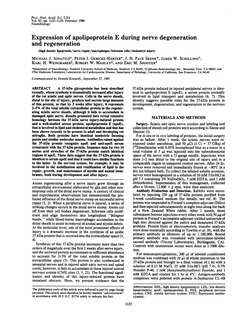
Antibody Characterization. To probe for the specific class-es of cells that secrete the 37-kDa protein and to furthercharacterize the protein itself, we have purified the 37-kDaprotein from injured peripheral nerves (8) and used thispurified protein to generate a monospecific rabbit antiserum.The antibody specifically precipitates metabolically labeled37-kDa protein from nerve-conditioned medium (Fig. 2 Left),establishing that the antibody, raised against NaDodSO4-denatured material, recognizes the native protein. A 20-kDapolypeptide is also precipitated from conditioned mediumunder these conditions; this polypeptide is likely to be afragment of the 37-kDa protein (see below). In electropho-retic transfer or immunoprotein blots, the antibody specifi-cally recognizes the 37-kDa protein from medium condi-tioned by injured nerves (Fig. 2 Right) at dilutions up to1:100,000. Barely detectable levels of the 37-kDa protein arepresent in conditioned medium from control nerves (Fig. 2Right).
Injury of rat optic nerves also induces synthesis ofa 37-kDaprotein (3) that is similar in its two-dimensional electropho-retic profile to sciatic nerve-derived 37-kDa protein. Anti-37-kDa serum recognizes this protein (Fig. 2 Right), furtherindicating its homology to the sciatic nerve 37-kDa protein.
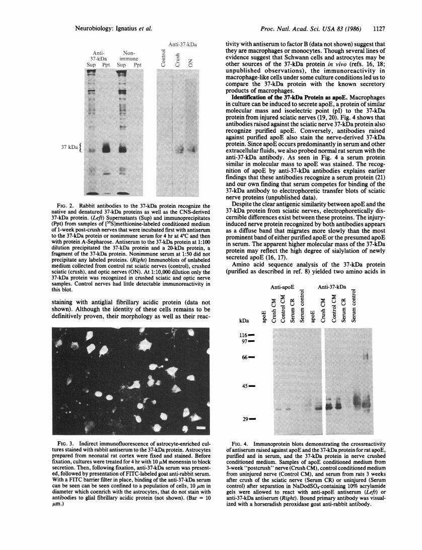
Cultured "Macrophage-Like" Cells Contain 37-kDa Immu-noreactivity. Since homologous 37-kDa proteins are inducedafter nerve injury in the peripheral nervous system (PNS) andCNS, our efforts to locate the cells secreting the 37-kDaprotein have included study of newborn rat brain culturesenriched for astroglia. Cultures 1-2 weeks old were treatedwith monensin to block secretion and allow intracellularaccumulation of the 37-kDa protein. The monensin-treatedcells were fixed and stained with antibodies to the 37-kDaprotein and a fluorescent second antibody. In astroglialcultures the 37-kDa immunofluorescence was localized to asubpopulation of small cells (Fig. 3), which in control,single-labeling experiments do not stain with the astrocytemarker antiglial fibrillary acidic protein (data not shown). Thepresence of astroglia in these cultures was confirmed by
In vitro accumulation* - IEF 8.5
... 11t
'0... t, :0
z
C ontrol Control-1,.... -
- -t : ;d
tv >' s
Al
Crush
FIG. 1. Two-dimensional electrophoret-ic analyses of proteins present in the distalsheath of uninjured and crushed rat sciatic
-OW nerves. (a and c) Fluorographs showing thenewly synthesized, NaDodSO4-soluble pro-teins collected in vivo after direct injection of[35S]methionine into the subperineurialspace of control and crushed nerves. (b andd) Coomassie-stained gels of the accumulat-ed proteins collected in vitro from equivalentsegments of control and crushed nerves.Crushed nerves were examined in vivo 1week after injury and in vitro nerves wereexamined 3 weeks after injury. In both in-stances, contralateral, uncrushed nervesserved as controls. The arrow defines theposition of the 37-kDa protein. IEF,isoelectric focusing.
C
Proc. Natl. Acad. Sci. USA 83 (1986)
..... .....
16.
Proc. Natl. Acad. Sci. USA 83 (1986) 1127
Anti-37-kDa
Sup PptVW
37 kDa I
Non-immuneSup Ppt
Anti-37-kDa
o 2 zv c 0
waB
_i~ - W101h
FIG. 2. Rabbit antibodies to the 37-kDa protein recognize thenative and denatured 37-kDa proteins as well as the CNS-derived37-kDa protein. (Left) Supernatants (Sup) and immunoprecipitates(Ppt) from samples of ['5S]methionine-labeled conditioned mediumof 1-week post-crush nerves that were incubated first with antiserumto the 37-kDa protein or nonimmune serum for 4 hr at 4°C and thenwith protein A-Sepharose. Antiserum to the 37-kDa protein at 1:100dilution precipitated the 37-kDa protein and a 20-kDa protein, afragment of the 37-kDa protein. Nonimmune serum at 1:50 did notprecipitate any labeled proteins. (Right) Immunoblots of unlabeledmedium collected from control rat sciatic nerves (control), crushedsciatic (crush), and optic nerves (ON). At 1:10,000 dilution only the37-kDa protein was recognized in crushed sciatic and optic nervesamples. Control nerves had little detectable immunoreactivity inthis blot.
staining with antiglial fibrillary acidic protein (data notshown). Although the identity of these cells remains to bedefinitively proven, their morphology as well as their reac-
FIG. 3. Indirect immunofluorescence of astrocyte-enriched cul-tures stained with rabbit antiserum to the 37-kDa protein. Astrocytesprepared from neonatal rat cortex were fixed and stained. Beforefixation, cultures were treated for 4 hr with 10 ,uM monensin to blocksecretion. Then, following fixation, anti-37-kDa serum was present-ed, followed by presentation of FITC-labeled goat anti-rabbit serum.With a FITC barrier filter in place, binding of the anti-37-kDa serumcan be seen can be seen confined to a population of cells, 10 ,m indiameter which coenrich with the astrocytes, that do not stain withantibodies to glial fibrillary acidic protein (not shown). (Bar = 10Am.)
tivity with antiserum to factor B (data not shown) suggest thatthey are macrophages or monocytes. Though several lines ofevidence suggest that Schwann cells and astrocytes may beother sources of the 37-kDa protein in vivo (refs. 16, 18;unpublished observations), the immunoreactivity inmacrophage-like cells under some culture conditions led us tocompare the 37-kDa protein with the known secretoryproducts of macrophages.
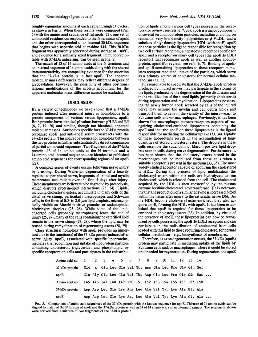
Identification of the 37-kDa Protein as apoE. Macrophagesin culture can be induced to secrete apoE, a protein of similarmolecular mass and isoelectric point (pl) to the 37-kDaprotein from injured sciatic nerves (19, 20). Fig. 4 shows thatantibodies raised against the sciatic nerve 37-kDa protein alsorecognize purified apoE. Conversely, antibodies raisedagainst purified apoE also stain the nerve-derived 37-kDaprotein. Since apoE occurs predominantly in serum and otherextracellular fluids, we also probed normal rat serum with theanti-37-kDa antibody. As seen in Fig. 4 a serum proteinsimilar in molecular mass to apoE was stained. The recog-nition of apoE by anti-37-kDa antibodies explains earlierfindings that these antibodies recognize a serum protein (21)and our own finding that serum competes for binding of the37-kDa antibody to electrophoretic transfer blots of sciaticnerve proteins (unpublished data).
Despite the clear antigenic similarity between apoE and the37-kDa protein from sciatic nerves, electrophoretically dis-cernible differences exist between these proteins. The injury-induced nerve protein recognized by both antibodies appearsas a diffuse band that migrates more slowly than the mostprominent band ofeither purified apoE or the presumed apoEin serum. The apparent higher molecular mass of the 37-kDaprotein may reflect the high degree of sialylation of newlysecreted apoE (16, 17).Amino acid sequence analysis of the 37-kDa protein
(purified as described in ref. 8) yielded two amino acids in
Anti-apoE Anti-37-kDa
0oU _U U _ )
kDa
7-,
C I°n,2 X E
116-97
66- 1
45-
29
FIG. 4. Immunoprotein blots demonstrating the crossreactivityofantiserum raised against apoE and the 37-kDa protein for rat apoE,purified and in serum, and the 37-kDa protein in nerve crushedconditioned medium. Samples of apoE conditioned medium from3-week "postcrush" nerve (Crush CM), control conditioned mediumfrom uninjured nerve (Control CM), and serum from rats 3 weeksafter crush of the sciatic nerve (Serum CR) or uninjured (Serumcontrol) after separation in NaDodSO4-containing 10%o acrylamidegels were allowed to react with anti-apoE antiserum (Left) oranti-37-kDa antiserum (Right). Bound primary antibody was visual-ized with a horseradish peroxidase goat anti-rabbit antibody.
Neurobiology: Ignatius et al.
W.wo-A. n
1128 Neurobiology: Ignatius et al.
roughly equimolar amounts at each cycle through 14 cycles,as shown in Fig. 5. When these results were compared (Fig.5) with the amino acid sequence of rat apoE (22), one set ofamino acid residues corresponded to the N terminus of apoEand the other corresponded to an internal 20-kDa sequencethat begins with aspartic acid at residue 145. This 20-kDafragment was apparently generated during storage at -80TC,and evidence for a similarly sized fragment, immunoprecipi-table with 37-kDa antiserum, can be seen in Fig. 2.The match of 13 of 14 amino acids at the N terminus and
an internal sequence of 14 amino acids along with the sharedimmunoreactivity are compelling evidence for the assertionthat the 37-kDa protein is in fact apoE. The apparentmolecular mass differences may reflect different degrees ofglycosylation. However, the possibility of other posttrans-lational modifications of the protein accounting for theapparent molecular mass difference cannot be excluded.
DISCUSSIONBy a variety of techniques we have shown that a 37-kDaprotein induced after neuronal injury is homologous to aprotein component of various serum lipoproteins, apoE.Both proteins have identical pI values between pH 5.3 and 5.5(6, 7, 19, 20) and similar, though not identical, apparentmolecular masses. Antibodies specific for the 37-kDa proteinrecognize apoE, and anti-apoE serum crossreacts with the37-kDa protein. This indirect evidence for homology betweenthe two proteins is further substantiated by direct comparisonofpartial amino acid sequences. Two fragments ofthe 37-kDaprotein-13 of 14 amino acids at the N terminus and a14-amino acid internal sequence-are identical to publishedamino acid sequences for corresponding regions of rat apoE(22).A complex series of events occurs following nerve injury
by crushing. During Wallerian degeneration of a heavilymyelinated peripheral nerve, fragments of axonal and myelinmembranes accumulate over the first 5 days after injury.These membranes are believed to be degraded by proteolysis,which disrupts protein-lipid interactions (23, 24). Lipids,including cholesteryl esters, accumulate in many cells of thedistal nerve stump, particularly macrophages and Schwanncells, in the form of 0.5- to 2.0-,um lipid droplets, microscop-ically visible as Marchi-positive granules or sudanophilic,birefringent droplets (25, 26). While some of the lipid-engorged cells (probably macrophages) leave the site ofinjury (25, 27), many ofthe cells containing the esterified lipidremain in the nerve stump (27, 28), where the lipid may bereused during remyelination of regenerating axons (28, 29).
Close structural homology with apoE provides an impor-tant clue to the function(s) ofthe 37-kDa protein induced afternerve injury. apoE, associated with specific lipoproteins,mediates the recognition and uptake of lipoprotein particlescontaining cholesterol, triglyceride, and phospholipid byspecific receptors on cells and participates in the redistribu-
tion of lipids among various cell types possessing the recep-tors (for review, see refs. 6, 7, 30). apoE is a major componentof several serum lipoprotein particles, including chylomicronremnants, very low density lipoproteins or 8-VLDL, and asubclass ofhigh density lipoproteins (HDL-with apoE). apoEon these particles is the ligand responsible for recognition bytwo cell surface receptors, a hepatocyte receptor specific forapoE and a receptor on many cell types [the apoB,E(LDL)receptor] that recognizes apoE as well as another apolipo-protein, apoB (for review, see refs. 6, 7). Binding of apoE-and apoB-containing lipoproteins to these receptors stimu-lates receptor-mediated uptake of the particles, which serveas a primary source of cholesterol for normal cellular me-tabolism (31, 32).
It is reasonable to speculate that the 37-kDa (apoE) proteinproduced by injured nerves may participate in the storage ofthe lipids produced by the degeneration ofthe distal axon andin the reutilization of the stored lipids (primarily cholesterol)during regeneration and myelination. Lipoproteins possess-ing the newly formed apoE secreted by cells of the injurednerve may acquire the myelin and cell debris lipids andtransport them to cells in the vicinity of the injury-e.g., toSchwann cells and/or macrophages. Previously, it has beenshown that macrophages possess receptors capable of rec-ognizing cholesterol-enriched lipoproteins containing theapoE and that the apoE on these lipoproteins is the ligandresponsible for mediating the cellular uptake (33, 34). Uptakeof these lipoproteins results in the accumulation of largequantities of stored cholesteryl esters. The droplets in thesecells resemble the sudanophilic, Marchi-positive lipid drop-lets seen in cells during nerve degeneration. Furthermore, ithas been shown that the cholesteryl esters stored in themacrophages can be mobilized from these cells when asuitable acceptor is present in the medium (33, 35). The mostwidely studied acceptor capable of acquiring the cholesterolis HDL. During this process of lipid mobilization thecholesteryl esters within the cells are hydrolyzed to freecholesterol, which is released from the cell. The cholesterolacquired by the HDL is then reesterified by the plasmaenzyme lecithin:cholesterol acyltransferase. [It is notewor-thy that the production of a similar enzyme is increased 5-foldwithin the tissue after injury to the rat sciatic nerve (36).] Asthe HDL become cholesteryl ester-enriched, they also ac-quire apoE, forming the HDL-with apoE. It has been estab-lished that apoE is required for these lipoproteins to beenriched in cholesteryl esters (33). In addition, by virtue ofthe presence of apoE, these lipoproteins can now be recog-nized by cells possessing the apoB,E(LDL) receptors and canparticipate in the redistribution of cholesterol from cellsloaded with this lipid to those requiring cholesterol for normalcellular metabolism-e.g., biosynthesis of membranes.
Therefore, as axon degeneration occurs, the 37-kDa (apoE)protein may participate in mediating uptake of the lipids bySchwann cells and/or macrophages, where it could be storeduntil needed for regeneration. During regeneration, the apoE
Amino acid no. 1 2 3 4 5 6 7 8 9 10 1 1 12 13 14
37-kDa protein Glu x Glu Leu Glu Val Thr Asp Gln Leu Pro Gly Gln Ser
apoE Glu Gly Glu Leu Glu Val Thr Asp Gln Leu Pro Gly Gln Ser .g
Amino acid no. 145 146 147 148 149 150 151 152 153 154 155 156 157 158
37-kDa protein Asp Asp Leu Gln Lys Arg Leu Ala Val Tyr Lys Ala Gly Ala
apoE Asp Asp Leu Gln Lys Arg Leu Ala Val Tyr Lys Ala Gly Ala
FIG. 5. Comparison of amino acid sequences of the 37-kDa protein with the known sequence for apoE. Thirteen of 14 amino acids can bealigned to match at the N termini of apoE and the 37-kDa protein as well as 14 of 14 amino acids in an internal fragment. The sequences shownwere derived from a mixture of two fragments of the 37-kDa protein.
Proc. Natl. Acad. Sci. USA 83 (1986)

Proc. Natl. Acad. Sci. USA 83 (1986) 1129
could participate in the redistribution of the cholesterol toneurons needing this lipid for axon growth and to Schwanncells for remyelination. This postulate is supported by thetemporal sequence of events following nerve injury: accu-mulation of the 37-kDa (apoE) protein (3, 37), accumulationof lipid-laden cells (25), and presence of an increase incholesterol-esterifying activity (23, 36). All three parametersbegin to increase 4-5 days after nerve injury and reach theirmaximum by 3-4 weeks. In the CNS, synthesis of the 37-kDaprotein is induced by nerve injury (3), but the accumulationof the protein is far less than in peripheral nerves (3, 5). Adirect role for the 37-kDa protein in the clearance of axonaland myelin membrane debris might explain the slower timecourse of degeneration in the CNS, where degradation ofmyelin debris and formation oflipid storage droplets continueover a period of several months (38), and cholesteryl estercontent does not change (39).
In addition to the possible role ofthe 37-kDa (apoE) proteinin lipid transport and metabolism, it may participate in theregulation of other aspects of cellular metabolism within thedegenerating and regenerating axon. For example, it has beenshown that apoE interacts with specific receptors on lym-phocyte membranes and renders these cells unresponsive tonormal mitogenic stimulation (40, 41). Therefore, it is pos-sible that the apoE regulates Schwann cell proliferation orother aspects of cellular metabolism, including axon growth.
Antibodies to glial fibrillary acidic protein were kindly provided byL. Eng, Veteran's Administration Hospital, Palo Alto, CA 94305. Wewould like to thank Jack Snipes and John Freeman, who havereached conclusions similar to our own (42), for sharing informationprior to publication. This research was supported by grants from theNational Institutes of Health (NS 04270, NS 20178), the Spinal CordSociety, and the Isabelle M. Niemala Fund. M.J.I. was supported byGrantMH 17047; P.J.G.-H. was supported in part by a grant from theMax Planck Society.
1. Forssman, J. (1898) Ziegler's Beitr. 24, 56-100.2. Benfey, M. & Aguayo, A. J. (1982) Nature (London) 296,
150-152.3. Skene, J. H. P. & Shooter, E. M. (1983) Proc. Natl. Acad.
Sci. USA 80, 4169-4173.4. Politis, M. J., Pellegrino, R. G., Oaklander, A. L. & Ritchie,
J. M. (1983) Brain Res. 273, 392-395.5. Muller, H. W., Gebicke-Harter, P. J., Hangen, D. H. &
Shooter, E. M. (1985) Science 228, 499-501.6. Mahley, R. W. & Innerarity, T. L. (1983) Biochim. Biophys.
Acta 737, 197-222.7. Mahley, R. W., Innerarity, T. L., Rall, S. C., Jr., & Weisgra-
ber, K. H. (1984) J. Lipid Res. 25, 1277-1294.8. Ignatius, M. J., Muller, H. W., Skene, J. H. P. & Shooter,
E. M. (1984) Soc. Neurosci. Abstr. 10, 1029.9. Towbin, H., Staehelin, T. & Gordon, J. (1979) Proc. Nati.
Acad. Sci. USA 76, 4350-4354.10. Raff, M. C., Fields, K. L., Hakomori, S. I., Mirsky, R.,
Pruss, R. M. & Winter, J. (1979) Brain Res. 174, 283-308.11. Hunkapiller, M. W. & Hood, L. E. (1983) Methods Enzymol.
91, 486-493.12. Laemmli, U. K. (1970) Nature (London) 227, 680-685.
13. O'Farrell, P. H. (1975) J. Biol. Chem. 250, 4007-4021.14. Fairbanks, G., Steck, T. L. & Wallach, D. F. (1971) Biochem-
istry 10, 2606-2617.15. Jen, G. & Thach, R. E. (1982) J. Virol. 43, 250-261.16. Boyles, J., Pitas, R. E., Wilson, E., Mahley, R. W. & Taylor,
J. M. (1985) J. Clin. Invest. 76, 1505-1513.17. Zannis, V. I., Breslow, J. L., Utermann, G., Mahley, R. W.,
Weisgraber, K. H., Havel, R. J., Goldstein, J. L., Brown,M. S., Schonfeld, G., Hazzard, W. R. & Blum, C. (1982) J.Lipid Res. 23, 911-914.
18. Halks-Miller, M. (1985) J. Neuropathol. Exp. Neurol. 44, 344(abstr.).
19. Basu, S. K., Brown, M. S., Ho, Y. K., Havel, R. J. & Gold-stein, J. L. (1981) Proc. Nati. Acad. Sci. USA 78, 7545-7549.
20. Basu, S. K., Ho, Y. K., Brown, M. S., Bilheimer, D. W.,Anderson, R. G. W. & Goldstein, J. L. (1982) J. Biol. Chem.257, 9788-9795.
21. Snipes, G. J. & Freeman, F. A. (1984) Soc. Neurosci. Abstr.10, 1029.
22. McLean, J. W., Fukazawa, C. & Taylor, J. M. (1983) J. Biol.Chem. 258, 8993-9000.
23. Rossiter, R. J. (1955) in Neurochemistry, eds. Elliot, K. A. C.,Page, I. H. & Quastel, J. H. (Thomas, Springfield, IL), 1st Ed.pp. 696-714.
24. Hallpike, J. F. & Adams, C. W. M. (1969) Histochem. J. 1,559-578.
25. Williams, P. L. & Hall, S. M. (1971) J. Anat. 109, 487-503.26. Bignami, A., Dahl, D., Nguyen, B. Y. & Crosby, C. J. (1981)
J. Neuropathol. Exp. Neurol. 40, 537-50.27. Cajal, S. R. Y. (1928) in Degeneration and Regeneration ofthe
Nervous System, ed. May, R. M. (Oxford Univ. Press, Lon-don), pp. 329-361, 368-375.
28. Rawlins, F. A., Hedley-Whyte, E. T., Villegas, G. & Uzman,B. G. (1970) Lab. Invest. 22, 237-240.
29. Rawlins, F. A., Villegas, G. M., Hedley-Whyte, E. T. & Uz-man, B. G. (1972) J. Cell Biol. 52, 615-625.
30. Brown, M. S., Kovanen, P. T. & Goldstein, J. L. (1981)Science 212, 628-635.
31. Goldstein, J. L. & Brown, M. S. (1974) J. Biol. Chem. 249,5153-5162.
32. Goldstein, J. L. & Brown, M. S. (1977) Annu. Rev. Biochem.46, 897-930.
33. Koo, C., Innerarity, T. L. & Mahley, R. W. (1985) J. Biol.Chem. 260, 11934-11943.
34. Innerarity, T. L., Arnold, K. S., Weisgraber, K. H. &Mahley, R. W. (1985) Arteriosclerosis, in press.
35. Gordon, V., Innerarity, T. L. & Mahley, R. W. (1983) J. Biol.Chem. 258, 6202-6212.
36. Yao, J. K. & Dyck, P. J. (1981) J. Neurochem. 37, 156-163.37. Muller, H. W., Ignatius, M. J. & Shooter, E. M. (1983) Soc.
Neurosci. Abstr. 9, 45.38. Franson, P. & Ronnevi, L. (1984) J. Comp. Neurol. 223,
138-151.39. Eto, Y., Suzuki, K. & Suzuki, K. (1971) J. Lipid Res. 12,
570-579.40. Hui, D. Y., Harmony, J. A. K., Innerarity, T. L. & Mahley,
R. W. (1980) J. Biol. Chem. 255, 11775-11781.41. Avila, E. M., Holdsworth, G., Sasaki, M., Jackson, B. L. &
Harmony, J. A. K. (1982) J. Biol. Chem. 257, 5900-5909.42. Snipes, G. J., McGuire, C. B., Norden, J. J. & Freeman, J. A.
(1986) Proc. Natl. Acad. Sci. USA 83, 1130-1134.
Neurobiology: Ignatius et al.