Embed Size (px)
Citation preview

UNIVERSIDADE ESTADUAL DE CAMPINAS
FACULDADE DE ODONTOLOGIA DE PIRACICABA
BRUNO AUGUSTO LINHARES ALMEIDA MARIZ
EXPRESSÃO IMUNO-HISTOQUÍMICA DE FGF-2 E FGFR-1 EM
CARCINOMA ESPINOCELULAR ORAL DE LÍNGUA
FGF-2 AND FGFR-1 IMMUNOEXPRESSION IN ORAL TONGUE
SQUAMOUS CELL CARCINOMA
Piracicaba
2018
BRUNO AUGUSTO LINHARES ALMEIDA MARIZ
EXPRESSÃO IMUNOISTOQUÍMICA DE FGF-2 E FGFR-1 EM
CARCINOMA ESPINOCELULAR ORAL DE LÍNGUA
FGF-2 AND FGFR-1 IMMUNOEXPRESSION IN ORAL TONGUE
SQUAMOUS CELL CARCINOMA
Dissertação apresentada à Faculdade de
Odontologia de Piracicaba da Universidade
Estadual de Campinas como parte dos requisitos
exigidos para a obtenção do título de Mestre em
Estomatopatologia, na Área de Patologia.
Dissertation presented to the Piracicaba Dental
School of the University of Campinas in partial
fulfilment of the requirements for the degree of
Master in Oral Medicine and Oral Pathology, in
Pathology area.
Orientador: Prof. Dr. Jacks Jorge Júnior
ESTE EXEMPLAR CORRESPONDE À VERSÃO FINAL DA DISSERTAÇÃO DEFENDIDA PELO ALUNO BRUNO AUGUSTO LINHARES ALMEIDA MARIZ, E ORIENTADA PELO PROF. DR. JACKS JORGE JÚNIOR.
Piracicaba
2018
Ficha Catalográfica

DEDICATÓRIA
Dedico este trabalho à minha mãe, Maria de Fatima Linhares, pelo amor
incondicional, pelo apoio constante, por ser a minha fortaleza; nunca serei capaz de
retribuir tudo o que renunciou para entregar a mim e aos meus irmãos; e ao meu pai,
Carlos Augusto de Almeida Mariz, que nos deixou antes de ver seus filhos
crescerem, mas cuja presença constante se mantem em nossos corações, sei que
hoje estaria muito orgulhoso.

AGRADECIMENTOS
Ao Prof. Dr. Jacks Jorge Júnior, pela orientação desse trabalho, a quem
agradeço profundamente por todos os ensinamentos, oportunidades e confiança que
vem me oferecendo.
Á Faculdade de Odontologia de Piracicaba da Universidade Estadual de
Campinas, na pessoa do seu Diretor, Prof. Dr. Henrique Guilherme Peçanha.
Ao Prof. Dr. Marcio Ajudarte Lopes, coordenador do programa de Pós-
Graduação em Estomatopatologia da Faculdade de Odontologia de Piracicaba da
Universidade Estadual de Campinas, pelo apoio e confiança constantemente
dedicados aos alunos.
Ao Prof. Dr. Oslei Paes de Almeida, pela oportunidade de participar da rotina
histopatológica no Laboratório de Patologia Oral da FOP-UNICAMP, por todo o
conhecimento ao qual nos contempla diariamente e pelo exemplo de força e
profissionalismo a ser seguido, sou extremamente grato.
Aos Profs. Drs. Alan Roger dos Santos Silva, Edgard Graner, Jacks Jorge
Júnior, Márcio Ajudarte Lopes, Oslei Paes de Almeida, Pablo Agustin Vargas e
Ricardo Della Coletta, professores das áreas de Patologia e Semiologia da
Faculdade de Odontologia de Piracicaba – UNICAMP, pelos valiosos ensinamentos
transmitidos durante esses dois anos de mestrado.
À Coordenação de Aperfeiçoamento de Pessoal de Nível Superior
(CAPES), pela concessão da bolsa de mestrado.
Aos meus colegas de programa, pela convivência diária, amizade e apoio: Ana
Camila, Anna, Bete, Celeste, Cinthia, Débora Bastos, Felipe, Florence, Gleyson,
Iara, Jamile, Juliana Kern, João, Lígia, Luan, Mariana, Paola, Patrícia, Rachel,
Raísa, Renata, Renato, Thayná, Vinícios e Wagner; e especialmente a Natália e
Rodrigo por me acolherem assim que cheguei e pelo apoio e amizade constantes
desde então, sempre os terei no meu coração; assim como a Carol, Diego, Débora
Lima, Isabel, Juliana Souza, Marisol, Maurício e Pedro que se tornaram mais que
amigos ao longo desses anos, vocês são a minha família piracicabana. Aos colegas
de orientação e amigos queridos Leonardo e Priscila, por tudo que me ensinaram, e

especialmente a Ciro: não tenho palavras para agradecer toda a ajuda, apoio e
confiança, te admiro muito e com certeza não estaria aqui sem a sua ajuda, muito
obrigado! Também aos que conheci e hoje traçam brilhantes jornadas: Camila,
Carine, Felipe Fonseca e Rebeca. Enfim, muito obrigado pela parceria.
A Fabiana Facco Casarotti e Adriano Luis Martins, técnicos do Laboratório
de Patologia Oral, por tudo que me ensinaram e pela ajuda constante nos momentos
que mais precisei.
Às professoras de Patologia Oral da Faculdade de Odontologia da
Universidade Federal da Paraíba – UFPB, Francineide Almeida, Hannah Verheul,
Marize Rosa e Socorro Aragão, sempre serei grato pela amizade, por todo o apoio
que sempre me deram e por terem me ajudado a descobrir minha vocação.
A toda minha família, por sonharem junto comigo e sempre apoiarem minhas
decisões. Vocês são meu porto seguro.
A Bruna Cavalcanti, pelo carinho e apoio constantes, pelo amor que nos
manteve unidos mesmo estando tão longe, tenho muita sorte de poder contar contigo.
E à Sandra Cavalcanti, por ter praticamente me adotado como um filho, muito
obrigado por tudo.

“Today we still yearn to know why we are
here and where we came from. Humanity's
deepest desire for knowledge is justification
enough for our continuing quest”
Stephen Hawking

RESUMO
O fator de crescimento fibroblástico 2 (FGF-2) e o receptor do fator de crescimento
fibroblástico 1 (FGFR-1) estão associados à maior capacidade de invasão tumoral,
proliferação celular, angiogênese e ao potencial de metástase. Este estudo teve como
objetivo investigar a expressão de FGF-2 e FGFR-1 na displasia epitelial oral (DEO)
e carcinoma espinocelular de língua (CECL). Foram selecionados retrospectivamente
cento e sessenta e sete casos, incluindo 85 espécimes cirúrgicos de pacientes com
CECL, provenientes do Hospital Onofre Lopes, Natal, Brasil, além de 46 biópsias
incisionais de CECL e 36 de DEO provenientes do arquivo do Laboratório de Patologia
Oral da Faculdade de Odontologia de Piracicaba (UNICAMP). Cortes de tecido
parafinado foram submetidas à reação imunoistoquímica para FGF-2 e FGFR-1. As
lâminas foram escaneadas e a marcação imunoistoquímica foi quantificada
digitalmente pelo software Aperio Positive Pixel Count v9. Cinco áreas iguais foram
selecionadas no estroma e no epitélio tumoral. Os resultados foram exportados e o
score final de cada lesão foi obtido através da soma da porcentagem de pixels fracos,
moderados e fortes, gerando um valor que variou de 100 a 300. Os casos foram
divididos como tendo “baixa expressão” ou “alta expressão” das proteínas de acordo
com a mediana dos grupos. FGF-2 e FGFR-1 foram mais expressos em DEO de alto
grau do que em DEO de baixo grau, tanto no epitélio, como nas células do estroma
(p<0.05). A sobrevivência doença específica (SDE) em 5 anos foi de 47,3% dos
pacientes com CECL. A alta expressão de FGF-2 nas células inflamatórias e
mesenquimatosas do estroma foi associada à invasão vascular e pior prognóstico.
Pacientes com alta expressão de FGF-2 no estroma apresentaram taxa de SDE em 5
anos de 36,7%, contra 59,3% dos pacientes com baixa expressão (HR: 2,272; IC95%:
1.213-4.254; p=0,008). A alta expressão de FGFR-1 no estroma foi correlacionada
com metástases linfonodais e metástases à distância. Pacientes com alta expressão
de FGFR-1 no epitélio apresentaram taxa de SDE em 5 anos de 22,9%, contra 75,6%
dos pacientes com baixa expressão (HR: 2,594; IC95%: 1,390-4,841; p=0,003). O
mesmo aconteceu com FGFR-1 no estroma, com taxa de SDE em 5 anos de 32,9%,
contra 64,0% (HR: 3,378; IC95%: 1,816-6,286; p=0,001). A análise multivariada de
Cox confirmou que a expressão de FGF-2 no estroma (HR: 2,197; IC95%: 1,128-
4,282; p=0,02), FGFR-1 no epitélio (HR: 3,178; IC95%: 1,505-6,709; p=0,002) e

FGFR-1 no estroma (HR: 3,041; IC95%: 1,454-6,356; p=0,003) estão fortemente
associadas a um maior risco de morte relacionada ao CECL. Em conjunto, nossos
achados demonstram que FGF-2 e FGFR-1 desempenham um papel importante na
DEO e no CECL, estando associados à presença de metástase e à sobrevivência dos
pacientes.
Palavras-chave: fator de crescimento fibroblástico, câncer oral, FGF-2, FGFR-1,
displasia epitelial oral
ABSTRACT
Fibroblast growth factor 2 (FGF-2) and fibroblast growth factor receptor 1
(FGFR-1) expression is associated with tumour invasiveness, cell proliferation,
angiogenesis and metastasis potential. This study aimed to investigate FGF-2 and
FGFR-1 expression in oral epithelial dysplasia (OED) and tongue squamous cell
carcinoma (TSCC). One hundred and sixty-seven cases were retrospectively selected,
including 85 surgical specimens of patients with TSCC, from Onofre Lopes Hospital,
Natal, Brazil, besides 46 TSCC and 36 OED incisional biopsies from the Laboratory of
Oral Pathology, Piracicaba Dental School, University of Campinas (UNICAMP). Tissue
sections were submitted to immunohistochemical reaction for FGF-2 and FGFR-1.
Slides were scanned and the immunostaining was digitally quantified by the Aperio
Positive Pixel Count v9 software. Five areas were selected from the stroma and the
epithelium. Results were exported and the final score of each lesion was obtained by
the sum of the percentage of weak, moderate and strong pixels, resulting in a value
ranging from 100 to 300. Cases were classified as “weak expression” or “high
expression” of the proteins, accordingly to the group median. FGF-2 and FGFR-1 were
more expressed in high-grade OED than in low-grade OED, either in the stroma or in
the epithelium (p<0.05). The 5-year disease-specific survival (DSS) rate was 47.3% of
the patients with TSCC. FGF-2 high expression in the inflammatory and mesenchymal
cells of the stroma was associated with vascular invasion and worse prognosis.
Patients with high expression of FGF-2 in the stroma had a 5-year DSS of 36.7%,
against 59.3% of patients with low expression (HR: 2.272; CI(95%): 1.213-4.254;
p=0.008). FGFR-1 high expression in the stroma was correlated with lymph node
metastasis and distant metastasis. Patients with high expression of FGFR-1 in the
tumour had a 5-year DSS of 22.9%, against 75.6% of patients with low expression
(HR: 2.594; CI(95%): 1.390-4.841; p=0.003). The same was observed with FGFR-1
in the stroma, with a 5-year DSS of 32.9%, against 64.0% (HR: 3.378; CI(95%): 1.816-
6.286; p=0.001). The Cox multivariate analysis confirmed that the expression of FGF-
2 in the stroma (HR: 2.197; CI(95%): 1.128-4.282; p=0.02), FGFR-1 in the tumour (HR:
3.178; CI(95%): 1.505-6.709; p=0.002) and FGFR-1 in the stroma (HR: 3.041;
CI(95%): 1.454-6.356; p=0.003) are strongly associated with a higher risk of death
related to TSCC. Taken together, our findings demonstrate that FGF-2 and FGFR-1

play an important role in OED and TSCC, and are associated with the presence of
metastasis and patients disease-specific survival.
Key Words: fibroblast growth factor, oral cancer, FGF-2, FGFR-1, oral epithelial
dysplasia

LISTA DE ABREVIATURAS E SIGLAS
AP – Adenoma Pleomorfo
CEC – Carcinoma Espinocelular
CI – Confidence interval
CxAP – Carcinoma ex-Adenoma Pleomorfo
DEO – Displasia epitelial oral
DSS – Disease-specific survival
DOPM – Desordens orais potencialmente malignas
EMT – Epithelial-mesenchymal transition
FGF-2 – Fibroblast growth factor 2
FGFR-1 – Fibroblast growth factor receptor 1
HPV – Papiloma vírus humano
HNSCC – Head and Neck Squamous cells carcinoma
HR – Hazard ratio
IC – Intervalo de confiança
LLLT – Low level laser therapy
LPO – Líquen plano oral
OED – Oral epithelial dysplasia
OSCC – Oral squamous cells carcinoma
PMD – Potentially malignant disorder
RNA – Ácido ribonucleico
SCC - Squamous cells carcinoma
TEM – Transição Epitélio Mesênquima
TSCC – Tongue squamous cells carcinoma

SUMÁRIO
1 INTRODUÇÃO........................................................................................................15
1.1 Expressão de FGF-2/FGFR-1 na mucosa oral normal
e durante o processo de reparo.................................................................16
1.2 Expressão de FGF-2/FGFR-1 nas Glândulas Salivares.............................18
1.3 Expressão de FGF-2/FGFR-1 em Desordens
Orais Potencialmente Malignas..................................................................19
1.4 Expressão de FGF-2/FGFR-1 em
Carcinoma Espinocelular Oral....................................................................20
2 ARTIGO: FGF-2 and FGFR-1 might be considered as prognostic
factors in Tongue Squamous Cell Carcinoma......………………………..22
3 CONCLUSÃO.........................................................................................................44
REFERÊNCIAS..........................................................................................................45
ANEXOS
Anexo 1: Certificado de submissão do artigo..................................................52
Anexo 2: Certificado do Comitê de Ética em Pesquisa...................................53
15
1 INTRODUÇÃO:
O câncer oral é uma doença multifatorial considerada um problema de saúde
pública, principalmente em países em desenvolvimento. Dados do projeto
GLOBOCAN apontam que em 2012 surgiram 300.373 casos de câncer na cavidade
oral e que mais de 145.000 pessoas morreram em decorrência da doença em todo o
mundo. No Brasil, a estimativa para o ano de 2018 é de 11.200 novos casos de câncer
na cavidade oral em homens e 3.500 casos em mulheres, tornando-o a quinta
neoplasia maligna mais comum em homens no Brasil, quando excluímos o câncer de
pele não-melanoma (INCA, 2018).
Os Carcinomas Espinocelulares (CEC) orais representam mais de 90% dos
casos de câncer oral em todo o mundo, sendo mais comum entre a sexta e sétima
décadas de vida e comumente associado a fatores de risco, como o tabagismo (Sloan
et al., 2017). O consumo de bebidas alcoólicas age juntamente com o hábito de fumar
como um fator de risco sinérgico. O sachê de bethel, associado ou não ao tabaco é
responsável por casos de câncer em regiões específicas onde esses hábitos são
culturais. No câncer de orofaringe, o HPV (principalmente o tipo 16) também é
considerado fator de risco, apesar de presente em uma pequena quantidade de casos
(Saraiya et al., 2015). Já no câncer de lábio, a radiação ultravioleta é o principal fator
de risco (Sloan et al., 2017). O CEC oral de língua é o tipo de câncer oral mais
agressivo e muitas vezes seu tratamento deve ser multimodal mesmo nos estágios
iniciais da doença, mostrando uma maior taxa de mortalidade que em outros sítios da
cavidade oral (Almangush et al., 2015; Kauppila et al., 2015).
O Fator de Crescimento Fibroblástico 2 (FGF-2, Fibroblast Growth Factor 2),
também conhecido como Fator de Crescimento Fibroblástico básico (b-FGF, basic
Fibroblast Growth Factor), é um dos principais constituintes da família FGF (Nayak et
al., 2015), que compreende 22 membros de proteínas secretadas agindo como fatores
parácrinos, autócrinos ou endócrinos (Giacomini et al., 2016) que sinalizam por meio
de dos receptores FGF (Turner & Grose, 2010).
A família dos receptores dos Fatores de Crescimento Fibroblásticos (FGFR) é
composta por quatro (FGFR-1-4) receptores transmembrana de tirosina quinase
expressos em diferentes tipos celulares (Ipenburg et al., 2016). A sinalização FGF-
FGFR é responsável por diferentes mecanismos celulares, incluindo comportamentos

16
malignos, como proliferação e invasão tumoral, aumento da angiogênese e
consequente impacto nas taxas de sobrevida (Turner e Grose, 2010).
FGF-2 é uma citocina multifuncional expressa em diversos tecidos e atua numa
grande variedade de atividades biológicas (Delrieu, 2000; Nayak et al., 2015), como
proliferação e diferenciação em vários tipos celulares (Delrieu, 2000). Além do seu
papel na embriogênese, ela regula funções homeostáticas na vida adulta (Giacomini
et al., 2016), participando de diferentes processos, incluindo angiogênese (Brizeno et
al., 2016) e reparo tecidual (Harada et al., 2017).
FGF-2 tem cinco isoformas criadas de um único RNA mensageiro por iniciação
de tradução alternativa (Delrieu, 2000). A proteína de baixo peso molecular tem 18kDa
e está presente no citoplasma, de onde é secretada, agindo por meio de seus
receptores (FGFR) (Martinez et al., 2010). Por outro lado, as quatro isoformas de alto
peso molecular estão localizadas no núcleo, onde seus sinais agem por meio de um
mecanismo intácrino, ou seja, independentemente dos FGFR (Delrieu, 2000).
FGFR-1 tem sido indicado como um potencial alvo molecular em diferentes
tipos de câncer, incluindo o CEC oral (Clauditz et al., 2017), e inibidores do FGFR-1
têm sido usados na tentativa de impedir os processos de carcinogênese e transição
epitélio-mesênquima (TEM) (Nguyen et al., 2013).
1.1 Expressão de FGF-2/FGFR-1 na mucosa oral normal e durante o processo de
reparo:
FGF-2 é normalmente expresso nas camadas basal e parabasal do epitélio da
mucosa oral (Forootan et al., 2000; Wakulich et al., 2002), enquanto FGFR-1 é
expresso nas camadas sobrejacentes (Forootan et al., 2000). Brizeno et al. (2016)
estudou os efeitos da diabetes no processo de reparo da mucosa oral, encontrando
uma menor expressão de FGF-2 em ratos diabéticos que em controles
normoglicêmicos, e portanto, um atraso significativo nos processos de angiogênese e
produção de colágeno, prejudicando o mecanismo de reparo de feridas. Fujisawa et
al. (2003) aplicou FGF-2 para tratar úlceras em coelhos, e foi capaz de promover a
cicatrização das lesões induzindo a proliferação de fibroblastos e ceratinócitos.

17
Usumez et al. (2014) usou terapia com laser de baixa intensidade (LLLT) para
tratar mucosite oral em ratos, mostrando que o tratamento acelerava o processo de
cicatrização da mucosite pelo aumento dos níveis de FGF-2. Harada et al. (2017)
também estudou mucosite oral em hamsters, induzida por 5-fluorouracil, e notou que
a maior expressão de FGF-2 estava relacionada a uma recuperação acelerada da
mucosa.
Morelli et al. (2011) notou a importância do FGF-2 durante os eventos iniciais
do reparo em pacientes tratados com um enxerto celular vivo como um procedimento
de aumento gengival, e uma pasta contendo FGF-2 foi usada clinicamente por Jiang
et al. (2013) para tratar estomatite aftosa menor recidivante, aliviando
substancialmente a intensidade da dor e acelerando a cicatrização das lesões nesses
pacientes.
Jansen et al. (2009) implantou enxertos de colágeno com FGF-2 no palato de
ratos, o que levou a um aumento na celularidade e vascularização. Além disso, houve
um número diminuído de miofibroblastos, diminuindo a contração das feridas, um dos
maiores problemas na cicatrização de feridas no palato. Estes resultados são
similares aos de Hata et al. (2008), que administrou FGF-2 em lesões no palato de
ratos, melhorando o suprimento vascular durante o reparo. Matsumoto et al. (2012)
usou uma esponja de colágeno contendo FGF-2 para regenerar defeitos ósseos em
cachorros, promovendo um reparo mais eficiente. Kanda et al. (2003) estudou a
imunoexpressão de FGFRs em feridas na mucosa palatina de ratos, e achou uma alta
expressão de FGFR-1 em miofibroblastos durante o processo de reparo.
Mullane et al. (2008) tratou polpas dentais com FGF-2, melhorando a
neovascularização, podendo ser usada como um tratamento tópico antes do
reimplante de dentes avulsionados. Além disso, FGF-2 é naturalmente secretado
pelas células da polpa dental, principalmente após algum trauma (Tran-Hung et al.,
2008). Além disso, a injeção de FGF-2 na câmara pulpar e canal radicular de dentes
humanos tratados endodonticamente por Kim et al. (2010) resultou na regeneração
de tecido semelhante à polpa dental após implantação ectópica in vivo, o que
representa uma possível alternativa futura durante o tratamento endodôntico.

18
1.2 Expressão de FGF-2/FGFR-1 nas Glândulas Salivares:
Nas glândulas salivares normais, FGF-2 é expresso na membrana basal dos
ductos intercalados, ácinos e células basais dos ductos excretores (Kusafuka et al.,
1998; Myoken et al., 1996), e como as glândulas salivares apresentam propriedades
regenerativas fracas, FGF-2 tem sido usado na regeneração tecidual de glândulas
submandibulares de ratos cirurgicamente danificadas, melhorando sua capacidade de
regeneração (Kobayashi et al., 2016).
FGF-2 também tem sido administrado para tentar reparar células das glândulas
salivares danificadas pela radioterapia (Guo et al., 2014; Kojima et al., 2011; Thula et
al., 2005). Thula et al. (2005) administrou FGF-2 antes e imediatamente após irradiar
células da glândula parótida de ratos, e encontrou um efeito radioprotetor associado
com aumento de parada do ciclo celular em G2, permitindo maior tempo para corrigir
danos ao DNA causados pela radioterapia. Kojima et al. (2011) administrou FGF-2
nas glândulas submandibulares irradiadas de camundongos, que mostraram um
aumento do número de células acinares no grupo tratado. Guo et al. (2014) testou um
adenovírus codificante de FGF-2 nas glândulas parótidas irradiadas de porcos,
limitando o declínio do fluxo salivar, causado pela proteção das células endoteliais da
microvasculatura da parótida pelo tratamento com FGF-2.
Em Adenomas Pleomórficos (AP), o tumor de glândula salivar mais comum
(Sloan et al., 2017), FGF-2 e FGFR-1 estão presentes na membrana basal dos ninhos
de células mioepiteliais, ao redor de células mioepiteliais nas áreas mixoides e nas
células das lacunas das áreas condroides (Kusafuka et al., 1998). Miguita et al. (2010)
encontrou expressão citoplasmática de FGF-2 e expressão nuclear de FGFR-1 em
células mioepiteliais de AP, enquanto Persson et al. (2008) encontrou fusão e
amplificação do gene FGFR-1 nessas lesões. Além disso, Soares et al. (2012)
encontrou alta expressão de FGF-2 em AP que recorreram. Em sua contraparte
maligna, o Carcinoma ex-Adenoma Pleomórfico (CxAP), as células mioepiteliais
benignas apresentaram alta expressão de FGF-2 (tanto a isoforma de baixo peso
molecular como de alto peso molecular) e ausência de expressão de FGFR-1. Já as
células epiteliais malignas mostraram menor expressão de FGF-2, mas apresentaram
expressão de FGFR-1, tanto citoplasmática como nuclear (Furuse et al., 2010;
Martinez et al., 2010). A presença do receptor de FGF-2 no epitélio maligno pode
indicar que existe uma resposta parácrina nessa lesão.
19
Myoken et al. (1996) encontrou maior expressão de FGF-2 e FGFR-1 em
tumores malignos de glândula salivar, e sugeriram que esses fatores podem contribuir
para a progressão do tumor de forma autócrina. Resultados similares foram
encontrados por Sumitomo et al. (1999), onde essas proteínas foram co-expressas
nas células que sofreram transformação maligna em CEC de glândulas
submandibulares de ratos. Além disso, níveis de FGF-2 e FGFR-1 estão aumentados
no soro e na saliva de pacientes com tumores de glândulas salivares benignos e
malignos (Huang et al., 2012). Adicionalmente, Ach et al. (2016) encontrou aberrações
no gene FGFR-1 em carcinomas de glândula salivar.
1.3 Expressão de FGF-2/FGFR-1 em Desordens Orais Potencialmente Malignas:
FGF-2 parece estar superexpresso no epitélio displásico de desordens orais
potencialmente malignas (DOPM) (Wakulich et al., 2002; Raimondi et al., 2006; Bishen
et al., 2008; Nayak et al., 2015). Wakulich et al. (2002) estudou a expressão de FGF-
2 em diferentes graus de displasia epitelial, carcinoma in situ e CEC oral, e encontrou
aumento da expressão de FGF-2 de acordo com a progressão das displasias para
CEC, principalmente nas camadas superiores do epitélio. Na fibrose submucosa oral
(FSO), uma DOPM caracterizada pela fibrose da mucosa que dificulta a abertura
bucal, FGF-2 é superexpresso nos fibroblastos e células endoteliais nos estágios
iniciais da doença, enquanto sua expressão na matriz do estroma aumenta nos
estágios finais da doença (Bishen et al., 2008).
Nayak et al. (2015) estudou 72 casos de DOPM, incluindo leucoplasia e FSO,
onde a expressão de FGF-2, tanto pela técnica de imunoistoquímica, como a nível
molecular (expressão gênica), estava associada com a transformação maligna de
DOPM em CEC oral. Gorugantula et al. (2012) encontrou altos níveis de FGF-2 na
saliva de pacientes com Líquen Plano Oral (LPO), em comparação com controles
saudáveis. Não existem estudos correlacionando a expressão de FGFR-1 com
DOPM.

20
1.4 Expressão de FGF-2/FGFR-1 em Carcinoma Espinocelular Oral:
A expressão de FGF-2 e FGFR-1 tem sido correlacionada com menor
diferenciação, maior potencial de invasão e pior prognóstico em pacientes com CEC
oral (Forootan et al., 2000; Hase et al., 2006; Freier et al., 2007; Harada et al., 2007;
Young et al., 2013; Nayak et al., 2015; Peng et al., 2015; Ozretić et al., 2016; Clauditz
et al., 2017), além de influenciar a TEM em linhagens celulares de CEC oral (Jiao et
al., 2015; Nguyen et al., 2013). Além disso, a expressão de FGF-2 e FGFR-1 em
fibroblastos no fronte de invasão de CEC oral foi correlacionado com maior potencial
invasivo, metástases linfonodais e pior prognóstico (Hase et al., 2006).
Myoken et al. (1994) inicialmente descreveu a expressão de FGF-2 em tecidos
e linhagens celulares de CEC oral. Forootan et al. (2000) mostrou que FGF-2 está
superexpresso em células mais atípicas de CEC oral e em linfócitos que infiltram o
tumor, estando significativamente associado ao grau de diferenciação do tumor
(Nayak et al., 2015) e à invasão linfovascular (Lassig et al., 2017). Interessantemente,
a supressão de FGF-2 por agentes anticancerígenos está correlacionada à diminuição
do crescimento e da neoangiogênese em linhagens celulares de CEC oral (Harada et
al., 2007).
Além disso, os níveis de FGF-2 na saliva de pacientes recentemente
diagnosticados com CEC oral encontram-se elevados em relação a pacientes em
remissão ou indivíduos saudáveis (Gorugantula et al., 2012).
A amplificação do gene FGFR-1 está associada à presença de metástases
distantes e pior prognóstico em pacientes com CEC oral (Peng et al., 2015). A
amplificação de FGFR-1 foi encontrada em 10% dos casos de CEC oral (Clauditz et
al., 2017), e em 5.6% dos CECs de orofaringe (Ozretić et al., 2016). Já Freier et al.
(2007) encontrou amplificação de FGFR-1 em 17.4% dos casos de CEC oral. Young
et al. (2013) correlacionou a amplificação de FGFR-1 com o hábito de fumar de
pacientes com CEC de língua.
A expressão de FGFR-1 foi relacionada ao processo da TEM em CEC oral
(Nguyen et al., 2013; Jiao et al., 2015). Nguyen et al. (2013) encontrou maior
expressão de FGFR-1 em CEC de cabeça e pescoço e linhagens celulares de TEM.
Sua maior expressão foi correlacionada com pleomorfismo nuclear, tumores mais
invasivos e menor diferenciação histológica. Quando as linhagens celulares de TEM
21
foram tratadas com um inibidor de FGFR-1, houve menor proliferação e invasão, além
de mudança do seu formato original fusiforme para uma morfologia poliédrica,
indicando que FGFR-1 tem um papel na TEM. Do mesmo modo, Jiao et al. (2015)
mostraram superexpressão de FGFR-1 em CEC de língua associada à menor
diferenciação e maior potencial de metástases. Quando a expressão de FGFR-1 foi
silenciada nas linhagens celulares, suas propriedades motoras e de invasão foram
altamente reduzidas.
Portanto, o objetivo deste estudo foi avaliar a expressão de FGF-2 e FGFR-2
em casos de Displasia Epitelial Oral e Carcinoma Espinocelular de língua, além de
correlacionar essa expressão com os dados clínico-patológicos dos pacientes.

22
2 ARTIGO:
Title: FGF-2 and FGFR-1 might be considered as prognostic factors in Tongue
Squamous Cell Carcinoma
Short Running Title: FGF-2 and FGFR-1 expression in oral cancer
Keywords: fibroblast growth factor, oral cancer, FGF-2, FGFR-1
Authors: Bruno Augusto Linhares Almeida Mariz1, Ciro Dantas Soares1, Maria
Goretti Freire de Carvalho2, Jacks Jorge-Júnior1
1Department of Oral Diagnosis, Piracicaba Dental School, University of Campinas, Piracicaba, Brazil 2Department of Oral Pathology, Potiguar University, Natal, Brazil
Corresponding author: Bruno Augusto Linhares Almeida Mariz
Address: Av. Limeira, 901, Areião, Piracicaba, São Paulo, Brazil, 13414-903. Oral
Pathology Laboratory, Department of Oral Diagnosis.
E-mail: [email protected]
Number: +5583996685380
Conflict of Interests Statement: the authors declare no conflicts of interest.
Word count: 2160 words.

23
Abstract:
Fibroblast growth factor-2 (FGF-2) and Fibroblast growth factor receptor-1
(FGFR-1) are associated with tumour invasiveness, cell proliferation, angiogenesis
and metastasis potential. Aims: investigate FGF-2 and FGFR-1 expression in oral
epithelial dysplasia (OED) and tongue squamous cell carcinoma (TSCC). Methods
and results: one hundred and sixty-seven cases were retrospectively selected,
including 85 surgical specimens of patients with TSCC, 46 incisional biopsies of TSCC
and 36 OED. Tissue sections were submitted to immunohistochemical staining for
FGF-2 and FGFR-1. FGF-2 and FGFR-1 were more expressed in high-grade OED
than in low-grade OED, either in the stroma or in the epithelium (p<0.05). The 5-year
disease-specific survival (DSS) rate was 47.3% of the patients with TSCC. FGF-2 high
expression in the inflammatory and mesenchymal cells of the stroma was associated
with vascular invasion and worse prognosis. Patients with high expression of FGF-2 in
the stroma had a 5-year DSS of 36.7%, against 59.3% of patients with low expression
(HR: 2.272; CI(95%): 1.213-4.254; p=0.008). FGFR-1 high expression in the stroma
was correlated with lymph node metastasis and distant metastasis. Patients with high
expression of FGFR-1 in the tumour had a 5-year DSS of 22.9%, against 75.6% of
patients with low expression (HR: 2.594; CI(95%): 1.390-4.841; p=0.003). The same
was observed with FGFR-1 in the stroma, with a 5-year DSS of 32.9%, against 64.0%
(HR: 3.378; CI(95%): 1.816-6.286; p=0.001). The Cox multivariate analysis confirmed
that the expression of FGF-2 in the stroma (HR: 2.197; CI(95%): 1.128-4.282; p=0.02),
FGFR-1 in the tumour (HR: 3.178; CI(95%): 1.505-6.709; p=0.002) and FGFR-1 in the
stroma (HR: 3.041; CI(95%): 1.454-6.356; p=0.003) are strongly associated with a
higher risk of death related to TSCC. Conclusions: taken together, our findings

24
demonstrate that FGF-2 and FGFR-1 play an important role in OED and TSCC, and
are associated with the presence of metastasis and patients disease-specific survival.
Keywords: fibroblast growth factor, oral cancer, FGF-2, FGFR-1 Introduction:
Tongue squamous cell carcinoma (TSCC) represents the type of oral SCC with
the worst prognosis1–3. Most of patients underwent multimodality therapy even in early
stages of the disease4. Oral epithelial dysplasia (OED) is a potentially malignant
disorder (PMD) caused by accumulation of genetic changes in the oral mucosa5,
associated with an increased risk of progression to oral cancer. However, few studies
have focused in stablishing useful key biological molecules or markers involved in OED
progression and with prognostic value in TSCC.
Fibroblast growth factor-2 (FGF-2) is a multifunctional cytokine involved in
several biologic activities, such as proliferation, differentiation, angiogenesis and tissue
repair. Under homeostatic situations, fibroblast growth factor receptor-1 (FGFR-1) is
signalled through growth factors, such as FGF-26. Changes in the FGF-2/FGFR-1
signalling has been identified in several cancers, including non-small cell lung cancer7,
breast cancer8,9, oesophageal cancer10,11, lymphoma12, hepatocellular carcinoma13,14
and glioblastoma15. Although the role of FGF2 and FGFR1 has been previously
investigated in oral SCC tumorigenicity and metastasis progression4,16,17, mainly with
experimental models, few studies evaluated the impact of these markers in the
outcome of patients with oral cancer18–20.

25
Thus, in this study, we evaluated FGF-2 and FGFR-1 immunoexpression in
TSCC and OED. The expression of these biological markers was further correlated
with clinicopathological parameters and survival data.
Materials and Methods:
Patients’ Cohorts:
A total of 85 cases of TSCC (surgical specimens) were retrieved from Onofre
Lopes Hospital, Natal, Brazil. Clinical data regarding patient’s age, sex, tumour
location, tumour stage, lymph node involvement, distant metastasis, treatment, clinical
status and follow-up were retrieved from patient’s files. The time between the diagnosis
and the death, or the last contact with the patient (when still alive) was used to perform
the survival analyses. Furthermore, 82 incisional biopsies from the Pathology
Department of the Piracicaba Dental School, University of Campinas (FOP/UNICAMP)
of TSCC (n=46) and Oral Epithelial dysplasia (n=36) were used to compare the
immunoexpression of FGF-2 and FGFR-1. The cases were reviewed by two
experienced pathologists and classified according to the World Health Organization
criteria21. Cases with insufficient material or incomplete clinical data were excluded.
The Ethics Committee of the Piracicaba Dental School, University of Campinas
(protocol 69181417.9.0000.5418), approved the ethical aspects of this study.
Immunohistochemistry:
Immunohistochemical reactions were performed on 3µm-thick sections of
paraffin-embedded tissues. Antigen retrieval was performed using EDTA/Tris solution
(pH 9.0) for 15 minutes in an electric pressure cooker (for FGF-2) and, using citrate
buffer solution (pH 6.0) in a microwave for 30 minutes (for FGFR-1). The endogenous

26
peroxidase activity was suppressed with 10% H2O2, in five cycles of 5 minutes each,
before the sections were incubated with the primary antibodies for 2 hours. Primary
antibodies utilized were mouse monoclonal anti-FGF-2 (clone G-2, 1:50 dilution; Santa
Cruz Biotechnology, Dallas, TX, USA) and rabbit polyclonal anti-FGFR-1 (1:50 dilution;
Santa Cruz Biotechnology). Immunohistochemical staining was performed with
Envision (for FGF-2) and Advance (for FGFR-1), both of which were purchased from
Dako and used following the manufacturer’s instructions. Sections were then exposed
to diaminobenzidine tetra- hydrochloride (DAB; Sigma-Aldrich, St Louis, Missouri,
USA) and counterstained with Carazzi’s haematoxylin. Breast carcinoma cases were
used as positive controls. For negative controls, the protocol was followed without
adding the primary antibody.
Immunohistochemical analysis:
After the immunohistochemical reactions, the slides were scanned into high-
resolution images using the Aperio Scanscope CS Slide Scanner (Aperio Technologies
Inc., Vista, California, USA). FGF-2 and FGFR-1 immunoexpression was digitally
assessed on a quantitative scale, by the Aperio Positive Pixel Count v9 software with
specific input parameters as follows: hue value, 0.1; hue width, 0.5; colour saturation
threshold, 0.04; and intensity threshold ranging from 100 to 175. Five areas were
selected from the epithelial cells and inflammatory cells in the stroma, and a separate
analysis was performed. These data were exported, and the final score of each tumour
was calculated as the sum of the percentage of each category multiplied by their
intensity scores using the following formula: [tumour score = (percentage weak × 1) +
(percentage moderate × 2) + (percentage strong × 3)]. The results ranged from 100 to
300, and cases were classified as having “weak expression” or “high expression” of
27
the proteins, accordingly to the group median, in agreement with previous studies from
our group22,23.
Statistical Analysis:
The scores obtained by digital analysis were submitted to normal distribution
tests (D’Agostino–Pearson and Shapiro–Wilk). Kruskal-Wallis test was used to
compare the differences between score means in the respective groups. For frequency
analysis in contingency tables, chi-square and Fisher’s exact test were performed.
Survival rate curves for disease-specific survival (DSS), based on clinicopathological
parameters and immunohistochemical staining, were constructed using the Kaplan–
Meier method and compared using the log-rank test. The multivariate Cox regression
model was used to estimate the hazard ratio and respective 95% confidence interval.
The level of significance considered was 5% (p≤0.05). All the tests were performed
using statistical software SPSS 22.0 (IBM, Armonk, New York, EUA).
Results:
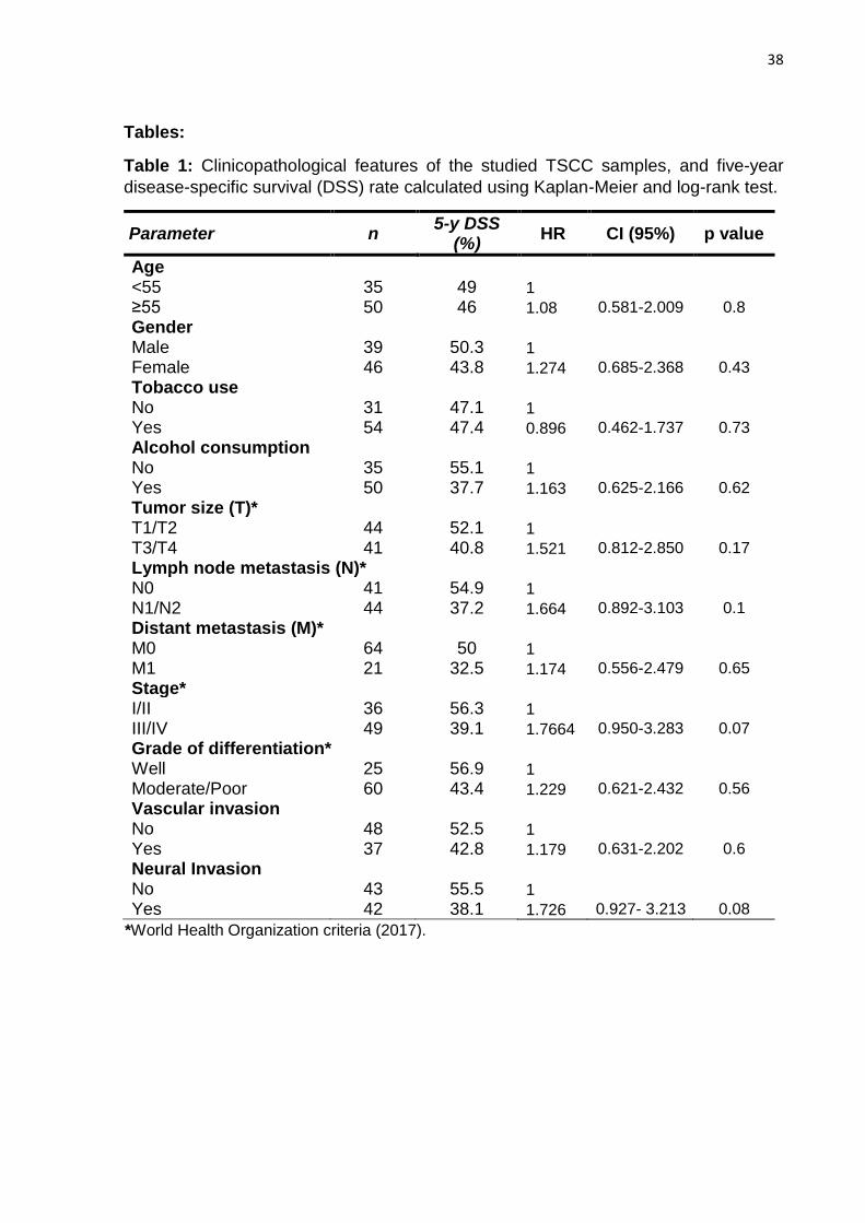
Clinicopathological characteristics of the patients:
A total of 85 cases of TSCC with complete clinical information was used in this
study (Table 1). The five-year disease-specific survival rate for the patients with TSCC
was 47.3%. A slight male predominance was found (M:F ratio of 1:0.8) with a mean
age of 55.5 years (range of 19-89 years). Most of the patients consumed tobacco
(63.5%) or alcohol (58.8%). Stage I/II tumours accounted for 42.4%, and stages III/IV
57.6% of the malignancies, with 51.8% of the patients presenting lymph node
metastasis and 24.7% presenting distant metastasis. Most of the patients (52/85,
61.2%) were solely treated with surgery. Radiotherapy as the single-modality treatment

28
was delivered to only 8/85 (9.4%) patients. Combined-modality treatment was
delivered to 21 patients (surgery plus radiotherapy and/or chemotherapy), and 4
patients received only palliative measures. Regarding the microscopic aspects of the
tumours, 29.4% were well differentiated, 37.7% moderate and 32.9% poorly
differentiated. Furthermore, vascular and neural invasion were present in 43.5% and
49.4%, respectively. Mean follow-up time was 40 months. The clinical data of the OED
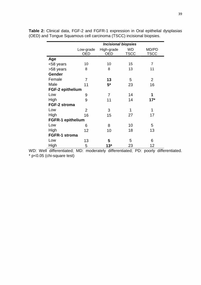
and TSCC incisional biopsies are shown in table 2.
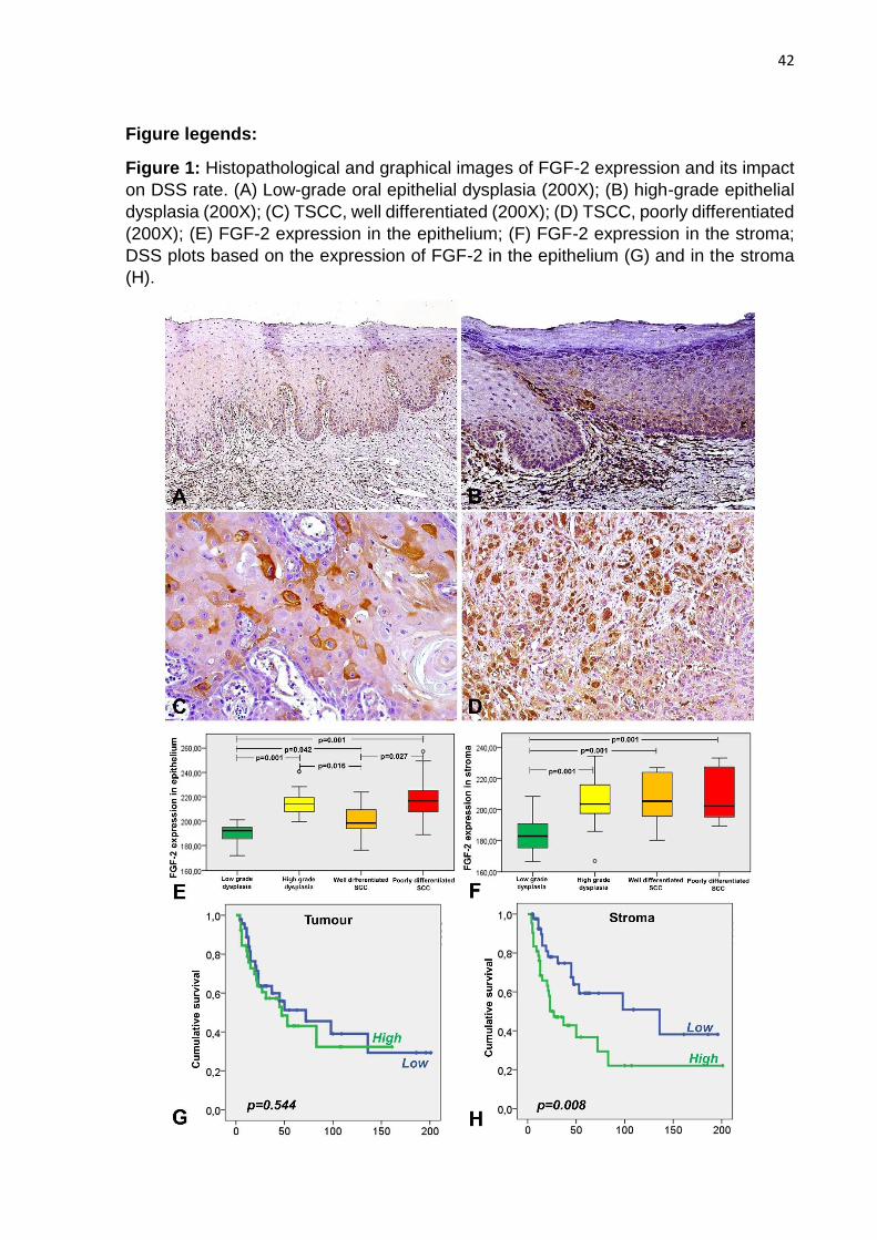
FGF-2 expression:
FGF-2 immunoexpression was assessed in the epithelial cells and in the
inflammatory cells within the stroma of the OED and TSCC. FGF-2 was expressed in
the basal and suprabasal layers of the epithelium in low-grade OED (Fig. 1A), while in
high-grade OED, this expression was more prominent and extended to the upper
layers of the epithelium (Fig. 1B). FGF-2 was expressed in the cytoplasm of malignant
cells in more well differentiated TSCC (Fig. 1C) and could be expressed in the nucleus
of these cells in poorly differentiated tumours (Fig. 1D). The immunoexpression digital
score was significantly higher in high-grade OED (p=0.001) when compared to low-
grade OED (Fig. 1E). This was also true when comparing well differentiated and poorly
differentiated TSCC (p=0.027) as shown in tables 2 and 3. FGF-2 high expression was
correlated with tumour size (p=0.024) and the presence of distant metastasis (p=0.007)
as shown in Table 3. However, epithelial expression of FGF-2 did not significantly
correlate with the DSS probabilities in the Kaplan-Meier analysis (Fig. 1G and Table
4).
Comparing the FGF-2 expression in the inflammatory cells, it was significantly
lower in the low-grade OED, when compared with the high-grade OED and the TSCC
lesions (p=0.001), but there was no difference between the other groups (Fig. 1F).

29
Cases with high expression of FGF-2 in the inflammatory and mesenchymal cells of
the stroma were associated with vascular invasion (Table 3). Patients with high
expression of FGF-2 in the stroma had a 5-year DSS of 36.7%, against 59.3% of
patients with low expression (HR: 2.272; CI(95%): 1.213-4.254; p=0.008). The Cox
multivariate analysis confirmed that the high expression of FGF-2 in the stroma (HR:
2.197; CI(95%): 1.128-4.282; p=0.02), is strongly associated with a higher risk of death
related to TSCC, as shown in Figure 1H and Table 4.
FGFR-1 expression:
FGFR-1 immunoexpression was also examined in the epithelial cells and in the
inflammatory cells within the stroma of OED and TSCC. FGFR-1 was expressed in the
upper layers of the epithelium of both low-grade and high-grade OED, but with much
more intensity in the high-grade lesions (Fig. 2A and 2B). This difference was
statistically significant (p=0.001), as in Fig. 2E. In the TSCC epithelial cells, the same
pattern of the FGF-2 analysis was observed, with cytoplasmic expression in the more
differentiated cases and nuclear expression in the poorly differentiated lesions (Fig. 2C
and 2D, respectively).
The FGFR-1 expression in the stroma was also significantly lower in the low-
grade OED when compared with the other groups (p<0.05), but no other differences
were seen (Fig. 2F). FGFR-1 high expression in the stroma were also correlated with
lymph node metastasis and distant metastasis (Table 3).
The survival curve rates using log-rank test showed that FGFR-1 high
expression either in the malignant cells or in the stroma cells were strongly correlated
with lower DSS (Fig. 2G and 2H). Patients with high expression of FGFR-1 in the
tumour had a 5-year DSS of 22.9%, against 75.6% of patients with low expression
30
(HR: 2.594; CI(95%): 1.390-4.841; p=0.003). The same was observed with FGFR-1
in the stroma, with a 5-year DSS of 32.9%, against 64.0% (HR: 3.378; CI(95%): 1.816-
6.286; p=0.001). The Cox multivariate analysis confirmed that the high expression of
FGFR-1 in the tumour (HR: 3.178; CI(95%): 1.505-6.709; p=0.002) and FGFR-1 in the
stroma (HR: 3.041; CI(95%): 1.454-6.356; p=0.003) are strongly associated with a
higher risk of death related to TSCC, as shown in Table 4.
Discussion:
FGF-2 and FGFR-1 aberrant expression has been associated with different
types of malignancies8,10–12,24 and has been suggested as interesting molecular
therapeutic targets. FGF-2 is a potent angiogenic factor and cells transfected with the
FGF-2 gene underwent malignant transformation25. Moreover, FGF-2 and FGFR-1
inhibitors have been suggested as therapeutic strategies in oral cancer26,27. Here, we
evaluated the expression of FGF-2 and FGFR-1 in TSCC and OED biopsies.
In patients with oral squamous cell carcinoma (OSCC), FGF-2 and FGFR-1
expression has been correlated with poorer differentiation, higher invasion potential
and worse prognosis3,16–18,28–32. Indeed, these proteins might influence the epithelial-
mesenchymal transition (EMT) in TSCC and OSCC cell lines4,33. Furthermore, FGF-2
is overexpressed in the dysplastic epithelium of oral potentially malignant disorders
(PMD)17,34,35 but there are no studies investigating the FGFR-1 expression in OED or
in any other oral PMD.
We found higher expression of FGF-2 and FGFR-1 in high grade OED when
comparing with the low-grade lesions, which is in agreement with previous studies17,34,
showing higher expression of FGF-2, as the severity of epithelial dysplasias increased,

31
mainly in the upper layers of the epithelium. FGF-2 expression has also been related
with the malignant transformation of oral leucoplakia into OSCC17. Our results also
suggest that the expression of FGF-2 and FGFR-1 in the stroma cells might be
important to understand the behaviour of these lesions, and needs further
investigation.
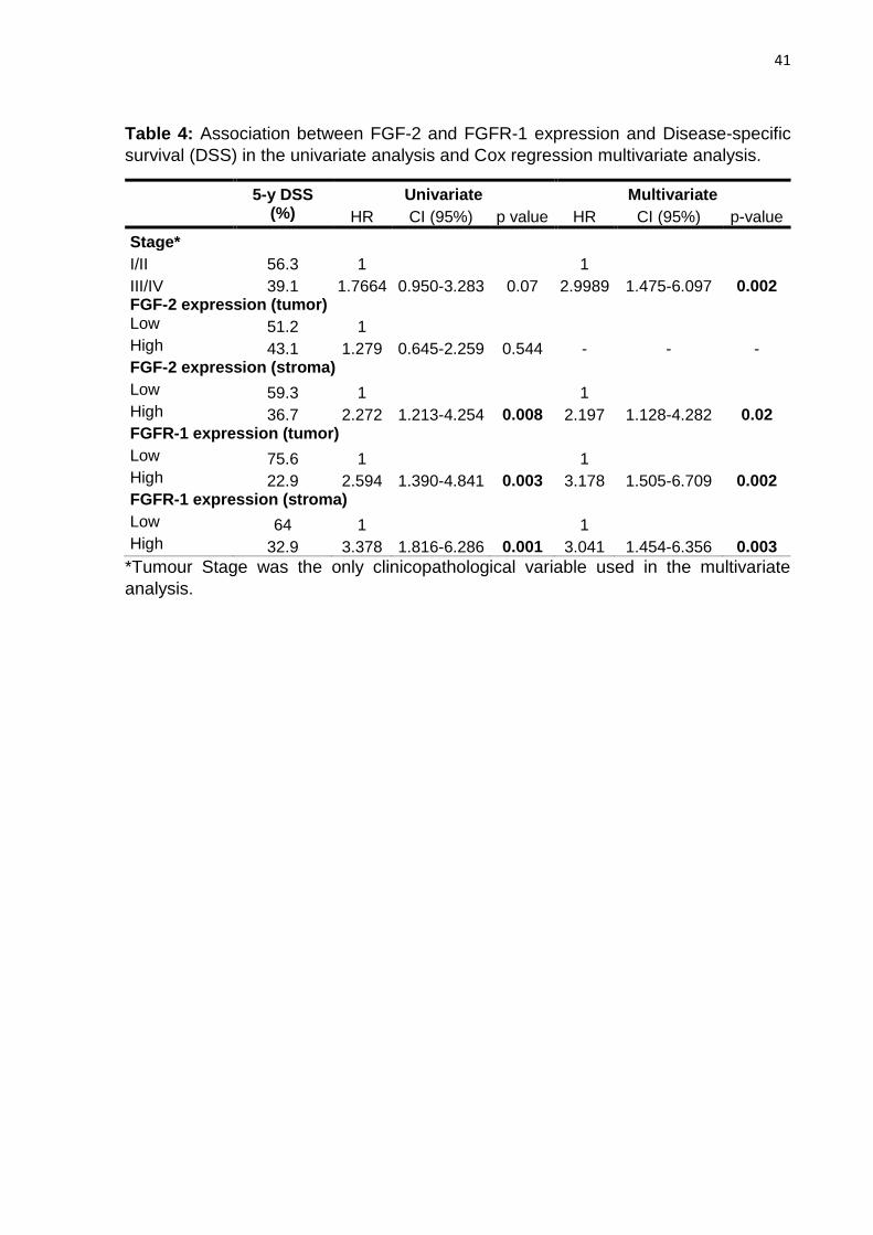
FGF-2 and FGFR-1 were associated with lower DSS both in univariate and
multivariate analysis of TSCC cases. Only one previous study correlated FGF-2
expression with a worse prognosis in 61 OSCC patients18, showing that FGF-2 high
expression in the stroma was correlated with the presence of lymph node metastasis
and a worse prognosis. However, their parameters to indicate the FGF-2 positivity were
merely visual, based on the “presence or absence” of expression. They also found a
correlation between FGF-2 expression and lymph node metastasis. Our results
indicate that patients with lymph node metastasis present a higher FGF-2 expression,
but without statistical significance. However, FGF-2 was significantly correlated with
the presence of distant metastasis in our cases, which was not covered by Hase et
al.18.
In previous studies, FGFR-1 expression has been associated with TSCC poor
differentiation and metastatic potential4,16,33. Our results show a correlation between
the expression of FGFR-1 in the stroma cells and the presence of lymph node
metastasis and distant metastasis, but there is no association, between its expression
in neoplastic cells and other clinical features. However, FGFR-1 expression, either in
the tumour or in the stroma, indicated higher risk of death in patients with TSCC. Hase
et al.18 also correlated FGFR-1 expression in fibroblasts of the invasion front with
impaired prognosis in OSCC patients. FGFR-1 overexpression was also correlated
with poor survival rates in HNSCC patients19,20, using a semi quantitative analysis of
32
the immunostaining. Conversely, FGFR-1 amplification has been found in
approximately 10-17% of OSCC cases16,29–31, but no significant correlation with patient
outcome has been found3.
FGF-FGFR axis can promote tumour development and progression by
downstream signal pathways, including MAPK/ERK and PI3K/AKT cascades,
promoting cancer cells proliferation and survival, besides supporting tumour
angiogenesis4,14,36. FGFR-1 expression has also been related with epithelial-
mesenchymal transition (EMT) in TSCC4,33, showing higher nuclear polymorphism,
more invasiveness, poor histopathological grade and higher metastasis potential.
Furthermore, treating the cells with FGFR-1 inhibitors or knocking down its expression
made the OSCC proliferate and invade less.
FGF-2 inhibitors have also been used to treat OSCC cells in vitro and in vivo27,37.
Furthermore, FGFR inhibitors reduced in vitro growth of head and neck SCC cell lines
expressing FGF-2, which is consistent with its autocrine fashion26. Hence, FGF/FGFR
inhibitors may represent a novel therapeutic modality for oral cancer, with potential as
molecular-targeted anticancer drugs for FGF-2/FGFR-1 dependent lesions.
In conclusion, overexpression of FGF-2 and FGFR-1 were correlated with the
presence of metastasis and worse outcome of patients with tongue squamous cell
carcinoma and might be considered as potential biomarkers to predict the prognosis
of TSCC patients.
Acknowledgments:
Bruno A.L.A. Mariz is grateful for the graduate scholarship grant provided by CAPES
(Coordination for the Improvement of Higher Education Personnel). The authors would

33
like to thank CAPES and FAPESP (processes #2015/25905-1 and #2017/16102-8) for
the financial support.
Author contributions:
BALAM performed the research and wrote the paper. CDS and JJ designed the
research study, analysed the data and reviewed the content of the paper. MGFC
provided cases and actively contributed with the revision of the paper.
34
References:
1. Almangush A, Coletta RD, Bello IO, et al. A simple novel prognostic model for
early stage oral tongue cancer. Int J Oral Maxillofac Surg. 2015;44(2):143-150.
doi:10.1016/j.ijom.2014.10.004.
2. Kauppila JH, Korvala J, Siirilä K, et al. Toll-like receptor 9 mediates invasion
and predicts prognosis in squamous cell carcinoma of the mobile tongue. J Oral
Pathol Med. 2015;44(8):571-577. doi:10.1111/jop.12272.
3. Young RJ, Lim AM, Angel C, et al. Frequency of Fibroblast Growth Factor
Receptor 1 gene amplification in oral tongue squamous cell carcinomas and
associations with clinical features and patient outcome. Oral Oncol. 2013;49(6):576-
581. doi:10.1016/j.oraloncology.2013.01.006.
4. Jiao J, Zhao X, Liang Y, Tang D, Pan C. FGF1–FGFR1 axis promotes tongue
squamous cell carcinoma (TSCC) metastasis through epithelial–mesenchymal
transition (EMT). Biochem Biophys Res Commun. 2015;466(3):327-332.
doi:10.1016/j.bbrc.2015.09.021.
5. El-Naggar A.K., Chan J.K.C., Grandis J.R., Takata T. SPJ, ed. WHO
Classification of Head and Neck Tumours. 4th ed. Lyon: IARC; 2017.
6. Yuan H, Li Z-M, Shao J, Ji W-X, Xia W, Lu S. FGF2/FGFR1 regulates
autophagy in FGFR1-amplified non-small cell lung cancer cells. J Exp Clin Cancer
Res. 2017;36(1):72. doi:10.1186/s13046-017-0534-0.
7. Pu D, Liu J, Li Z, Zhu J, Hou M. Fibroblast Growth Factor Receptor 1
(FGFR1), Partly Related to Vascular Endothelial Growth Factor Receptor 2
(VEGFR2) and Microvessel Density, is an Independent Prognostic Factor for Non-
Small Cell Lung Cancer. Med Sci Monit. 2017;23:247-257.
doi:10.12659/MSM.899005.
8. Turner N, Pearson A, Sharpe R, et al. FGFR1 amplification drives endocrine
therapy resistance and is a therapeutic target in breast cancer. Cancer Res.
2010;70(5):2085-2094. doi:10.1158/0008-5472.CAN-09-3746.
9. Chen H, Singh RR, Lu X, et al. Genome-wide copy number aberrations and
HER2 and FGFR1 alterations in primary breast cancer by molecular inversion probe
microarray. Oncotarget. 2017;8(7):1-13. doi:10.18632/oncotarget.14802.
10. Song Q, Liu Y, Jiang D, et al. High amplification of FGFR1 gene is a delayed
poor prognostic factor in early stage ESCC patients. Oncotarget. 2017;8(43):74539-
74553. doi:10.18632/oncotarget.20215.
11. Maehara O, Suda G, Natsuizaka M, et al. Fibroblast growth factor-2–mediated
FGFR/Erk signaling supports maintenance of cancer stem-like cells in esophageal
squamous cell carcinoma. Carcinogenesis. 2017;38(11):1073-1083.
doi:10.1093/carcin/bgx095.

35
12. Cowell JK, Qin H, Hu T, Wu Q, Bhole A, Ren M. Mutation in the FGFR1
tyrosine kinase domain or inactivation of PTEN is associated with acquired
resistance to FGFR inhibitors in FGFR1-driven leukemia/lymphomas. Int J Cancer.
2017;141(9):1822-1829. doi:10.1002/ijc.30848.
13. Wang F, Yang L, Shi L, et al. Nuclear translocation of fibroblast growth factor-
2 (FGF2) is regulated by Karyopherin-β2 and Ran GTPase in human glioblastoma
cells. Oncotarget. 2015;6(25):21468-21478. doi:10.18632/oncotarget.4097.
14. Wang W-M, Xu Y, Wang Y, et al. HOXB7 promotes tumor progression via
bFGF-induced activation of MAPK/ERK pathway and indicated poor prognosis in
hepatocellular carcinoma. Oncotarget. 2017;8(29):1-15.
doi:10.18632/oncotarget.17004.
15. Wang F, Yang L, Sun J, et al. Tumor suppressors microRNA-302d and
microRNA-16 inhibit human glioblastoma multiforme by targeting NF-κB and FGF2.
Mol Biosyst. 2017;13(7):1345-1354. doi:10.1039/C7MB00139H.
16. Peng C-H, Liao C-T, Ng K-P, et al. Somatic copy number alterations detected
by ultra-deep targeted sequencing predict prognosis in oral cavity squamous cell
carcinoma. Oncotarget. 2015;6(23):19891-19906. doi:10.18632/oncotarget.4336.
17. Nayak S, Goel MM, Makker A, et al. Fibroblast Growth Factor (FGF-2) and Its
Receptors FGFR-2 and FGFR-3 May Be Putative Biomarkers of Malignant
Transformation of Potentially Malignant Oral Lesions into Oral Squamous Cell
Carcinoma. Tang C-H, ed. PLoS One. 2015;10(10):e0138801.
doi:10.1371/journal.pone.0138801.
18. Hase T, Kawashiri S, Tanaka A, et al. Correlation of basic fibroblast growth
factor expression with the invasion and the prognosis of oral squamous cell
carcinoma. J Oral Pathol Med. 2006;35(3):136-139. doi:10.1111/j.1600-
0714.2006.00397.x.
19. Koole K, Brunen D, van Kempen PMW, et al. FGFR1 Is a Potential Prognostic
Biomarker and Therapeutic Target in Head and Neck Squamous Cell Carcinoma.
Clin Cancer Res. 2016;22(15):3884-3893. doi:10.1158/1078-0432.CCR-15-1874.
20. Koole K, Clausen MJ, van Es RJ, et al. FGFR Family Members Protein
Expression as Prognostic Markers in Oral Cavity and Oropharyngeal Squamous Cell
Carcinoma. Mol Diagn Ther. 2016 Aug;20(4):363-74. doi: 10.1007/s40291-016-0204-
5.
21. El-Naggar AK, Chan JKC, Grandis JR, Takata T SP, ed. WHO Classification of
Head and Neck Tumours. 4th ed. Lyon: International Agency for Research on
Cancer; 2017.
22. Fonseca FP, Bingle L, Santos-Silva AR, et al. Semaphorins and neuropilins
expression in salivary gland tumors. J Oral Pathol Med. 2016;45(2):119-126.
doi:10.1111/jop.12341.
23. Soares CD, Borges CF, Sena-Filho M, et al. Prognostic significance of
cyclooxygenase 2 and phosphorylated Akt1 overexpression in primary nonmetastatic

36
and metastatic cutaneous melanomas. Melanoma Res. 2017;27(5):448-456.
doi:10.1097/CMR.0000000000000368.
24. Chen D, Persson A, Sun Y, et al. Better Prognosis of Patients with Glioma
Expressing FGF2-Dependent PDGFRA Irrespective of Morphological Diagnosis.
PLoS One. 2013;8(4):1-14. doi:10.1371/journal.pone.0061556.
25. Sasada R, Kurokawa T, Iwane M, Igarashi K. Transformation of mouse
BALB/c 3T3 cells with human basic fibroblast growth factor cDNA. Mol Cell Biol.
1988;8(2):588-594. doi:10.1111/j.1600-0714.2006.00394.x.
26. Marshall ME, Hinz TK, Kono SA, et al. Fibroblast growth factor receptors are
components of autocrine signaling networks in head and neck squamous cell
carcinoma cells. Clin Cancer Res. 2011;17(15):5016-5025. doi:10.1158/1078-
0432.CCR-11-0050.
27. Shintani T, Takatsu F, Rosli SNZ, et al. Eldecalcitol (ED-71), an analog of
1α,25(OH)2D3, inhibits the growth of squamous cell carcinoma (SCC) cells in vitro
and in vivo by down-regulating expression of heparin-binding protein 17/fibroblast
growth factor-binding protein-1 (HBp17/FGFBP-1) and FGF. Vitr Cell Dev Biol - Anim.
2017;53(9):810-817. doi:10.1007/s11626-017-0183-9.
28. Harada K, Supriatno, Kawashima Y, Yoshida H, Sato M. S-1 inhibits
tumorigenicity and angiogenesis of human oral squamous cell carcinoma cells by
suppressing expression of phosphorylated Akt, vascular endothelial growth factor
and fibroblast growth factor-2. Int J Oncol. 2007;30(2):365-374. doi:10.3892/ijo-
00000417.
29. Clauditz TS, Böttcher A, Hanken H, et al. Prevalence of fibroblast growth
factor receptor 1 (FGFR1) amplification in squamous cell carcinomas of the head and
neck. J Cancer Res Clin Oncol. 2017;1. doi:10.1007/s00432-017-2528-x.
30. Ozretić L, Wagner S, Huebbers CU, et al. FGFR1 amplification and co-
overexpression of c-MYC in oropharyngeal squamous cell carcinoma. Oral Oncol.
2016;54:e7-e9. doi:10.1016/j.oraloncology.2015.12.006.
31. Freier K, Schwaenen C, Sticht C, et al. Recurrent FGFR1 amplification and
high FGFR1 protein expression in oral squamous cell carcinoma (OSCC). Oral
Oncol. 2007;43(1):60-66. doi:10.1016/j.oraloncology.2006.01.005.
32. Forootan SS, Ke Y, Jones AS, Helliwell TR. Basic fibroblast growth factor and
angiogenesis in squamous carcinoma of the tongue. Oral Oncol. 2000;36(5):437-
443. doi:10.1016/S1368-8375(00)00032-4.
33. Nguyen PT, Tsunematsu T, Yanagisawa S, et al. The FGFR1 inhibitor
PD173074 induces mesenchymal–epithelial transition through the transcription factor
AP-1. Br J Cancer. 2013;109(8):2248-2258. doi:10.1038/bjc.2013.550.
34. Wakulich C, Jackson-Boeters L, Daley TD, Wysocki GP. Immunohistochemical
localization of growth factors fibroblast growth factor-1 and fibroblast growth factor-2
and receptors fibroblast growth factor receptor-2 and fibroblast growth factor
receptor-3 in normal oral epithelium, epithelial dysplasias, and sq. Oral Surgery, Oral

37
Med Oral Pathol Oral Radiol Endodontology. 2002;93(5):573-579.
doi:10.1067/moe.2002.124461.
35. Bishen KA, Radhakrishnan R, Satyamoorthy K. The role of basic fibroblast
growth factor in oral submucous fibrosis pathogenesis. J Oral Pathol Med.
2008;37(7):402-411. doi:10.1111/j.1600-0714.2008.00649.x.
36. Turner N, Grose R. Fibroblast growth factor signalling: from development to
cancer. Nat Rev Cancer. 2010;10(2):116-129. doi:10.1038/nrc2780.
37. Rosli SNZB, Shintani T, Toratani S, Usui E, Okamoto T. 1α,25(OH)2D3
inhibits FGF-2 release from oral squamous cell carcinoma cells through down-
regulation of HBp17/FGFBP-1. Vitr Cell Dev Biol - Anim. 2014;50(9):802-806.
doi:10.1007/s11626-014-9787-5.
38
Tables:
Table 1: Clinicopathological features of the studied TSCC samples, and five-year
disease-specific survival (DSS) rate calculated using Kaplan-Meier and log-rank test.
Parameter n 5-y DSS
(%) HR CI (95%) p value
Age
<55 35 49 1
≥55 50 46 1.08 0.581-2.009 0.8
Gender
Male 39 50.3 1
Female 46 43.8 1.274 0.685-2.368 0.43
Tobacco use
No 31 47.1 1
Yes 54 47.4 0.896 0.462-1.737 0.73
Alcohol consumption
No 35 55.1 1
Yes 50 37.7 1.163 0.625-2.166 0.62
Tumor size (T)*
T1/T2 44 52.1 1
T3/T4 41 40.8 1.521 0.812-2.850 0.17
Lymph node metastasis (N)*
N0 41 54.9 1
N1/N2 44 37.2 1.664 0.892-3.103 0.1
Distant metastasis (M)*
M0 64 50 1
M1 21 32.5 1.174 0.556-2.479 0.65
Stage*
I/II 36 56.3 1
III/IV 49 39.1 1.7664 0.950-3.283 0.07
Grade of differentiation*
Well 25 56.9 1
Moderate/Poor 60 43.4 1.229 0.621-2.432 0.56
Vascular invasion
No 48 52.5 1
Yes 37 42.8 1.179 0.631-2.202 0.6
Neural Invasion
No 43 55.5 1
Yes 42 38.1 1.726 0.927- 3.213 0.08
*World Health Organization criteria (2017).
39
Table 2: Clinical data, FGF-2 and FGFR-1 expression in Oral epithelial dysplasias
(OED) and Tongue Squamous cell carcinoma (TSCC) incisional biopsies.
Incisional biopsies
Low-grade OED
High-grade OED
WD TSCC
MD/PD TSCC
Age
<58 years 10 10 15 7
>58 years 8 8 13 11
Gender
Female 7 13 5 2 Male 11 5* 23 16 FGF-2 epithelium
Low 9 7 14 1
High 9 11 14 17*
FGF-2 stroma
Low 2 3 1 1
High 16 15 27 17
FGFR-1 epithelium
Low 6 8 10 5
High 12 10 18 13
FGFR-1 stroma
Low 13 5 5 6
High 5 13* 23 12
WD: Well differentiated; MD: moderately differentiated; PD: poorly differentiated.
* p<0.05 (chi-square test)

40
Table 3: Number of TSCC cases with high expression of FGF-2 and FGFR-1 according
to clinicopathological data.
Number and percentage (%) of cases with high protein expression
FGF-2 epithelium
FGF-2 stroma
FGFR-1 epithelium
FGFR-1 stroma
Tumour size
T1/T2 15 (34.1)* 20 (44.5) 22 (50.0) 23 (52.3)
T3/T4 24 (58.5)* 22 (53.7) 20 (48.8) 19 (46.3)
Lymph node metastasis
No 17 (41.5) 21 (51.2) 22 (53.7) 25 (61.0)*
Yes 22 (50.0) 21 (47.7) 20 (45.5) 17 (38.6)*
Distant metastasis
No 24 (37.5)* 32 (50.0) 33 (51.6) 36 (56.3)*
Yes 15 (71.4)* 10 (47.6) 9 (42.9) 6 (28.6)*
Stage
I/II 14 (38.9) 18 (50.0) 20 (55.5) 21 (58.3)
III/IV 25 (51.0) 24 (49.0) 22 (44.9) 21 (43.9)
Vascular invasion
No 21 (50.0) 8 (19.0)* 19 (45.2) 20 (47.6)
Yes 18 (41.9) 34 (79.0)* 23 (53.5) 22 (51.2)
Neural invasion
No 18 (48.6) 14 (37.9) 15 (40.5) 16 (43.2)
Yes 21 (43.8) 28 (58.3) 27 (56.2) 26 (54.2)
* p<0.05 (chi-square test)
41
Table 4: Association between FGF-2 and FGFR-1 expression and Disease-specific
survival (DSS) in the univariate analysis and Cox regression multivariate analysis.
5-y DSS (%)
Univariate Multivariate
HR CI (95%) p value HR CI (95%) p-value
Stage*
I/II 56.3 1 1
III/IV 39.1 1.7664 0.950-3.283 0.07 2.9989 1.475-6.097 0.002 FGF-2 expression (tumor) Low 51.2 1 High 43.1 1.279 0.645-2.259 0.544 - - - FGF-2 expression (stroma) Low 59.3 1 1 High 36.7 2.272 1.213-4.254 0.008 2.197 1.128-4.282 0.02 FGFR-1 expression (tumor) Low 75.6 1 1 High 22.9 2.594 1.390-4.841 0.003 3.178 1.505-6.709 0.002 FGFR-1 expression (stroma) Low 64 1 1 High 32.9 3.378 1.816-6.286 0.001 3.041 1.454-6.356 0.003
*Tumour Stage was the only clinicopathological variable used in the multivariate
analysis.
42
Figure legends:
Figure 1: Histopathological and graphical images of FGF-2 expression and its impact
on DSS rate. (A) Low-grade oral epithelial dysplasia (200X); (B) high-grade epithelial
dysplasia (200X); (C) TSCC, well differentiated (200X); (D) TSCC, poorly differentiated
(200X); (E) FGF-2 expression in the epithelium; (F) FGF-2 expression in the stroma;
DSS plots based on the expression of FGF-2 in the epithelium (G) and in the stroma
(H).

43
Figure 2: Histopathological and graphical images of FGFR-1 expression and its impact
on DSS rate. (A) Low-grade oral epithelial dysplasia (200X); (B) high-grade epithelial
dysplasia (200X). (C) TSCC, well differentiated (200X); (D) TSCC, poorly differentiated
(200X); (E) FGFR-1 expression in the epithelium; (F) FGFR-1 expression in the stroma;
DSS plots based on the expression of FGFR-1 in the epithelium (G) and in the stroma
(H).
44
3 CONCLUSÃO:
A expressão de FGF-2 e FGFR-1 aumenta de acordo com o grau de displasia
epitelial oral.
Carcinoma Espinocelular de língua com superexpressão de FGF-2 e FGFR-1
apresentam pior prognóstico, com maior risco de morte dos pacientes.
O uso dos marcadores FGF-2 e FGFR-1 parece ser útil como marcador de
prognóstico de lesões de Carcinoma Espinocelular de língua, potencialmente
determinando formas de tratamento individualizadas de acordo com as características
clínicas e biológicas de cada tumor. Esse potencial ainda necessita confirmação por
meio de estudos futuros.

45
RERERÊNCIAS*:
Ach T, Schwarz-Furlan S, Ach S, et al. Genomic aberrations of MDM2, MDM4,
FGFR1 and FGFR3 are associated with poor outcome in patients with salivary gland
cancer. J Oral Pathol Med. 2016;45(7):500-509.
Almangush A, Coletta RD, Bello IO, et al. A simple novel prognostic model for early
stage oral tongue cancer. Int J Oral Maxillofac Surg. 2015;44(2):143-150.
Bishen KA, Radhakrishnan R, Satyamoorthy K. The role of basic fibroblast growth
factor in oral submucous fibrosis pathogenesis. J Oral Pathol Med. 2008;37(7):402-
411.
Brizeno LAC, Assreuy AMS, Alves APNN, et al. Delayed healing of oral mucosa in a
diabetic rat model: Implication of TNF-α, IL-1β and FGF-2. Life Sci. 2016;155:36-47.
Clauditz TS, Böttcher A, Hanken H, et al. Prevalence of fibroblast growth factor
receptor 1 (FGFR1) amplification in squamous cell carcinomas of the head and neck.
J Cancer Res Clin Oncol. 2017;1.
Delrieu I. The high molecular weight isoforms of basic fibroblast growth factor (FGF-
2): an insight into an intracrine mechanism. FEBS Lett. 2000;468(1):6-10.
Forootan SS, Ke Y, Jones AS, Helliwell TR. Basic fibroblast growth factor and
angiogenesis in squamous carcinoma of the tongue. Oral Oncol. 2000;36(5):437-
443.
1 * De acordo com as normas da UNICAMP/FOP, baseadas na padronização do International Committee of
Medical Journal Editors - Vancouver Group. Abreviatura dos periódicos em conformidade com o PubMed.
46
Freier K, Schwaenen C, Sticht C, et al. Recurrent FGFR1 amplification and high
FGFR1 protein expression in oral squamous cell carcinoma (OSCC). Oral Oncol.
2007;43(1):60-66.
Fujisawa K, Miyamoto Y, Nagayama M. Basic fibroblast growth factor and epidermal
growth factor reverse impaired ulcer healing of the rabbit oral mucosa. J Oral Pathol
Med. 2003;32(6):358-366.
Furuse C, Miguita L, Rosa ACG, et al. Study of growth factors and receptors in
carcinoma ex pleomorphic adenoma. J Oral Pathol Med. 2010;39(7):540-547.
Giacomini A, Chiodelli P, Matarazzo S, Rusnati M, Presta M, Ronca R. Blocking the
FGF/FGFR system as a “two-compartment” antiangiogenic/antitumor approach in
cancer therapy. Pharmacol Res. 2016;107(March):172-185.
Gorugantula LM, Rees T, Plemons J, Chen H-S, Cheng Y-SL. Salivary basic
fibroblast growth factor in patients with oral squamous cell carcinoma or oral lichen
planus. Oral Surg Oral Med Oral Pathol Oral Radiol. 2012;114(2):215-222.
Guo L, Gao R, Xu J, et al. AdLTR2EF1α-FGF2-mediated prevention of fractionated
irradiation-induced salivary hypofunction in swine. Gene Ther. 2014;21(10):866-873.
Harada K, Ferdous T, Kobayashi H, Ueyama Y. Elemental Diet Accelerates the
Recovery From Oral Mucositis and Dermatitis Induced by 5-Fluorouracil Through the
Induction of Fibroblast Growth Factor 2. Integr Cancer Ther.
2017:153473541772101.
Harada K, Supriatno, Kawashima Y, Yoshida H, Sato M. S-1 inhibits tumorigenicity
and angiogenesis of human oral squamous cell carcinoma cells by suppressing
expression of phosphorylated Akt, vascular endothelial growth factor and fibroblast
growth factor-2. Int J Oncol. 2007;30(2):365-374.
Hase T, Kawashiri S, Tanaka A, et al. Correlation of basic fibroblast growth factor
expression with the invasion and the prognosis of oral squamous cell carcinoma. J
Oral Pathol Med. 2006;35(3):136-139.
47
Hata Y, Kawanabe H, Hisanaga Y, Taniguchi K, Ishikawa H. Effects of Basic
Fibroblast Growth Factor Administration on Vascular Changes in Wound Healing of
Rat Palates. Cleft Palate-Craniofacial J. 2008;45(1):63-72.
Huang Y-Q, Li Y-D, Li G-K, Jin Z, Ma J. The Evaluation of Basic Fibroblast Growth
Factor and Fibroblastic Growth Factor Receptor 1 Levels in Saliva and Serum of
Patients with Salivary Gland Tumor. DNA Cell Biol. 2012;31(4):520-523.
INCA. Estimativas 2018: Incidência de Câncer no Brasil. Rio de Janeiro: Instituto
Nacional do Câncer; 2018.
Ipenburg NA, Koole K, Liem KS, et al. Fibroblast Growth Factor Receptor Family
Members as Prognostic Biomarkers in Head and Neck Squamous Cell Carcinoma: A
Systematic Review. Target Oncol. 2016;11(1):17-27.
Jansen RG, van Kuppevelt TH, Daamen WF, Kuijpers-Jagtman AM, Von den Hoff
JW. FGF-2-loaded collagen scaffolds attract cells and blood vessels in rat oral
mucosa. J Oral Pathol Med. 2009;38(8):630-638.
Jiang X-W, Zhang Y, Zhang H, Lu K, Yang S-K, Sun G-L. Double-blind, randomized,
controlled clinical trial of the effects of diosmectite and basic fibroblast growth factor
paste on the treatment of minor recurrent aphthous stomatitis. Oral Surg Oral Med
Oral Pathol Oral Radiol. 2013;116(5):570-575.
Jiao J, Zhao X, Liang Y, Tang D, Pan C. FGF1–FGFR1 axis promotes tongue
squamous cell carcinoma (TSCC) metastasis through epithelial–mesenchymal
transition (EMT). Biochem Biophys Res Commun. 2015;466(3):327-332.
Kanda T, Funato N, Baba Y, Kuroda T. Evidence for fibroblast growth factor
receptors in myofibroblasts during palatal mucoperiosteal repair. Arch Oral Biol.
2003;48(3):213-221.
Kauppila JH, Korvala J, Siirilä K, et al. Toll-like receptor 9 mediates invasion and
predicts prognosis in squamous cell carcinoma of the mobile tongue. J Oral Pathol
Med. 2015;44(8):571-577.

48
Kim JY, Xin X, Moioli EK, et al. Regeneration of Dental-Pulp-like Tissue by
Chemotaxis-Induced Cell Homing. Tissue Eng Part A. 2010;16(10):3023-3031.
Kobayashi F, Matsuzaka K, Inoue T. The effect of basic fibroblast growth factor on
regeneration in a surgical wound model of rat submandibular glands. Int J Oral Sci.
2016;8(1):16-23.
Kojima T, Kanemaru S, Hirano S, et al. The protective efficacy of basic fibroblast
growth factor in radiation-induced salivary gland dysfunction in mice. Laryngoscope.
2011;121(9):1870-1875.
Kusafuka K, Yamaguchi A, Kayano T, Takemura T. Immunohistochemical
localization of fibroblast growth factors (FGFs) and FGF receptor-1 in human normal
salivary glands and pleomorphic adenomas. J Oral Pathol Med. 1998;27(7):287-292.
Lassig AAD, Joseph AM, Lindgren BR, Yueh B. Association of Oral Cavity and
Oropharyngeal Cancer Biomarkers in Surgical Drain Fluid With Patient Outcomes.
JAMA Otolaryngol Neck Surg. 2017;143(7):670-678.
Martinez EF, Demasi APD, Miguita L, Altemani A, Araújo NS, Araújo VC. FGF-2 is
overexpressed in myoepithelial cells of carcinoma ex-pleomorphic adenoma in situ
structures. Oncol Rep. 2010;24(1):155-160.
Matsumoto G, Hoshino J, Kinoshita Y, et al. Alveolar bone regeneration using poly-
(lactic acid-co-glycolic acid-co-ε-caprolactone) porous membrane with collagen
sponge containing basic fibroblast growth factor: An experimental study in the dog. J
Biomater Appl. 2012;27(4):485-493.
Miguita L, Martinez EF, Araújo NS De, Araújo VC De. FGF-2, TGFβ-1, PDGF-A and
respective receptors expression in pleomorphic adenoma myoepithelial cells: an in
vivo and in vitro study. J Appl Oral Sci. 2010;18(1):83-91.
Morelli T, Neiva R, Nevins ML, et al. Angiogenic Biomarkers and Healing of Living
Cellular Constructs. J Dent Res. 2011;90(4):456-462.
49
Mullane EM, Dong Z, Sedgley CM, et al. Effects of VEGF and FGF2 on the
Revascularization of Severed Human Dental Pulps. J Dent Res. 2008;87(12):1144-
1148.
Myoken Y, Myoken Y, Okamoto T, et al. Immunohistochemical study of
overexpression of fibroblast growth factor-1 (FGF-1), FGF-2, and FGF receptor-1 in
human malignant salivary gland tumours. J Pathol. 1996;178(4):429-436.
Myoken Y, Myoken Y, Okamoto T, Sato JD, Takada K. Immunocytochemical
localization of fibroblast growth factor-1 (FGF-1) and FGF-2 in oral squamous cell
carcinoma (SCC). J Oral Pathol Med. 1994;23(10):451-456.
Nayak S, Goel MM, Makker A, et al. Fibroblast Growth Factor (FGF-2) and Its
Receptors FGFR-2 and FGFR-3 May Be Putative Biomarkers of Malignant
Transformation of Potentially Malignant Oral Lesions into Oral Squamous Cell
Carcinoma. Tang C-H, ed. PLoS One. 2015;10(10):e0138801.
Nguyen PT, Tsunematsu T, Yanagisawa S, et al. The FGFR1 inhibitor PD173074
induces mesenchymal–epithelial transition through the transcription factor AP-1. Br J
Cancer. 2013;109(8):2248-2258.
Ozretić L, Wagner S, Huebbers CU, et al. FGFR1 amplification and co-
overexpression of c-MYC in oropharyngeal squamous cell carcinoma. Oral Oncol.
2016;54:e7-e9.
Peng C-H, Liao C-T, Ng K-P, et al. Somatic copy number alterations detected by
ultra-deep targeted sequencing predict prognosis in oral cavity squamous cell
carcinoma. Oncotarget. 2015;6(23):19891-19906.
Pérez-Sayáns M, Suárez-Peñaranda J-M, Gayoso-Diz P, Barros-Angueira F,
Gándara-Rey J-M, García-García A. The role of p21Waf1/CIP1 as a Cip/Kip type
cell-cycle regulator in oral squamous cell carcinoma (Review). Med Oral Patol Oral
Cir Bucal. 2013;18(2):e219-25.
50
Persson F, Winnes M, Andrén Y, et al. High-resolution array CGH analysis of salivary
gland tumors reveals fusion and amplification of the FGFR1 and PLAG1 genes in
ring chromosomes. Oncogene. 2008;27(21):3072-3080.
Raimondi a R, Molinolo a a, Itoiz ME. Fibroblast growth factor-2 expression during
experimental oral carcinogenesis. Its possible role in the induction of pre-malignant
fibrosis. J Oral Pathol Med. 2006;35(4):212-217.
Saraiya M, Unger E, Thompson T, Lynch C, Hernandez BY, Lyu CW, et al. US
assessment of HPV types in cancers: implications for current and 9-valent HPV
vaccines. Journal of the National Cancer Institute. 2015; 107(6): djv086.
Soares AB, Demasi AP, Tincani AJ, Martins AS, Altemani A, de Araújo VC. The
increased PDGF-A, PDGF-B and FGF-2 expression in recurrence of salivary gland
pleomorphic adenoma. J Clin Pathol. 2012;65(3):272-277.
Sumitomo S, Okamoto Y, Mizutani G, Kudeken W, Mori M, Takai Y.
Immunohistochemical study of fibroblast growth factor-2 (FGF-2) and fibroblast
growth factor receptor (FGF-R) in experimental squamous cell carcinoma of rat
submandibular gland. Oral Oncol. 1999;35(1):98-104.
Sloan P, Gale N, Hunter K, Lingen MW, Nylander K, Reibel J, et al. Malignant
surface epithelial tumours. In: El-Naggar AK, Chan JK, Grandis Jennifer R, Takata T,
Slootweg PJ, organizadores. WHO Classif Head Neck Tumours. Fourth. 2017;109–
11.
Thula TT, Schultz G, Tran-Son-Tay R, Batich C. Effects of EGF and bFGF on
Irradiated Parotid Glands. Ann Biomed Eng. 2005;33(5):685-695.
Tran-Hung L, Laurent P, Camps J, About I. Quantification of angiogenic growth
factors released by human dental cells after injury. Arch Oral Biol. 2008;53(1):9-13.
Turner N, Grose R. Fibroblast growth factor signalling: from development to cancer.
Nat Rev Cancer. 2010;10(2):116-129.

51
Usumez A, Cengiz B, Oztuzcu S, Demir T, Aras MH, Gutknecht N. Effects of laser
irradiation at different wavelengths (660, 810, 980, and 1,064 nm) on mucositis in an
animal model of wound healing. Lasers Med Sci. 2014;29(6):1807-1813.
Wakulich C, Jackson-Boeters L, Daley TD, Wysocki GP. Immunohistochemical
localization of growth factors fibroblast growth factor-1 and fibroblast growth factor-2
and receptors fibroblast growth factor receptor-2 and fibroblast growth factor
receptor-3 in normal oral epithelium, epithelial dysplasias, and squamous cell
carcinoma. Oral Surgery, Oral Med Oral Pathol Oral Radiol Endodontology.
2002;93(5):573-579.
Young RJ, Lim AM, Angel C, et al. Frequency of Fibroblast Growth Factor Receptor 1
gene amplification in oral tongue squamous cell carcinomas and associations with
clinical features and patient outcome. Oral Oncol. 2013;49(6):576-581.

52
ANEXO 1:
53
ANEXO 2:


![artigo de imuno[1]](https://img.pdfslide.us/doc/110x75/577d20271a28ab4e1e921acf/artigo-de-imuno1.jpg)