Embed Size (px)
Citation preview
shaun carpenter, md, fapwca, cwsp
stephen davis
ryan fitzgerald, dpm, facfas
marie gehling, aprn, msn, np-c, cwocn
daniel kapp, md
jeffrey karr, dpm
john samies, md
gregory shultz, phd
adam teichman, dpm, facfas
dot weir, rn, cwon, cws
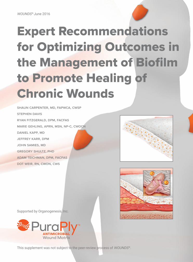
Expert Recommendations for Optimizing Outcomes in the Management of Biofilm to Promote Healing of Chronic Wounds
WOUNDS® June 2016
This supplement was not subject to the peer-review process of WOUNDS®.
Supported by Organogenesis, Inc.
Expert Recommendations for Optimizing Outcomes in the Management of Biofilm to Promote Healing of Chronic Wounds
2
Biofilm is a common form of wound contamination that is now recognized to be a major factor in delaying the healing of wounds. However, awareness of
the biology of biofilm, its prevalence, its clinical significance, and optimal treatment approaches needs to be improved.
Ten experienced wound care specialists reviewed the current status of evidence-based management of biofilm with a focus on the optimal use of PuraPly™
Antimicrobial, a purified collagen-based wound care matrix containing the antimicrobial polyhexamethylene biguanide (PHMB).
The overarching goal of this meeting is to empower clinicians caring for patients with chronic wounds to optimize clinical outcomes by providing an edu-
cational resource about the management of biofilm that is concise and useful in their clinical practices.
The meeting participants represented a variety of perspectives including academic settings, teaching hospitals, and stand-alone wound care clinics. Gregory
Schultz, PhD, from the University of Florida and Director of the Institute for Wound Research, and Professor Stephen C. Davis, from the University of Miami
Department of Dermatology and Cutaneous Surgery, led the discussions. Following the conference, a summary of the presentations and group discussions was
written and finalized with input from all meeting participants.
Meeting Participants
Purpose of this Publication
GREGORY SHULTZ, PHDUNIVERSITY OF FLORIDA
Dr. Schultz is Professor of Obstetrics and Gynecology, and Direc-
tor of the Institute for Wound Research at the University of Florida.
Dr. Schultz completed a PhD in biochemistry from Oklahoma State
University and postdoctoral training in cell biology at Yale University.
Dr. Schultz’s research focuses on the molecular regulation of wound
healing with an emphasis on anti-scarring therapies and the roles of
elevated proteases and bacterial biofilms in chronic wounds. Dr. Schultz
has authored over 320 scientific publications that have been cited more
than 11,700 times, is principal investigator or co-investigator on grants
totaling over $35 million, is an inventor on 26 patents and a co-founder
of two biotech companies. He was recognized by Time magazine as an
Innovation Leader in 2006. He served as a member of the National
Pressure Ulcer Advisory Panel from 2007 to 2010, and served as Presi-
dent of the Wound Healing Society from 1999-2001.
STEPHEN C. DAVISUNIVERSITY OF MIAMI
Professor Davis is a professor in the Department of Dermatology
and Cutaneous Surgery at the University of Miami. He has over 32
years experience using various wound healing and infection models in
swine. He has worked with a large number of companies in research
and development of various products that are on the market today (e.g.
DuoDerm® dressing, Iodosorb®, Procellera®, and Kerlix A.M.D.®). He
has authored several book chapters, and over 100 published articles and
abstracts on wound healing and infection. He is currently on the edito-
rial advisory board for the journal WOUNDS, and is a reviewer for sev-
eral journals including the Journal of Clinical and Experimental Dermatol-
ogy, the British Journal of Dermatology, and Wound Repair & Regeneration.
Some of his interests include: occlusive therapy, electrical stimulation,
low energy light therapy, and bacterial biofilm formation. For the past
several years, he has been funded by the Defense Advanced Research
Projects Agency (DARPA), Canadian Defense, United States Army, Of-
fice of Naval Research, National Institutes of Health and the National
Science Foundation (NSF).
SHAUN CARPENTER, MD, FAPWCA, CWSPBATON ROUGE GENERAL MEDICAL CENTER, BATON ROUGE, LA
Dr. Carpenter is a Certified Wound Specialist Physician, specializing
in wound care and hyperbaric medicine since 2004. He graduated from
the University of California at Berkeley, majoring in molecular and cell
biology. He received his medical doctorate from Tulane Medical School,
and subsequently became board-certified in emergency medicine af-
ter completing his residency at LSU Charity Hospital in New Orle-
ans. Over the past 10 years, Dr. Carpenter has developed an aggressive
wound management program for hospitals, nursing homes, and clinics.
He is the inventor of TeleWoundTM, a process by which homebound pa-
tients can receive aggressive wound care via telemedicine in the home.
He also has several patents pending for wound care products and de-
vices, including the Carpenter Curette and the True-See Digital Image
Calibration system. He serves as Director of Wound Care for North-
shore Specialty Hospital, Baton Rouge General Medical Center, and
Promise Hospital of Baton Rouge. Dr. Carpenter is the founder and
CEO of MedCentris, a comprehensive wound management company
3
with hospital contracts throughout Southeastern Louisiana and Texas.
He is also the founder and president of Total Wound Management, a
comprehensive wound management company dedicated to helping
hospitals, nursing homes, and clinics achieve maximum healing rates
while maintaining cost-effectiveness.
RYAN FITZGERALD, DPM, FACFASGREENVILLE HEALTH SYSTEM, GREENVILLE, SC
Dr. Fitzgerald is a podiatric surgeon with extensive training in elec-
tive and reconstructive surgery of the foot and ankle. Dr. Fitzgerald’s
expertise includes minimal incision arthroscopic surgery, diabetic foot
care, lower extremity wound care, plastic surgery techniques, external
and internal fixation, and reconstructive limb salvage. As a founding
member of the Greenville Health System (GHS) Center for Amputa-
tion Prevention, a collaborative clinical and research alliance at GHS,
Dr. Fitzgerald is dedicated to advancing the care of lower extremity ul-
cerations and preventing amputations in high-risk patient populations.
Additionally, Dr. Fitzgerald serves as Assistant Professor of Surgery at the
University of South Carolina School of Medicine-Greenville, where his
responsibilities include lower extremity reconstruction and limb salvage
techniques, wound care, research and medical education.
Dr. Fitzgerald has authored numerous peer-reviewed journal articles
and trade publications, and he has been featured as a speaker at both
national and international conferences. Dr. Fitzgerald is board- certified
in reconstructive rearfoot and ankle surgery as well as foot surgery by
the American Board of Foot and Ankle Surgery. He is a Fellow of the
American College of Foot and Ankle Surgeons (ACFAS), and a mem-
ber of the American Podiatric Medical Association (APMA).
MARIE GEHLING, APRN, MSN, NP-C, CWOCN REGIONAL MEDICAL CENTER, ORANGEBURG, SC
Ms. Gehling is a board-certified family nurse practitioner with over
20 years of experience in wound care. She has been credentialed as a
CWOCN since 1992 and practices with Dr. Samies in wound care and
infectious disease medicine at the Center for Advanced Wound Care
and Hyperbaric Medicine at the Regional Medical Center in Orange-
burg, SC. She was elected Southeast Region WOCN Nurse of the Year
for 2012, and has presented numerous educational lectures, research
posters and publications on advanced wound care topics.
DANIEL L. KAPP, MDJUPITER MEDICAL CENTER, JUPITER, FL
Dr. Kapp is the Medical Director of the Jupiter Medical Center in
Jupiter, Florida. He is a board certified plastic surgeon. His practice
focuses and complex reconstructive surgery including oncologic re-
construction, soft tissue reconstruction and limb salvage. His active areas
of interest include the management of soft tissue radionecrosis and flap
surgery. He is an active member of the American Society of Plastic
Surgeons.
JEFFREY C. KARR, DPM, CWS, ACCPPS, FAPWCA, FCCWSLAKELAND REGIONAL HEALTH, LAKELAND, FL
Dr. Karr is the founder and medical director of the Osteomyeli-
tis Center of Central Florida, and the Central Florida Limb Salvage
Alliance. Dr. Karr is the former medical director of the Wound Care
Center at the Lakeland Regional Health, an 820-bed, level two trau-
ma center. Dr. Karr serves as the chairman of the Institutional Review
Board (IRB) at Lakeland Regional Health. Dr. Karr is board-certified
in podiatric surgery and medicine, and is a Fellow of the American
Professional Wound Care Association (APWCA), and the College of
Certified Wound Specialists (CCWS). Dr. Karr has authored numerous
publications and lectures extensively domestically as well as interna-
tionally on advanced wound care, limb salvage and reconstruction, and
osteomyelitis. Dr. Karr has performed surgery in five countries in three
different continents outside of the United States.
JOHN H. SAMIES, MD, CWSREGIONAL MEDICAL CENTER, ORANGEBURG, SC
Dr. Samies is a board-certified specialist in infectious diseases and in-
ternal medicine with more than 25 years of experience in the treatment
of acute and chronic wounds. He is the hospital epidemiologist and Di-
rector of the Center for Advanced Wound Care and Hyperbaric Medi-
cine at the Regional Medical Center in Orangeburg, SC. He is a Fellow
of the Society for Healthcare Epidemiology of America, and a Certified
Wound Specialist. He has an active clinical practice in infectious disease
medicine and wound care. His clinical and research interests are in in-
fection control in wound care. He has published numerous articles and
lectures on infection control, wound care and infectious disease topics.
ADAM TEICHMAN, DPM, FACFASSACRED HEART HOSPITAL, ALLENTOWN, PA
Dr. Teichman is a board-certified foot and ankle surgeon, and is a
Fellow of the American College of Foot and Ankle Surgeons. A graduate
of the California College of Podiatric Medicine, he completed a 3-year
surgical foot and ankle reconstruction residency at St. Mary’s Hospital
in Hoboken, NJ. Dr. Teichman is Chief of Podiatric Surgery at Sacred
Heart Hospital in Allentown, PA. He currently serves on the limb sal-
vage team at Sacred Heart Hospital, where he is also a panel physician
specializing in lower extremity limb preservation and management. Dr.
Teichman has been in private practice in Allentown, PA since 2005, and
is co-founder of PA Foot and Ankle Associates. His current practice is
focused on conservative and surgical management of the diabetic foot.
He lectures nationally on wound management and limb preservation.
Expert Recommendations for Optimizing Outcomes in the Management of Biofilm to Promote Healing of Chronic Wounds
4
This article is supported by Organogenesis. The authors would like to thank Edward Perper, MD for editorial and manuscript assistance.
SHAUN CARPENTER, MD, FAPWCA, CWSP
Dr. Carpenter indicates participation on the speakers bureau for
Organogenesis.
RYAN FITZGERALD, DPM, FACFAS
Dr. Fitzgerald indicates that he is a consultant and speaker for Organo-
genesis, Osiris Therapeutics, and Integra LifeSciences.
STEPHEN C. DAVIS
Professor Davis indicates that he is on an advisory board for Organo-
genesis. As an employee of the University of Miami, he also conducts
many studies with a variety of companies and agencies.
MARIE GEHLING, APRN, MSN, NP-C, CWOCN
Ms. Gehling indicates that she is on the speakers bureau for Organ-
ogenesis and provides education through the clinical experience
program for Organogenesis field tissue regeneration specialists (TRS).
DANIEL L. KAPP, MD
No conflicts of interest reported.
JEFFREY C. KARR, DPM
No conflicts of interest reported.
JOHN H. SAMIES, MD, CWS
Dr. Samies indicates that he is a speaker and participates in a clinical
experience program for Organogenesis.
GREGORY SHULTZ, PHD
Dr. Schultz indicates advisory board participation or consulting with
Organogenesis, Acelity, Smith & Nephew, Medline Industries, Bio-
Monde, Hollister, and Quick-Med Technologies.
ADAM TEICHMAN, DPM, FACFAS
Dr. Teichman is a consultant, speaker, and participates in a clinical
experience program for Organogenesis.
DOROTHY (DOT) WEIR, RN, CWON, CWS
Ms. Weir indicated speakers bureau participation with Organogenesis,
Smith & Nephew, Acelity, Hollister, Lohmann & Rauscher, Organ-
ogenesis, Osiris Therapeutics, and Molnlycke Health Care. She also
has indicated advisory board/consultant participation with Molnlycke
Health Care, Corstrata, Aplion Medical, and Medline Industries.
Acknowledgements
Conflicts of Interest
Meeting Participants (Continued)
DOROTHY (DOT) WEIR, RN, CWON, CWSOSCEOLA REGIONAL MEDICAL CENTER, KISSIMMEE, FL
Ms. Weir has been a registered nurse for 40 years; 36 of those dedicat-
ed to the practice of wound and ostomy care. She has practiced in acute
care, home care and long term care, spent seven years in industry, and has
practiced in outpatient care since 2001. She has been board-certified by
the Wound, Ostomy and Continence Nursing Certification Board since
1985 and the American Board of Wound Management since 2004. She
practices outpatient wound management at Osceola Regional Medical
Center in Kissimmee, FL, and at Health Central Hospital in Ocoee, FL.
Ms. Weir is the Co-Chair of the Symposium on Advanced Wound Care,
was on the founding Board of the Association for the Advancement of
Wound Care and held the positions of the first Treasurer and the third
President. She has been on the faculty of the Wound Certification Prep
Course since 2004. She has been a member of the Wound, Ostomy
and Continence Nurses Society since 1980, the Florida Association for
Enterostomal Therapists since 1979, and has held regional board posi-
tions with both. She has been a member of the Wound Healing Society
since 2008, and was one of the Founding Editors of the journal Today’s
Wound Clinic. Ms. Weir is a frequent lecturer on all aspects of wound
management, has authored and co-authored many journal articles and
eight book chapters. She is on the Speakers Bureau and medical advi-
sory boards with several manufacturers.
5
1|Biofilm Impedes Healing of Chronic Wounds
Chronic non-healing wounds remain a major challenge for all wound care practitioners. These include diabetic ul-cers, venous ulcers, pressure ulcers, vas-cular ulcers, trauma wounds and surgical wounds. It is axiomatic among expe-rienced wound care practitioners that wound infection can delay (or complete-ly prevent) wound healing. However, it
is now known that an important factor that delays wound healing in many pa-tients is biofilm, a typically polymicrobial infection in a protective proteinaceous matrix.1
There have been significant advances in our understanding of why biofilms are so prevalent, how they resist interven-tions to eliminate them, and how they slow wound healing. This knowledge has resulted in the development of novel wound therapies that promise to improve management of chronic wounds.
Biofilm is the most common type of wound bioburden and represents an intermediate stage in a continuum of bioburden that ranges from simple contamination to localized infection to sepsis. In simple contaminations, mi-croorganisms within the wound are free-floating (planktonic), but as they multiply, these microbes firmly attach to the wound’s surfaces, differentiate, and change (“switch”) their gene ex-pression patterns to a biofilm phenotype that promotes their survival.2 Virtually all
Clotting
Clotting
Vasc
ular R
esponse
Vasc
ular R
esponse
Scar F
ormat
ion
Scar F
ormat
ion
Epitheli
al Hea
ling
Epitheli
al Hea
ling
Contract
ion
Contract
ion
Scar R
emodeli
ng
Scar R
emodeli
ng
Month
s
Month
s
12 10 8 64
12 10 8 6 4
25
20
15
10
0
25
20
15
10
0
Days
Days
Normal Wound Healing
Chronic Wound with Delayed Healing
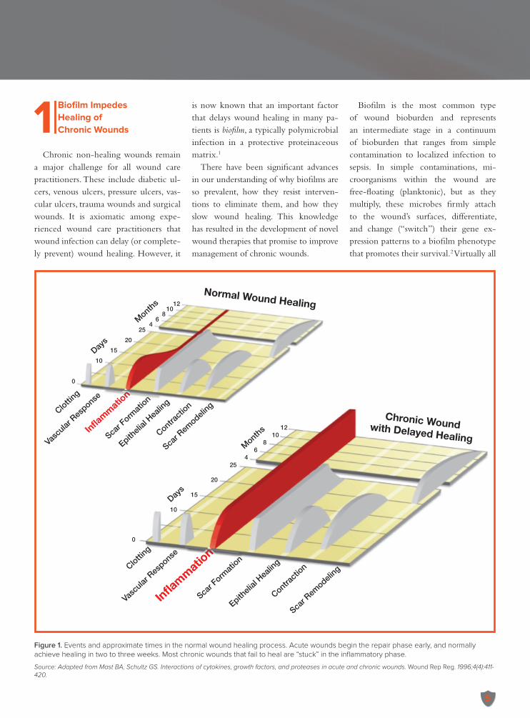
Figure 1. Events and approximate times in the normal wound healing process. Acute wounds begin the repair phase early, and normally achieve healing in two to three weeks. Most chronic wounds that fail to heal are “stuck” in the inflammatory phase.
Source: Adapted from Mast BA, Schultz GS. Interactions of cytokines, growth factors, and proteases in acute and chronic wounds. Wound Rep Reg. 1996;4(4):411-420.
Expert Recommendations for Optimizing Outcomes in the Management of Biofilm to Promote Healing of Chronic Wounds
6
wounds contain a combination of both free-floating microbes and biofilm; how-ever, biofilm is much harder to eradicate (see section #3 “Why Biofilm is Hard to Kill” on page 9) and delays wound heal-ing to a greater degree.3
Most chronic wounds that fail to heal are “stuck” in the inflammatory phase of the normal healing process (see Figure 1 on page 5). In this in-flammatory phase—which follows the normal clotting and vascular response phases—neutrophils and macrophages
are attracted to the wound site where they secrete large quantities of a variety of enzymes, including matrix metallo-proteinases (MMPs) and elastases, that break down damaged matrix. As invad-ing microbes and damaged tissues are cleared, inflammatory cells present in the wound die off, the influx of addi-tional inflammatory cells stops, and in-flammation subsides.4 This is followed by the formation of new collagen, re-epithelialization, contraction, and ultimately scar formation.
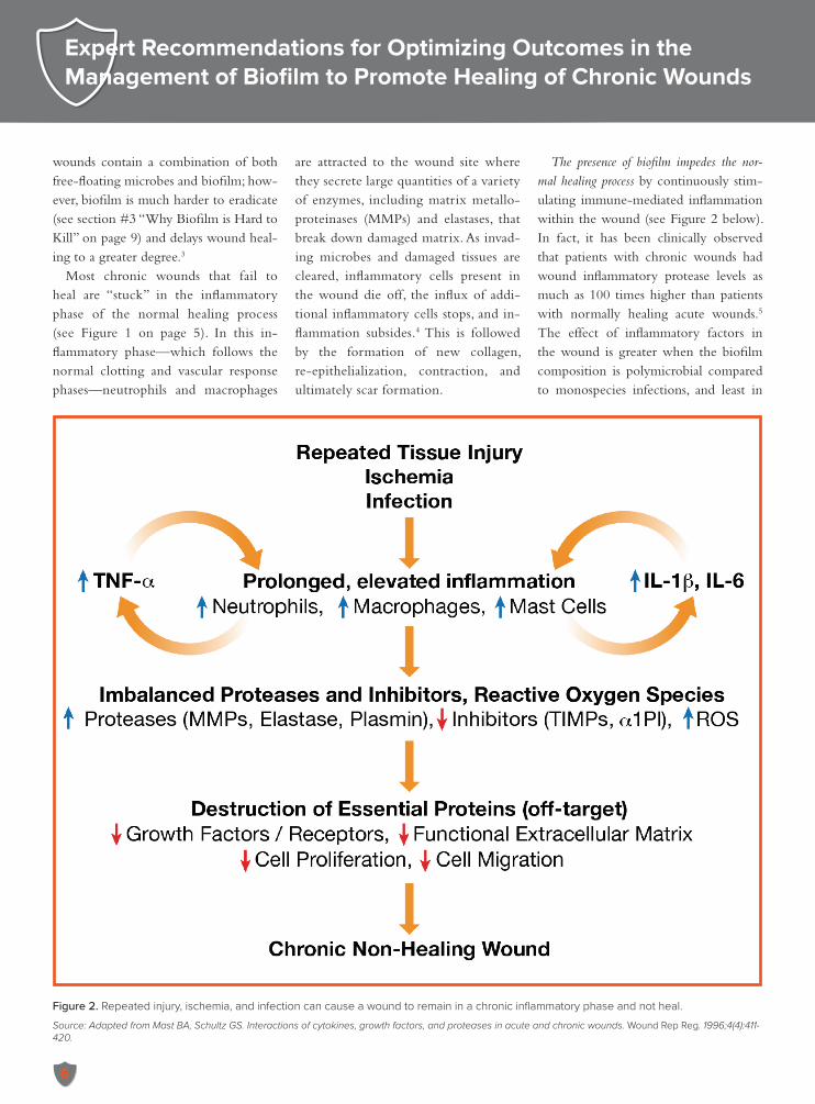
The presence of biofilm impedes the nor-mal healing process by continuously stim-ulating immune-mediated inflammation within the wound (see Figure 2 below). In fact, it has been clinically observed that patients with chronic wounds had wound inflammatory protease levels as much as 100 times higher than patients with normally healing acute wounds.5 The effect of inflammatory factors in the wound is greater when the biofilm composition is polymicrobial compared to monospecies infections, and least in
Figure 2. Repeated injury, ischemia, and infection can cause a wound to remain in a chronic inflammatory phase and not heal.
Source: Adapted from Mast BA, Schultz GS. Interactions of cytokines, growth factors, and proteases in acute and chronic wounds. Wound Rep Reg. 1996;4(4):411-420.
7
wounds that do not contain biofilm.6
In addition to stimulating wound in-flammation, biofilm slows healing by serving as a microbial reservoir for in-fection of healthy tissue, and by com-peting with normal cells for oxygen and nutrients.
Importantly, studies at Northwestern University have demonstrated that bio-film is a direct cause of delayed wound healing, and that the association between biofilm and delayed healing is not sec-
ondary or coincidental.7 Polymicrobial biofilms, which are associated with most wounds, have also been shown to reduce healing more as compared to single bac-terial strains.8
EXPERT PANEL DISCUSSION AND RECOMMENDATIONS
The panel members agreed that al-though the term biofilm is often rec-ognized by wound care practitioners, its biology and clinical significance are often
not well appreciated. A key point is that even when biofilm is not visually appar-ent and the wound “looks good,” biofilm is frequently present and delays wound healing significantly.
In addition, it was also discussed that high proteolytic activity within wounds is often a critical factor in non-healing wounds, but this fact is underappreci-ated by many practitioners. The panel concluded that there is an urgent need for education in this regard because
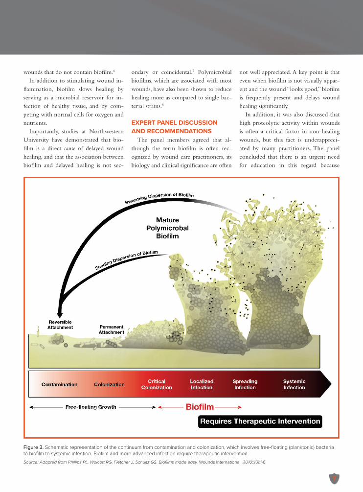
Figure 3. Schematic representation of the continuum from contamination and colonization, which involves free-floating (planktonic) bacteria to biofilm to systemic infection. Biofilm and more advanced infection require therapeutic intervention.
Source: Adapted from Phillips PL, Wolcott RG, Fletcher J, Schultz GS. Biofilms made easy. Wounds International. 2010;1(3):1-6.
Expert Recommendations for Optimizing Outcomes in the Management of Biofilm to Promote Healing of Chronic Wounds
8
there are important implications for therapy: proteolytic enzymes generated by biofilm can degrade collagen dress-ings when bioburden is not adequately controlled.
2|Biofilm Is Common But Difficult to Diagnose
Many practitioners correctly believe that essentially all chronic wounds are contaminated or colonized with bacteria (i.e., they are not sterile), but these low levels of planktonic bacteria typically do not stop wounds from healing. However, when planktonic bacteria convert into communities of biofilm, as discussed above, the biofilm becomes clinically significant and bioburden control be-
comes critical to wound management (see Figure 3 on page 7). But how com-mon is biofilm in real world settings? The answer is that a large majority of chronic wounds (60% to 90%) contain biofilm, while only 6% of acute wounds do.2,3,9 In fact, it is likely that almost all chronic wounds have biofilm on at least part of the wound bed.2
Importantly, the presence of biofilm should not be excluded based on the ab-sence of gross signs of infection. Unfor-tunately, biofilm can be underdiagnosed precisely because it is not associated with visual evidence of infection.
Practitioners can not necessarily rely on standard wound cultures performed by clinical microbiology labs to de-termine if biofilm is present. Typically, wound samples are processed by addi-
tional techniques to first kill all plank-tonic bacteria by brief exposure to a microbicidal antiseptic such as bleach, followed by neutralization of the an-tiseptic and dispersal of biofilms into single cells, followed by plating on cul-ture dishes. From a practical perspective, the definitive determination of biofilm is probably unnecessary, if all chronic wounds are treated on the presump-tion that biofilm is already present or there is high risk of biofilm formation. Culturing the wound may not provide useful results in this regard, as standard cultures detect free-floating microbes on the wound surface and not biofilm mi-crobes that lie deeper within the wound site. In fact, wound cultures may point to antimicrobial treatment that will not be effective against the biofilm.2
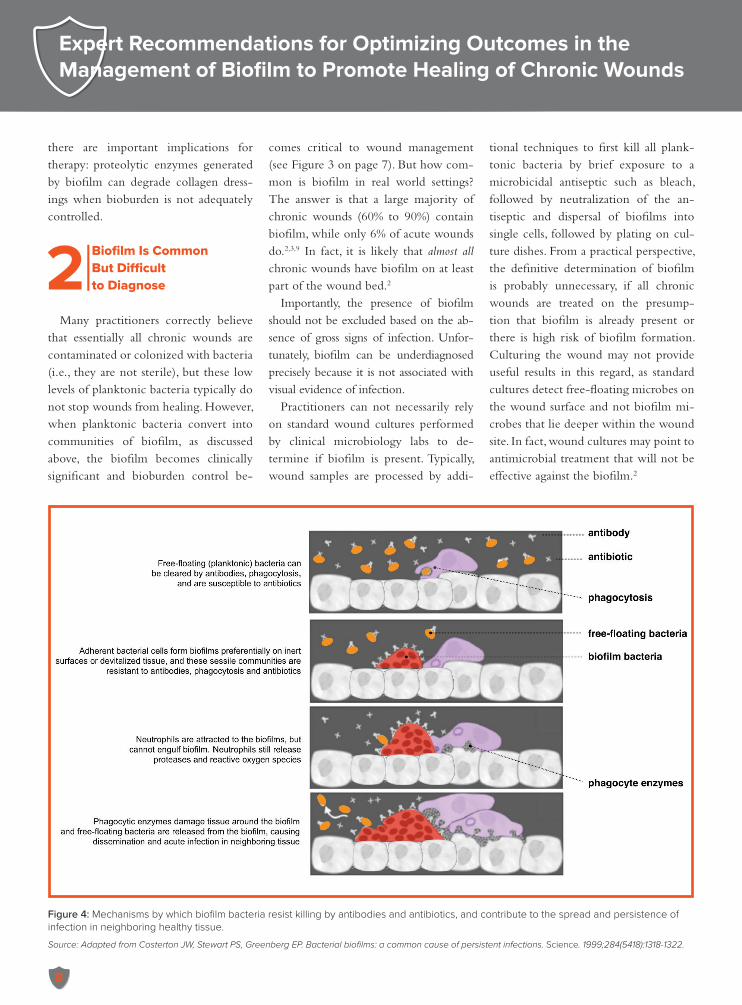
Figure 4: Mechanisms by which biofilm bacteria resist killing by antibodies and antibiotics, and contribute to the spread and persistence of infection in neighboring healthy tissue.
Source: Adapted from Costerton JW, Stewart PS, Greenberg EP. Bacterial biofilms: a common cause of persistent infections. Science. 1999;284(5418):1318-1322.
9
Therefore, when faced with wounds that are slow to heal, it is reasonable for practitioners to assume that all non-heal-ing wounds contain biofilm, and to treat the patient accordingly.
EXPERT PANEL DISCUSSION AND RECOMMENDATIONS
There was a strong consensus among the panel members that practitioners need to be more aware that wound bio-film is extremely common (i.e., affecting up to 80% of wounds). Panelists agreed that commonly there is failure to rec-ognize that essentially all open wounds will likely develop biofilm within three days. In addition, it is also important to understand that while wound cultures are helpful in guiding antibiotic therapy of single free-floating (planktonic) bacte-ria within wounds, such cultures do not
provide reliable information on biofilm bacteria, which “hide” more deeply within the wound bed. A key teaching point here is that wiping the surface of a wound with a swab samples only free-floating bacteria, but does not sam-ple biofilm-associated bacteria. Given these considerations, the panel recom-mends that essentially all wounds should be treated on the assumption that bio-film is already present, or is highly likely to develop if adequate bioburden man-agement is not achieved.
3|Why Biofilm Is Hard to Kill
Studies have shown that biofilm can be very difficult to eradicate.10-13
While free-floating bacteria are usually
eliminated by the immune system and/or antibiotics, biofilm poses a much greater challenge for several reasons (see Figure 4 on page 8).
1. Extracellular Polymeric Substance – Biofilm-associated microbes pro-duce a protective substance known as extracellular polymeric substance (EPS) that accelerates biofilm for-mation. EPS is a very dense collec-tion of proteins, sugars, and other factors that impair diffusion of an-timicrobial molecules, including antibodies and antibiotics. In addi-tion, due to its highly negative ion-ic structure, EPS acts as an “anionic screen” that blocks many cationic agents, such as silver and many anti-biotics. EPS may also act as a diffu-sion barrier against small molecule antimicrobial substances released by
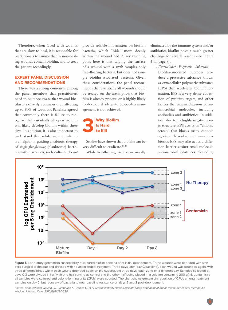
Figure 5: Laboratory gentamicin susceptibility of cultured biofilm bacteria after initial debridement. Three wounds were debrided with stan-dard surgical technique and dressed with no antimicrobial treatment. Three days later (day 0/baseline), each wound was debrided again, with three different zones within each wound debrided again on the subsequent three days, each zone on a different day. Samples collected at days 0-3 were divided in half with one half serving as control and the other half being placed in a solution containing 200 g/mL gentamicin; all samples were cultured and colony-forming units (CFUs) were counted. The chart shows gentamicin reduction of CFUs among treatment samples on day 2, but recovery of bacteria to near baseline resistance on days 2 and 3 post-debridement.
Source: Adapted from Wolcott RD, Rumbaugh KP, James G, et al. Biofilm maturity studies indicate sharp debridement opens a time-dependent therapeutic window. J Wound Care. 2010;19(8):320-328.
Expert Recommendations for Optimizing Outcomes in the Management of Biofilm to Promote Healing of Chronic Wounds
10
inflammatory cells.2 2. Persister Bacteria - Biofilm contains
“persister bacteria” that have low metabolic activity. Since antibiot-ics only kill metabolically active microbes by inhibiting metabolic enzyme systems, inactive bacteria not using these systems may not be sensitive to antibiotics.14
3. Anaerobic Bacteria - Oxygen diffu-sion to the center of the biofilm is reduced, creating a hypoxic envi-ronment in which more anaerobic bacteria can thrive.15 Anaerobic bacteria are generally more re-sistant to antimicrobial therapies. One study found that 60% of bio-
film bacterial species in pressure wounds were strict anaerobes.16
4. Synergism Between Bacteria – Ex-amples of such synergism include MRSA that secrete resistance pro-teins within the biofilm that help other wound bacteria survive,17 and Pseudomonas that secrete cat-alase, which destroys cytotoxic hy-drogen peroxide.18
EXPERT PANEL DISCUSSION AND RECOMMENDATIONS
The panel members discussed that wound care practitioners need to ap-preciate that biofilm-associated bac-teria can be extremely difficult to kill
for the reasons described above. A key teaching point here is that antibiotics are effective only against metabolically active bacteria, which explains why they are not effective against biofilm-associ-ated bacteria, many of which are metabol-ically inactive. This is in contrast to the antimicrobial agent PHMB, which is capable of killing quiescent biofilm-as-sociated bacteria by “punching holes” through their membranes (see section #6 “PHMB: An Excellent Topical Anti-microbial” on page 13).
An important distinction that was discussed was that resistance to therapy and tolerance to therapy are not synony-mous. Resistance means there has been a permanent genetic alteration within the bacteria’s DNA that allows it to resist killing. In tolerance, there is no genet-ic change but the bacteria are less sus-ceptible to killing. Both resistance and tolerance may cause antimicrobials, both topical and systemic, to be ineffective against biofilm.
4|Aggressive Debridement is Essential but Biofilm Reforms Rapidly
Successful management of biofilm re-quires two therapeutic objectives to be achieved: (1) remove biofilm that has already formed, and (2) prevent biofilm reformation.
Chronic wounds usually have both free-floating, single, planktonic bacte-ria and biofilm-associated microbes on their surface. These can be wiped or washed off, but beneath the surface of the wound, there are potentially many additional biofilm colonies. Aggressive debridement is necessary to remove the deeper biofilm. Fortunately, effective de-bridement can reduce bacterial counts to less than 1/100th of baseline.
However, even with aggressive debride-
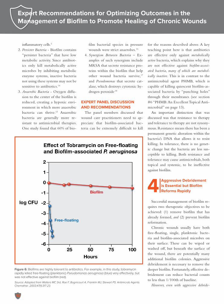
Figure 6: Biofilms are highly tolerant to antibiotics. For example, in this study, tobramycin rapidly killed free-floating (planktonic) Pseudomonas aeruginosa (blue) very effectively, but was not effective against biofilm (red).
Source: Adapted from Walters MC 3rd, Roe F, Bugnicourt A, Franklin MJ, Stewart PS. Antimicrob Agents Chemother. 2003;47(1):317-23.
11
ment, biofilm can start to reform within 24 hours and can fully mature within 3 days (see Figure 5 on page 9).10
In a porcine biofilm model, it has been shown that various debridement methods can initially reduce bio-film-associated bacteria by a few log CFU/g. However, high levels of these bacteria can remain in the wound for several days.11
Frequent aggressive debridement is a necessary part of wound care and bio-film management. Practitioners com-monly perform frequent debridements to care for wounds, however, due to the rapid reformation of biofilm, debride-ment alone is not sufficient to control bioburden.
EXPERT PANEL DISCUSSION AND RECOMMENDATIONS
A key point of consensus is that suc-cessful wound management requires suc-cessful biofilm management. Treating and preventing biofilm formation allows the wound to utilize the normal healing mechanisms. While some utilize a “step-
up” approach in which increasingly po-tent therapies are utilized starting with the least potent, the alternative may be a better option, namely, the “step down” approach in which aggressive treatment (surgical debridement followed by anti-microbial therapy) is used initially. There was agreement that in many wound care centers, debridement is performed on a once-weekly basis. However, this was characterized as a “disconnect” because, as discussed above, biofilm starts to re-form within 24 hours of debridement. In other words, there is a “window of op-portunity” after debridement in which it is critical to prevent biofilm formation. Practitioners must address this by en-suring that topical antimicrobial wound therapy is applied that will prevent bio-film formation between debridement visits (i.e., as a “bridge” to the next debride-ment). If not, biofilm is likely to recur and interfere with wound healing. In addi-tion, there was consensus that in wounds that are not healing well, it is critical to control bioburden before advancing to bioengineered living cell-based thera-
pies (Dermagraft® and Apligraf®). The panelists agreed that complete wound bed preparation with adequate control of biofilm must be achieved before cell-based therapies are utilized. If adequate biofilm control is not achieved, the graft can be “consumed” by high levels of mi-crobial enzymes and host inflammato-ry cell enzymes (MMPs and neutrophil elastase) and reactive oxygen species, which may explain why these therapies fail in some patients.
5|Limitations of Antimicrobial Therapy of Biofilm
Given that biofilm is formed by the activity of microbes, antimicrobial treat-ment of wounds is appropriate. Antimi-crobial products are commonly used, both topically and systemically, but the ability of certain topical and systemic products to effectively manage biofilm is limited in several ways.
Topical antibiotics. Several studies demonstrate that treatment of biofilm
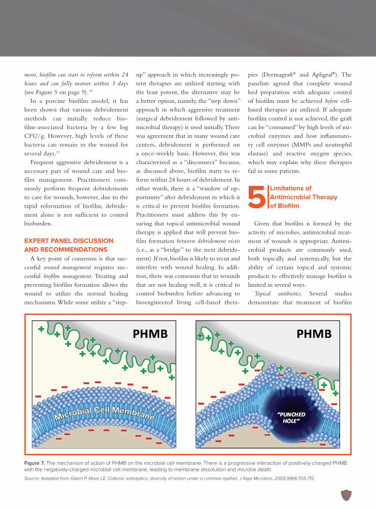
Figure 7: The mechanism of action of PHMB on the microbial cell membrane. There is a progressive interaction of positively-charged PHMB with the negatively-charged microbial cell membrane, leading to membrane dissolution and microbe death.
Source: Adapted from Gibert P, More LE. Cationic antiseptics: diversity of action under a common epithet. J Appl Microbiol. 2005;99(4):703-715.
Expert Recommendations for Optimizing Outcomes in the Management of Biofilm to Promote Healing of Chronic Wounds
12
with topical antibiotics can be ineffec-tive. For example, an in vivo study of two topical antibiotics in pig wounds inoculated with S. aureus showed that these preparations were effective at sup-pressing free-floating, single, planktonic bacteria but neither product was able to completely eradicate the biofilm.19 An in vitro study of topical tobramycin and ciprofloxacin against P. aeruginosa showed similar results (see Figure 6 on page 10). Antibiotics penetrated the biofilm but only eradicated the organisms in the outer layers where there was exposure to oxygen and the bacteria were metaboli-cally active.13
Systemic antibiotics. Clearly, systemic antibiotics do have a role when infec-tion has progressed beyond biofilm to a gross infection, but such treatment is not targeting the biofilm per se. As discussed earlier, extracellular polymeric substance (EPS) secreted by biofilm in the wound protects bacteria from antibiotic expo-sure.2 It also harbors large numbers of inactive microbes, and such antibiotics act primarily against metabolically ac-tive organisms.14 Furthermore, as is al-ways the case with antibiotic use, the presence and emergence of genetically resistant organisms in a polymicrobial infection is a common occurrence. For
example, methicillin-resistant S. aureus (MRSA) has been isolated from chronic wounds.20
Topical antimicrobials. A number of top-ical antimicrobials are available to re-duce wound bioburden and these can be highly effective. However, some topical antimicrobial agents can have deleteri-ous effects on healthy cells in or adjacent to the wound (cytotoxicity), which may contribute to poor wound healing. Sil-ver, for example, has been shown to be non-specific in its mode of action, kill-ing both bacteria and host keratinocytes and fibroblasts, leading to delayed heal-ing. In addition, not all topical agents
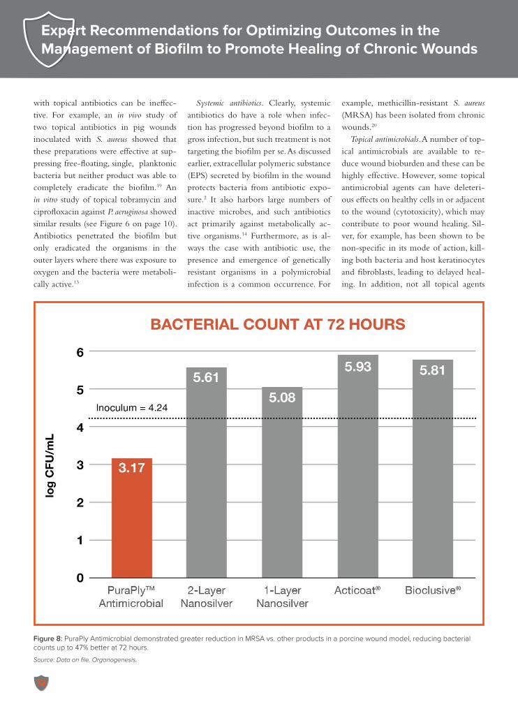
Figure 8: PuraPly Antimicrobial demonstrated greater reduction in MRSA vs. other products in a porcine wound model, reducing bacterial counts up to 47% better at 72 hours.
Source: Data on file, Organogenesis.
13
have a broad antimicrobial spectrum, which may limit their clinical utility, and bacterial resistance to certain antimicro-bial agents has been documented.
EXPERT PANEL DISCUSSION AND RECOMMENDATIONS
The panel discussion focused on the limitations of topical antimicrobials, and that this needs to be more deeply ap-preciated by wound care practitioners. A key point that the panel wished to emphasize is that while some topical an-timicrobials are very effective at killing bacteria, they may also have cytotoxic effects on normal cells. For example, bleach-containing agents and some sil-ver dressings have been shown to have cytotoxic effects.
6|PHMB: An Excellent Topical Antimicrobial
In light of the foregoing discussion of the challenges that biofilm poses, the ideal topical antimicrobial for combat-ting biofilm should have the following properties:21
• Broad antimicrobial spectrum – Bio-film is often polymicrobial, includ-ing both bacteria, fungi, and un-identified pathogens
• No microbial resistance – Ideally, the mechanism of action would target a universal trait of all biofilm mi-crobes that cannot be genetically modified
• Low microbial tolerance – It should have a proven ability to thwart the strategies microbes use to tolerate antimicrobials
• High tissue compatibility – Micro-bicidal effects should not damage healthy tissue
• Deliver antimicrobial therapy over a
sufficiently prolonged period of time and remain in the wound to pre-vent biofilm reformation
• Well tolerated by host wound cells
Polyhexamethylene biguanide (PHMB), a positively-charged polymer that has been extensively studied for more than 25 years21, has all of the above character-istics. Its mechanism of action has been well characterized. PHMB kills microbes through direct physical contact rather than chemical reactions (as is the case for silver and most antibiotics). The positive-ly-charged polymer interacts with nega-tively-charged phospholipids in microbi-al membranes, forming “punched holes” within the membrane. This results in loss of cellular integrity and is quickly fol-lowed by bacterial cell death, while main-taining low cytotoxicity to surrounding mammalian cells.22, 23 (See Figure 7 on page 11.)
This mechanism has additional ad-vantages. Since it does not rely on cellular activity (unlike antibiotics), PHMB is effective against quiescent cells within biofilm. Since the mech-anism is a direct effect on microbial membranes, there is no known micro-bial resistance.22 Given that virtually all microbes have negatively-charged phospholipids in their cell membranes, PHMB has a broad antimicrobial spec-trum that includes gram-positive bac-teria (including MRSA), gram-negative bacteria (including P. aeruginosa and E. coli), anaerobic bacteria, spore-forming bacteria, intracellular bacteria (includ-ing Chlamydiae and Mycoplasma), and fungi (including C. albicans and Asper-gillus niger).21
EXPERT PANEL DISCUSSION AND RECOMMENDATIONS
There was consensus among the panel members that many wound
care practitioners are not sufficiently aware of the advantages of PHMB as a biofilm barrier, despite the fact that it has been extensively studied for many years. In addition, it is important to un-derstand that the mechanism of action of PHMB is very different from other antimicrobials, and that this results in significant clinical advantages, includ-ing broad antimicrobial spectrum, no resistance, and high tissue compatibil-ity. Unlike antibiotics, which only kill metabolically active microbes, PHMB kills metabolically inactive microbes as well. In addition, PHMB kills both free-floating and biofilm-associated microbes.
7|The Importance of Natural Collagen Matrix in Wound Healing
Although in the past collagen was thought to act only as a passive support structure, it is now known that intact collagen plays an active role in all phases of wound healing—hemostasis, inflamma-tion, proliferation and remodeling. As a wound heals, a network of collagen fi-bers is formed, serving as a framework for fibroblasts to migrate along and close the wound. Collagen controls many cel-lular functions, including cell shape and differentiation, migration, and protein synthesis.24
As noted earlier, in chronic wounds, the repair phase can be thwarted by ex-cessive chronic inflammation caused by biofilm. Proteinases (such as MMPs and neutrophil elastase) released by inflam-matory cells, which target denatured and damaged proteins, can also damage intact proteins. An imbalance in pro-teases plays a critical role in the disor-dered remodeling of extracellular matrix during delayed wound healing. Studies have shown that collagen with native
Expert Recommendations for Optimizing Outcomes in the Management of Biofilm to Promote Healing of Chronic Wounds
14
structural and functional characteristics can effectively address the protease im-balance seen in chronic wounds.25
EXPERT PANEL DISCUSSION AND RECOMMENDATIONS
Several points were made during the panel discussion regarding the use of col-lagen products in wound management. Collagen can be used to sequester pro-teolytic enzymes (such as MMPs), how-ever this may not be effective if biobur-den is not controlled because bacteria will produce additional MMPs that will consume the collagen. It was discussed that there is some evidence that colla-gen with native structure inhibit a wider range of MMPs, and are therefore more effective at addressing the proteolytic en-vironment than collagens that are recon-stituted. There was consensus that if ade-quate bioburden control is not achieved, collagen products may be less effective in supporting wound healing.
8|PuraPly AM: Sustained PHMB Delivery Via Intact Collagen Matrix
characteristics and benefitsPuraPlyTM Antimicrobial Wound Ma-
trix (PuraPly AM) is composed of two layers of intact Type I collagen sheets that are saturated with the antimicrobial PHMB.
Intact type I collagen is purified from a porcine source. The collagen materi-al is treated chemically to remove cells and non-collagen materials that could cause an inflammatory or immunologic response, and the process also inactivates viruses and bacteria. This proprietary pu-rification process preserves the natural structure of the collagen matrix, which is important for strength, function, and bio-compatibility during wound healing. The two sheets of intact collagen are cross-
linked with ethyl dimethylamine car-boxylic acid (EDC) and then laminated together. This combination of cross-link-ing and lamination increases resistance to enzymatic degradation and breakdown within the wound.
Prior to lamination, the cross-lipnked collagen sheets are soaked in PHMB. The subsequent double layering provides a greater surface area for PHMB availabili-ty in the product. The collagen matrix is fenestrated to allow for wound drainage through the dressing.
The collagen matrix in PuraPly AM supports healing while the PHMB is an effective barrier against microbial colo-nization, and has been shown to reduce biofilm formation in preclinical studies.26
indications and contraindications
PuraPly Antimicrobial is approved for the management of wounds as an effec-tive barrier to resist microbial coloniza-tion within the dressing, and reduce mi-crobes penetrating through the dressing. It is indicated for multiple types of wounds:
• Partial and full-thickness wounds • Pressure ulcers • Venous ulcers • Diabetic ulcers • Chronic vascular ulcers • Tunneled or undermined wounds • Surgical wounds (e.g., donor
sites/grafts, post-Mohs surgery, post-laser surgery, podiatric, wound dehiscence)
• Trauma wounds (e.g., abrasions, lacerations, second-degree burns, skin tears)
• Draining wounds
Contraindications are a known sensi-tivity to porcine material or PHMB, and use of the product for the treatment of third-degree burns.
preclinical studies of puraply am
In a preclinical wound infection mod-el, 36 partial thickness wounds in an an-imal model were infected with MRSA and covered for 24 hours to promote biofilm formation. Various treatments were then applied to the wounds: PuraPly Antimicrobial, 2-layer nanosilver, 1-layer nanosilver, Acticoat® and Bioclusive. At 72 hours, PuraPly Antimicrobial reduced bacterial counts to a greater extent com-pared to the silver dressings and Bioclu-sive film.26 (See Figure 8 on page 12.)
EXPERT PANEL DISCUSSION AND RECOMMENDATIONS
There was panel agreement that Pu-raPly AM offers the advantage of having both a collagen matrix and a highly effec-tive antimicrobial (PHMB) in one prod-uct. Panel members discussed that this has the potential for providing a sustained re-lease of microbicide rather than the burst release that may be less effective; however, there is no data on this issue at present. PuraPly AM can therefore be useful in preventing biofilm reformation which, as discussed above, can begin within 24 hours after wound debridement. For patients who are receiving frequent de-bridements, PuraPly AM can be used as a “bridge” between debridement visits.
There was also discussion regarding the use of PuraPly AM in diabetic foot ulcers (DFUs) and venous leg ulcers (VLUs). The panel recommended PuraPly AM as a first-line therapy to address biobur-den and high proteolytic activity in such wounds. If the wound responds adequate-ly, then PuraPly AM should be continued. However, if the DFU or VLU does not adequately shrink in size as expected, de-spite adequate bioburden control, clini-cians should consider advancing therapy to bioengineered living cell-based prod-ucts (Dermagraft and Apligraf).
15
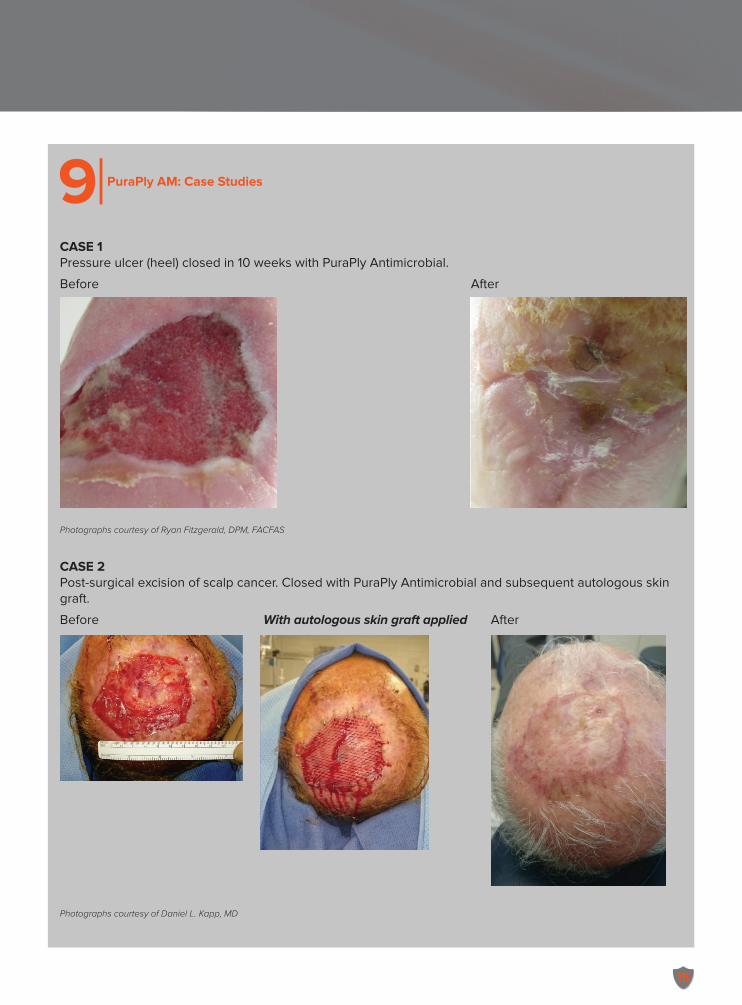
9| PuraPly AM: Case Studies
CASE 1Pressure ulcer (heel) closed in 10 weeks with PuraPly Antimicrobial.
Before After
Photographs courtesy of Ryan Fitzgerald, DPM, FACFAS
CASE 2Post-surgical excision of scalp cancer. Closed with PuraPly Antimicrobial and subsequent autologous skin graft.
Before With autologous skin graft applied After
Photographs courtesy of Daniel L. Kapp, MD
Expert Recommendations for Optimizing Outcomes in the Management of Biofilm to Promote Healing of Chronic Wounds
16
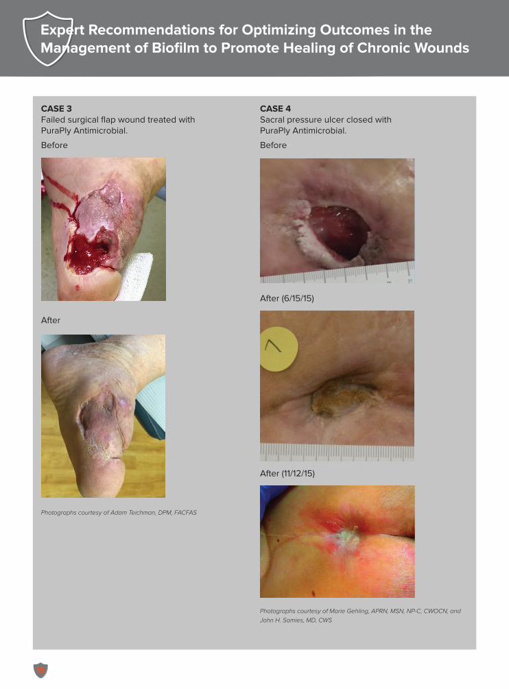
CASE 3Failed surgical flap wound treated with PuraPly Antimicrobial.
Before
After
Photographs courtesy of Adam Teichman, DPM, FACFAS
CASE 4Sacral pressure ulcer closed with PuraPly Antimicrobial.
Before
After (6/15/15)
After (11/12/15)
Photographs courtesy of Marie Gehling, APRN, MSN, NP-C, CWOCN, and John H. Samies, MD, CWS
17
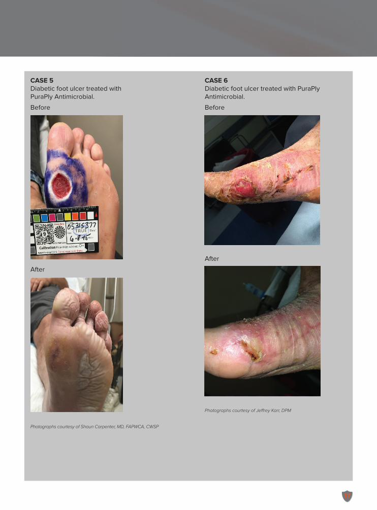
CASE 5Diabetic foot ulcer treated with PuraPly Antimicrobial.
Before
After
Photographs courtesy of Shaun Carpenter, MD, FAPWCA, CWSP
CASE 6Diabetic foot ulcer treated with PuraPly Antimicrobial.
Before
After
Photographs courtesy of Jeffrey Karr, DPM
Expert Recommendations for Optimizing Outcomes in the Management of Biofilm to Promote Healing of Chronic Wounds
18
10|Summary
Biofilm is the most common biobur-den in wounds and lies at an intermediate stage in the continuum of infection se-verity from light contamination to sepsis. Biofilm is clinically significant because it has been shown to delay wound healing by causing wounds to become “stuck” in the inflammatory phase of healing.
Virtually all chronic wounds have or are at risk of developing biofilm, which is a thick, sticky mass of pathogens, of-ten polymicrobial, residing in a protective extracellular polymeric matrix (EPM) that protects the microbes from both endogenous (e.g., antibodies) as well as exogenous (e.g., antibiotics) antimicrobi-al attack. This makes biofilm difficult to eradicate.
Aggressive debridement is critical in biofilm management, but studies show that biofilm starts reforming within 24 hours and mature biofilm can form by three days post-debridement. There-fore, antimicrobial therapy is essential. However, both topical antimicrobial and systemic antibiotics have significant lim-itations in effectively managing biofilm. Antibiotics may not penetrate the bio-film, and have limited effect as biofilm microbes tend to be inactive and antibi-otics require metabolically active targets. Resistance, tolerance, and cytotoxicity to normal tissues also pose challenges with some topical antimicrobial products.
PHMB is a topical antimicrobial with many advantages. It has a wide antimi-crobial spectrum, no mechanism by which resistance can emerge, very low cytotoxicity to normal tissues, and excel-lent tolerability.
Collagen-based dressings have been shown to promote wound healing, but collagen products are not interchange-
able. Research has shown that products that retain the native collagen architec-ture are able to inhibit a wider range of proteolytic enzymes compared to recon-stituted collagen. PuraPly Antimicrobial combines PHMB within an intact type I collagen matrix. The collagen matrix supports healing while the PHMB acts as an effective barrier against microbes, and has been shown to reduce biofilm.
PuraPly Antimicrobial has been ap-proved for use in a wide variety of wound types. It is also an appropriate choice for the initial phases of chronic DFU and VLU care to control biofilm in those wounds that may require a bioen-gineered cell-based therapy to promote wound healing and closure.
references1. Phillips PL, Yang Q, Sampson EM, et al.
Antimicrobial dressing efficacy against
mature Pseudomonas aeruginosa biofilm
on porcine skin explants. Int Wound J.
2013;12(4):469-483.
2. Phillips PL, Wolcott RG, Fletcher J, Schul-
tz GS. Biofilms made easy. Wounds Interna-
tional. 2010;1(3):1-6.
3. James GA, Swogger E, Wolcott R, et al.
Biofilms in chronic wounds. Wound Repair
Regen. 2008;16(1):37-44.
4. Mast BA, Schultz GS. Interactions of cy-
tokines, growth factors, and proteases in
acute and chronic wounds. Wound Rep
Reg. 1996;4(4):411-420, 1996.
5. Trengove NJ, Stacey MC, MacAuley S,
et al. Analysis of the acute and chronic
wound environments: the role of proteases
and their inhibitors. Wound Repair Regen.
1999;7(6):442-452.
6. Seth AK, Geringer MR, Hong SJ, Leung
KP, Galiano RD, Mustoe TA. Comparative
analysis of single-species and polybac-
terial wound biofilms using a quantita-
tive, in vivo, rabbit ear model. PLoS One.
2012;7(8):e42897.
7. Schierle CF, De la Garza M, Mustoe
TA, Galiano RD. Staphylococcal bio-
films impair wound healing by delaying
summary of expert panel key recommendations 3 Be highly aware that biofilm
affects virtually all wounds and delays wound healing.
3 Treat all wounds assuming that biofilm is present, even when wounds “look good.”
3 Focus on controlling bioburden as the first step in successful wound management.
3 Don’t rely on wound cultures since they are not accurate in diagnosing biofilm.
3 Debride wounds aggressively but realize debridement alone is insufficient.
3 Remember that biofilm starts reforming with 24 hours of de-bridement.
3 Use topical antimicrobials to prevent biofilm reformation be-tween debridement visits.
3 PHMB is an excellent topical antimicrobial agent with sever-al advantages—a broad spec-trum of activity, no resistance, and high tissue compatibility—that can act as a barrier against biofilm.
3 PuraPly Antimicrobial provides both PHMB and a natural colla-gen matrix in one wound care product.
3 In DFUs and VLUs that fail to adequately progress despite bioburden control, transition to bioengineered living cell thera-pies.
3 Do not use collagen products in presence of uncontrolled wound infection.
3 Do not advance to bioengi-neered living cell-based prod-ucts until bioburden is ade-quately controlled.
3 Keys to success: Control bioburden and support heal-ing.
19
reepithelialization in a murine cutane-
ous wound model. Wound Repair Regen.
2009;17(3):354-359.
8. Pastar I, Nusbaum AG, Gil J, et al. Interac-
tions of methicillin-resistant Staphylococcus
aureus USA300 and Pseudomonas aeruginosa
in polymicrobial wound infection. PLOS
One. 2013;8(2):e56846.
9. Yang Q, Phillips PL, Sampson E, et al. De-
velopment of a novel in vitro porcine skin
explant model for the assessment of ma-
ture bacterial biofilms. Wound Repair Regen.
2013;21(5):704-714.
10. Wolcott RD, Rumbaugh KP, James G.
Biofilm maturity studies indicate sharp
debridement opens a time-depen-
dent therapeutic window. J Wound Care.
2010;19(8):320–328.
11. Nusbaum AG, Gil J, Rippy MK, et al. Ef-
fective method to remove wound bacteria:
comparison of various debridement mo-
dalities in an in vivo porcine model. J Surg
Res. 2012:176(2):701-7.
12. Davis SC, Ricotti C, Cazzaniga AL, Welch
E, Mertz PM. Microscopic and physiolog-
ical evidence for biofilm-associated wound
colonization in-vivo. Wound Rep Reg.
2008;16(1):23-29.
13. Walters MC 3rd, Roe F, Bugnicourt A,
Franklin MJ, Stewart PS. Contributions of
antibiotic penetration, oxygen limitation,
and low metabolic activity to tolerance of
Pseudomonas aeruginosa biofilms to cipro-
floxacin and tobramycin. Antimicrob Agents
Chemother. 2003;47(1):317-323.
14. Lewis K. Platforms for antibiotic discovery.
Nat Rev Drug Dis. 2013;12(5):371-387.
15. de Beer D, Stoodley P, Roe F, Lewandows-
ki Z. Effects of biofilm structure on oxygen
distribution and mass transport. Biotechnol
Bioeng. 1994;43(11):1131-1138.
16. Dowd SE, Sun Y, Secor PR, et al. Survey of
bacterial diversity in chronic wounds us-
ing pyrosequencing, DGGE, and full ribo-
some shotgun sequencing. BMC Microbiol.
2008;8:43.
17. van Sorge NM, Beasley FC, Gusarov I, et
al. Methicillin-resistant Staphylococcus aureus
bacterial nitric-oxide synthase affects anti-
biotic sensitivity and skin abscess develop-
ment. J Biol Chem. 2013;288(9):6417-6426.
18. Shin DH, Choi YS, Cho YH. Unusual
properties of catalase A (KatA) of Pseudo-
monas aeruginosa PA14 are associated with
its biofilm peroxide resistance. J Bacteriology.
2008;190(8):2663-2670.
19. Davis SC, Ricotti C, Cazzaniga AL, Welch
E, Mertz PM. Microscopic and physiolog-
ical evidence for biofilm-associated wound
colonization in-vivo. Wound Rep Reg.
2008;16(1): 23-29.
20. Mertz PM, Oliveira-Gandia MF, Davis
SC. The evaluation of a cadexomer io-
dine wound dressing on methicllin resis-
tant Staphylococcus aureus (MRSA) in acute
wounds. Dermatol Surg. 1999;25(2):89-93.
21. Hübner NO, Kramer A. Review on the
efficacy, safety and clinical applications of
polihexanide, a modern wound antisep-
tic. Skin Pharmacol Physiol. 2010;23(Sup-
pl):17-27.
22. Gilbert P, Moore LE. Cationic antiseptics:
diversity of action under a common epi-
thet. J Appl Microbiol. 2005;99(4):703–715.
23. Muller G, Kramer A, Schmitt J, Harden
D, Koburger T. Reduced cytotoxicity of
polyhexamethylene biguanide hydrochlo-
ride (PHMB) by egg phosphatidylcholine
while maintaining antimicrobial efficacy.
Chem Biol Interact. 2011;190(2-3):171-178.
24. Hochstein AO, Bhatia A. Collagen: its role
in wound healing. Podiatry Management.
2014;33(6):103-110.
25. Negron L, Lun S, May BC. Ovine fores-
tomach matrix biomaterial is a broad spec-
trum inhibitor of matrix metalloprotein-
ases and neutrophil elastase. Int Wound J.
2014;11(4):392-397.
26. Solis M, Gil J, Valdes J, Higa A, Davis SC.
Evaluation of a purified collagen ma-
trix with PHMB on the proliferation of
methicillin resistant Staphylococcus aureus
(MRSA) in a porcine wound model. Poster
presented at the Symposium on Advanced
Wound Care; April 13-17, 2016; Atlanta,
GA.
©2016 HMP Communications, LLC (HMP). All rights reserved. Reproduction in whole or in part prohibited. Opinions expressed by authors are their own and not necessarily those of
HMP Communications, the editorial staff, or any member of the editorial advisory board. HMP Communications is not responsible for accuracy of dosages given in articles printed here-
in. HMP Communications disclaims responsibility for any injury to persons or property resulting from any ideas or products referred to in the articles. Content may not be reproduced
in any form without written permission. Reprints of articles are available. Rights, permission, reprint, and translation information are available at: www.hmpcommunications.com.
HMP Communications, LLC (HMP) is the authoritative source for comprehensive information and education servicing healthcare professionals. HMP’s products include peer-reviewed
and non–peer-reviewed medical journals, national tradeshows and conferences, online programs, and customized clinical programs. HMP is a wholly owned subsidiary of HMP
Communications Holdings LLC. Discover more about HMP’s products and services at www.hmpcommunications.com.
www.hmpcommunications.com 130-550LLC
an HMP Communications Holdings Company,
™
The citation for this publication is as follows: Carpenter S, Davis S, Fitzgerald R, et al. Expert recommendations for optimizing outcomes in the management of biofilm to promote healing of chronic wounds. WOUNDS. 2016;28(6 Suppl):S1-S20.