Embed Size (px)
Citation preview


Experiments on Biological Structure
- Optical Microscopy- Electron Microscopy
- X-ray crystallography- NMR Spectroscopy- Fluorescence Spectroscopy
45 minutes

Myosin - Optical Microscopy
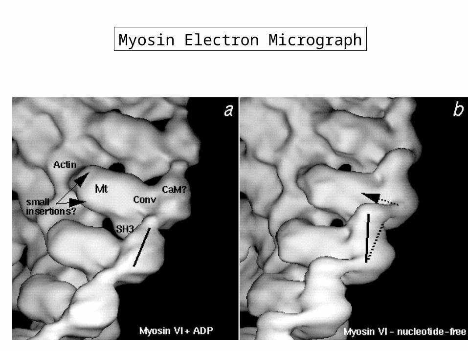
Myosin Electron Micrograph

Wilhelm Conrad Röntgen (1845-1923)
Nobel prize in Physics 1901 in recognition of the extraordinary services he has rendered by the discovery of the remarkable rays subsequently named after him
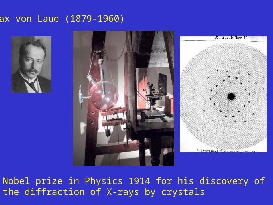
Max von Laue (1879-1960)
Nobel prize in Physics 1914 for his discovery of the diffraction of X-rays by crystals
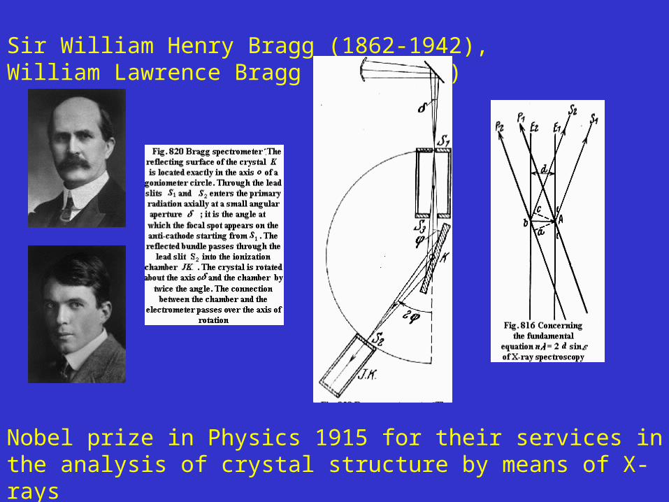
Sir William Henry Bragg (1862-1942), William Lawrence Bragg (1890-1971)
Nobel prize in Physics 1915 for their services in the analysis of crystal structure by means of X-rays
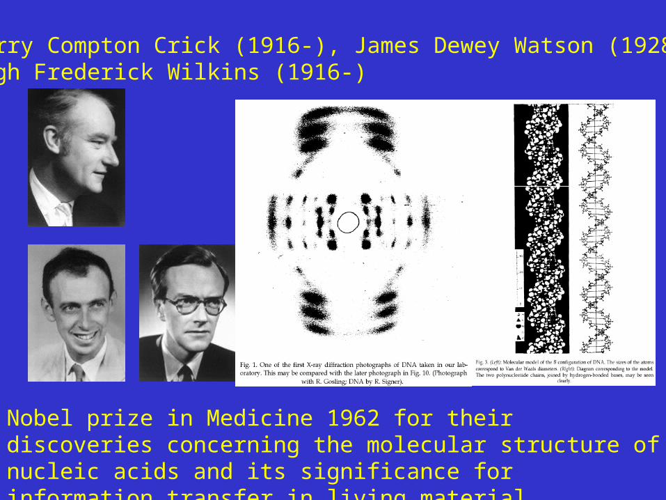
Francis Harry Compton Crick (1916-), James Dewey Watson (1928-),Maurice Hugh Frederick Wilkins (1916-)
Nobel prize in Medicine 1962 for their discoveries concerning the molecular structure of nucleic acids and its significance for information transfer in living material
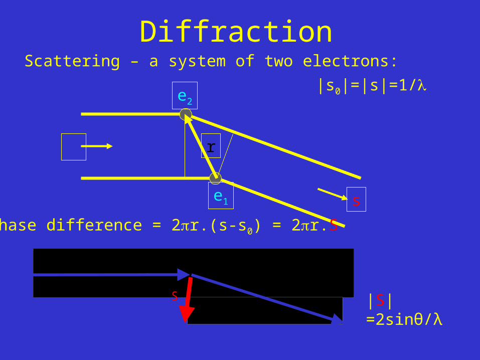
DiffractionScattering – a system of two electrons:
e1
e2
s0
s
r
phase difference = 2r.(s-s0) = 2r.S
|s0|=|s|=1/
-s 0
ss 0S
-s 0
ss 0S
|S|=2sinθ/λ
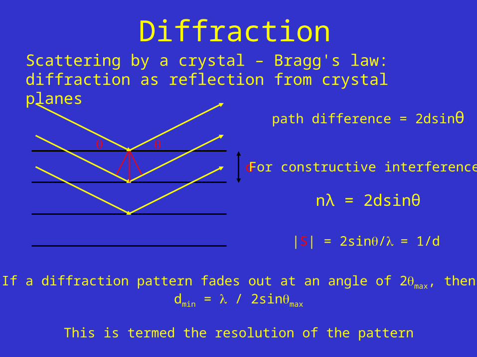
DiffractionScattering by a crystal – Bragg's law: diffraction as reflection from crystal planes
d
path difference = 2dsinθ
For constructive interference,
nλ = 2dsinθ
|S| = 2sin/= 1/d
If a diffraction pattern fades out at an angle of 2max, thendmin = / 2sinmax
This is termed the resolution of the pattern

End of Lecture 1
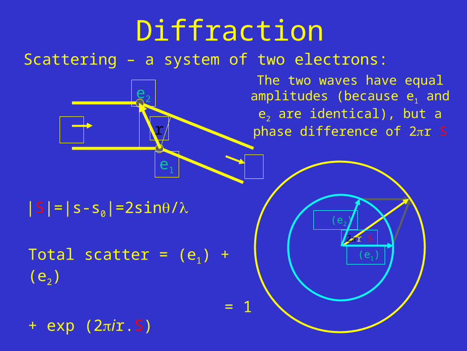
DiffractionScattering – a system of two electrons:
The two waves have equal amplitudes (because e1 and e2 are identical), but a phase difference
of 2r.S
|S|=|s-s0|=2sin/
(e1)Total scatter = (e1) + (e2)
= 1 + exp (2ir.S)
2r.S
(e2)
e1
e2
s0
s
r
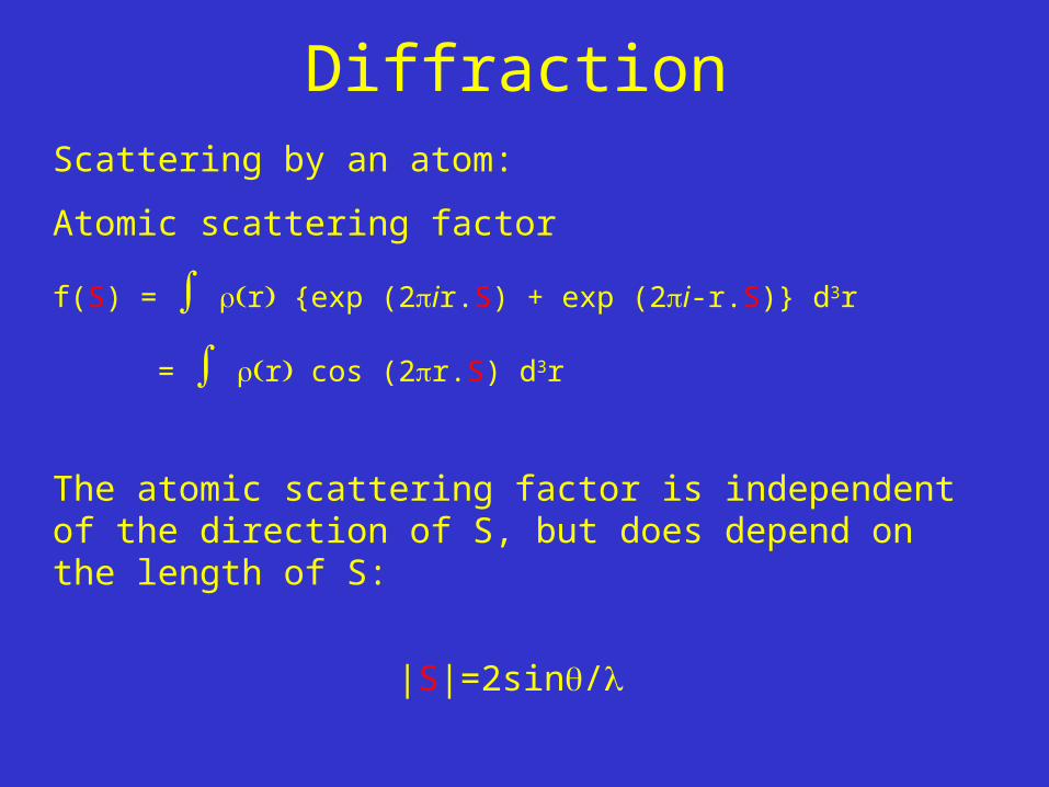
DiffractionScattering by an atom:
Atomic scattering factor
f(S) = r {exp (2ir.S) + exp (2i-r.S)} d3r
= r cos (2r.S) d3r
The atomic scattering factor is independent of the direction of S, but does depend on the length of S:
|S|=2sin/
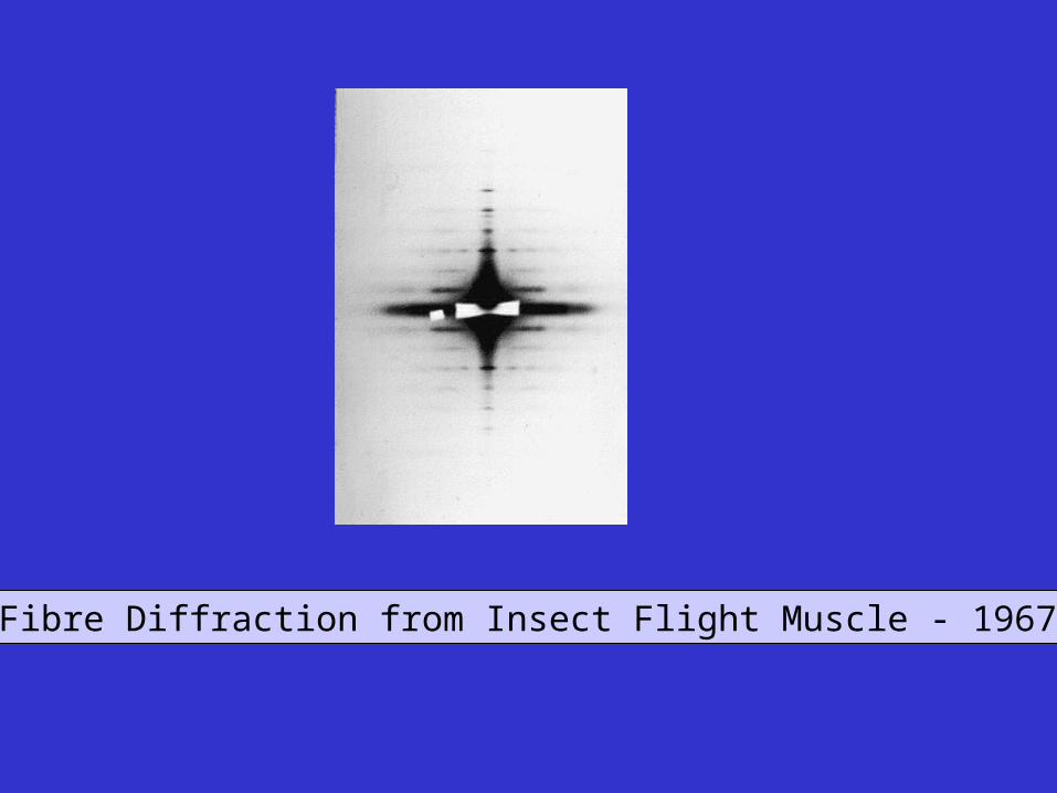
Fibre Diffraction from Insect Flight Muscle - 1967

The Beginning of Molecular Biology.
Francis Crick and James D Watson
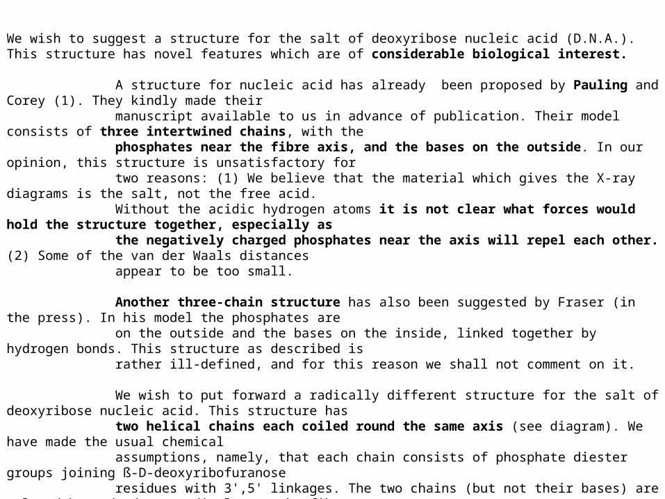
We wish to suggest a structure for the salt of deoxyribose nucleic acid (D.N.A.). This structure has novel features which are of considerable biological interest.
A structure for nucleic acid has already been proposed by Pauling and Corey (1). They kindly made their manuscript available to us in advance of publication. Their model consists of three intertwined chains, with the phosphates near the fibre axis, and the bases on the outside. In our opinion, this structure is unsatisfactory for two reasons: (1) We believe that the material which gives the X-ray diagrams is the salt, not the free acid. Without the acidic hydrogen atoms it is not clear what forces would hold the structure together, especially as the negatively charged phosphates near the axis will repel each other. (2) Some of the van der Waals distances appear to be too small.
Another three-chain structure has also been suggested by Fraser (in the press). In his model the phosphates are on the outside and the bases on the inside, linked together by hydrogen bonds. This structure as described is rather ill-defined, and for this reason we shall not comment on it.
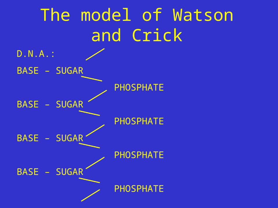
We wish to put forward a radically different structure for the salt of deoxyribose nucleic acid. This structure has two helical chains each coiled round the same axis (see diagram). We have made the usual chemical assumptions, namely, that each chain consists of phosphate diester groups joining ß-D-deoxyribofuranose residues with 3',5' linkages. The two chains (but not their bases) are related by a dyad perpendicular to the fibre axis. Both chains follow right- handed helices, but owing to the dyad the sequences of the atoms in the two chains run in opposite directions. Each chain loosely resembles Furberg's model No. 1; that is, the bases are on the inside of the helix and the phosphates on the outside. The configuration of the sugar and the atoms near it is close to Furberg's 'standard configuration', the sugar being roughly perpendicular to the attached base. There is a residue on each every 3.4 A. in the z-direction. We have assumed an angle of 36° between adjacent residues in the same chain, so that the structure repeats after 10 residues on each chain, that is, after 34 A. The distance of a phosphorus atom from the fibre axis is 10 A. As the phosphates are on the outside, cations have easyaccess to them.
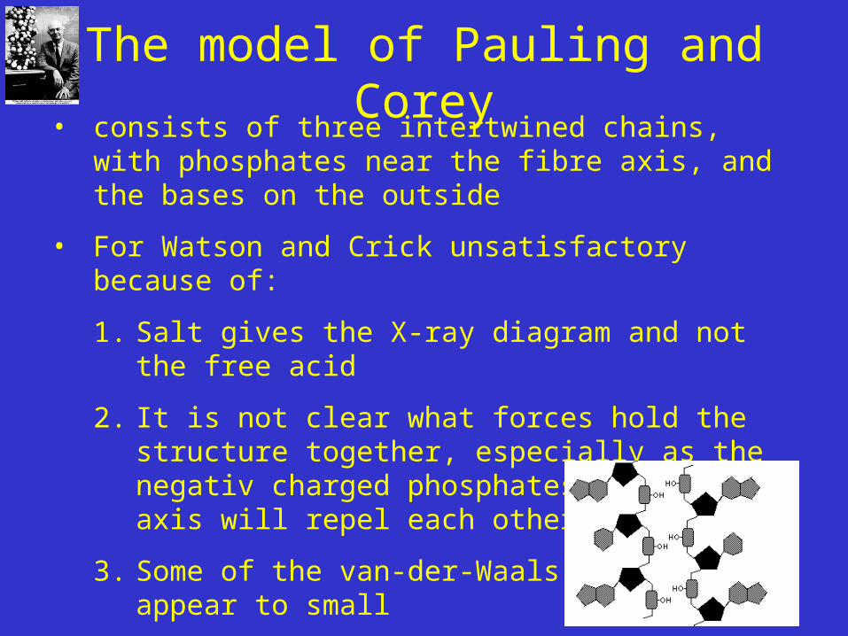
The model of Pauling and Corey• consists of three intertwined chains, with phosphates
near the fibre axis, and the bases on the outside
• For Watson and Crick unsatisfactory because of:
1. Salt gives the X-ray diagram and not the free acid
2. It is not clear what forces hold the structure together, especially as the negativ charged phosphates near the axis will repel each other
3. Some of the van-der-Waals distance appear to small

The structure is an open one, and its water content is rather high. At lower water contents we would expect the bases to tilt so that the structure could become more compact.
The novel feature of the structure is the manner in which the two chains are held together by the purine and pyrimidine bases. The planes of the bases are perpendicular to the fibre axis. The are joined together in pairs, a single base from the other chain, so that the two lie side by side with identical z-co-ordinates. One of the pair must be a purine and the other a pyrimidine for bonding to occur. The hydrogen bonds are made as follows : purine position 1 to pyrimidine position 1 ; purine position 6 to pyrimidine position 6.If it is assumed that the bases only occur in the structure in the most plausible tautomeric forms (that is, with the keto rather than the enol configurations) it is found that only specific pairs of bases can bond together. These pairs are : adenine (purine) with thymine (pyrimidine), and guanine (purine) with cytosine (pyrimidine).
In other words, if an adenine forms one member of a pair, on either chain, then on these assumptions the other member must be thymine ; similarly for guanine and cytosine. The sequence of bases on a single chain does not appear to be restricted in any way. However, if only specific pairs of bases can be formed, it follows that if the sequence of bases on one chain is given, then the sequence on the other chain is automatically determined .
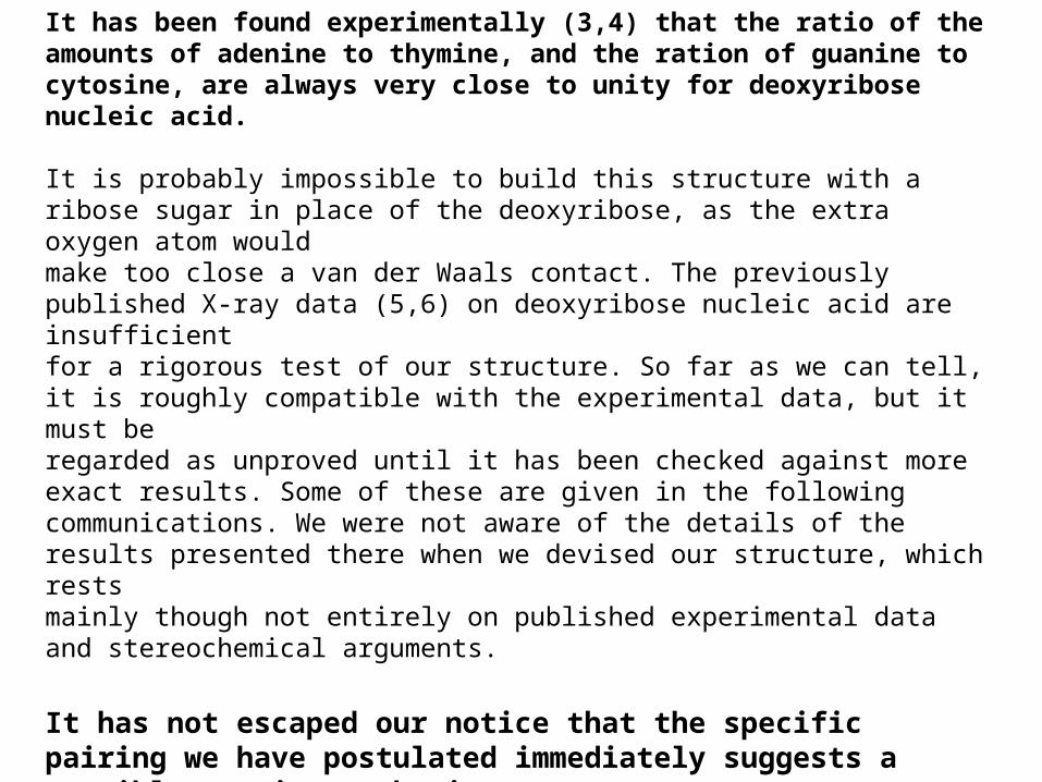
It has been found experimentally (3,4) that the ratio of the amounts of adenine to thymine, and the ration of guanine to cytosine, are always very close to unity for deoxyribose nucleic acid.
It is probably impossible to build this structure with a ribose sugar in place of the deoxyribose, as the extra oxygen atom wouldmake too close a van der Waals contact. The previously published X-ray data (5,6) on deoxyribose nucleic acid are insufficientfor a rigorous test of our structure. So far as we can tell, it is roughly compatible with the experimental data, but it must beregarded as unproved until it has been checked against more exact results. Some of these are given in the followingcommunications. We were not aware of the details of the results presented there when we devised our structure, which restsmainly though not entirely on published experimental data and stereochemical arguments.
It has not escaped our notice that the specific pairing we have postulated immediately suggests a possible copying mechanismfor the genetic material.
Full details of the structure, including the conditions assumed in building it, together with a set of co-ordinates for the atoms,will be published elsewhere.

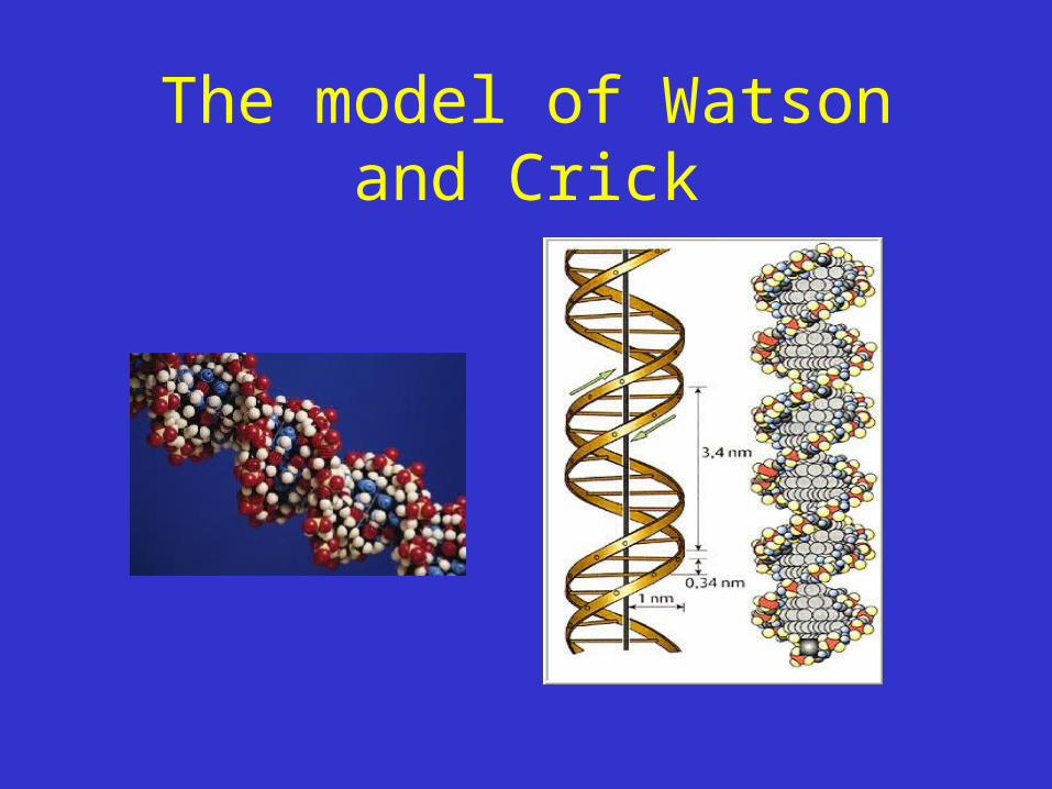
The model of Watson and Crick
• two helical chains each coiled round the same axis
• Right handed helices
• Residue on each chain every 3,4 Å in z direction
• Angle of 36° between the residues of the same chain
• Structure repeats after 10 residues, after 34 Å
• Bases are joined in pairs via hydrogen bond
• Pairs: adenine (purine) – thymine (pyrimidin), guanine (purine) – cytosine (pyrimidin)
The model of Watson and Crick
D.N.A.:
BASE – SUGAR
PHOSPHATE
BASE – SUGAR
PHOSPHATE
BASE – SUGAR
PHOSPHATE
BASE – SUGAR
PHOSPHATE

The model of Watson and Crick

The model of Watson and Crick
Crystallography
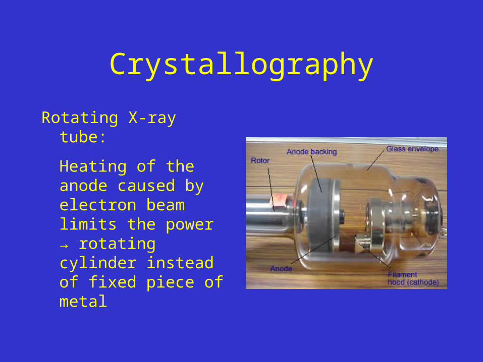
Crystallography
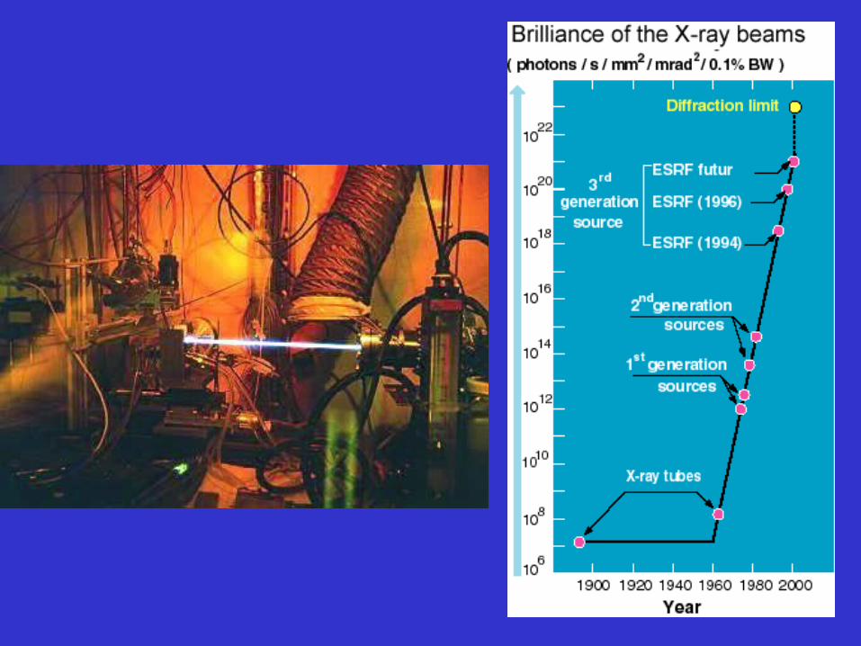
Rotating X-ray tube:
Heating of the anode caused by electron beam limits the power → rotating cylinder instead of fixed piece of metal
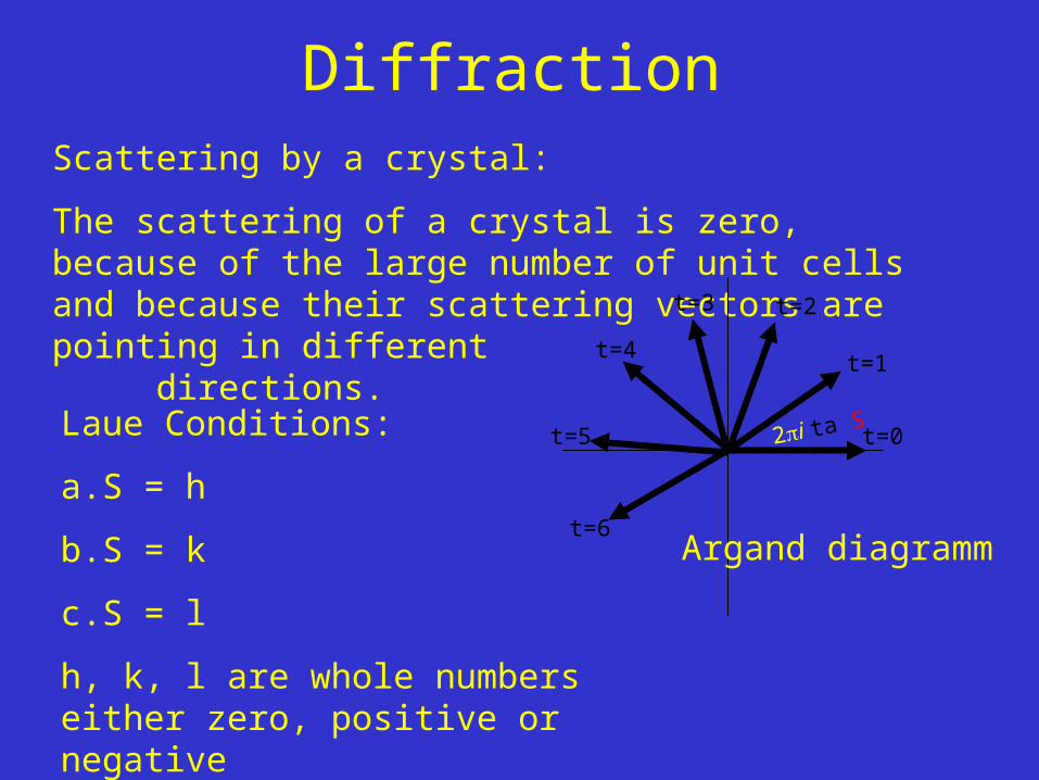
DiffractionScattering by a crystal:
The scattering of a crystal is zero, because of the large number of unit cells and because their scattering vectors are pointing in differentdirections.
t=0
t=1
2i ta.S
t=2t=3
t=4
t=5
t=6Argand diagramm
Laue Conditions:
a.S = h
b.S = k
c.S = l
h, k, l are whole numbers either zero, positive or negative

James Batcheller Sumner (1887-1955)Nobel prize in Chemistry 1946 for his discovery that enzymes can be crystallized
John Howard Northrop (1891-1987), Wendell Meredith Stanley (1904-1971) Nobel prize in Chemistry 1946 for their preparation of enzymes and virus proteins in a pure form
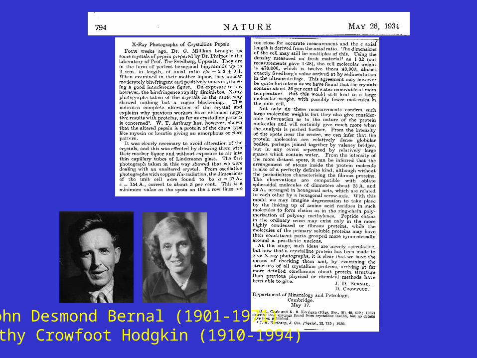
John Desmond Bernal (1901-1971)Dorothy Crowfoot Hodgkin (1910-1994)
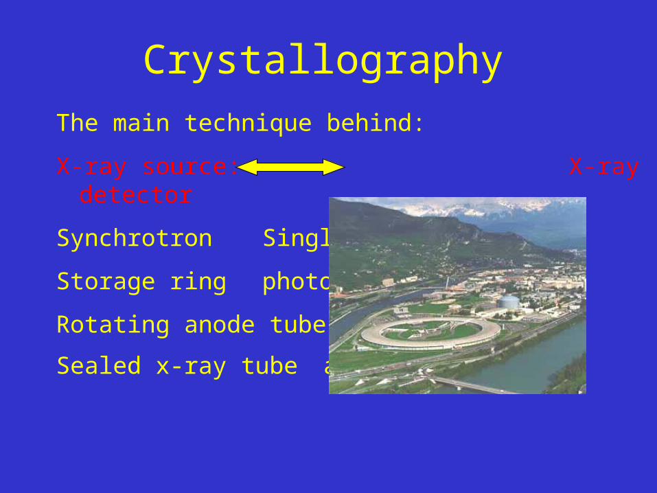
Crystallography
The main technique behind:
X-ray source: X-ray detector
Synchrotron Single photon counter
Storage ring photographic film
Rotating anode tube image plates
Sealed x-ray tube area detectors

X-ray crystallography - first purify and crystallize

Crystallography - Crystals
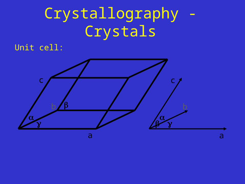
Crystallography - Crystals
Unit cell:
a
b
c
a
b
c
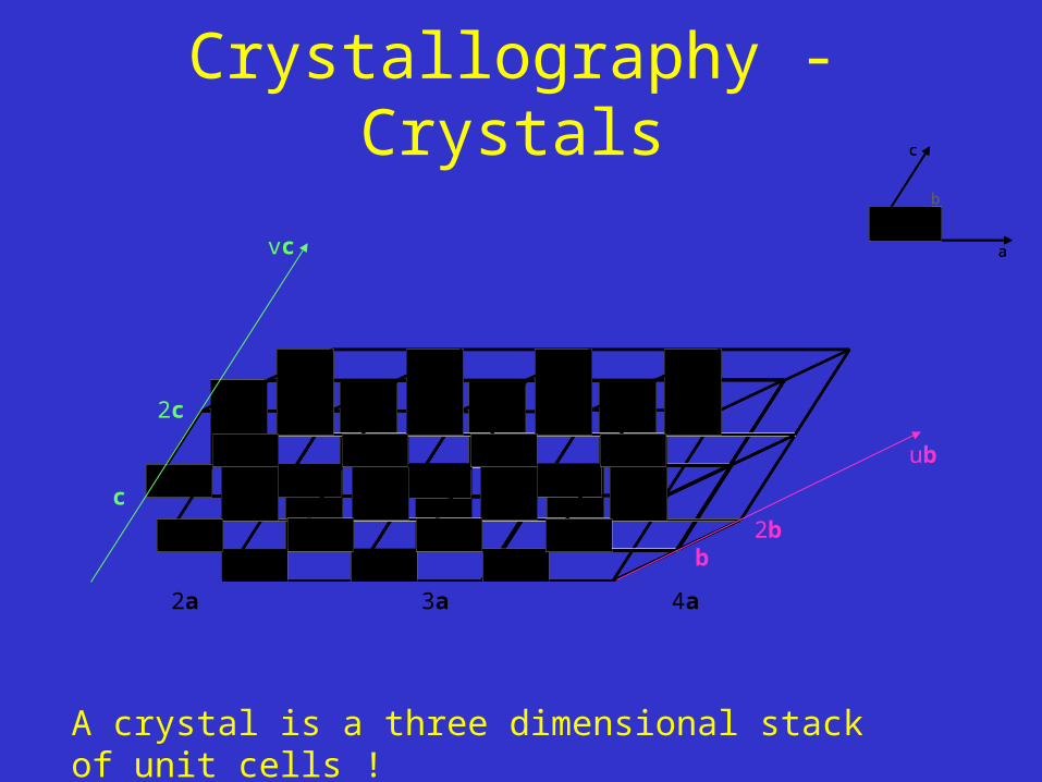
Crystallography - Crystals
a
b
c
a
b
c
0 a 2a 3a 4a ta
b2b
c
2c
vc
ub
A crystal is a three dimensional stack of unit cells !

Crystallography - CrystalsDifferent unit cells:
a primitive unit cell
a unit cell centered in the planes
a body-centered unit cell
a face-centered unit cell
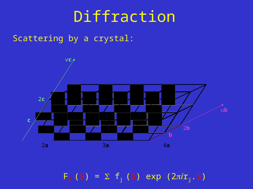
DiffractionScattering by a crystal:
F0 (S) = fj (S) exp (2irj.S)
0 a 2a 3a 4a ta
b2b
c
2c
vc
ub
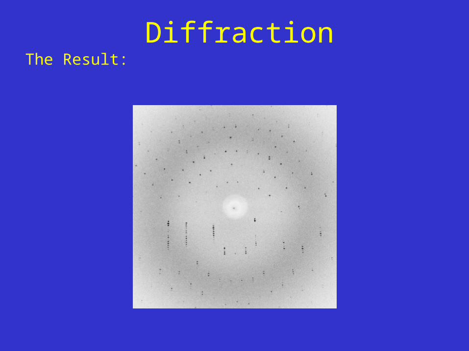
DiffractionThe Result:

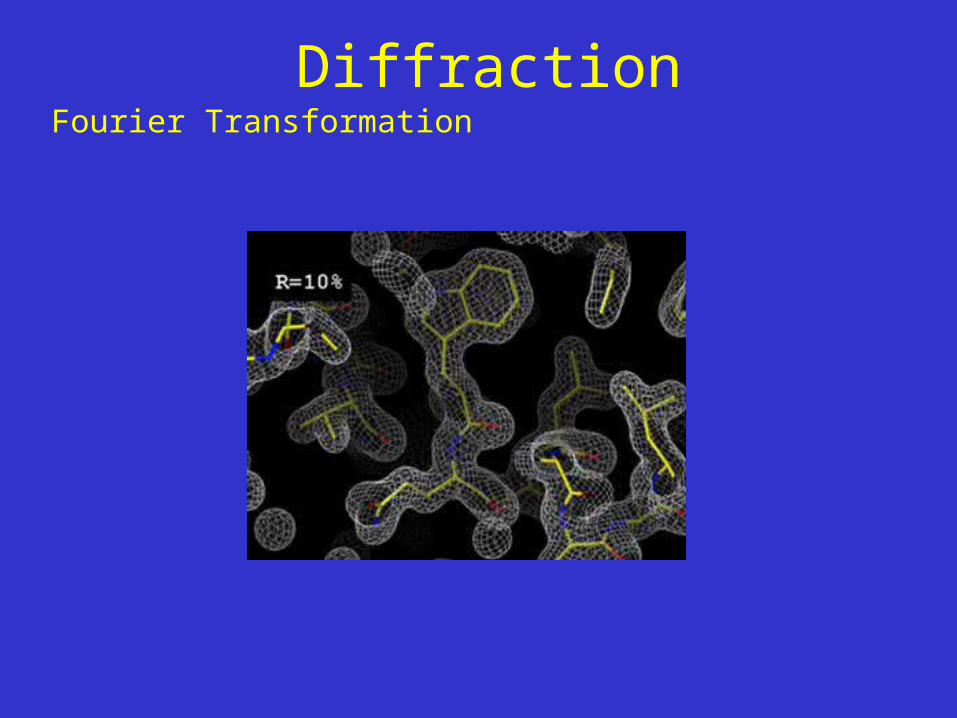
DiffractionFourier Transformation
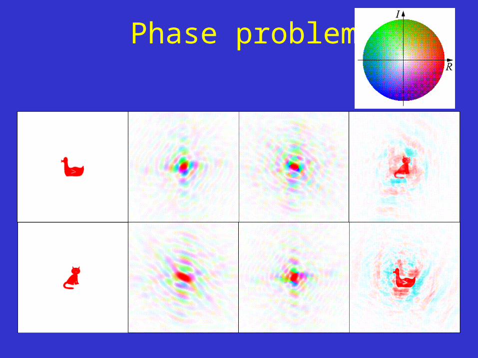
Phase problem


-Stamp Collecting.
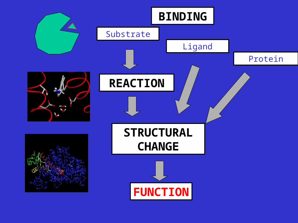
Substrate
Protein
Ligand
BINDING
REACTION
FUNCTION
STRUCTURAL CHANGE
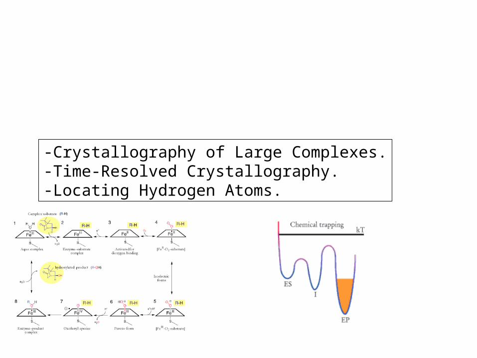
-Crystallography of Large Complexes.-Time-Resolved Crystallography.-Locating Hydrogen Atoms.
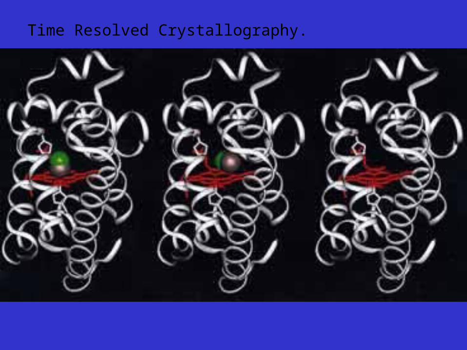
Time Resolved Crystallography.

Mouse Prion Protein (PrPc)
NMR Structure
Protein Structure

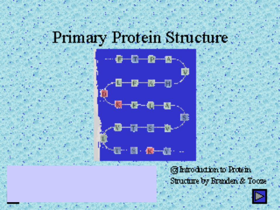

The Peptide Backbone Chain
Crosslinking - the disulphide bridge
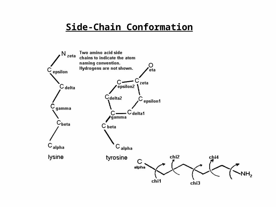
Side-Chain Conformation

Linus Carl Pauling (1901-1994)
Nobel prize in Chemistry 1954 for his research into the nature of the chemical bond and its application to the elucidation of the structure of complex substances

The Alpha Helix (Pauling & Corey, from alpha keratin)


Supersecondary Structure
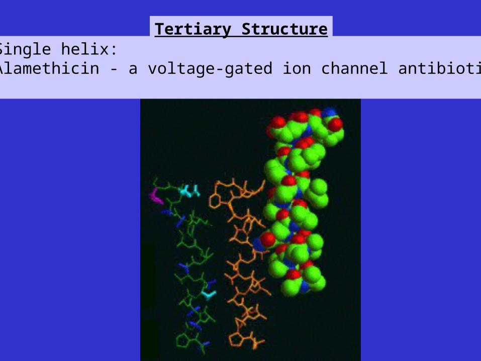
Single helix:Alamethicin - a voltage-gated ion channel antibiotic
Tertiary Structure

Helix-turn Helix:Rop (RNA-Binding Protein) Four-Helix Bundle

Two Greek Keys (gamma crystallin)
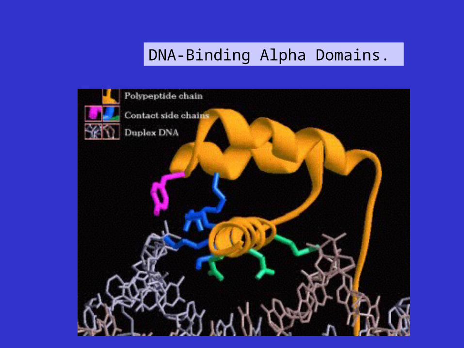
DNA-Binding Alpha Domains.
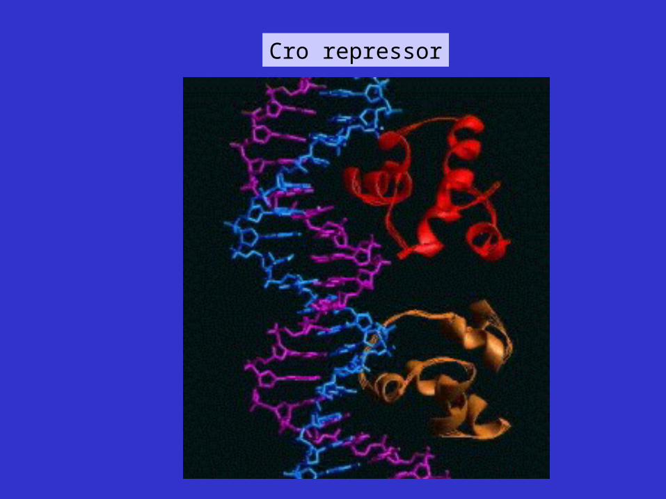
Cro repressor
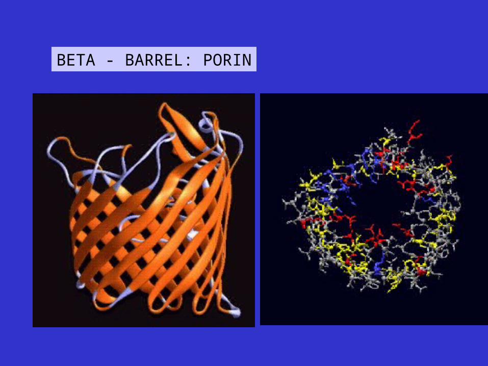
BETA - BARREL: PORIN

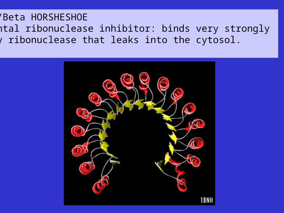
Alpha/Beta HORSHESHOEPlacental ribonuclease inhibitor: binds very strongly to any ribonuclease that leaks into the cytosol.
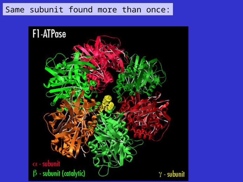
Same subunit found more than once:
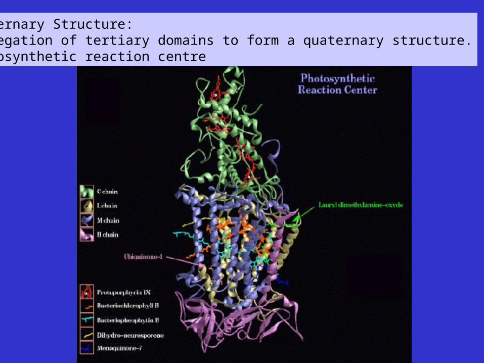
Quaternary Structure:Aggregation of tertiary domains to form a quaternary structure.Photosynthetic reaction centre
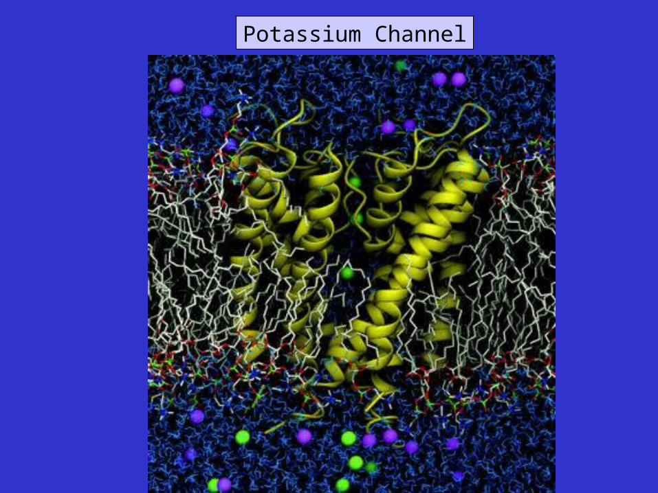
Potassium Channel
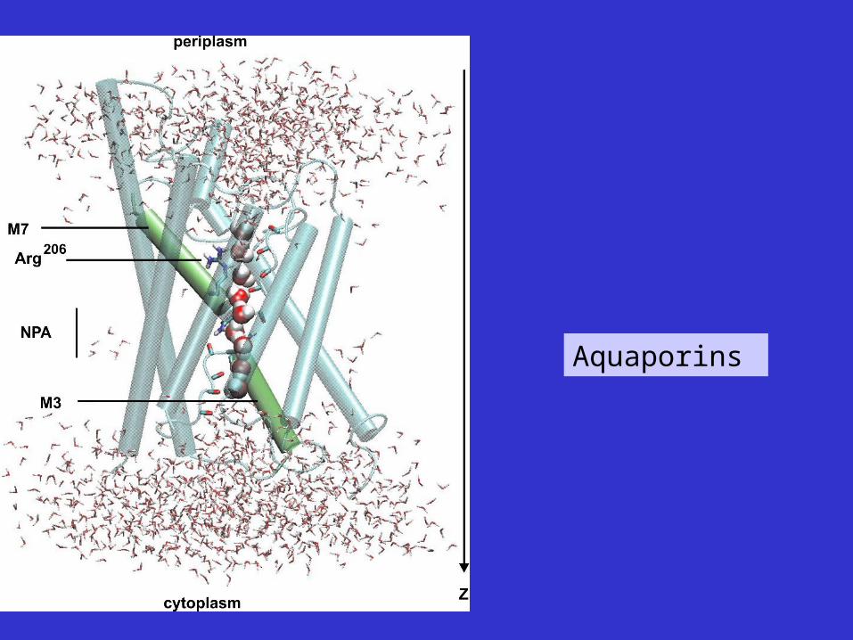
Aquaporins

Strands from different protein chains associate to form 2o struct.

Same subunit associates