Embed Size (px)
Citation preview

EXPERIMENTAL SHOCK DECOMPOSITION OF SIDERITE AND THE ORIGIN OF MAGNETITE IN MARTIAN METEORITE ALH84001
M.S. BELL1,2*
1Geosciences Department, The University of Houston, Houston, Texas 77204, 2Jacobs Sverdrup@Johnson Space Center, Bldg. 31, Room 222, Mail Code KT, Houston, TX 77058
*Corresponding author. E-mail: [email protected]
Abstract
Shock recovery experiments to determine whether magnetite could be produced by the
decomposition of iron-carbonate were initiated. Naturally occurring siderite was first
characterized by electron microprobe (EMP), transmission electron microscopy (TEM),
Mossbauer spectroscopy, and magnetic susceptibility measurements to be sure that the starting
material did not contain detectable magnetite. Samples were shocked in tungsten-alloy holders
(W=90%, Ni=6%, Cu=4%) to further insure that any iron phases in the shock products were
contributed by the siderite rather than the sample holder. Each sample was shocked to a specific
pressure between 30 to 49 GPa. Previously reported results of TEM analyses on 49 GPa
experiments indicated the presence of nano-phase spinel-structured iron oxide. Transformation
of siderite to magnetite as characterized by TEM was found in the 49 GPa shock experiment.
Compositions of most magnetites are > 50% Fe+2 in the octahedral site of the inverse spinel
structure. Magnetites produced in shock experiments display the same range of single-domain,
superparamagnetic sizes (~50 – 100 nm), compositions (100% magnetite to 80% magnetite-20%
magnesioferrite), and morphologies (equant, elongated, euhedral to subhedral) as magnetites
synthesized by Golden et al. (2001) or magnetites grown naturally by MV1 magnetotactic
bacteria, and as the magnetites in Martian meteorite ALH84001. Fritz et al. (2005) previously
concluded that ALH84001 experienced ~32 GPa pressure and a resultant thermal pulse of ~100 -
110oC. However, ALH84001 contains evidence of local temperature excursions high enough to
1

melt feldspar, pyroxene, and a silica-rich phase. This 49 GPa experiment demonstrates that
magnetite can be produced by the shock decomposition of siderite as a result of local heating to
> 470oC. Therefore, magnetite in the rims of carbonates in Martian meteorite ALH84001 could
be a product of shock devolatilization of siderite as well.
2

Introduction
The debate about fossil life on Mars currently concentrates on magnetites of
specific sizes and habits in the Fe-rich portions of carbonate concretions in Martian
meteorite ALH 84001 (Golden et al., 2001; Thomas-Keprta et al., 2000). Golden et al.
(2001) were able to demonstrate inorganic synthesis of these compositionally zoned
concretions from aqueous solutions of variable ion-concentrations. They further
demonstrated the formation of magnetite from siderite upon heating at 470oC under a
Mars-like, CO2-rich atmosphere according to 3FeC03 = Fe3O4 + 2CO2 + CO (French,
1970) and they postulated that the carbonates in ALH 84001 were heated to these
temperatures in an unspecified shock event. Devolatilization of the ALH84001
carbonates would have occurred in a closed system or at least in the absence of free
surfaces. Langenhorst et al. (2000) found bubbles or channels in ALH84001 carbonates
which they interpret to represent shock-induced degassing structures. Brearley (2003)
described the presence of magnetite associated with voids in ALH84001 carbonate which
could be evidence of CO2 loss. Barber and Scott (2006) describe “fine-grained carbonate
that is partially decomposed to coarse-grained magnetite” in ALH84001. A number of
differential thermal analysis (DTA) studies of siderite (Bagin and Rybak, 1970; Bell et
al., 2000; Criado et al., 1988; Gallagher and Warne, 1981; Lin et al., 2000; Pan, 2000;
Zakharov and Adonyi, 1986) conclude that siderite decomposes at about 580oC and the
resultant FeO becomes oxidized to Fe3O4 in a CO2 atmosphere at about 600oC. Brearley
(2003) describes the thermal decomposition relationships between end-member
carbonates that represent the solid solution compositions found to comprise ALH 84001
carbonates. Observations based on experimental data (Harker and Tuttle, 1955; Koziol
3
and Newton, 1995) show conclusively that siderite is much less stable than calcite,
magnesite, or rhodochrosite, which are the three other significant solid solution
components in carbonates in ALH84001 ((Brearley, 2003).
The average shock pressure deduced for ALH 84001, substantially based on the
refractive index of diaplectic feldspar glasses (Fritz et al., 2005), is 32 GPa; associated
temperatures increase by 100 - 110oC. However, some of the feldspar, pyroxene, and a
silica-rich phase was melted (Barber and Scott, 2006; Bell et al., 1999; Mittlefehldt,
1994; Schwandt et al., 1999a, 1999b; Shearer and Adock, 1998) requiring local pressure
deviations as high as 45-50 GPa. Langenhorst et al. (2000) observe the carbonates in
ALH 84001 to be melted locally, requiring pressures in excess of 60 GPa and
temperatures > 600oC. Combining these shock studies with the above inorganic synthesis
of zoned carbonates it would seem possible to produce the ALH 84001 magnetites by the
shock-induced decomposition of siderite.
The extreme physical conditions of pressure, temperature, and strain imposed by
transient shock waves during an impact event produce unique effects (e;g;, mineral
deformation, melting) in the rocks and mineral grains through which they pass. Although
sediments, sedimentary rocks (including carbonates), water, and ice are major
constituents of the outer crust of the Earth, most experimental and theoretical cratering
studies have been directed toward volatile-free rocks. Whereas volatile-free rocks (such
as quartz) may preserve unambiguous signatures of shock metamorphism, volatile-rich
rocks such as carbonates may be completely vaporized. However, features caused by
shock metamorphism may be preserved in rock textures or as new mineral phases. It is
therefore possible that the magnetite in ALH84001 could have been formed by shock
4

devolatilization of siderite according to the equation from French (1970). This could
explain the lack of siderite in the ALH84001 zoned carbonate rims containing magnetite
(McKay et al., 1996).
This hypothesis is tested here experimentally by exposing siderite to experimental
shock. As reported earlier by Bell et al. (2002), intimately mixed zones of nanocrystalline
spinel-structured Fe-Mg oxides were produced, based on preliminary transmission
electron microscope (TEM) analyses and selected area electron diffraction (SAED)
patterns (Figure 1), in experimentally shocked siderite at 49 GPa. The magnetic
components of this mix have now been concentrated by isolating magnetic grains from
the bulk shocked material for chemical and other analyses of the shock products. Using a
hand magnet drawn beneath weighing paper upon which the sample powder had been
placed, magnetic particles were extracted from the bulk and subjected to dissolution
using the techniques of Golden et al. (2001) for more detailed TEM identification and
characterization of the shock products.
Analytical Methods
Natural Copper Lake (Antigonish County, Nova Scotia, Canada) siderite was
crushed, ground in alcohol and sieved to obtain the 120 – 250 μm size fraction. For the
shock experiments approximately 70 mg aliquots were encased in tungsten-alloy holders
(W=90%, Ni=6%, Cu=4%) to avoid any contribution of Fe from the sample holder to the
reaction products. The experimental set up of the shock experiment is described by
(Gibbons et al., 1975; Horz, 1970). Samples were shocked at pressures from 30 to 49
5
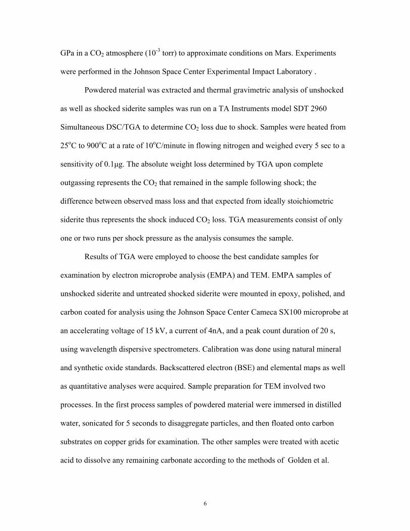
GPa in a CO2 atmosphere (10-3 torr) to approximate conditions on Mars. Experiments
were performed in the Johnson Space Center Experimental Impact Laboratory .
Powdered material was extracted and thermal gravimetric analysis of unshocked
as well as shocked siderite samples was run on a TA Instruments model SDT 2960
Simultaneous DSC/TGA to determine CO2 loss due to shock. Samples were heated from
25oC to 900oC at a rate of 10oC/minute in flowing nitrogen and weighed every 5 sec to a
sensitivity of 0.1μg. The absolute weight loss determined by TGA upon complete
outgassing represents the CO2 that remained in the sample following shock; the
difference between observed mass loss and that expected from ideally stoichiometric
siderite thus represents the shock induced CO2 loss. TGA measurements consist of only
one or two runs per shock pressure as the analysis consumes the sample.
Results of TGA were employed to choose the best candidate samples for
examination by electron microprobe analysis (EMPA) and TEM. EMPA samples of
unshocked siderite and untreated shocked siderite were mounted in epoxy, polished, and
carbon coated for analysis using the Johnson Space Center Cameca SX100 microprobe at
an accelerating voltage of 15 kV, a current of 4nA, and a peak count duration of 20 s,
using wavelength dispersive spectrometers. Calibration was done using natural mineral
and synthetic oxide standards. Backscattered electron (BSE) and elemental maps as well
as quantitative analyses were acquired. Sample preparation for TEM involved two
processes. In the first process samples of powdered material were immersed in distilled
water, sonicated for 5 seconds to disaggregate particles, and then floated onto carbon
substrates on copper grids for examination. The other samples were treated with acetic
acid to dissolve any remaining carbonate according to the methods of Golden et al.
6
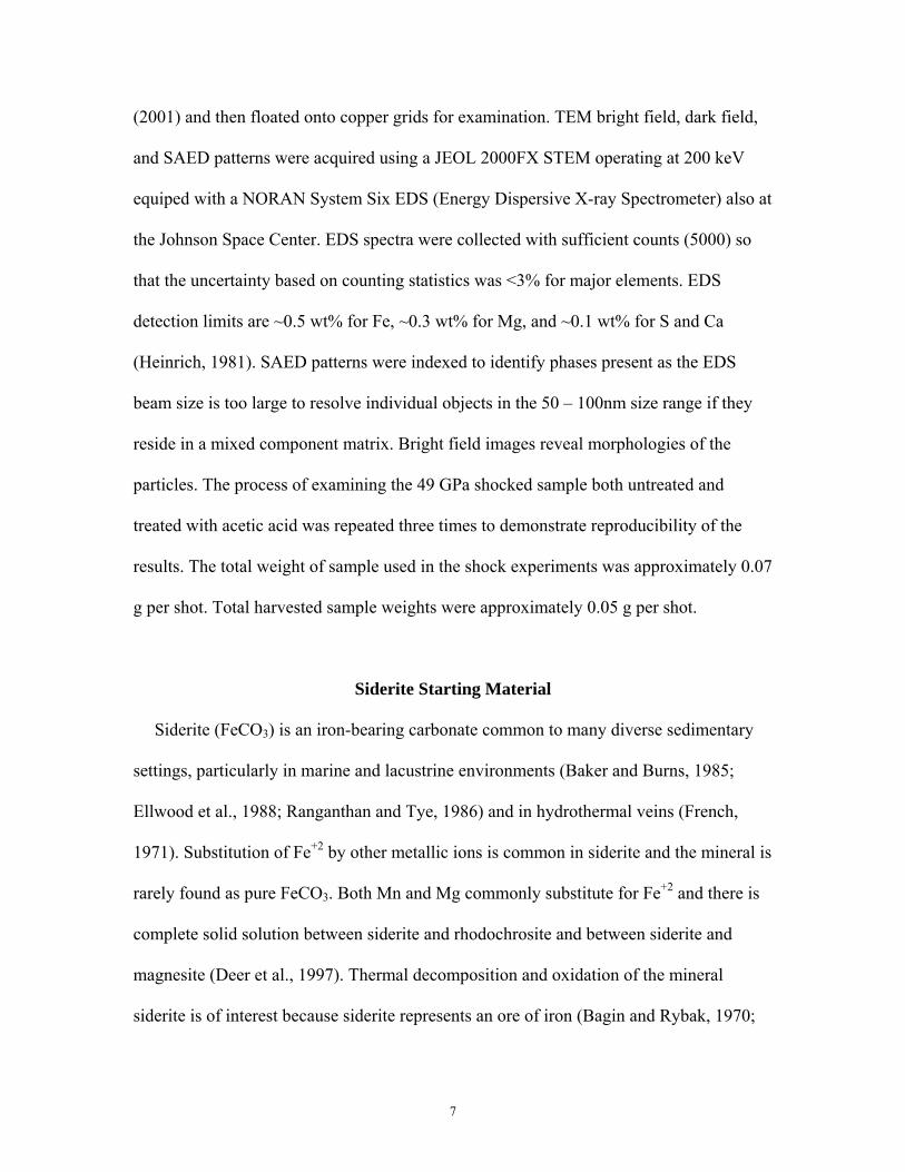
(2001) and then floated onto copper grids for examination. TEM bright field, dark field,
and SAED patterns were acquired using a JEOL 2000FX STEM operating at 200 keV
equiped with a NORAN System Six EDS (Energy Dispersive X-ray Spectrometer) also at
the Johnson Space Center. EDS spectra were collected with sufficient counts (5000) so
that the uncertainty based on counting statistics was <3% for major elements. EDS
detection limits are ~0.5 wt% for Fe, ~0.3 wt% for Mg, and ~0.1 wt% for S and Ca
(Heinrich, 1981). SAED patterns were indexed to identify phases present as the EDS
beam size is too large to resolve individual objects in the 50 – 100nm size range if they
reside in a mixed component matrix. Bright field images reveal morphologies of the
particles. The process of examining the 49 GPa shocked sample both untreated and
treated with acetic acid was repeated three times to demonstrate reproducibility of the
results. The total weight of sample used in the shock experiments was approximately 0.07
g per shot. Total harvested sample weights were approximately 0.05 g per shot.
Siderite Starting Material
Siderite (FeCO3) is an iron-bearing carbonate common to many diverse sedimentary
settings, particularly in marine and lacustrine environments (Baker and Burns, 1985;
Ellwood et al., 1988; Ranganthan and Tye, 1986) and in hydrothermal veins (French,
1971). Substitution of Fe+2 by other metallic ions is common in siderite and the mineral is
rarely found as pure FeCO3. Both Mn and Mg commonly substitute for Fe+2 and there is
complete solid solution between siderite and rhodochrosite and between siderite and
magnesite (Deer et al., 1997). Thermal decomposition and oxidation of the mineral
siderite is of interest because siderite represents an ore of iron (Bagin and Rybak, 1970;
7
Criado et al., 1988; French, 1971; Gallagher and Warne, 1981, Zakharov and Adonyi,
1986). The initial decomposition temperature is dependent on substitution of Mn+2 and
Mg+2 for Fe+2 but even a few per cent MnO or MgO is not enough to noticably increase
the temperature (Kulp et al., 1951).
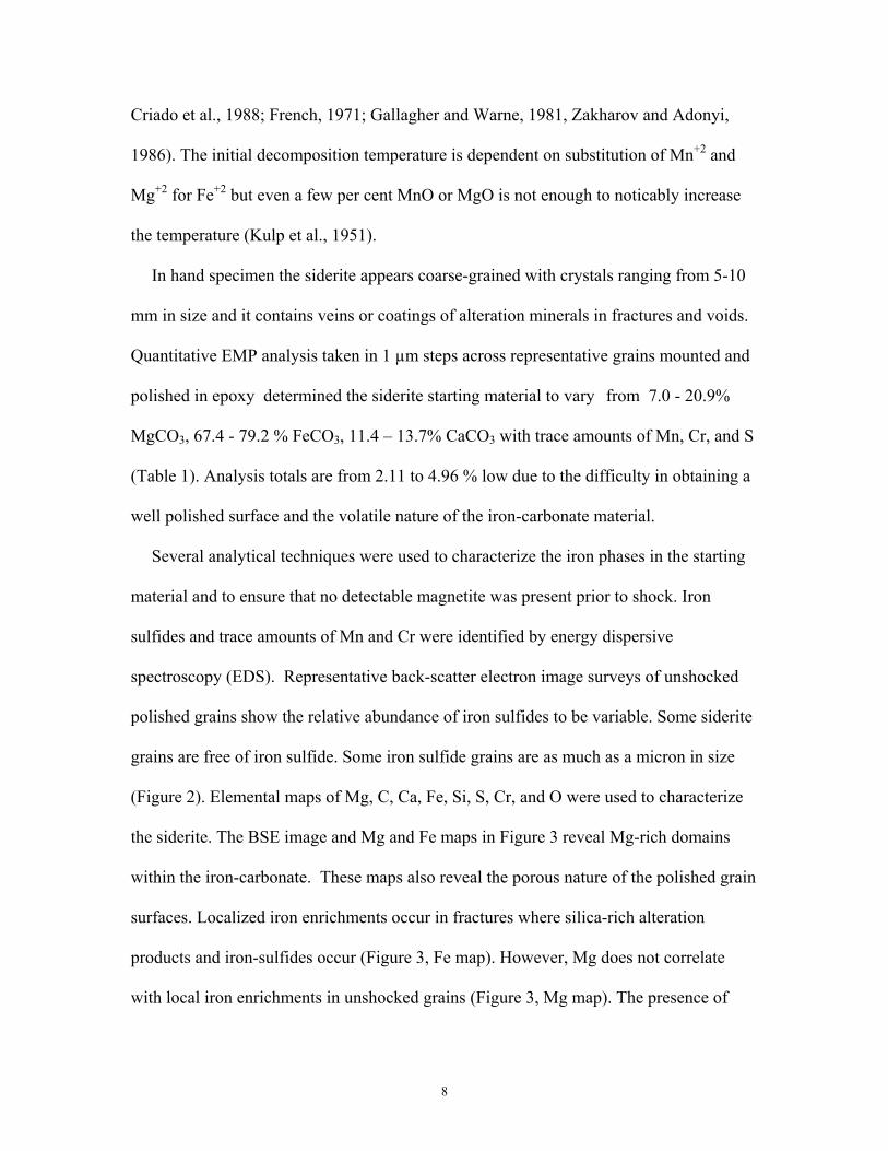
In hand specimen the siderite appears coarse-grained with crystals ranging from 5-10
mm in size and it contains veins or coatings of alteration minerals in fractures and voids.
Quantitative EMP analysis taken in 1 µm steps across representative grains mounted and
polished in epoxy determined the siderite starting material to vary from 7.0 - 20.9%
MgCO3, 67.4 - 79.2 % FeCO3, 11.4 – 13.7% CaCO3 with trace amounts of Mn, Cr, and S
(Table 1). Analysis totals are from 2.11 to 4.96 % low due to the difficulty in obtaining a
well polished surface and the volatile nature of the iron-carbonate material.
Several analytical techniques were used to characterize the iron phases in the starting
material and to ensure that no detectable magnetite was present prior to shock. Iron
sulfides and trace amounts of Mn and Cr were identified by energy dispersive
spectroscopy (EDS). Representative back-scatter electron image surveys of unshocked
polished grains show the relative abundance of iron sulfides to be variable. Some siderite
grains are free of iron sulfide. Some iron sulfide grains are as much as a micron in size
(Figure 2). Elemental maps of Mg, C, Ca, Fe, Si, S, Cr, and O were used to characterize
the siderite. The BSE image and Mg and Fe maps in Figure 3 reveal Mg-rich domains
within the iron-carbonate. These maps also reveal the porous nature of the polished grain
surfaces. Localized iron enrichments occur in fractures where silica-rich alteration
products and iron-sulfides occur (Figure 3, Fe map). However, Mg does not correlate
with local iron enrichments in unshocked grains (Figure 3, Mg map). The presence of
8

iron-sulfides and other alteration minerals as evidenced in the iron, silica, and sulfur
maps in Figure 4 is consistent with compositions in published reports of natural siderite
(see above references). Magnetic susceptibility of this natural siderite is ~8 x 10⁻⁷ m3/kg
(mid-range for published values, 4-12 x 10-7 m3/kg). This corresponds to < 1.6 ppt of
some possible combination of paramagnetic, ferromagnetic, and or diamagnetic minerals
in the starting material and is three orders of magnitude less than values for crustal rocks
with low magnetic susceptibility (Telford et al., 1976). Mössbauer spectroscopy was used
to analyse a powdered sample of the starting material. No magnetite was detected as
determined from the 57Fe Mössbauer spectra (to a detection limit of 0.5%). A powdered
sample of unshocked siderite was dissolved in acetic acid according to the methods of
Golden et al., (2001) (soaked in a 20% acetic acid bath at 65oC for 72 hrs) and the residue
was examined by TEM to further characterize the material used for these shock
experiments. The iron-bearing phase in the residue from the dissolved unshocked siderite
is hematite. No spinel-structured iron oxides were identified in the siderite starting
material.
Results of Shock Experiments
Cursory examination of the experimentally shocked siderite indicates alteration of
the starting material (Figure 5). The natural, unshocked siderite is reddish in color but all
shocked samples have been transformed to gray, grading to black with increasing shock
pressure. Magnetic particles can readily be separated from the powdered material
shocked to pressures >39 GPa with a hand magnet. This was not evident in the samples
shocked to lower pressures. Initial results of TGA are not systematic. Mass loss does
9

occur with increasing shock pressure but the 24.8 GPa experiment shows no appreciable
mass loss (Figure 6); yet the 30.2 GPa sample has the greatest mass loss, whereas the
sample shocked to the highest pressure (49GPa) has one of lowest mass losses. After re-
running TGA on new aliquots of two samples, the 30.2 GPa sample resulted in a similar
mass loss as the original run. However, the 49 GPa sample resulted in an increased mass
loss more in line with the expected trend of increasing mass loss with increasing shock
pressure. The highest pressure experiment was chosen for further examination assuming
its shock effects to be greatest based on its color change from red-orange to black and the
ease of extracting enough magnetic particles for TEM analysis from the recovered
material compared to the lower pressure shock experiments.
EMP maps and BSE images show that grains in the 49 GPa sample are smaller in
size than those of the starting powder and that the abundance of large iron sulfide grains
is reduced (Figure 2). Shocked siderite grains are highly fractured, some along cleavage
planes but also in irregular patterns (Figure 7). Shocked siderite grains retain the porous
surface appearance of unshocked grains. Iron and magnesium enrichments correlate in
veins and pockets although Fe >> Mg and areas of iron concentrations free of Mg can be
seen in Figure 7. Compositional boundaries between Fe and Mg-enrichments and
surrounding siderite are sharp (Figure 7). Carbon blebs <10 μm in size were detected in
cracks and veins where iron is also concentrated (Figure 7d). Chromium and sulfur were
not detected in many shocked grains although iron sulfide blebs can be found preserved
in veinlets within remaining siderite grains. Textures indicative of melt (irregular blebs,
globules, embayments, vessiculation, flow and spinifex textures) observed in shocked
carbonate from the Haughton impact structure (Osinski and Spray, 2001) were not
10

observed in siderite grains shocked to 49 GPa. Some carbonate remains intact in all
shocked siderite grains examined as evidenced by the EMP elemental maps.
Figures 8-14 show TEM images of shocked siderite from the 49 GPa experiment.
Interiors of residual carbonates in untreated samples contain extensive regions hundreds
of nanometers in size of electron-dense crystals in the ~50 - 100 nm size range. These
crystals occur in clusters within and on margins of remaining siderite (Figure 8). Diffuse
and weakly streaked diffraction spots in SAED patterns of untreated samples as well as d-
spacings (lattice spacings) characteristic of both siderite and another phase indicate a
mixture of many non-randomly oriented crystallites (Figure 9a). However, SAED
patterns acquired on grain margins are characteristic of a single phase and are a good fit
to solid solution spinel (Figures 9b and 10). Many magnetites in the <50 to 100 nm size
range possess both equant and elongated forms (Figure 11 at arrows). Some grains have
euhedral forms but most do not. Elongated euhedral magnetite crystals can be seen in
Figures 11 & 12. The two largest crystals in Figure 12 are either intimately inter-grown
or are magnetically attracted to each other. These magnetite crystals lack microstructure
suggesting they nucleated in the shock experiment whereas the residual siderite has
mottled contrast from lattice strain acquired during shock.
Acid-treated samples shocked to 49 GPa contain the same sizes and
morphologies of shock-produced magnetite crystals as the untreated samples, however
the SAED patterns of samples treated to remove siderite are characteristic of one phase
and lack the characteristic d-spacings of siderite (Figs.13 & 14). TEM examination of the
shock-produced crystals before and after the acetic acid treatments indicates that the acid
extraction did not alter the size, shape, or surface textures of the shock-produced crystals.
11

EDS analyses of shock-produced oxides result in a range of compositions (See
figure 15). Fetot ranges from 80.69% -100% of total cations in the nucleating phase; the
other major cation detected in some single crystals is Mg. Crystals of all shapes and sizes
have the same range of compositions. Trace amounts of Si, Ca, Mn, Cr, and S can be
detected in dense areas of untreated samples where multiple phases are indicated by
SAED patterns.
Discussion of Shock Experiments
After shock, the color change of the siderite suggests that a systematic alteration
effect has occurred on the macro scale, but microanalysis reveals the alteration to be
incomplete. Intact siderite is present in EMP maps of shocked grains. Iron and
magnesium enrichments are present in highly fractured zones (Figure 7). Residual iron
carbonate mixed with crystals of another iron-rich phase is detectable in SAED patterns
from TEM examination of both acid-treated and untreated shocked samples (Figure 9).
However, SAED patterns acquired from electron-dense crystals on the margins of
residual iron carbonate indicate the presence of only a single phase.
Table 2 compares d-spacings measured from SAED patterns of the shocked
crystals with published d-spacings for magnetite and magnesioferrite (Joint Committee
for Powder Diffraction, 1980). Even within a measurement uncertainty of ~1%, the
SAED patterns for magnetite, magnesioferrite, and these shock products are
indistinguishable (Brearley, 2003).
EDS measurements of the shocked crystals detected Fe in concentrations >80% of
the total cation concentration. Figure 15 is a histogram of seven EDS measurements taken
12

from a variety of spinel morphologies found within and at margins of remaining siderite.
These analyses represent the range of compositions in shock-produced spinels. A
horizontal line at 83% represents the dividing line between < 50% Fe+2 and >50% Fe+2 in
the octahedral site of the spinel structure. EDS analyses of the spinels produced in these
shock experiments show that most contain >50% Fe+2 and Mg is not detected in some
measurements. Magnetite and magnesioferrite are isostructural Fd3m inverse spinels of
the magnetite series solid solution (Deer et al., 1997). For solid solutions, the “50% Rule”
applies (Nickel, 1992; Nickel and Grice, 1998). A complete solid solution series without
structural order of the ions defining the end members is arbitrarily divided at 50 mol %
and the two portions are given different names. To indicate a significant (but minor)
amount of Mg, for instance, a Schaller adjective modifier may be assigned such as
magnesian magnetite (Schaller, 1930). For magnetite, the “50 % Rule” applies to the
Fe+2 cation site (octahedral site). Therefore, most spinels analysed here by EDS are
magnesian magnetite and some are “pure” magnetite.
The only published data on the compositional purity of ALH84001 magnetites,
one of the five criteria for biogenicity following Thomas-Keprta et al.(2000), can be
found in Thomas-Keprta et al. (2000). All subsequent publications (Thomas-Keprta et al.,
2001; Thomas-Keprta et al., 2002) refer to these data. Brearley (2003) states “the
experimental conditions for other spectra reported by Thomas-Keprta et al. (2000) are
somewhat different from those of the standard spectra” and that “these data cannot be
used to argue unequivocally for or against the presence of minor or trace concentrations
of Mg, Al, etc. in the elongated prismatic magnetites from ALH84001.” (See Brearley
(2003) for further discussion of chemically pure magnetite in ALH84001). ALH84001
13
magnetite was extracted by Golden et al. (2006), and analysed using the same instrument
as Thomas-Keprta et al. (2000) and this study. Golden et al. (2006) found ALH
magnetites to contain from 3.0 mol % Mg to an amount below detection after acquiring
5000 counts. Magnetite created in this study ranges from “pure” Fe3O4 (Mg not detected
in EDS) to compositions containing 19.31 mol % Mg. These amounts closely reflect
compositions of the siderite starting material for the shock experiments which was
composed of local areas with iron concentrations and no associated Mg to as much as
20.9 mol % MgCO3. Comparatively, magnetite-bearing carbonate layers in ALH84001
have compositions from ~10% MgCO3 to ~60% MgCO3 (Treiman, 2003). Golden et al.
(2006) have shown that magnetite created by heating Mg-rich siderite + pyrite to 350oC
in a closed system and kept at set temperature for approximately nine days was
compositionally indistinguishable from magnetite in ALH84001.
The starting material for these shock experiments does contain iron sulfides.
However, the duration of the localized temperature excursion in these shock experiments
is short – less than 100 μs. This short time scale precludes large scale cation diffusion and
mitigates any effect of fO2 created by the presence of iron sulfides in the siderite thereby
allowing the inclusion of Mg, if present locally, into the magnetite structure. However,
the decomposition experiments of Brearley (1986) demonstrate that under disequilibrium
conditions, the earliest phase that forms from mixed component phases is a simple binary
oxide rather than a more complex solid solution because it is kinetically the easiest to
nucleate. Formation of magnetite by shock experiment is a disequilibrium process which
results in rapid heating and cooling of the shocked material. The earliest stages of
incipient decomposition produce many small nucleating crystals as observed.
14

The bulk of the iron-carbonate is not completely devolatilized in the 49 GPa
shock experiment. The spatial relationships of magnesium to iron after the shock
experiment at 49 GPa is similar to that before the experiment. Shocked siderite grains
from these experiments show no compositional gradients in EMP elemental maps which
would indicate diffusion of cations from the siderite-composition domains towards iron
enrichments. Magnetite crystals from the shock experiments may be produced at the
expense of iron-rich phases concentrated in alteration zones (fractures and voids) or in
more end-member like iron-rich siderite domains in the natural siderite (or both) as iron
carbonate compositions are the first to thermally decompose. In the case of pure FeCO3
domains, the spinel shock product would be pure magnetite. Magnesium detected by EDS
in the spinel-bearing material may be present in the spinel structure, in residual siderite,
or represent a separate oxide phase such as periclase but has yet to be resolved (the 400
and 440 diffraction rings for magnetite occur at nearly the same position as the 200 and
220 rings for periclase). Barber and Scott (2002) report occurrences of nanocrystalline
periclase associated with Mg-rich carbonate in ALH84001. Si and trace elements
detected by EDS may also reside in residual siderite. Elevated carbon found associated
with iron concentrations in the shocked sample is consistent with the magnetite +
graphite assemblage described by French (1970) from siderite stability experiments in
controlled fO2 atmospheres. Hematite detected in the siderite starting material could also
contribute to magnetite formation according to the equilibrium reaction:
Carbonate + hematite = magnetite +CO2
for pure siderite and siderite60magnesite40 (Koziol, 2004). It should be noted that
“chemical purity” has not been the sole property of biogenic magnetite since Maher and
15

Taylor (1988) demonstrated that impurity-free magnetite microcrystals with perfect
crystal faces only 10-30 nm across are produced pedogenically and can be reproduced in
controlled laboratory simulations of pedogenesis.
Local target conditions such as porosity, in this case on the scale of tens of
micrometers, are believed to play a large role in pressure (and resulting temperature)
excursions which determine whether or not melting or de-volatilizations occurs. Original
porosity variations could explain the heterogeneous distribution of shock produced nano-
phase magnetite in siderite interiors. No textural, structural, or compositional evidence
for melting was found in siderite shocked to 49 GPa. These are the first experiments to
provide information on the earliest stages of incipient shock decomposition in carbonates.
Discussion of Magnetite Formation in ALH84001
Shock damage in rocks is a function of many material properties including bulk
density, porosity, modal mineral compositions, compressibility, and shock impedance
variations between adjacent minerals, variations in volatile content, and macroscopic and
microscopic structural features. The peak pressure at which specific types of deformation
occur (fracturing, plastic deformation, phase transformations, melting, vaporization) is a
complex function of material properties coupled with the character of the shock event
(Lyzenga et al., 1983; Boslough, 1988). The material properties of ALH84001 include a
mixture of phases which vary on the scale of tens of micrometers and include carbonates
with a range of decomposition temperatures. Proponents of magnetite formation as a
result of shock metamorphism in ALH84001 have invoked two scenarios: simple heating
of siderite (Brearley, 2003; Golden et al, 2004) and partial decomposition and a scenario
16

in which carbonate crystallized from a shock-formed fluid followed by decomposition on
cooling (Barber and Scott, 2006; Scott et al, 1997).
Brearley 2003 describes carbonate in ALH84001 as “exhibiting pronounced
domainal microstructure, complex strain contrast, and a mottled appearance that is caused
by the presence of myriad magnetite crystals and voids” formed by CO2 loss and draws
the conclusion that the carbonate pre-dates the shock event that produced the feldspathic
glass. Bradley et al. 1998 also observed voids associated with magnetite in Fe-rich
carbonate and that some carbonate rosettes in ALH84001 are extensively penetrated by
networks of feldspathic glass-containing veins. Barber and Scott 2006 observed a
carbonate disk that they claim shows no evidence for shock deformation but which
exhibits a substructure of elongated, slightly misoriented subcells in the exterior regions,
leading them to conclude that the carbonate crystallized from a shock-formed fluid
followed by decomposition on cooling. However, the “spectacular zoning” described by
Shearer and Adcock 1998 is shown as evidence for carbonate precipitation by non-
equilibrium crystallization at low temperature in an open-fracture system and reflects
changes in the openness of the hydrothermal system. The precipitation products were
then disrupted by a succeeding shock event during which silicate melt detached and
shattered many carbonate concretions (Schwandt et al., 1999, Shearer and Adcock, 1998)
and thermally decomposed the siderite-rich rims to magnetite and associated void space.
That the magnetites in ALH84001 formed by decomposition can be further attested to by
the presence of cracks or fractures present where the Fe-rich compositions of the
carbonate globules are adjacent to the feldspathic glass. These fractures represent a
volume decrease resulting from decomposition leaving a porous aggregate of magnetites
17

and are not present where glass is in contact with less Fe-rich compositions (Brearley,
2003, Shearer and Adcock, 1998). The magnesite layers show no textural (voids) or
compositional (diffusion or depletions) evidence of corrosion effects at their margins.
(see Brearley 2003, Figure 13). Finally, Eiler et al., 2002 conclude from oxygen isotope
and trace element analysis that formation of “carbonate concretions” in ALH84001 is
inconsistent with models involving high temperature hydrothermal alteration,
metamorphism, or carbonate-melt injection. It could be argued that the misoriented
subcells in ALH84001 carbonate described by Barber and Scott are also evidence of
deformation during the shock event that mobilized feldspar and decomposed siderite.
Therefore, impact shock metamorphism could provide the thermal input to
convert siderite to magnetite in any of several impact events to which ALH84001 has
been subjected (Treiman, 1998). The petrological and mineralogical changes induced by
impacts, including phase transformations, depend upon shock pressure and duration
(Duvall and Graham, 1977). If ejection is a low pressure phenomenon (Melosh, 1995),
how or when could the ALH material be heated to produce magnetite? Possible scenarios
for prolonging thermal pulse duration after impact event(s) include insulation in a suevite
layer (Bell et al., 1996; Dodd, 1981; Simonds, 1978) after impact to bring cumulate rock
(like ALH84001) from depth to Mars’ surface, or during impact events the material may
have suffered before ejection from Mars (Treiman, 2003). In the pressure range from 10
to 40 GPa, in experimental shock studies, localized zones of very high temperature have
been observed (Lyzenga et al, 1983; Boslough 1988). These observations suggest that
shock events in this pressure range produce very intense localized deformation with
associated high temperatures even though the bulk temperatures do not increase
18

dramatically. Therefore, change to the refractive index of plagioclase feldspar in
ALH84001 my indicate a “bulk” peak pressure of ~32 GPa. However, Barber and Scott
(2006) found no evidence of recrystallization or recovery in deformed orthopyroxene
crystals in fracture zones in ALH84001 and concluded that no global heating event
occurred in the meteorite. Consequently, the formation of magnetite from decomposed
iron carbonate in ALH84001 must have occurred in the most severe shock event to which
it was subjected for local thermal excursions to be great enough to decompose siderite.
The magnetite rich zone in ALH84001 is very porous on the nanometer scale
(Barber and Scott, 2001; Scott and Barber, 2002). TEM reveals this material to consist of
many nanometer sized magnetites and elemental carbon left from the decomposition of
iron carbonate – the iron carbonate being a likely component of the natural carbonate
precipitation sequence (Golden et al., in press). If the magnetite in ALH84001 was
allocthonous, there is no reason for the magnetite to be concentrated only in two zones
sandwiching the magnesite layer and not distributed throughout the meteorite, or at least
distributed throughout the carbonate, given the many pathways created by the various
deformation events described as possible (See Treiman 1998 for a synopsis).
Golden et al. (2004) found that “thermal decomposition of of Fe-bearing
carbonate produces magnetite crystals that are identical to those found in ALH84001”
and more importantly they found that “most of the purported biogenic magnetite crystals
in ALH84001 do not have the reported [111]-THO morphology, and so are not identical
to those from the MV-1 bacterium". Furthermore, Golden et al. (2006) analysed 11
ALH84001 magnetites and found their compositions to contain from undetectable Mg in
the spinel structure to nearly 7 mole %. These observations indicate that ALH84001
19
magnetites do not share the characteristics of chemical purity or the [111]-THO crystal
morphology with MV-1 bacterium produced magnetites and should not be considered
”biomarkers”.
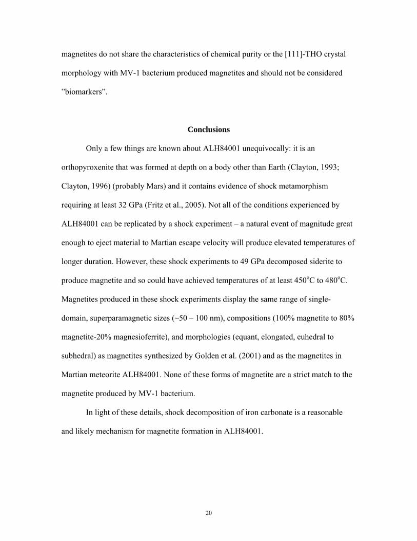
Conclusions
Only a few things are known about ALH84001 unequivocally: it is an
orthopyroxenite that was formed at depth on a body other than Earth (Clayton, 1993;
Clayton, 1996) (probably Mars) and it contains evidence of shock metamorphism
requiring at least 32 GPa (Fritz et al., 2005). Not all of the conditions experienced by
ALH84001 can be replicated by a shock experiment – a natural event of magnitude great
enough to eject material to Martian escape velocity will produce elevated temperatures of
longer duration. However, these shock experiments to 49 GPa decomposed siderite to
produce magnetite and so could have achieved temperatures of at least 450oC to 480oC.
Magnetites produced in these shock experiments display the same range of single-
domain, superparamagnetic sizes (~50 – 100 nm), compositions (100% magnetite to 80%
magnetite-20% magnesioferrite), and morphologies (equant, elongated, euhedral to
subhedral) as magnetites synthesized by Golden et al. (2001) and as the magnetites in
Martian meteorite ALH84001. None of these forms of magnetite are a strict match to the
magnetite produced by MV-1 bacterium.
In light of these details, shock decomposition of iron carbonate is a reasonable
and likely mechanism for magnetite formation in ALH84001.
20

Acknowledgements- This work is supported by Lockheed Martin, Jacobs Sverdrup, and
NASA’s Cosmochemistry Program. Gerry Hanes performed the shock experiments at
Johnson Space Center and his work is appreciated. D.C. Golden is thanked for his help
with TEM. Craig Schwandt is thanked for his help with EMP analysis and for valuable
critiques. Dick Morris is thanked for the siderite sample and Mossbauer analysis. Stuart
Hall provided the magnetic susceptibility measurements and he is thanked. Fred Horz,
Mike Zolensky, Arch Reid, and Doug Ming are thanked for their enlightening discussions
which improved this paper immensely. Detailed and useful reviews by E.R.D. Scott, D.
Barber and two anonymous reviewers are gratefully acknowledged.
21

Page intentionally left blank

Figure 1. TEM image of microtomed grain mount from 49 GPa shocked siderite experiment (Bell et al., 2002). Black areas are domains of nano-phase spinel structure iron oxide within surviving iron carbonate identified by characteristic d-spacing in SAED pattern. Grouped spots in the SAED pattern are the manifestation of nanocrystalline spinels in non-random orientation. Enlargement shows the complex mottled contrast in siderite (S) due to extensively strained submicron domains where the crystal lattice is distorted.
200 nm
23
S
S
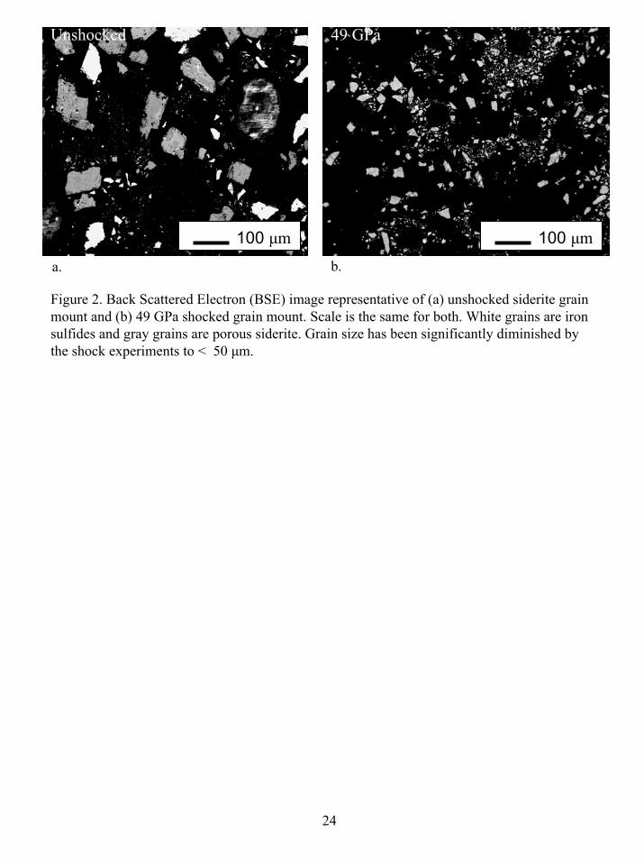
Figure 2. Back Scattered Electron (BSE) image representative of (a) unshocked siderite grain mount and (b) 49 GPa shocked grain mount. Scale is the same for both. White grains are iron sulfides and gray grains are porous siderite. Grain size has been significantly diminished by the shock experiments to < 50 μm.
100 μm
24
a. b.
100 μm
Unshocked 49 GPa

Figure 3. EMPA backscattered and elemental maps of an unshocked siderite grain showing typicaldistribution of iron and magnesium-rich domains. (a) The BSE image illustrates the porous nature of the grain surfaces. Note Fe enrichments associated with voids contain no Mg (b,c). All three images are the same area of the same grain.
50 µm
BSE Fe Mg
a. b. c.
25

SiFe
Figure 4. Electron microprobe elemental maps of a representative unshocked siderite grain. (a) Localizediron enrichments occur along micro-fractures and are probably haematite which was found in the residueafter dissolution of the carbonate (b). A silica-rich phase, probably alteration of the siderite, is located insome of the same fractures as iron (c). Sulfur map shows the presence of iron sulfides in the startingmaterial. Chromium was analysed for, but not detected. All three images are the same siderite grain.
100 µm
S
a. b. c.
26

Figure 5. Unshocked and shocked siderite powders photographed in vials. The unshockedsample of starting material is reddish in color. Shocked samples were progressively changedin color with increasing shock pressure from dark gray to black.
Unshocked 24 GPa
35 GPa 49 GPa
27

02468
10
20 25 30 35 40 45 50
Figure 6. Thermal Gravimetric Analysis results of siderite experiments to 24.8, 30.2, 34.3, 35, 35.7 36.7, 39, 44, and 49 GPa. The mass loss of CO2 due to shock ranges from below detection to 9 Wt % but does not increase systematically with increasing shock pressure (note the low R2 value for the trendline). Measurements are + 3.16 Wt%.
R2 = 0.0207
Shock Pressure (GPa)
Mas
s Los
s (W
t %)
28

50 μm
BSE
a.
Fe
c.
Fe
b.
C M
d.
g
e.
Mg
f.
Figure 7. Elemental maps of typical siderite grains shocked to 49 GPa. (a) Fractures in the lower rightHalf of the grain follow cleavage patterns but are more irregular in the upper left portion of the grain.Localized iron (b,c) and magnesium (e,f) enrichments are detected in portions of these grains but Mg isnot always associated with Fe enrichments (boxes) and may indicate the presence of periclase. Carboncan be detected above background (d). Scale is the same for all images.
29
Mg

Figure 8. TEM bright-field image of untreated magnetite. Interior of residualcarbonate (arrow) contains electron dense regions of nucleating magnetite in the<100nm size range.
100 nm
30

50 nm
Figure 9. TEM bright-field image of untreated shocked siderite sample. SAED pattern (a) from theimaged area is characteristic of a mixture of siderite (S) and magnetite crystals. The mottled contrastof strained siderite is in marked contrast to the defect free contrast of the magnetite crystals (at arrows). The offset diffraction spots in SAED pattern (b) are indicative of several magnetite crystalites in slightly preferred orientation. Arrows points to <50 nm, equant, euhedral to anhedral magnetite crystals.
31
S
a
b

100 nm
Figure10. TEM bright-field image of untreated magnetite on the margin of residual siderite. Equantand elongated magnetites, some euhedral, are visible at arrows. SAED pattern is characteristic ofmagnetite.
32

ab
Figure 11. TEM bright-field image of untreated magnetite. To the upper right is siderite (S) containing nucleating crystallites of magnetite. (a) Equant ,(b) euhedral elongated,and (C) subhedral elongated magnetite crystals are in the lower left of the micrograph.
c 50 nm
S
Mt
33

Figure 12. TEM bright-field image of untreated magnetite produced in ourexperiments. Magnetite crystals (Mt) on edge of residual siderite (S) display defect free contrast. Elongated euhedral magnetite grains at right appear to be intergrownepitaxially (at arrow).
50 nm
Mt
Mt
Mt
Mt
S
S
34
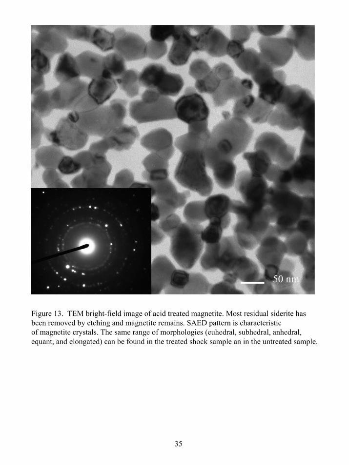
Figure 13. TEM bright-field image of acid treated magnetite. Most residual siderite has been removed by etching and magnetite remains. SAED pattern is characteristicof magnetite crystals. The same range of morphologies (euhedral, subhedral, anhedral, equant, and elongated) can be found in the treated shock sample an in the untreated sample.
35
50 nm

Figure 14. TEM bright-field image of acid treated shock product and SAED pattern characteristicof magnetite. Image contains equant, euhedral, and anhedral randomly oriented magnetite crystalsin the < 100 nm size range.
100 nm
36

0
10
20
30
40
50
60
70
80
90
100
1 2 3 4 5 6 7
EDS measurement #
Feto
tala
s % o
f tot
al c
atio
ns80.69 83.67 87.68 83.69 83.60 83.28 100
Figure 15. Histogram of EDS measurements for Iron as a percent of the total cations in the shock produced spinels. Compositions with > 83 mol % Fetotalhave > 50% FeII in the spinel structure (magnetite composition). With theexception of analysis 1, these shock products are magnetite. Mg is theonly other major cation detected in these magnetite crystals.
37
83%
Mol % Fetotal

Table 1. EMP quantitative analysis of unshocked siderite, the starting material for the experiments. Trace Mn was detected in analysis 4. Cr and S were detected in trace amounts in analyses 3and 8. See text for instrument conditions.
1 2 3 4 5 6 7 8 9 10 111 cationFe 0.674 0.676 0.681 0.687 0.676 0.675 0.677 0.677 0.675 0.675 0.792Mg 0.206 0.207 0.202 0.197 0.206 0.207 0.206 0.207 0.209 0.208 0.070Ca 0.120 0.118 0.114 0.117 0.118 0.118 0.117 0.116 0.117 0.117 0.137
Weight %FeO 46.1 45.38 45.7 46.02 44.93 45.34 45.77 45.09 45.24 44.83 45.13MgO 14.12 13.88 13.56 13.19 13.68 13.91 13.92 13.79 13.98 13.84 4.01CaO 8.19 7.91 7.84 7.81 7.85 7.96 7.89 7.72 7.83 7.76 7.81CO2 29.44 28.57 28 28.71 28.9 28.83 28.3 28.39 28.76 28.65 28.87
Totals 97.85 95.73 95.11 95.72 95.35 96.03 95.89 95 95.8 95.09 95.83
38

Table 2. D-spacing measurements from SAED’s (Å) of shock experiment productscompared to published values for magnetite and magnesioferrite. Within 1%uncertainty in the measurements the d-spacing values are indistinguishable.
39
(a) Joint Committee for Powder Diffraction file 19-629. (b) High temperature form. (c) Joint Committee for Powder Diffraction file 17-465.
IntensityMiller Indices
Interplanar spacings for magnetite (a)
Interplanar spacings for
magnesioferrite (b)(c)
Shocked sample
1
Shocked sample
2
Shocked sample
3
35 220 2.967 2.969 2.97 2.99 2.91100 311 2.536 2.532 2.48 2.49 2.5325 400 2.099 2.099 2.11 2.09 2.1530 511 1.616 1.616 1.62 1.61 1.6040 440 1.485 1.485 1.48 1.46 1.48


![A Domain Decomposition Non-Intrusive Reduced Order Model ...inavon/pubs/DDNIROM.pdf · cia et al. [33,34] rstly introduced the subdomain idea into ROM for tracking a moving shock](https://img.pdfslide.us/doc/110x75/60361cf84fe73f56b81da98a/a-domain-decomposition-non-intrusive-reduced-order-model-inavonpubsddnirompdf.jpg)