Embed Size (px)
Citation preview
Experimental model of tooth movement in mice: A standardized protocol forstudying bone remodeling under compression and tensile strains
Silvana Rodrigues de Albuquerque Taddei a,d,1, Adriana Pedrosa Moura a,d,1, Ildeu Andrade Jr.b,Gustavo Pompermaier Garlet c, Thiago Pompermaier Garlet c, Mauro Martins Teixeira d,Tarcılia Aparecida da Silva a,d,n
a Department of Oral Pathology, Faculty of Dentistry, Universidade Federal de Minas Gerais, Belo Horizonte, Minas Gerais, Brazilb Department of Orthodontics, School of Dentistry, Pontifıcia Universidade Catolica de Minas Gerais (PUC Minas), Belo Horizonte, Minas Gerais, Brazilc Department of Biological Sciences, School of Dentistry of Bauru, S ~ao Paulo University, Bauru, S~ao Paulo, Brazild Laboratory of Immunopharmacology, Department of Biochemistry and Immunology, Instituto de Ciencias Biologicas, Universidade Federal de Minas Gerais, Belo Horizonte, MinasGerais, Brazil
a r t i c l e i n f o
Article history:Accepted 7 September 2012
Keywords:Orthodontic tooth movementBone remodelingCytokinesProteolytic enzymes
a b s t r a c t
During orthodontic tooth movement (OTM), alveolar bone is resorbed by osteoclasts in compressionsites (CS) and is deposited by osteoblasts in tension sites (TS). The aim of this study was to develop astandardized OTM protocol in mice and to investigate the expression of bone resorption and depositionmarkers in CS and TS. An orthodontic appliance was placed in C57BL6/J mice. To define the idealorthodontic force, the molars of the mice were subjected to forces of 0.1 N, 0.25 N, 0.35 N and 0.5 N. Theexpression of mediators that are involved in bone remodeling at CS and TS was analyzed using a Real-Time PCR. The data revealed that a force of 0.35 N promoted optimal OTM and osteoclast recruitmentwithout root resorption. The levels of TNF-a, RANKL, MMP13 and OPG were all altered in CS and TS.Whereas TNF-a and Cathepsin K exhibited elevated levels in CS, RUNX2 and OCN levels were higher inTS. Our results suggest that 0.35 N is the ideal force for OTM in mice and has no side effects. Moreover,the expression of bone remodeling markers differed between the compression and the tension areas,potentially explaining the distinct cellular migration and differentiation patterns in each of these sites.
& 2012 Elsevier Ltd. All rights reserved.
1. Introduction
Bone remodeling is essential for bone turnover and occursprincipally as a result of osteoclast and osteoblast activity(Riancho and Delgado-Calle, 2011; Tanaka et al., 2005). Thecoordinated action of these two cell types leads to bone resorp-tion and deposition, in response to stress and mechanical loading(Hadjidakis and Androulakis, 2006).
Models of orthodontic tooth movement (OTM) are effective forthe study of mechanical loading-induced bone remodeling (Vernaet al., 2004). Different animal models (e.g., rats, dogs, monkeys andmice) have been described in recent decades (Ren et al., 2007).Nevertheless, the availability of murine knock out strains hasencouraged the study of protein and receptor functions duringOTM in these animals (Andrade Jr et al., 2007, 2009; Taddei et al.,
2012; Yoshimatsu et al., 2006). However, only a small number ofstudies have been performed in mice, and the methodologies thathave been used for these analyses, including the amount of appliedforce, have varied (Andrade Jr et al., 2007; Brooks et al., 2009; Huanget al., 2009; Taddei et al., 2012; Yoshimatsu et al., 2006). Theachievement of an optimal force is essential for obtaining themaximum rate of OTM without deleterious effects on the root, thePDL and the alveolar bone (Krishnan and Davidovitch, 2006; Renet al., 2007). Moreover, a standardized methodology is important toallow the comparison of different studies.
There is a general agreement in the literature that the mechan-ical loading that is provided by an orthodontic appliance generatestwo different strains in the periodontal ligament (PDL): compres-sion and tension. In compression site (CS), the force that isgenerated by the root against the alveolar bone induces boneresorption. In tension site (TS); however, PDL fibers are strainedand bone tissue is deposited (Krishnan and Davidovitch, 2006;Masella and Meister, 2006). The alteration of PDL vascularity incombination with strain-induced mechanosensitivity promotesthe release of several molecules, such as chemokines/cytokines,neurotransmitters, growth factors and arachidonic acid metabo-lites (Alhashimi et al., 1999; Davidovitch et al., 1988; Krishnan and
Contents lists available at SciVerse ScienceDirect
journal homepage: www.elsevier.com/locate/jbiomechwww.JBiomech.com
Journal of Biomechanics
0021-9290/$ - see front matter & 2012 Elsevier Ltd. All rights reserved.http://dx.doi.org/10.1016/j.jbiomech.2012.09.006
n Corresponding author at: Faculdade de Odontologia, Universidade Federal deMinas Gerais (UFMG), Av. Presidente Antonio Carlos 6627, Campus Pampulha CEP30270-901, Belo Horizonte, Minas Gerais, Brazil. Tel.: !55 31 3499 2402;fax: !55 31 3399 2430.
E-mail address: [email protected] (T.A. da Silva).1 Contributed equally to the work presented in this paper.
Journal of Biomechanics 45 (2012) 2729–2735
Davidovitch, 2006). The distinct expression of these mediators inCS and TS may be responsible for the specific pattern of cellmigration and bone remodeling that is observed to occur follow-ing OTM (Cattaneo et al., 2005; Garlet et al., 2007; Masella andMeister, 2006).
The aim of this study was to develop a standardized OTMprotocol in mice and to analyze the expression of bone remodel-ing markers in CS and TS.
2. Material and methods
2.1. Experimental animals
Ten-week-old C57BL6/J wild-type mice were used in this experiment. Theethical regulations of the Institutional Ethics Committee were followed for all ofthe treatments. No significant weight loss was observed in the mice.
2.2. Experimental protocol
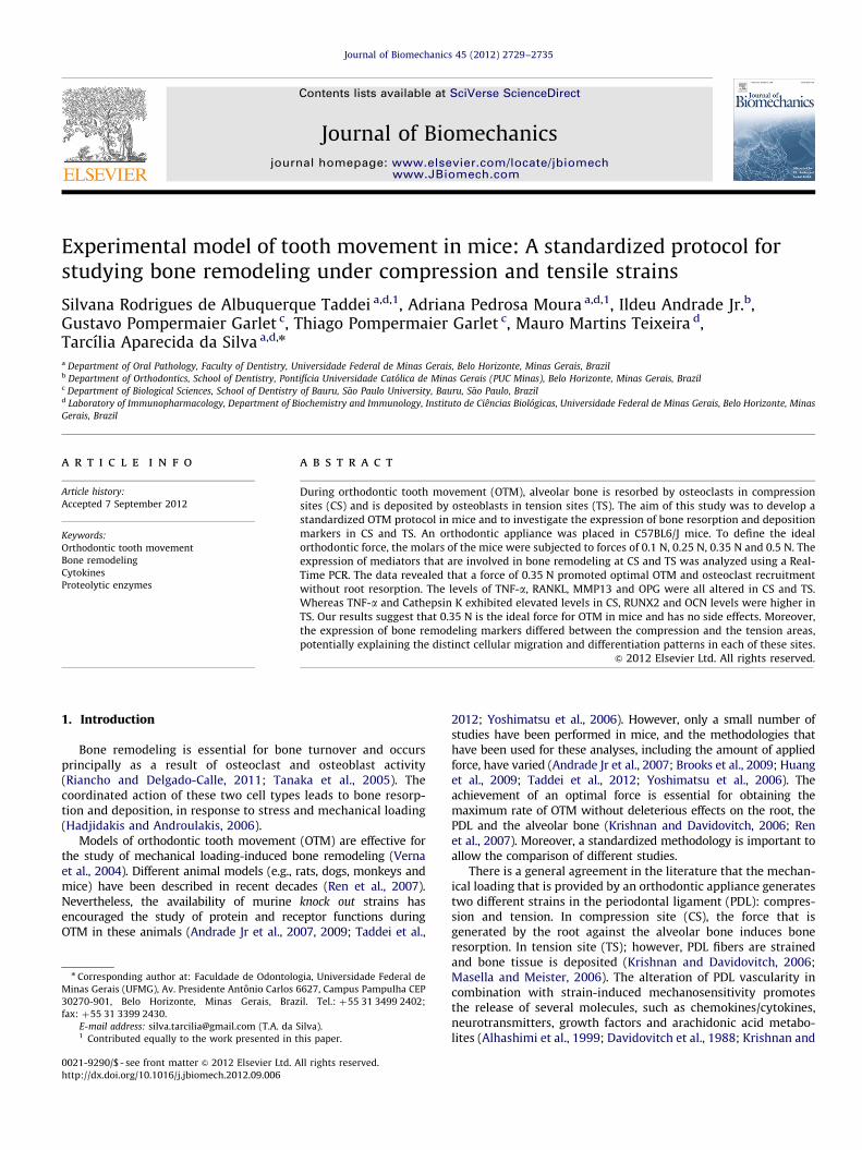
The mice were anesthetized i.e. with 0.2 mL of a xylazine (0.02 mg/mL) andketamine (50 mg/mL) solution. The mice were then placed in the dorsal decubitusposition, with the 4 limbs affixed to a surgical table (Fig. 1a). To permit the fullvisualization of intra-oral structures, a mouth-opener (m) was developed for thisexperiment and was affixed to the surgical table with a 0.08 mm wire to inhibitany forward movement of the head (w) (Fig. 1b).
A stereomicroscope (Quimis Aparelhos Cientıficos Ltd, Diadema, S~ao Paulo,Brazil) (s) and an optical light system (Multi-Position Fiber Optic Illuminator
System, Cole-Parmer Instrument Company Ltd., London, England) (o) were used tobetter visualize the intra-oral structures (Fig. 1c). The right first maxillary molarand incisors surface were cleaned using acetone and were etched using a self-etching primer (Unitek/3 M, Minneapolis, USA). The distal end of an eight-loop,nickel–titanium open-coil spring 0.25"0.76 mm (Lancer Orthodontics, SanMarcos, CA, USA) (c) (Fig. 1d) was bonded to the occlusal surface of the right firstmaxillary molar (f) using a light-cured resin (Transbond, Unitek/3 M, Monrova, CA,USA) (Fig. 1d).
To activate the coil, the surgical table was attached to a specially designedapparatus that contained a rail (r) and a crank (cr) to allow the table to slide backand forth. A tension gauge (t) allowed for the measurement of the amount ofdelivered force (Fig. 1e). A 0.08 mm round wire was used to connect the anteriorportion of the coil spring to the tension gauge (h) (Fig. 1e). Therefore, when thecrank (cr) was activated, the surgical table slid along the rail (r) until thetensiometer registered the desired force (Fig. 1e). Next, the anterior portion ofthe coil was bonded to both of the upper incisors (Fig. 1f) to prevent their furthereruption and anchorage loss (Beertsen et al., 1982). No reactivation was performedduring the entire experimental period. The left side of the maxilla (without anorthodontic appliance) was used as a control.
The experiment was divided into two portions (1) to determine the optimalorthodontic force and (2) to determine the molecular profile in CS and TSfollowing OTM in mice. First, 20 mice were divided into 4 groups, for whichdifferent levels of force were used (0.10 N, 0.25 N, 0.35 N and 0.50 N).
The mice were sacrificed after 0, 12 h, 72 h (for molecular analysis) or 6 days(for histopathological analysis). For each set of experiments, 5 animals were usedat each time-point.
2.3. Histopathological analysis
As was previously described (Taddei et al., 2012), the right and the leftmaxillae halves were fixed in 10% buffered formalin (pH 7.4), decalcified in 14%
Fig. 1. (A) Mice positioned in the dorsal decubitus on the surgical table, developed to restrict animal movement and to permit intra-oral access. (B) Mouth-opener in position. (C)Surgical table placed under a streromicroscope and a fiber optic ligthing device. (D) Occlusal view of the maxillary molar region. Note the distal/posterior end of the Ni–Ti opencoil spring bonded to the occlusal surface of the upper right first molar. (E) A tension gauge attached to the surgical table showing 0.35 N of mechanical loading. (F) The coil springbonded to both upper incisors. Note the stainless steel wire positioned between the mesial/anterior end of the coil. Upper screw (u), lower screw (l), Wire (w), mouth-opener (m),stereomicroscope (s) optical fiber device (o), upper first molar (f), coil spring (c), tension gauge hook (h), rail (r), crank (cr) and tension gauge (t).
S.R.d.A. Taddei et al. / Journal of Biomechanics 45 (2012) 2729–27352730
EDTA (pH 7.4) and paraffin-embedded. 5-mm thick sagittal sections were stainedfor tartrate resistant acid phosphatase (TRAP; Sigma-Aldrich, Saint Louis, MO,USA), counterstained with hematoxylin and used for the histological examina-tions. The mesial periodontal site of the distal-buccal root of the first molar wasused for the osteoclasts counts. Five sections were taken per animal. Theosteoclasts were identified as TRAP-positive, multinucleated cells on the bonesurface. The slides were evaluated by two examiners who were blinded to thegroup status.
2.4. Measurement of OTM
Images of the first and second molars were obtained using an opticalmicroscope (Axioskop 40, Carl Zeiss, Gottingen, Germany) and an adapted digitalcamera (PowerShot A620, Canon, Tokyo, Honshu, Japan). Image J software(National Institutes of Health) was used to quantify the degree of OTM bymeasuring the distance between the cementum–enamel junction (CEJ) of the firstmolar and the second molar on the right hemi-maxilla in relation to the samemeasurements for the left hemi-maxilla. Five vertical sections per animal wereevaluated, and three measurements were conducted for each evaluation.
2.5. RNA extraction and real-time PCR
The PDL and the surrounding alveolar bone were extracted from the upperfirst molars region using a stereomicroscope. All of the gingival tissue, oral mucosaand teeth were discarded. The periodontal tissues and the alveolar bone that wasextracted from the distal area of the distal buccal root of the first maxillary molarwere considered TS. The mesial area of the same root was considered CS. The CSand TS tissues were submitted to RNA extraction using TRIZOL reagent (Invitro-gen, Carlsbad, CA, USA). Complementary DNA (cDNA) was synthesized using 2 mgof RNA through a reverse transcription reaction (Superscript II, Invitrogen). Real-time PCR analysis was performed in a Mini Opticon (BioRad, Hercules, CA, USA)using a SYBR-green fluorescence quantification system (Applied Biosystems,Foster City, CA, USA). The standard PCR conditions were 95 1C (10 min) followed40 cycles of 94 1C (1 min), 58 1C (1 min) and 72 1C (2 min). These cycles werefollowed by the standard denaturation curve. The primer sequences are listed inTable 1. The mean Ct values from duplicate measurements were used to calculatethe expression levels of the target gene, with normalization to an internal control(b-actin) using the 2#DDCt formula.
2.6. Statistical analyses
The data were expressed as the mean7SEM. The comparison between thegroups was performed using one-way analysis of variance (ANOVA) followed bythe Newman–Keuls multiple comparison test. Po0.05 was considered statisticallysignificant.
3. Results
3.1. Definition of the optimal orthodontic force
The histological analysis revealed distal movement of the teethin the control group, with TRAP activity observed on the distalalveolar bone and no activity observed on the mesial side of thedistal-buccal root (Fig. 2c).
Following 6 days of mechanical loading, the experimentalgroup exhibited a significant increase in TRAP activity and,
consequently, greater bone resorption on the mesial bone surfacethen was observed in the control group, (Fig. 2 e–j). The greatestdegree of OTM and osteoclast recruitment was observed with0.35 and 0.50 N of mechanical loading (Fig. 2a and b). However,significant root resorption was observed when 0.50 N was applied(Fig. 2i and j). Therefore, the results suggested that 0.35 N was theideal force for this model, given that this magnitude promoted themaximum rate of OTM and relatively minor tissue damage.
3.2. Bone resorption markers are preferentially expressed in CS
Increased mRNA levels of tumor necrosis factor-alfa (TNF-a)(Fig. 3a), receptor activator of nuclear factor kappa B ligand(RANKL) (Fig. 3b), receptor activator of nuclear factor kappa B(RANK) (Fig. 3c) and metalloproteinase 13 (MMP-13) (Fig. 3e),were observed in both CS and TS compared to the control group.Cathepsin K expression was only significantly increased in the CS(Fig. 3d). Greater levels of TNF-a and Cathepsin K were observedin the CS than in the TS at both time points. However, increasedlevels were only observed at specific time-points for RANKL(at 12 h), MMP13 and RANK (both 72 h) (Fig. 3a–e).
3.3. Expression levels of both osteoblast markers and the boneresorption inhibitors were increased at TS
Runt-related transcription factor 2 (RUNX2) (Fig. 4a) andosteocalcin (OCN) expression were only significantly augmentedin TS (Fig. 4b). Moreover, IL-10 and OPG expression was increasedin TS relative to CS after 72 and 12 h, respectively (Fig. 4c and d).
4. Discussion
This study proposed a standardized protocol to examine thecellular and molecular mechanisms of bone remodeling duringOTM. This protocol allows for limited operator interference and,through a tension gauge and a specially designed apparatus,standardizes 0.35 N as the optimal force for OTM in a mousemodel.
OTM models trigger bone remodeling by means of a mechan-ical loading that is applied to the teeth using a coil spring(Andrade Jr et al., 2009; Braga et al., 2011; Pavlin et al., 2000).The achievement of an optimal force is essential to promote anadequate biological response from periodontal tissues (Krishnanand Davidovitch, 2006). There is no agreement regarding theamount of optimal force for OTM in mice given that the appliedforce has varied between 0.10 and 0.35 N in previous works(Andrade Jr et al., 2007; Brooks et al., 2009; Huang, et al., 2009;Pavlin et al., 2000; Yoshimatsu et al., 2006). This range of forceapplication is due to the use of different orthodontic appliancesin these studies. The same diversity is also observed when a
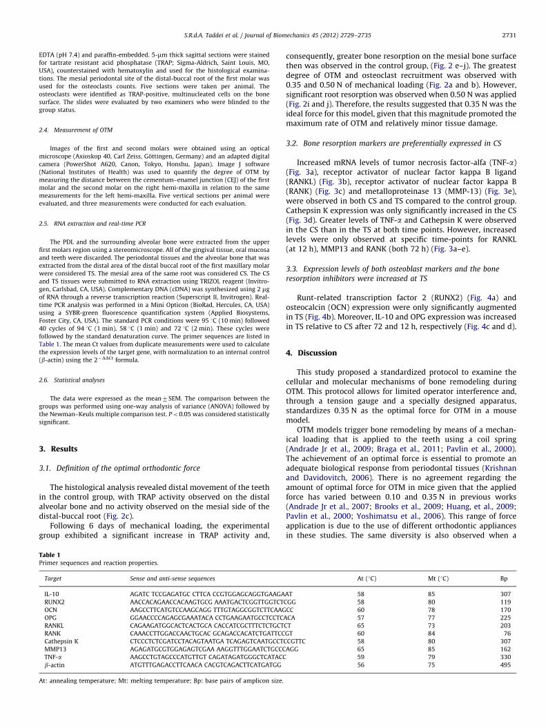
Table 1Primer sequences and reaction properties.
Target Sense and anti-sense sequences At (1C) Mt (1C) Bp
IL-10 AGATC TCCGAGATGC CTTCA CCGTGGAGCAGGTGAAGAAT 58 85 307RUNX2 AACCACAGAACCACAAGTGCG AAATGACTCGGTTGGTCTCGG 58 80 119OCN AAGCCTTCATGTCCAAGCAGG TTTGTAGGCGGTCTTCAAGCC 60 78 170OPG GGAACCCCAGAGCGAAATACA CCTGAAGAATGCCTCCTCACA 57 77 225RANKL CAGAAGATGGCACTCACTGCA CACCATCGCTTTCTCTGCTCT 65 73 203RANK CAAACCTTGGACCAACTGCAC GCAGACCACATCTGATTCCGT 60 84 76Cathepsin K CTCCCTCTCGATCCTACAGTAATGA TCAGAGTCAATGCCTCCGTTC 58 80 307MMP13 AGAGATGCGTGGAGAGTCGAA AAGGTTTGGAATCTGCCCAGG 65 85 162TNF-a AAGCCTGTAGCCCATGTTGT CAGATAGATGGGCTCATACC 59 79 330b-actin ATGTTTGAGACCTTCAACA CACGTCAGACTTCATGATGG 56 75 495
At: annealing temperature; Mt: melting temperature; Bp: base pairs of amplicon size.
S.R.d.A. Taddei et al. / Journal of Biomechanics 45 (2012) 2729–2735 2731
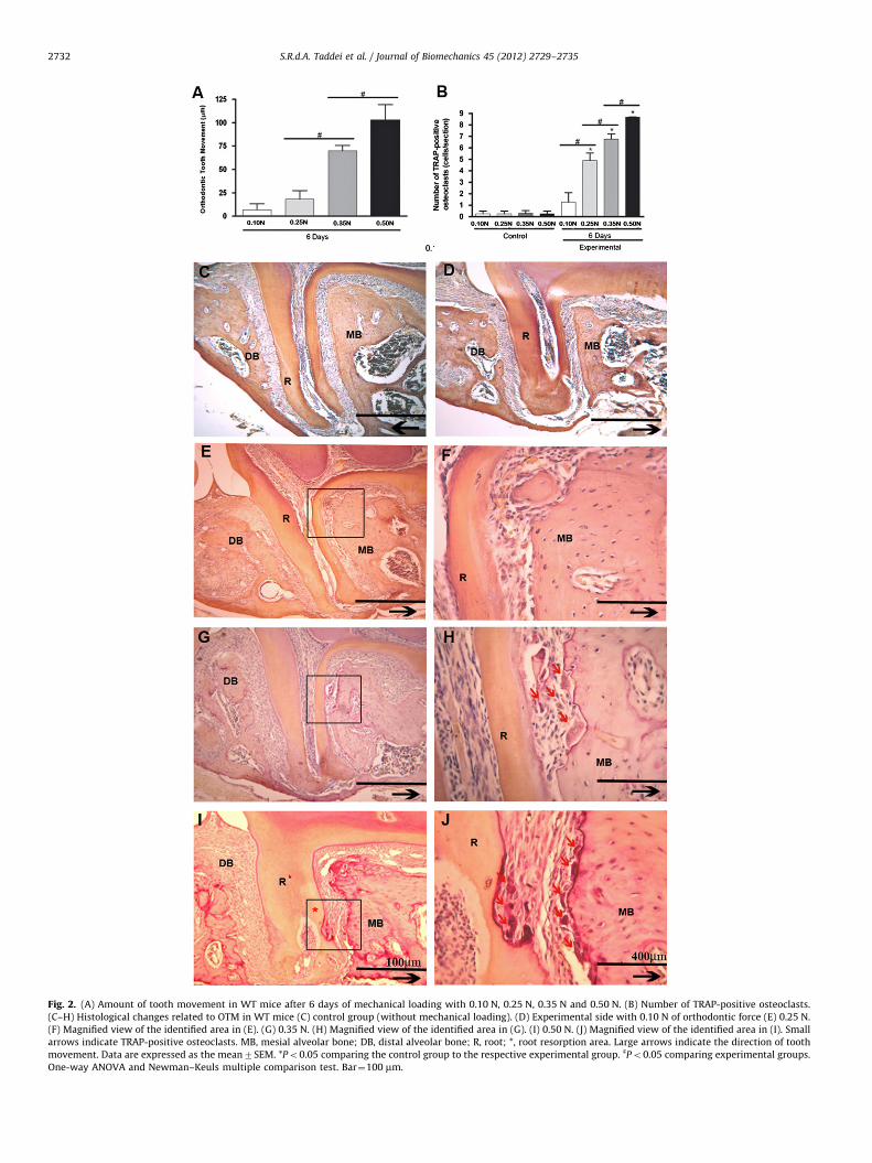
Fig. 2. (A) Amount of tooth movement in WT mice after 6 days of mechanical loading with 0.10 N, 0.25 N, 0.35 N and 0.50 N. (B) Number of TRAP-positive osteoclasts.(C–H) Histological changes related to OTM in WT mice (C) control group (without mechanical loading). (D) Experimental side with 0.10 N of orthodontic force (E) 0.25 N.(F) Magnified view of the identified area in (E). (G) 0.35 N. (H) Magnified view of the identified area in (G). (I) 0.50 N. (J) Magnified view of the identified area in (I). Smallarrows indicate TRAP-positive osteoclasts. MB, mesial alveolar bone; DB, distal alveolar bone; R, root; *, root resorption area. Large arrows indicate the direction of toothmovement. Data are expressed as the mean7SEM. *Po0.05 comparing the control group to the respective experimental group. ]Po0.05 comparing experimental groups.One-way ANOVA and Newman–Keuls multiple comparison test. Bar$100 mm.
S.R.d.A. Taddei et al. / Journal of Biomechanics 45 (2012) 2729–27352732

coil-spring is used given that there are different alloys anddiameters of wire, which themselves can have different lengths(Andrade Jr et al., 2007; Brooks et al., 2009; Huang et al., 2009;Pavlin et al., 2000; Yoshimatsu et al., 2006).
The calibration of the delivered force is also extremely impor-tant for the reproducibility of experiments. For this reason, asurgical table was developed with an attached tension gauge toallow for the measurement of the desired force. This designabolished the possibility of any operator interference, such ashand vibrations or heating of the tension gauge. Several studieshave proposed that the coil opens in a mesial direction, thusobtaining a specific extended length, resulting in a specificdelivered force that is based on force/deflection rate of coil(Brooks et al., 2009; Pavlin et al., 2000). However, this approachmay be influenced by undesirable movement from the manip-ulator’s hand when bonding the coil, which alters the force.Notably, even when different coils stretch to the same lengthwhile activating, the original size of the coil also influences theamount of force that is delivered. For example, if the original sizeof wire is 2.88 mm (Ni–Ti 0.25"0.76 mm) and the coil-spring isstretched up to 1 mm, the delivered force will be 40 g. However,this force can be 20 g if the original size of coil is 5.29 mm, as wasdemonstrated in our pilot study. Therefore, the protocol that is
described here used a standardized original size, wire diameter,alloy, length and diameter of the chosen coil.
Considering that OTM creates both TS and CS in the period-ontium, protein expression, bone resorption and deposition mar-kers were analyzed separately. The results of the present analysisconfirmed a differential expression of marker for these processes,which allows for the creation of a positive microenvironment forbone resorption or deposition at specific sites. Higher expressionlevels of the cytokines TNF-a and RANKL and the bone resorptionmarkers Cathepsin K and MMP13were observed in CS relative toTS at both of the examined time-points. In agreement with theseresults, previous reports described elevated levels of TNF-a andRANKL in CS following mechanical loading (Brooks et al., 2009;Garlet et al., 2007; Nishijima, et al., 2006). Given that the RANKL/RANK axis and TNF-a participate in osteoclastogenesis by up-regulating osteoclast activity, it can be concluded that a moreresorptive microenvironment was created in CS. Moreover, TNF-ais an apoptotic factor for osteoblasts and osteocytes (Suda et al.,2001; Yamashita et al., 2007; Yano et al., 2005). This cytokine mayalso be a signal for osteoclast recruitment and, consequently,bone resorption (Burger et al., 2003). Another important factorthat is expressed in resorption sites is Cathepsin K becauseresorption itself depends on the secretion of this protease from
Fig. 3. mRNA expression of (A) TNF-a, RANKL (B), RANK (C), Cathepsin K(D) and MMP13 (E) in WT mice periodontium after 12 and 72 h of mechanical loading. Data areexpressed as mean7SEM. *Po0.05 comparing control to the respective experimental group. One-way ANOVA and Newman–Keuls multiple comparison test.
S.R.d.A. Taddei et al. / Journal of Biomechanics 45 (2012) 2729–2735 2733

osteoclasts (Troen, 2004). Together, these results point to apossible explanation for the higher osteoclast count and theconsequent greater bone resorption in this area.
Our data revealed higher levels of OCN and RUNX2 (osteoblastdifferentiation markers) in TS compared to controls and CS. Theseresults are in agreement with a previous study that also demon-strated higher OCN levels in TS than in CS (Garlet et al., 2008).This result may be due to the increased promotion of bonedeposition in this area by mature osteoblasts.
In addition, we observed higher expression of IL-10 and OPG(negative regulators of osteoclasts) in TS compared to CS atdifferent time-points. IL-10 may affect the development of osteo-clast precursors by decreasing RANK calcium-dependent signaling(Park-Min et al., 2009). A previous study reported higher IL-10expression in TS and associated this finding with reduced osteo-clast activity and increased osteoblast activity in these sites(Garlet et al., 2007). In addition to IL-10, high OPG levels alsodecrease osteoclastogenesis via RANKL inhibition during inducedmechanical stress (Kanzaki et al., 2004). Therefore, TS exhibited adifferential expression pattern of anti-resorptive mediators thatare known to be involved in bone formation.
5. Conclusions
This study developed of a standardized OTM protocol thatcontrols the external variables that can influence the amount offorce delivered. In this model, 0.35 N is the optimal force for OTM,with less-evident tissue damage than was observed for higherforces. CS and TS exhibit differential expression of mediators thatare involved in bone remodeling. The observed increase in thelevels of these mediators is essential for osteoclast and osteoblastfunction and differentiation and may account for increased boneresorption or deposition at specific sites.
Conflict of interest statement
We also affirm that this study is free of conflict of interest.
Acknowledgments
We are grateful to the Fundac- ~ao de Amparo a Pesquisas doEstado de Minas Gerais (FAPEMIG, Brazil), the Coordenac- ~ao deAperfeic-oamento de Pessoal de Nıvel Superior (CAPES) and theConselho Nacional de Desenvolvimento Cientıfico e Tecnologico(CNPq, Brazil) for financial support.
References
Alhashimi, N., Frithiof, L., Brudvik, P., Bakhiet, M., 1999. Chemokines are upregu-lated during orthodontic tooth movement. Journal of Interferon and CytokineResearch 19, 1047–1052.
Andrade Jr, I., Silva, T.A., Silva, G.A., Teixeira, A.L., Teixeira, M.M., 2007. The role oftumor necrosis factor receptor type 1 in orthodontic tooth movement. Journalof Dental Research 86, 1089–1094.
Andrade Jr, I., Taddei, S.R., Garlet, G.P., Garlet, T.P., Teixeira, A.L., Silva, T.A.,Teixeira, M.M., 2009. CCR5 down-regulates osteoclast function in orthodontictooth movement. Journal of Dental Research 88, 1037–1041.
Beertsen, W., Everts, V., Niehof, A., Bruins, H., 1982. Loss of connective tissueattachment in the marginal periodontium of the mouse following blockage oferuption. Journal of Periodontal Research 17, 640–656.
Braga, S.M., Taddei, S.R., Andrade Jr, I., Queiroz-Junior, C.M., Garlet, G.P., Repeke,C.E., Teixeira, M.M., da Silva, T.A., 2011. Effect of diabetes on orthodontic toothmovement in a mouse model. European Journal of Oral Sciences 119, 7–14.
Brooks, P.J., Nilforoushan, D., Manolson, M.F., Simmons, C.A., Gong, S.G., 2009.Molecular markers of early orthodontic tooth movement. Angle Orthodontist79, 1108–1113.
Burger, E.H., Klein-Nulend, J., Smit, T.H., 2003. Strain-derived canalicular fluid flowregulates osteoclast activity in a remodelling osteon—a proposal. Journal ofBiomechanics 36, 1453–1459.
Cattaneo, P.M., Dalstra, M., Melsen, B., 2005. The finite element method: a tool tostudy orthodontic tooth movement. Journal of Dental Research 84, 428–433.
Davidovitch, Z., Nicolay, O.F., Ngan, P.W., Shanfeld, J.L., 1988. Neurotransmitters,cytokines and the control of alveolar bone remodeling in orthodontics. DentalClinics of North America 32, 411–435.
Fig. 4. mRNA expression of RUNX2 (A), OCN (B), IL-10 (C) and OPG (D) in WT periodontium after 12 and 72 h of mechanical loading. Data are expressed as mean7SEM.*Po0.05 comparing control group to the respective experimental group. One-way ANOVA and Newman–Keuls multiple comparison test.
S.R.d.A. Taddei et al. / Journal of Biomechanics 45 (2012) 2729–27352734
Garlet, T.P., Coelho, U., Repeke, C.E., Silva, J.S., Cunha, F., Garlet, G.P., 2008.Differential expression of osteoblast and osteoclast chemmoatractants incompression and tension sides during orthodontic movement. Cytokine 42,330–335.
Garlet, T.P., Coelho, U., Silva, J.S., Garlet, G.P., 2007. Cytokine expression pattern incompression and tension sides of the periodontal ligament during orthodontictooth movement in humans. European Journal of Oral Sciences 115, 355–362.
Hadjidakis, D.J., Androulakis, I.I., 2006. Bone remodeling. Annals of the New YorkAcademy of Sciences 1092, 385–396.
Huang, X.F., Zhao, Y.B., Zhang, F.M., Han, P.Y., 2009. Comparative study of geneexpression during tooth eruption and orthodontic tooth movement in mice.Oral Diseases 15, 573–579.
Kanzaki, H., Chiba, M., Takahashi, I., Haruyama, N., Nishimura, M., Mitani, H., 2004.Local OPG gene transfer to periodontal tissue inhibits orthodontic toothmovement. Journal of Dental Research 83, 920–925.
Krishnan, V., Davidovitch, Z., 2006. Cellular, molecular, and tissue-level reactionsto orthodontic force. American Journal of Orthodontics Dentofacial Orthope-dics 129, 469.e1–469.e32.
Masella, R.S., Meister, M., 2006. Current concepts in the biology of orthodontictooth movement. American Journal of Orthodontics Dentofacial Orthopedics129, 458–468.
Nishijima, Y., Yamaguchi, M., Kojima, T., Aihara, N., Nakajima, R., Kasai, K., 2006.Levels of RANKL and OPG in gingival crevicular fluid during orthodontic toothmovement and effect of compression force on releases from periodontalligament cells in vitro. Orthodontics and Craniofacial Research 9, 63–70.
Park-Min, K.H., Ji, J.D., Antoniv, T., Reid, A.C., Silver, R.B., Humphrey, M.B.,Nakamura, M., Ivashkiv, L.B., 2009. IL-10 suppresses calcium-mediated costi-mulation of receptor activator NF-kappa B signaling during human osteoclastdifferentiation by inhibiting TREM-2 expression. Journal of Immunology 183,2444–2455.
Pavlin, D., Goldman, S., Gluhak-Heinrich, J., Magness, M., Zadro, R., 2000. Ortho-dontically stressed periodontium of transgenic mouse as a model for studyingmechanical response in bone: the effect on the number of osteoblasts. ClinicalOrthodontics and Research 3, 55–66.
Ren, Y., Hazemeijer, H., de Haan, B., Qu, N., de Vos, P., 2007. Cytokine profiles increvicular fluid during orthodontic tooth movement of short and long dura-tions. Journal of Periodontology 78, 453–458.
Riancho, J.A., Delgado-Calle, J., 2011. Osteoblast-osteoclast interaction mechan-isms. Reumatologıa Clınica. (Suppl. 2), S1–S4.
Suda, T., Kobayashi, K., Jimi, E., Udagawa, N., Takahashi, N., 2001. The molecularbasis of osteoclast differentiation and activation. Novartis Foundation sympo-sium 232, 235–247.
Taddei, S.R., Andrade Jr, I., Queiroz-Junior, C.M., Garlet, T.P., Garlet, G.P., Cunha, Q.,Teixeira, M.M., da Silva, T.A., 2012. Role of CCR2 in orthodontic tooth move-ment. American Journal of Orthodontics Dentofacial Orthopedics 141,153–160.
Tanaka, Y., Nakayamada, S., Okada, Y., 2005. Osteoblasts and osteoclasts in boneremodeling and inflammation. Current Drug Targets Inflammation and Allergy4, 325–328.
Troen, B.R., 2004. The role of cathepsin K in normal bone resorption. Drug Newsand Perspectives 17, 19–28.
Verna, C., Dalstra, M., Lee, T.C., Cattaneo, P.M., Melsen, B., 2004. Microcracks in thealveolar bone following orthodontic tooth movement: a morphological andmorphometric study. European Journal of Orthodontics 26, 459–467.
Yamashita, T., Yao, Z., Li, F., Zhang, Q., Badell, I.R., Schwarz, E.M., Takeshita, S.,Wagner, E.F., Noda, M., Matsuo, K., Xing, L., Boyce, B.F., 2007. NF-kappaB p50and p52 regulate receptor activator of NF-kappaB ligand (RANKL) and tumornecrosis factor-induced osteoclast precursor differentiation by activating c-Fosand NFATc1. Journal of Biological Chemistry 282, 18245–18253.
Yano, S., Mentaverri, R., Kanuparthi, D., Bandyopadhyay, S., Rivera, A., Brown, E.M.,Chattopadhyay, N., 2005. Functional expression of b-chemokine receptors inosteoblasts: role of regulated upon activation, normal T cell expressed andsecreted (RANTES) in osteoblasts and regulation of its secretion by osteoblastsand osteoclasts. Endocrinology 146, 2324–2335.
Yoshimatsu, M., Shibata, Y., Kitaura, H., Chang, X., Moriishi, T., Hashimoto, F.,Yoshida, N., Yamaguchi, A., 2006. Experimental model of tooth movement byorthodontic force in mice and its application to tumor necrosis factor receptor-deficient mice. Journal of Bone and Mineral Metabolism 24, 20–27.
S.R.d.A. Taddei et al. / Journal of Biomechanics 45 (2012) 2729–2735 2735

![Research Article The Optimal Design Method and ...The Optimal Design Method and Standardized Mathematical Model of Tooth Profile Modification of Spur Gear ... Tang et al. [] give the](https://img.pdfslide.us/doc/110x75/60dfd840cc25097d672b0b79/research-article-the-optimal-design-method-and-the-optimal-design-method-and.jpg)