Embed Size (px)
Citation preview

Research Article Open Access
Novel Physiotherapies Borges et al., J Nov Physiother 2015, 5:4
http://dx.oi.org/10.4172/2165-7025.1000267
Volume 5 • Isue 4 • 1000267J Nov PhysiotherISSN: 2165-7025 JNP, an open access journal
Experimental Model of Suit Therapy with Traction Bands in Vertebral Bone Remodeling in Wistar RatsMarcia Cristina Dias Borges, Lucinéia de Fátima Chasko Ribeiro, Rose Meire Costa Brancalhão and Gladson Ricardo Flor Bertolini*
Laboratory of Cellular Biology of UNIOESTE, Universitária St., 2069. Jd. Universitário, Cascavel – Paraná – Brazil
Keywords: Therasuit; Pediasuit; Adelisuit; Intensive therapy; Bone mineral density; Cerebral palsy
IntroductionThere are several children neurological manifestations with motor
impairments. Among the most prevalent is the Cerebral Palsy (CP), being reported as 1.2 to 3.6 per 1,000 live births [1]. In these infants, the slowed growth decreases bone mineral density (BMD) below the normal range [2,3] which added to the absence or reduction of weight-bearing and poor muscles load results in significant bone fragility [4,5]. Thus, osteopenia and osteoporosis, are very prevalent in this category of patients [3].
Due to the importance of determining effective means for bone development, preventing weight loss and the incidence of fractures in these patients [5,6], exercise has been recognized as an effective strategy for the adequacy of bone mass through bone tissue metabolic changes [7–11] and so it becomes logical to introduce the weight support on the skeleton and exercise, aiming the improvement of bone health in children with CP [1,12,13].
In the last decade there have been methods and protocols in the area of neurofunctional physiotherapy, which have in common the term “suit” in its nomenclatures to characterize the existence of garments that work as dynamic orthoses, which were developed from an original model for astronauts [14–16]. The basic concept of this therapy is to create a support unit to align the body as close as possible to normal, improve and adjust the proprioceptive system using the pressure exerted on the skeleton, muscles and ligaments by means of adjustable elastic bands [14,15]. Thus, it can be applied axially on the body, a load of 15 to 40 kg [14,16]. Therefore, the available methods that use the suit therapy, are unanimous in pointing the improvement in BMD [14–17]. However, the suit therapy has undergone many changes from the original form to fit within the therapeutic needs of neurorehabilitation, and at present, little attention has been given to the therapeutic effects of this theurapeutic device regarding the changes on the skeletal system of children with neuromotor dysfunction.
Animals used in experiments, are extremely useful tools to analyze morphological features that could hardly be observed in human
studies, not only because of technical impracticability, but, especially in regard to the principles of bioethics, facing the investigation need of some condition of human life. Given the above, the objective of this study was to analyze histomorphometrically the bone tissue of dorsal and lumbar vertebrae, as well as to quantify serum levels of Osteocalcin (OC), after using an experimental model of suit and suit therapy associated with mechanical load for vertebral compression in rats and also provide a model to suit therapy in animal studies as the lack of studies with such characteristics.
Materials and MethodsAnimals
The sample consisted of 30 male Wistar rats, approximately eight weeks old. There were no restrictions on feeding, being offered food and natural water ad libitum, with controlled temperature (24 ± 1°C) and 12-hour dark/light cycle. This research was approved by the Ethics Committee on Animal Use (CEUA), ---- University -------. It is included only animals that did not show any noticeable change in behavior or physical.
Experimental model of suit therapy
It was made for the animals, an experimental model of therapeutic jumpsuit dress type, based on the original model used in suit therapy.
AbstractObjectives: Osteopenia and osteoporosis are quite prevalent in children with Cerebral Palsy and the percentage of
these children with a history of fractures is high. In the past decade there have been methods and protocols within the suit therapy characterized by garments with adjustable elastic bands and the possibility of applying load on the human skeleton for improved bone mineral density. The aim of this study was to analyze the histomorphometric bone of dorsal and lumbar vertebrae, and to quantify serum levels of osteocalcin, after using an experimental model of suit therapy in Wistar rats.
Methods: The sample included 30 male Wistar rats. It was made for animals, an experimental model of the “suit”, adapted and arranged in two elastic “X” to approach the spine by means of traction load resulting in 50% of the weight of the animal. They remained two hours daily with the suit, 5 consecutive days and four weeks of trial, a total of 40 hours.
Results: There was no statistical result for cortical thickness, and osteocytes counts and percentages of the trabecular porous area in the dorsal and lumbar vertebrae.
Conclusion: There was no change in bone tissue of the dorsal and lumbar vertebrae with the spinal compression performed by the traction bands of the suit in this experimental model.
*Corresponding author: Flor Bertolini RG, Universitária St., 2069. Jd. Universitário. Zip Code: 85819-110. Post-box: 711. Cascavel – Paraná – Brazil, Tel: 554532207330; E-mail: [email protected]
Received June 22, 2015; Accepted July 09 2015; Published July 15, 2015
Citation: Borges MCD, de Fátima Chasko Ribeiro L, Brancalhão RMC, Bertolini GRF (2015) Experimental Model of Suit Therapy with Traction Bands in Vertebral Bone Remodeling in Wistar Rats. J Nov Physiother 5: 267. doi:10.4172/2165-7025.1000267
Copyright: © 2015 Borges MCD, et al. This is an open-access article distributed under the terms of the Creative Commons Attribution License, which permits unrestricted use, distribution, and reproduction in any medium, provided the original author and source are credited.
Citation: Borges MCD, de Fátima Chasko Ribeiro L, Brancalhão RMC, Bertolini GRF (2015) Experimental Model of Suit Therapy with Traction Bands in Vertebral Bone Remodeling in Wistar Rats. J Nov Physiother 5: 267. doi:10.4172/2165-7025.1000267
Page 2 of 6
Volume 5 • Isue 4 • 1000267J Nov PhysiotherISSN: 2165-7025 JNP, an open access journal
study, all the rats were adapted for three days regarding the placement of the garment.
The experiment lasted four weeks, held on five consecutive days, with a break of two days, totaling 40 hours of permanence with the suit (G3 to G5) and without the garment (G1 and G2). On the day following the last day of the experiment, the animals were anesthetized with ketamine (50 mg/kg) and xylazine (10 mg/kg), and under the effect of these anesthetics, were decapitated at the guillotine for vertebral and blood analysis.
Bone histomorfometry
Histomorphometric analysis was performed on thoracic (T3-T6) and lumbar region vertebrae (L4-L6), without having a specific segment of these regions. The bones were fixed in 10% buffered formalin for 24 hours at room temperature. Then descaling was performed in 5% trichloroacetic acid for ten days. After this time, the bones were washed, cleaved longitudinally, dehydrated, diaphanized, and embedded in histological paraffin for sectioning in seven micrometers thickness (µm), using a histological microtome (Olympus CUT 4055). After making the slides, it was stained with hematoxylin-eosin (HE). The images were obtained by photomicrography, saved in JPEG format and analyzed with Image-Pro Plus version 6.0 (Media Cybernetics, Inc.).
For osteocytes counting and cortical thickness measurement, each vertebrae was photographed in four quadrants: two posterior (upper and lower) and two anterior (upper and lower). The images of the four quadrants were obtained at 400X magnification, immediately below the articular cartilage and the apparent concentration of chondrocytes to upper images, and immediately above the articular cartilage and the apparent concentration of chondrocytes to lower images.
The counting of osteocytes number was adapted from Ando et al. [18] and Hagiwara et al. [19]. Therefore, it was chosen an interest area (rectangle of 100 μm width by 200 μm length) which was overlaid on the cortical layer. The counting was performed only in cells that were within this space, considering the top and left edges of the rectangle, as lines of exclusion and the right and inferior edges, lines of inclusion for this analyzes.
For measurement of the cortical layer, a line was drawn in this area, keeping a distance of 5.195 µm from the image top edge to the upper quadrants and the same distance from the lower edge for the analysis of images of the lower quadrants. The four values from both analysis (osteocytes counting and cortical thickness) were summed to provide a final value per vertebrae for each category of analysis.
The percentage measurement of the porous area and trabecular area followed the methodology adapted from Carvalho et al. [20]. For this, the images were obtained at 100X magnification, upper quadrant below the articular cartilage and the lower quadrant above the articular cartilage of each vertebrae, being attached to them, a grid containing 150 points. Medullar regions (porosity) and trabecular which have been under the points were marked and counted, and their measurements (%) were determined according to the following formulas:
Porosity = number of points in the cavity ×100 /
Total number of points
Trabecular bone density = number of points on the trabecular × 100 /
Total number of points
Serum level of osteocalcin
For the OC analysis, a blood sample was collected from each
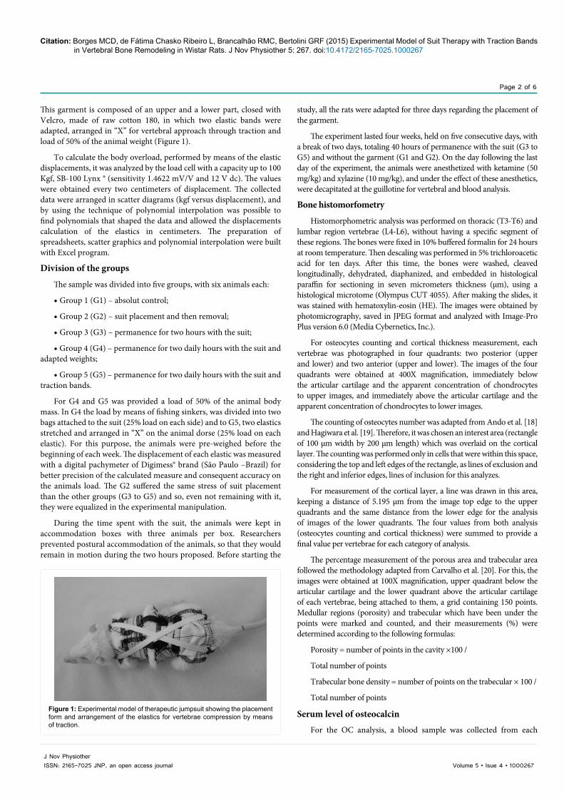
This garment is composed of an upper and a lower part, closed with Velcro, made of raw cotton 180, in which two elastic bands were adapted, arranged in “X” for vertebral approach through traction and load of 50% of the animal weight (Figure 1).
To calculate the body overload, performed by means of the elastic displacements, it was analyzed by the load cell with a capacity up to 100 Kgf, SB-100 Lynx ® (sensitivity 1.4622 mV/V and 12 V dc). The values were obtained every two centimeters of displacement. The collected data were arranged in scatter diagrams (kgf versus displacement), and by using the technique of polynomial interpolation was possible to find polynomials that shaped the data and allowed the displacements calculation of the elastics in centimeters. The preparation of spreadsheets, scatter graphics and polynomial interpolation were built with Excel program.
Division of the groups
The sample was divided into five groups, with six animals each:
• Group 1 (G1) – absolut control;
• Group 2 (G2) – suit placement and then removal;
• Group 3 (G3) – permanence for two hours with the suit;
• Group 4 (G4) – permanence for two daily hours with the suit and adapted weights;
• Group 5 (G5) – permanence for two daily hours with the suit and traction bands.
For G4 and G5 was provided a load of 50% of the animal body mass. In G4 the load by means of fishing sinkers, was divided into two bags attached to the suit (25% load on each side) and to G5, two elastics stretched and arranged in “X” on the animal dorse (25% load on each elastic). For this purpose, the animals were pre-weighed before the beginning of each week. The displacement of each elastic was measured with a digital pachymeter of Digimess® brand (São Paulo –Brazil) for better precision of the calculated measure and consequent accuracy on the animals load. The G2 suffered the same stress of suit placement than the other groups (G3 to G5) and so, even not remaining with it, they were equalized in the experimental manipulation.
During the time spent with the suit, the animals were kept in accommodation boxes with three animals per box. Researchers prevented postural accommodation of the animals, so that they would remain in motion during the two hours proposed. Before starting the
Figure 1: Experimental model of therapeutic jumpsuit showing the placement form and arrangement of the elastics for vertebrae compression by means of traction.
Citation: Borges MCD, de Fátima Chasko Ribeiro L, Brancalhão RMC, Bertolini GRF (2015) Experimental Model of Suit Therapy with Traction Bands in Vertebral Bone Remodeling in Wistar Rats. J Nov Physiother 5: 267. doi:10.4172/2165-7025.1000267
Page 3 of 6
Volume 5 • Isue 4 • 1000267J Nov PhysiotherISSN: 2165-7025 JNP, an open access journal
animal at the euthanasia time, with a minimum fasting of eight hours. Subsequently, the blood was centrifuged to separate the serum. Once separated and frozen it was analyzed by Immulite®/Immulite® 1000 osteocalcin (Siemens Medical Solutions Diagnostics – USA) kit, considering the reference value of < 2.0 to 21 ng/ml.
Statistical analysis
The Shapiro-Wilk test for verification of data normality was used. Then, one-way ANOVA for analysis between groups was applied, with t post hoc. The significance level was 5%.
ResultsBody mass
Only G4 showed significant variation in body weight, with 65.6 grams less than G2, and 62.76 grams less than G1 (Table 1).
There was a significant increase in total body weight of the animals until the fourth week of intervention. At the end of the fourth week the weight remained with no significant difference between groups.
Bone histomorfometry
There were no significant differences between the groups in comparison to the percentage of the porous and trabecular area, as well as the osteocytes count. For cortical thickness, G3 showed significant difference from the G4 to the dorsal vertebra (Table 2).
Bone tissue morphology
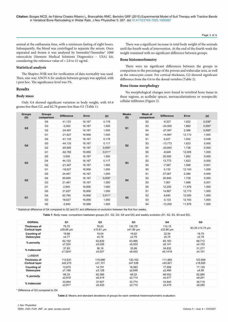
No morphological changes were found in vertebral bone tissue in these regions, as acellular spaces, neovascularizations or nonspecific cellular infiltrates (Figure 2).
Groups(G)
Groups comparison Difference Error (p) Weeks
(S)Week of
comparison Difference Error (p)
G5
G4 41,133 16,167 0,178
S1
S2 -6,227 1,932 0,036*G3 -3,000 16,167 1,000 S3 -20,000 1,892 0,000*G2 -24,467 16,167 1,000 S4 -27,067 2,386 0,000*G1 -21,627 16,956 1,000 S5 -14,867 12,173 1,000
G4
G5 -41,133 16,167 0,178
S2
S1 6,227 1,932 0,036G3 -44,133 16,167 0,117 S3 -13,773 1,623 0,000G2 -65,600 16,167 0,005* S4 -20,840 1,738 0,000G1 -62,760 16,956 0,011* S5 -8,640 12,005 1,000
G3
G5 3,000 16,167 1,000
S3
S1 20,000 1,892 0,000G4 44,133 16,167 0,117 S2 13,773 1,623 0,000G2 -21,467 16,167 1,000 S4 -7,067 1,568 0,001G1 -18,627 16,956 1,000 S5 5,133 12,163 1,000
G2
G5 24,467 16,167 1,000
S4
S1 27,067 2,386 0,000G4 65,600 16,167 0,005* S2 20,840 1,738 0,000G3 21,467 16,167 1,000 S3 7,067 1,568 0,001G1 2,840 16,956 1,000 S5 12,200 11,979 1,000
G1
G5 21,627 16,956 1,000
S5
S1 14,867 12,173 1,000G4 62,760 16,956 0,011* S2 8,640 12,005 1,000G3 18,627 16,956 1,000 S3 -5,133 12,163 1,000G2 -2,840 16,956 1,000 S4 -12,200 11,979 1,000
* Statistical difference of G4 compared to G2 and G1 and difference on evolution between the first four weeks.
Table 1: Body mass: comparison beetween groups (G1, G2, G3, G4 and G5) and weekly evolution (S1, S2, S3, S4 and S5).
DORSAL G1 G2 G3 G4 G5Thickness of Cortical layer
79,70 ±29,68 µm
78,53±15,81 µm
130,75*±41,90 µm
110,15±33,80 µm 83,35 ±14,75 µm
Counting ofOsteocytes
19,88±4,77
19,04±4,79
16,62±2,79
22,54±5,75
18,75±2,79
% porosity 62,162±7,524
63,830±5,026
63,995±6,422
65,163±6,141
68,712±4,103
% trabecular 37,83±7,5241
36,16±5,027
35,99±6,422
34,832±6,1418
31,277±4,101
LUMBARThickness of Cortical layer
113,631±42,275
119,696±31,101
130,163±47,536
111,965±45,821
123,949±18,620
Counting ofOsteocytes
13,875±7,186
14,791±3,128
18,583±2,648
17,958±2,466
17,125±4,56
% porosity 66,33±2,818
62,385±6,419
66,22±2,714
66,052±5,477
62,885±8,201
% trabecular 33,664±2,817
37,607±6,420
33,774±2,712
33,940±5,479
36,718±8,282
* Difference of G3 compared to G4
Table 2: Means and standard deviations of groups for each vertebral histomorphometric evaluation.

Citation: Borges MCD, de Fátima Chasko Ribeiro L, Brancalhão RMC, Bertolini GRF (2015) Experimental Model of Suit Therapy with Traction Bands in Vertebral Bone Remodeling in Wistar Rats. J Nov Physiother 5: 267. doi:10.4172/2165-7025.1000267
Page 4 of 6
Volume 5 • Isue 4 • 1000267J Nov PhysiotherISSN: 2165-7025 JNP, an open access journal
Quantitative analysis of serum osteocalcin
There was no estimated OC quantity on blood serum that could indicate changes in bone metabolism. All samples presented results of <2.0 ng/mL.
DiscussionThe suit therapy was designed in the project known as “Penguin
suit” developed by the Russian space program. This garment was used by astronauts on long space voyages to neutralize the effects of lack of gravity and consequent body hypokinesia [14]. The fact that it could be worn for long periods of time was the basis for creation of the intensive therapy using the suit [16,17], which consists of 80 hours of therapeutic exercises program performed in four hours daily over five consecutive days, for a period of four weeks. During this period the patient remains for up to three hours with the garment [14].
The experimental method of this research aimed to approximate at maximum to the existing protocol of intensive therapy, to do so, the animals remained in quadrupedal and bipedal support, without remaining lying. It was not given a protocol of body activity to be performed during the two hours proposed. Therefore, there was no musculoskeletal demand beyond the one imposed by the garment and elastics, which may contribute for not having muscle and bone overload enough to interfere with bone remodeling. Thus, it was found no change in the osteocytes number, trabecular volume and presence of serum expression of OC.
According to Maimoun and Sultan [18], as the changes in bone mass result from slow metabolic action, biochemical bone markers are useful to investigate the acute effects of mechanical loading on bone remodeling, however, the results of OC on serum in this study, confirm the absence of bone formation even in initial process. In contrast, G3, showed an increase to cortical layer thickness compared to G4. This finding may be a chance occurrence, considering the other results presented and the lack of change in body mass for this group pointing to an increase in muscle mass due to higher contraction labor and consequent influence on bone formation. Another hypothesis could be the higher animal body activity of G3, once that without the bands and weights adapted to the suit, and with the increase in sensory inputs, mainly tactile and proprioceptive provided by the suit, the animals may have felt freer and sensorially stimulated to movement.
On bipedal posture, the gravity distribution on the spine works
in balance with the muscular forces that are directly proportional to the spine direction and pelvic tilt [19]. Solomonow [21], describes that the muscles associated with the joints have an important role as limiters of the movement and therefore as joint stabilizers, and in some joints, such as intervertebral, the muscle function as stabilizer is amplified. Based on this assumption, on all fours posture, there is no unidirectional gravitational action with the muscle fibers of the trunk, neither unloading of this weight compatible with the spinal direction. Thus, the muscular activity could supply the load imposed by the traction bands, without there being vertebral compression sufficient to generate changes in the bone tissue metabolism of these structures.
In a retrospective analysis of researches about bone density regarding the frequency and duration of mechanical stimuli, can be highlighted the study carried out by Iwamoto et al. [22], which in a sample of 40 rats, found that after eight and 12 weeks of treadmill walking there was a significant increase in the rate of mineral apposition and bone formation at the proximal and distal tibia shafts of these animals. They also observed that the rate increasing was greater after 12 weeks of physical activity. In a study carried out by Boreham and McKay [23], with humans, it became evident an increase of 1.2% BMD at the femoral trochanteric region after eight months of jumps incorporated to the school physical education program. Likewise, Gunter et al. [24], reported that children 7-9 years of age showed 7.9% higher bone mineral content at the spine and 8.4% more in BMD at the hip than the control group, after an intervention with jumps for seven months. Finally, Meyer et al. [25], observed that children after nine months of exercises, including in physical education at least 10 minutes of jumping or strength training, showed BMD 4.7% higher at the spine and 5.4 % at the hip than the control group. In this sense, the researches have shown that changes in bone tissue by physical interventions occur with short stimuli for prolonged periods and at high frequency. Comparing with the present study, the time of this experiment was four weeks and may be an inefficient period to occur morphological changes in bone tissue.
Erinckson and Vukovich [26], in a pilot study with men, evaluated changes in bone markers during a eight week jump training program. The results showed that the bone tissue of adult men, did not respond to high-impact exercises. Furthermore, they concluded that the use of recovery periods between exercise sessions performed on the same day can result in positive changes in bone renovation. This conclusion was based on a previous study carried out by Robling et al. [27] who investigated the recovery time to restore the mechano sensitivity in bone dynamic loads. For these authors, given the knowledge that the bone cell is able to sense and respond to mechanical forces, starting to reduce the mechano sensitivity right after starting the stimulus, under constant mechanical stimulation, the bone tends to become insensitive to morphological and chemical changes. In this study, the mechanical stimulus was constant (two hours), daily and with high magnitude load (50% of the animal weight) which may have provided, desensitization of mechanoreceptors.
Some treatment methods that employ suit therapy advocate the use of these garments with traction bands out of the clinical environment, encouraging its manipulation by caregivers, providing the use of therapeutic device for a longer time [15]. This statement may be more effective for the quality of bone remodeling, since it increases the time and period of mechanical stimulation on bone tissue compared to the protocol of intensive therapy that uses the suit for a maximum time of three hours for a limited period of four weeks. Apart from longer stimulus time, should be considered that in order to allow normal
Figure 2: Photomicrograph of a lumbar vertebrae of Wistar rat showing bone tissues without Morphological changes: in A, cortical area in 400x magnification and B, trabecular area in 100x magnification.

Citation: Borges MCD, de Fátima Chasko Ribeiro L, Brancalhão RMC, Bertolini GRF (2015) Experimental Model of Suit Therapy with Traction Bands in Vertebral Bone Remodeling in Wistar Rats. J Nov Physiother 5: 267. doi:10.4172/2165-7025.1000267
Page 5 of 6
Volume 5 • Isue 4 • 1000267J Nov PhysiotherISSN: 2165-7025 JNP, an open access journal
domestic activities, the mechanical load will be applied in a mild to moderate way and in various directions.
According to Wallace et al. [28], in an experimental study with rats, boosting usual activities with bone loading in different directions can positively affect a growing skeleton, suggesting that low-intensity activities that produce loads in various directions may represent an alternative more feasible for the quality of bone tissue development than strenuous high-impact exercises. Still, Chen et al. [29] found in a home therapy program of 12 weeks for CP, with light increasing loads, that there was an increase in femoral BMD by increasing muscle strength, and claim that physical exercise in a natural environment, can increase the participation of children. Therefore dynamic activities with daily doses of short duration, low magnitude, high frequency, with rest breaks [2,30,31], different mechanical stimuli in different directions [20], are more effective for increasing bone strength.
It is important to consider all aspects of bone response to the exercise so that they can be reliably used as a treatment in a clinical enviroment. Given this, any form of scientific evidence that may result from the use of physical activity protocols in living beings should be carefully analyzed and adequate to a musculoskeletal system with disordered development. It is worth remembering that, as confirmed by Riquelme et al. [32], lesions in the central nervous system, such as the CP, the change in muscle tone will be present, as well as impaired recruitment of motor units to generate adequate reactive power to the loads imposed daily to body. Given this assumption, appropriate muscular strengthening exercise programs for this population, rather than providing direct mechanical load on skeletal, may contribute indirectly to the desired bone formation. According Mullender et al. [33], the forces generated by the muscles during exercise, sufficiently overload the bone tissue, producing adaptation with increasing in bone metabolism. This hypothesis was recently reinforced by Noble et al. [34], in a study with CP in which they observed the relation of bone endurance with muscle volume, concluding that there is significance in the development of therapeutic programs for strengthening and improvement in muscle volume to improve bone strength and reduce fracture risk in CP.
There were some limitations to this study, as the animals present themselves on all fours being the trunk with different gravitational loads than in the bipedal posture of the human; and the scarce researches that addressed issues related to mechanical loads or suit therapy as vertebral articular approximation forms, with no guiding methodologies for the experimental process, requiring not only the creation of an experimental suit model, but also experimental parameters which were based on the existing suit therapy protocol and on methods of histological analyzes adapted. After searching the literature it was found that this is an unpublished study, pointing to the need for quantitative and qualitative research regarding the suit therapy, with extensive investigations and that can include other structures of the locomotor system, especially the muscles for checking its response to this method of physical therapeutical intervention.
Important to stress that the discussion generated here aims neither to induce nor to conclude that the concepts of suit therapy have no scientific and secure basis for its results not be considered a reliable and efficient intervention. What is proposed is the beginning of a discussion based on scientific evidences guiding to physical interventions where there is the qualitative and quantitative knowledge necessary to achieve real goals.
Conclusion There were no changes in bone tissue of the dorsal and lumbar
vertebras of Wistar rats with the spinal compression performed by the traction bands of this experimental model of suit and suit therapy. Similarly, no morphological changes that indicate some form of degenerative process of the bone tissue were observed.
References
1. Sheung-Tung H (2012) Review of fractures and low bone mass in children with cerebral palsy. J Orthop, Trauma Rehab 16: 45–50.
2. Finbråten AK, Syversen U, Skranes J, Andersen GL, Stevenson RD, et al. (2015) Bone mineral density and vitamin D status in ambulatory and non-ambulatory children with cerebral palsy. Osteoporos Int 26: 141-150.
3. Grossberg R, Blackford MG, Kecskemethy HH, Henderson R, Reed MD, et al. (2015) Longitudinal assessment of bone growth and development in a facility-based population of young adults withcerebral palsy. Dev Med Child Neurol.
4. Mughal MZ (2014) Fractures in children with cerebral palsy. Curr Osteoporos Rep 12: 313-318.
5. Chen C-L, Ke J-Y, Lin K-C, Wang C-J, Wu C-Y,et al. (2011) Anthropometric and fitness variables associated with bone mineral density and broadband ultrasound attenuation in ambulatory children with cerebral palsy. J Child Neurol 26: 552–559.
6. Hough JP, Boyd RN, Keating JL (2010) Systematic review of interventions for low bone mineral density in children with cerebral palsy. Pediatrics 125: e670–678.
7. Lester ME, Urso ML, Evans RK, Pierce JR, Spiering B a, et al. (2009) Influence of exercise mode and osteogenic index on bone biomarker responses during short-term physical training. Bone 45: 768–776.
8. Janz KF, Letuchy EM, Gilmore JME, Burns TL, Torner JC, et al. (2010) Early physical activity provides sustained bone health benefits later in childhood. Med Sci Sport Exerc 42: 1072–1078.
9. Tenforde AS, Fredericson M (2011) Influence of sports participation on bone health in the young athlete: a review of the literature. PMR 3: 861–867.
10. Vicente WS, dos Reis LM, Graciolli RG, Graciolli FG, Dominguez WV, et al. (2013) Bone plasticity in response to exercise is sex-dependent in rats. PLoS One 8: e64725.
11. Pieles GE, Horn R, Williams CA, Stuart AG (2014) Paediatric exercise training in prevention and treatment. Arch Dis Child 99: 380–385.
12. Chad KE, Bailey DA, Mckay HA, Zello GA, Snyder RE, et al. (1999) The effect of a weight-bearing physical activity program on bone mineral content and estimated volumetric density in children with spastic cerebral palsy. J Pediatr 135: 115–117.
13. Zwier JN, van Schie PEM, Becher JG, Smits D-W, Gorter JW, et al. (2010) Physical activity in young children with cerebral palsy. Disabil Rehabil 32: 1501–1508.
14. Assis R (2012) Condutas práticas em fisioterapia neurológica. 1. ed. Manole S Paulo.
15. Therasuit. About TheraSuit [Internet]. TheraSuit Method. 2014.
16. ADELI. ADELI, rehabilitation and training center [Internet]. 2014.
17. Oliveira L, Pedrozo L, Thomaz J (2012) Manual de treinamento do protocolo Pediasuit.
18. Ando A, Suda H, Hagiwara Y, Onoda Y, Chimoto E, et al. (2011) Reversibility of immobilization-induced articular cartilage degeneration after remobilization in rat knee joints. Tohoku J Exp Med 224: 77–85.
19. Hagiwara Y, Ando A, Chimoto E, Saijo Y, Ohmori-Matsuda K, et al.(2009) Changes of articular cartilage after immobilization in a rat knee contracture model. J Orthop Res 27: 236–242.
20. Carvalho ACB, Henriques HN, Pantaleão JAS, Pollastri CE, Fernandes GVO, et al. (2010) Histomorfometria do tecido ósseo em ratas castradas tratadas com tibolona. J Bras Patol Med Lab 46: 235–243.
21. Solomonow M (2009) Ligaments: a source of musculoskeletal disorders. J Bodyw Mov Ther 13: 136–154.
22. Iwamoto J, Yeh JK, Aloia JF (1999) Differential effect of treadmill exercise on three cancellous bone sites in the young growing rat. Bone. 24: 163–169.

Citation: Borges MCD, de Fátima Chasko Ribeiro L, Brancalhão RMC, Bertolini GRF (2015) Experimental Model of Suit Therapy with Traction Bands in Vertebral Bone Remodeling in Wistar Rats. J Nov Physiother 5: 267. doi:10.4172/2165-7025.1000267
Page 6 of 6
Volume 5 • Isue 4 • 1000267J Nov PhysiotherISSN: 2165-7025 JNP, an open access journal
23. Boreham CA, McKay HA (2011) Physical activity in childhood and bone health. Br J Sport Med. 45: 877–879.
24. Gunter K, Baxter-Jones ADG, Mirwald RL, Almstedt H, Fuller A, et al. (2008) Jump starting skeletal health: a 4-year longitudinal study assessing the effects of jumping on skeletal development in pre and circum pubertal children. Bone 42: 710–718.
25. Meyer U, Romann M, Zahner L, Schindler C, Puder JJ, et al. (2011) Effect of a general school-based physical activity intervention on bone mineral content and density: a cluster-randomized controlled trial. Bone 48: 792–797.
26. Erickson CR, Vukovich MD (2010) Osteogenic index and changes in bone markers during a jump training program: a pilot study. Med Sci Sport Exerc 42: 1485–1492.
27. Robling AG, Burr DB, Turner CH (2001) Recovery periods restore mechanosensitivity to dynamically loaded bone. J Exp Biol 204: 3389–3399.
28. Wallace IJ, Kwaczala AT, Judex S, Demes B, Carlson KJ, et al. (2013) Physical activity engendering loads from diverse directions augments the growing skeleton. J Musculoskelet Neuronal Interact 13: 283–288.
29. Chen C-L, Chen C-Y, Liaw M-Y, Chung C-Y, Wang C-J, et al. (2013) Efficacy of home-based virtual cycling training on bone mineral density in ambulatory children with cerebral palsy. Osteoporos Int 24: 1399–1406.
30. Roussouly P, Pinheiro-Franco JL (2011) Biomechanical analysis of the spino-pelvic organization and adaptation in pathology. Eur Spine J 20: S609–618.
31. Solomonow M (2009) Ligaments: a source of musculoskeletal disorders. J Bodyw Mov Ther 13: 136–154.
32. Riquelme I, Cifre I, Muñoz M a, Montoya P (2014) Altered corticomuscular coherence elicited by paced isotonic contractions in individuals with cerebral palsy: A case-control study. J Electromyogr Kinesiol 24: 928–933.
33. Mullender M, El Haj a J, Yang Y, van Duin M a, Burger EH, et al. (2004) Mechanotransduction of bone cells in vitro: mechanobiology of bone tissue. Med Biol Eng Comput 42: 14–21.
34. Noble JJ, Fry N, Lewis AP, Charles-Edwards GD, Keevil SF, et al. (2014) Bone strength is related to muscle volume in ambulant individuals with bilateral spastic cerebral palsy. Bone 66: 251–255.
Citation: Borges MCD, de Fátima Chasko Ribeiro L, Brancalhão RMC, Bertolini GRF (2015) Experimental Model of Suit Therapy with Traction Bands in Vertebral Bone Remodeling in Wistar Rats. J Nov Physiother 5: 267. doi:10.4172/2165-7025.1000267
Submit your next manuscript and get advantages of OMICS Group submissionsUnique features:
• Userfriendly/feasiblewebsite-translationofyourpaperto50world’sleadinglanguages• AudioVersionofpublishedpaper• Digitalarticlestoshareandexplore
Special features:
• 400OpenAccessJournals• 30,000editorialteam• 21daysrapidreviewprocess• Qualityandquickeditorial,reviewandpublicationprocessing• IndexingatPubMed(partial),Scopus,EBSCO,IndexCopernicusandGoogleScholaretc• SharingOption:SocialNetworkingEnabled• Authors,ReviewersandEditorsrewardedwithonlineScientificCredits• Betterdiscountforyoursubsequentarticles
Submityourmanuscriptat:http://www.omicsonline.org/submission/