Embed Size (px)
Citation preview
J. Embryol. exp. Morph. 74, 1-14 (1983)Printed in Great Britain © The Company of Biologists Limited 1983
Experimental analysis of control mechanisms insomite segmentation in avian embryos.
I. Reduction of material at the blastula stage in Coturnixco turnix japonica
By MARIANNE VEINI1 AND RUTH BELLAIRS2
From the Department of Anatomy & Embryology, University College London
SUMMARYThe blastulae of unincubated eggs of the quail, Coturnix coturnix japonica, have been
bisected in ovo, using the technique of Lutz (1949). Some embryos were harvested after 24 hand found to possess two primitive streaks. Most were fixed at 48 h or 72 h. Some were foundto have regulated to form almost normal single axes, whilst others had developed intoduplicitas anterior embryos, separate twins or collided axes. All three types of twinned em-bryos were smaller than the control embryos. The number of somites was not however reducedin the shorter embryos. This finding corresponds with a similar result obtained by Cooke(1975) who reported that if a Xenopus blastula is reduced in size, it nevertheless develops thecorrect number of somites.
The quail however adjusts the shape of the individual somites so that they fit into thereduced body length, whereas Xenopus reduces the size of somites. No miniaturized somiteswere ever seen in these quail embryos.
As a result of the present experiments, it was concluded that the length of incubation timedoes not directly control the rate of somite formation, because different numbers of somiteswere found in twins which possessed identical genomes and had developed in almost the sameenvironment for identical periods. In addition, the size of the area pellucida does not appearto control somite formation. Probably, the most important influence is the regression of thenode.
INTRODUCTION
The number of somites that form in an embryo determines the number ofvertebrae that subsequently develop. It is essential therefore that only theappropriate number of somites should segment. (In the chick embryo this isabout 52, although the last 10 pairs, which are in the tail region, soon disappear:Hamilton, 1952.)
One of the fundamental problems of Developmental Biology is that of how theembryo ensures that only the appropriate numbers of somites segment. Curious-ly, little attention has been paid to this problem in the chick, despite the fact that
1 Author's address: Zoological Laboratory of the University of Athens, Greece.2 Author's address: Department of Anatomy & Embryology, University College London,
Gower Street, London WC1E 6BT, U.K.
2 M. VEINI AND R. BELLAIRS
many papers have been published on other aspects of somitogenesis (e.g.Bellairs, 1979; Bellairs & Veini, 1980). It has received a little more analysis inthe mouse (Flint, Ede, Wilby & Proctor, 1978; Tarn, 1981) but has been tackledmost thoroughly in amphibians.
A theoretical explanation, the 'Clock and Wavefront Model' was put forwardfor amphibian embryos by Cooke & Zeeman (1976). This model which will bediscussed more fully in our second paper in this series was based to some extenton Cooke's (1975) demonstration that if the amount of tissue was reduced in aXenopus blastula, the appropriate number of somites developed though eachwas less than normal size. Thus in some way the embryo had been able to adjustthe segmentation pattern to the reduced body length.
It has been sometimes suggested (Cooke, 1977) that a similar control systemoperates in avian embryos, especially since miniature somites have been repor-ted as a result of experimental interference (Spratt, 1955,1957). In our opinionhowever, these particular tiny somites are not valid evidence since they havewithout exception been obtained by interference with the embryo at a timewhen the segmentation pattern is already programmed even though the mainmorphological changes have yet to take place (Meier, 1979; Bellairs, 1980). Theexperiments described in the present paper have therefore been designed to testwhether reduction of material in the blastula stages of birds leads to productionof smaller somites as it does in amphibians. It should be noted however thatalthough these experiments are similar to those performed on amphibians, theyare not totally analogous, since a bird blastoderm contains some extraembryonictissues in addition to the embryonic ones, whilst an amphibian blastula consistsentirely of embryonic tissues.
Cooke & Zeeman's theory has received support from experiments in which thepattern of segmentation has been disrupted by treating amphibian embryos withheat shocks (Elsdale, Pearson & Whitehead, 1976; Pearson & Elsdale, 1979;Cooke, 1977). Similar experiments carried out on the chick embryo will bediscussed in our second paper in this series.
MATERIALS AND METHODS
For technical reasons, in ovo operations proved to be simpler to carry out onquail embryos than on chicks. All the in ovo experiments described in this papertherefore utilised quails. A small additional series of experiments, not describedhere, demonstrated however that comparable results could be obtained withchick embryos. In staging the quails we have followed the chick normal tables ofHamburger & Hamilton (1951) and of Eyal-Giladi & Kochav (1976).
A window was cut in the shell of each unincubated quail egg and an assessmentwas made, if possible, of the stage of development. Usually, however unin-cubated quail embryos were found to be difficult to stage unless dissected fromthe yolk; most of them were about stage XII of Eyal-Giladi & Kochav (1976) so
Somites in small avian embryos 3
that no clear-cut orientation was yet apparent. The embryos were therefore atthe blastula stage.
Experiments consisted of bisecting the blastoderm in the manner described byLutz (1949). The operation does not damage the vitelline membrane over theembryo. The knife cuts extended right across the area opaca as well as the areapellucida, so that each of the two pieces was about half the size of an unoperatedembryo. After the operation, the shell was resealed with Scotch tape and the eggincubated.
No operation was performed on control embryos but the eggs were opened andresealed in the same way.
Most eggs were then incubated for either 48 h or 72 h before the embryos wereremoved for photography and fixation. A few eggs were however opened after18-24 h of incubation so that the early stages of development could be inspected.Eight of these embryos were dissected from the yolk and explanted in culture bythe technique of New (1955) so that their development could be followed.
The length of each embryo was measured after fixation, at 40x magnification,using an eyepiece graticule in a Nikon dissecting binocular microscope. Theregion measured was the maximum distance from the anteriormost tip of theembryo to the posterior limit of the area pellucida. Until about stage 13 (19 pairsof somites) this anterior point is the tip of the forebrain, but as cranial flexuretakes place, it comes to be situated in the mesencephalon.
The sizes of the somites were measured on whole embryos after fixation, at85x magnification, using an eyepiece graticule in a Wild binocular microscope.Only the last two pairs of somites were measured. The width and the length ofeach somite was recorded and the area of its dorsal surface was then calculated.The height was compared in a limited number of whole-mount embryos bymeasuring the shift in depth of focus from the lower to the upper surface of thesomite on a calibrated microscope.
Serial sections were cut at 8 jum through seven pairs of twins and through sixcontrol embryos and these were stained with Harris' haematoxylin and eosin.The numbers of cells were counted in the middle section of the last pair of somitesin each series.
The area of the area pellucida was measured by means of a planimeter, fromdrawings made at 30x magnification with a Wild camera-lucida apparatus. Un-fortunately, however, it is not always easy to define the precise border betweenarea opaca and area pellucida in embryos after the extraembryonic circulationhas begun, so that only a limited number of results (25), could be obtained.
RESULTS
I. Types of embryos
(a) Control embryos
Thirty-three quail eggs which had been opened and resealed gave rise to
4 M. VEINI AND R. BELLAIRS
normal embryos. Those examined after 48 h incubation had usually reachedabout stage 11 and possessed about 15 pairs of somites, whilst those examinedafter 72 h were usually at stages 15 or 16 and possessed about 20 pairs of somites.Two further embryos were found to be abnormal and were discarded. Twelveadditional controls were fixed at times ranging from 24-84 h incubation. Thesewere from eggs which had not been opened at all until the time of fixation.
(b) Operated embryos
Thirty-one embryos were examined 18-24 h after the operation, and in everycase the cut was found to have healed. Five of the blastoderms each possessedtwo primitive streaks, whilst each of the remaining 26 had either regulated toform a single streak or had failed to form a streak at all. Of those embryos whichwere explanted in culture, one which possessed two primitive streaks anteriorlybut only one posteriorly subsequently formed a duplicitas anterior embryo. Twoothers which each possessed a single primitive streak anteriorly but two primitivestreaks posteriorly subsequently formed single axes. Those which had alreadyregulated to form a single primitive streak developed a single axis.
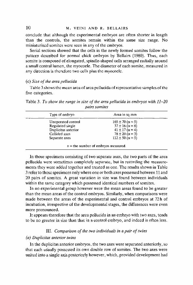
Not all the embryos examined after 48 or 72 h had formed double axes.Twenty-three were discarded either because they were so distorted that theycould not be interpreted, or because they had died. Thirty-six had each regulatedto form what was principally a single axis, although abnormalities such asplatyneuria and diplocardia were often present; a disorderly arrangement of thesomites was also a feature of these embryos (Fig. 1).
Forty-two embryos had each formed well-defined double axes. These were ofthree main types:-
i) Duplicitas anterior (16 pairs of twins)These are composed of two separate heads which share a common trunk. The
level at which the two axes become united varies. Figure 2 illustrates a specimenin which the somites are not yet shared in the trunk.
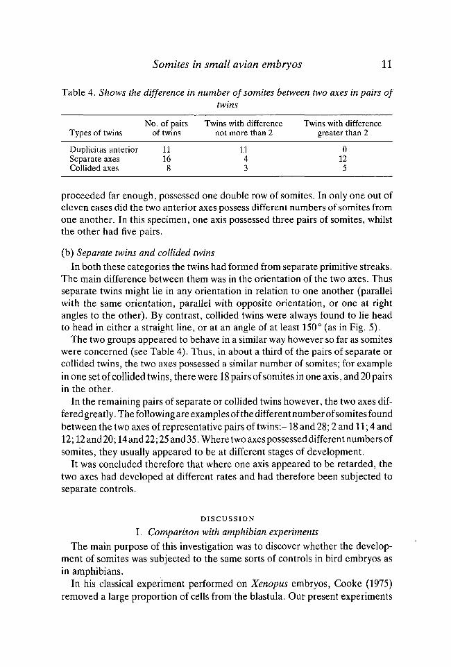
ii) Collided axes (8 pairs of twins)These appear to have resulted from two initially separate axes which have
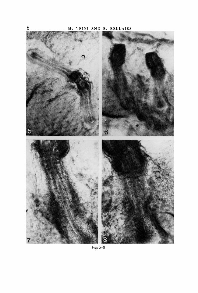
collided in the head region (Fig. 5). The heads are therefore often distorted.
iii) Separate axes (18 pairs of twins)These may be orientated in a variety of ways in relation to one another. Those
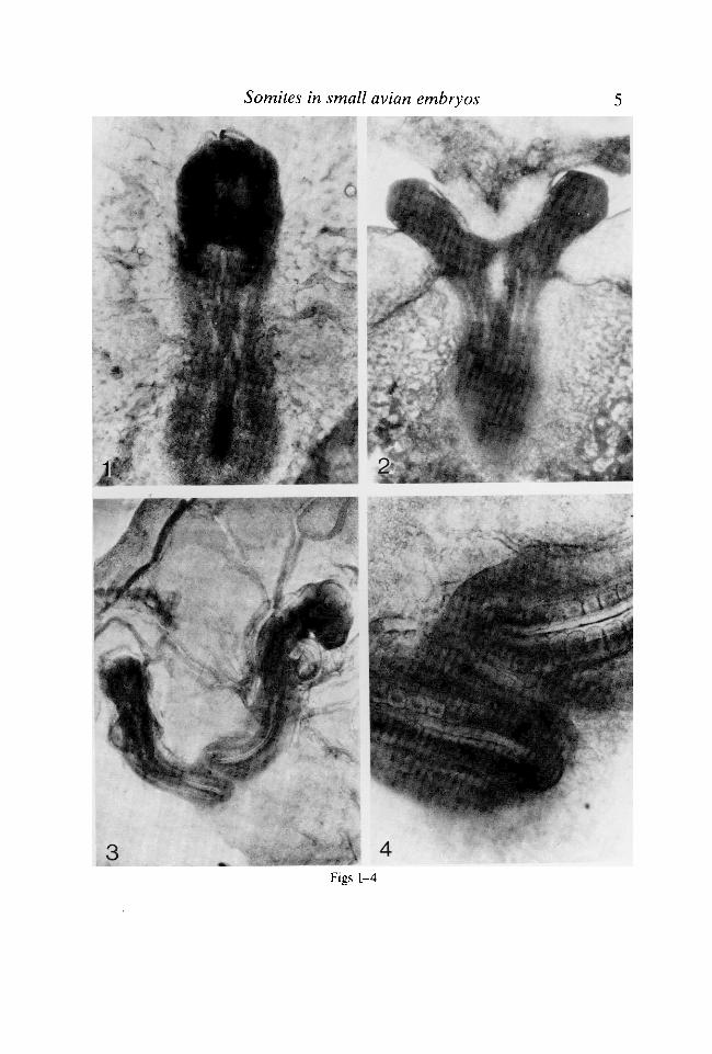
Fig. 1. Partially regulated embryo suffering from platyneuria and a disturbed ar-rangement of somites in the trunk. Fixed at 48 h. x40.Fig. 2. Duplicitas anterior embryo in which the two axes are not yet fully united.Each axis possesses the same number of somites. Fixed at 48 h. x40.Fig. 3. Separate twinned axes lying at right angles to one another. The left possesses14 pairs somites, the right 22 pairs. Fixed at 72h. x20.Fig. 4. Enlargement of the tail buds of the embryos illustrated in Fig. 3, to show thatthe two axes are separate from one another. x60.
Somites in small avian embryos•i
* * •
Figs 1-4
M. VEINI AND R. BELLAIRS
•*»:** t ».
. v i*»
f '*f
Somites in small avian embryos 1
Table 1. The table shows the range of stages (Hamburger & Hamilton) reachedafter 72 h incubation
Type of embryo
ControlRegulated singleDuplicitas anteriorCollided axes*Separate axes
Stages after 72 h
13-1511-168-139-137-13
Pairs of somites
19-2513-274-194-351-19
* It was not always possible to determine the precise stage of development of collided axessince the heads were frequently distorted. The figures are therefore based on three pairs onlyof collided axes.
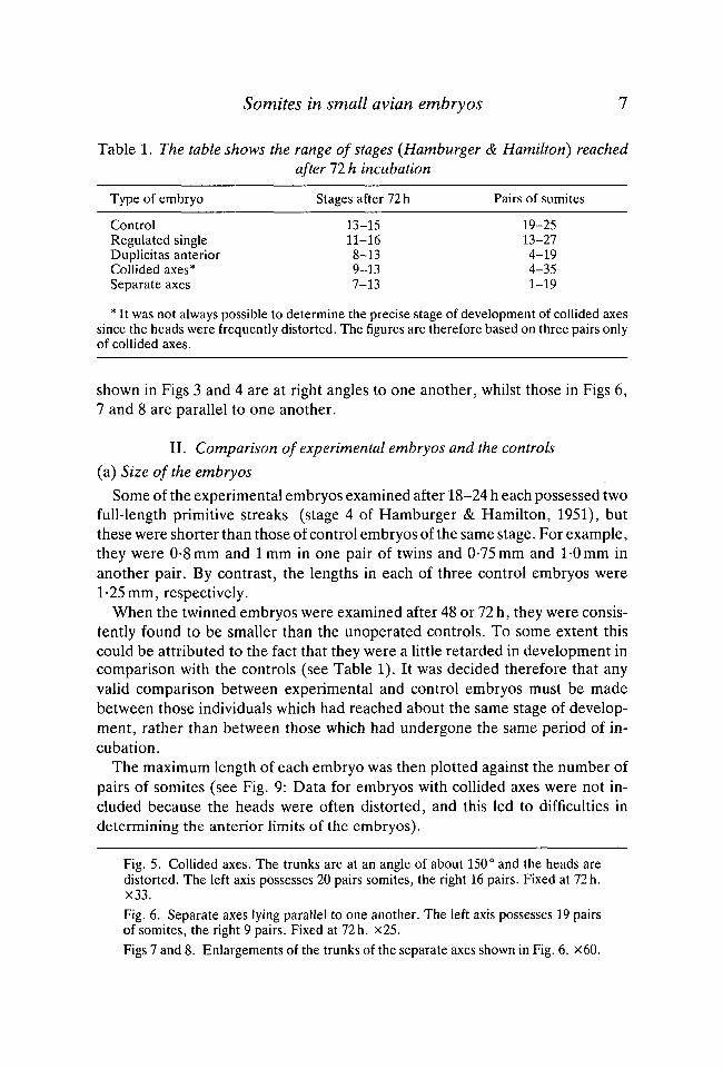
shown in Figs 3 and 4 are at right angles to one another, whilst those in Figs 6,7 and 8 are parallel to one another.
II. Comparison of experimental embryos and the controls
(a) Size of the embryos
Some of the experimental embryos examined after 18-24 h each possessed twofull-length primitive streaks (stage 4 of Hamburger & Hamilton, 1951), butthese were shorter than those of control embryos of the same stage. For example,they were 0-8 mm and lmm in one pair of twins and 0-75 mm and 1-0 mm inanother pair. By contrast, the lengths in each of three control embryos were1-25 mm, respectively.
When the twinned embryos were examined after 48 or 72 h, they were consis-tently found to be smaller than the unoperated controls. To some extent thiscould be attributed to the fact that they were a little retarded in development incomparison with the controls (see Table 1). It was decided therefore that anyvalid comparison between experimental and control embryos must be madebetween those individuals which had reached about the same stage of develop-ment, rather than between those which had undergone the same period of in-cubation.
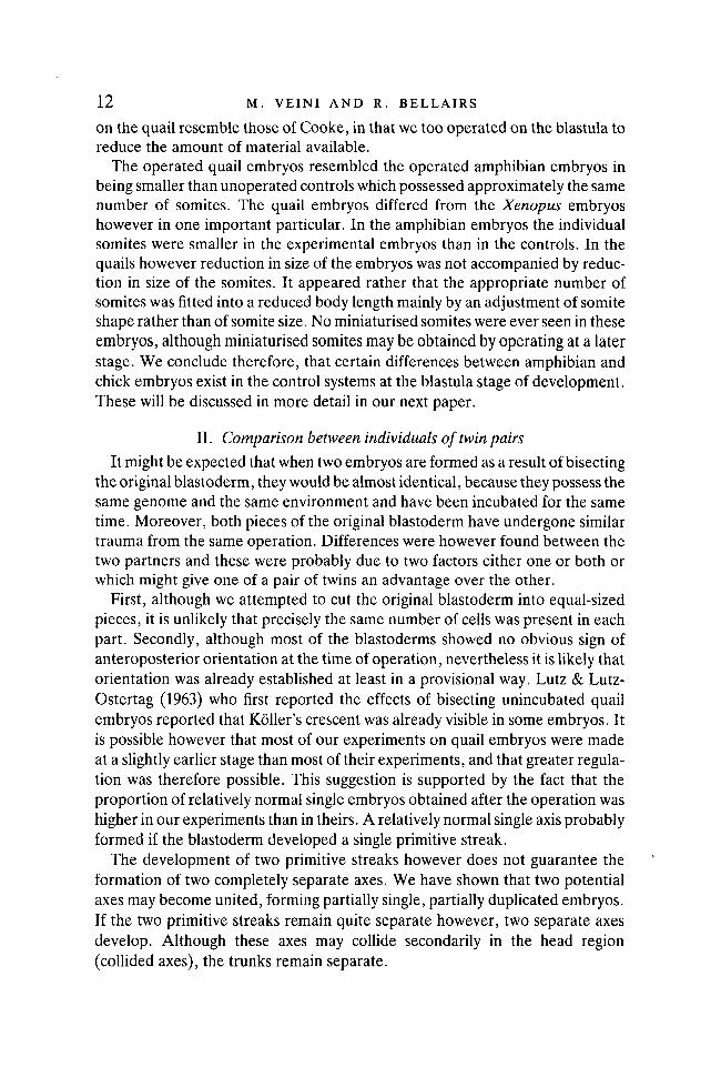
The maximum length of each embryo was then plotted against the number ofpairs of somites (see Fig. 9: Data for embryos with collided axes were not in-cluded because the heads were often distorted, and this led to difficulties indetermining the anterior limits of the embryos).
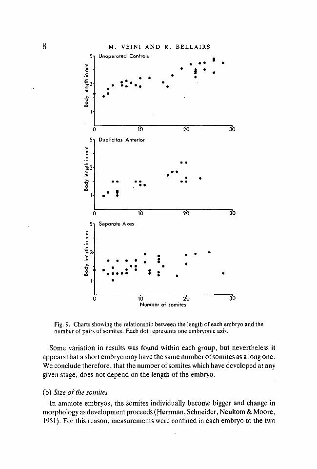
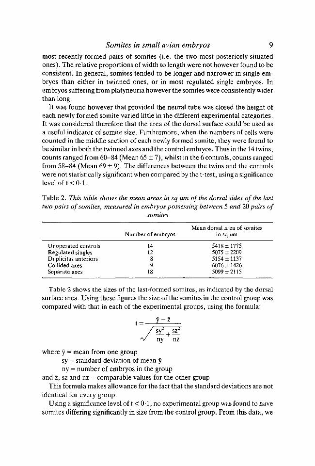
Fig. 5. Collided axes. The trunks are at an angle of about 150° and the heads aredistorted. The left axis possesses 20 pairs somites, the right 16 pairs. Fixed at 72h.X33.Fig. 6. Separate axes lying parallel to one another. The left axis possesses 19 pairsof somites, the right 9 pairs. Fixed at 72 h. x25.Figs 7 and 8. Enlargements of the trunks of the separate axes shown in Fig. 6. x60.
EE
_c
"o>3-
M. VEINI AND R. BELLAIRSUnoperated Controls
0 10
5 i Duplicitas Anterior
20 30
"5)3-0)
10 20 30
5-1
in m
m
-C"oS3-c
Bod
y 1
1-
Separate Axes
•
i • • • •
•
s • •• •
10 20Number of somites
30
Fig. 9. Charts showing the relationship between the length of each embryo and thenumber of pairs of somites. Each dot represents one embryonic axis.
Some variation in results was found within each group, but nevertheless itappears that a short embryo may have the same number of somites as a long one.We conclude therefore, that the number of somites which have developed at anygiven stage, does not depend on the length of the embryo.
(b) Size of the somites
In amniote embryos, the somites individually become bigger and change inmorphology as development proceeds (Herrman, Schneider, Neukom & Moore,1951). For this reason, measurements were confined in each embryo to the two
Somites in small avian embryos 9most-recently-formed pairs of somites (i.e. the two most-posteriorly-situatedones). The relative proportions of width to length were not however found to beconsistent. In general, somites tended to be longer and narrower in single em-bryos than either in twinned ones, or in most regulated single embryos. Inembryos suffering from platyneuria however the somites were consistently widerthan long.
It was found however that provided the neural tube was closed the height ofeach newly formed somite varied little in the different experimental categories.It was considered therefore that the area of the dorsal surface could be used asa useful indicator of somite size. Furthermore, when the numbers of cells werecounted in the middle section of each newly formed somite, they were found tobe similar in both the twinned axes and the control embryos. Thus in the 14 twins,counts ranged from 60-84 (Mean 65 ± 7), whilst in the 6 controls, counts rangedfrom 58-84 (Mean 69 ± 9). The differences between the twins and the controlswere not statistically significant when compared by the t-test, using a significancelevel of t < 0 1 .
Table 2. This table shows the mean areas in sq \xm of the dorsal sides of the lasttwo pairs of somites, measured in embryos possessing between 5 and 20 pairs of
somites
Unoperated controlsRegulated singlesDuplicitas anteriorsCollided axesSeparate axes
Number of embryos
141289
18
Mean dorsal area of somitesin sq jUm
5418 ±17755075 ± 22095154 ±11376076 ± 14265099 ± 2115
Table 2 shows the sizes of the last-formed somites, as indicated by the dorsalsurface area. Using these figures the size of the somites in the control group wascompared with that in each of the experimental groups, using the formula:
ny nz
where y = mean from one groupsy = standard deviation of mean yny = number of embryos in the group
and z, sz and nz = comparable values for the other groupThis formula makes allowance for the fact that the standard deviations are not
identical for every group.Using a significance level of t < 0-1, no experimental group was found to have
somites differing significantly in size from the control group. From this data, we
10 M. VEINI AND R. BELLAIRS
conclude that although the experimental embryos are often shorter in lengththan the controls, the somites remain within the same size range. Nominiaturised somites were seen in any of the embryos.
Serial sections showed that the cells in the newly formed somites follow thepattern described for normal chick embryos by Bellairs (1980). Thus, eachsomite is composed of elongated, spindle-shaped cells arranged radially arounda small central lumen, the myocoele. The diameter of each somite, measured inany direction is therefore two cells plus the myocoele.
(c) Size of the area pellucida
Table 3 shows the mean area of area pellucida of representative samples of thefive categories.
Table 3. To show the range in size of the area pellucida in embryos with 11-20pairs somites
Type of embryo Area in sq mm
Unoperated control 165 ± 70 (n = 5)Regulated single 37 ± 16 (n = 8)Duplicitas anterior 41 ± 17 (n = 4)Collided axes 78 ± 20 (n = 3)Separate axes 112±50(n = 5)
n = the number of embryos measured.
In those specimens consisting of two separate axes, the two parts of the areapellucida were sometimes completely separate, but in recording the measure-ments they were added together and treated as one. The results shown in Table3 refer to those specimens only where one or both axes possessed between 11 and20 pairs of somites. A great variation in size was found between individualswithin the same category which possessed identical numbers of somites.
In no experimental group however were the mean areas found to be greaterthan the mean areas of the control embryos. Similarly, when comparisons weremade between the areas of the experimental and control embryos at 72 h ofincubation, irrespective of the developmental stages, the differences were evenmore pronounced.
It appears therefore that the area pellucida in an embryo with two axes, tendsto be no greater in size than that in a control embryo, and indeed is often less.
III. Comparison of the two individuals in a pair of twins
(a) Duplicitas anterior twins
In the duplicitas anterior embryos, the two axes were separated anteriorly, sothat each usually possessed its own double row of somites. The two axes wereunited into a single axis posteriorly however, which, provided development had
Somites in small avian embryos 11
Table 4. Shows the difference in number of somites between two axes in pairs oftwins
No. of pairs Twins with difference Twins with differenceTypes of twins of twins not more than 2 greater than 2
Duplicitas anterior 11 11 0Separate axes 16 4 12Collided axes 8 3 5
proceeded far enough, possessed one double row of somites. In only one out ofeleven cases did the two anterior axes possess different numbers of somites fromone another. In this specimen, one axis possessed three pairs of somites, whilstthe other had five pairs.
(b) Separate twins and collided twinsIn both these categories the twins had formed from separate primitive streaks.
The main difference between them was in the orientation of the two axes. Thusseparate twins might lie in any orientation in relation to one another (parallelwith the same orientation, parallel with opposite orientation, or one at rightangles to the other). By contrast, collided twins were always found to lie headto head in either a straight line, or at an angle of at least 150° (as in Fig. 5).
The two groups appeared to behave in a similar way however so far as somiteswere concerned (see Table 4). Thus, in about a third of the pairs of separate orcollided twins, the two axes possessed a similar number of somites; for examplein one set of collided twins, there were 18 pairs of somites in one axis, and 20 pairsin the other.
In the remaining pairs of separate or collided twins however, the two axes dif-fered greatly. The following are examples of the different number of somites foundbetween the two axes of representative pairs of twins:-18 and 28; 2 and 11; 4 and12; 12 and 20; 14 and 22; 25 and 35. Where two axes possessed different numbers ofsomites, they usually appeared to be at different stages of development.
It was concluded therefore that where one axis appeared to be retarded, thetwo axes had developed at different rates and had therefore been subjected toseparate controls.
DISCUSSION
I. Comparison with amphibian experiments
The main purpose of this investigation was to discover whether the develop-ment of somites was subjected to the same sorts of controls in bird embryos asin amphibians.
In his classical experiment performed on Xenopus embryos, Cooke (1975)removed a large proportion of cells from the blastula. Our present experiments
12 M. VEINI AND R. BELLAIRS
on the quail resemble those of Cooke, in that we too operated on the blastula toreduce the amount of material available.
The operated quail embryos resembled the operated amphibian embryos inbeing smaller than unoperated controls which possessed approximately the samenumber of somites. The quail embryos differed from the Xenopus embryoshowever in one important particular. In the amphibian embryos the individualsomites were smaller in the experimental embryos than in the controls. In thequails however reduction in size of the embryos was not accompanied by reduc-tion in size of the somites. It appeared rather that the appropriate number ofsomites was fitted into a reduced body length mainly by an adjustment of somiteshape rather than of somite size. No miniaturised somites were ever seen in theseembryos, although miniaturised somites may be obtained by operating at a laterstage. We conclude therefore, that certain differences between amphibian andchick embryos exist in the control systems at the blastula stage of development.These will be discussed in more detail in our next paper.
II. Comparison between individuals of twin pairs
It might be expected that when two embryos are formed as a result of bisectingthe original blastoderm, they would be almost identical, because they possess thesame genome and the same environment and have been incubated for the sametime. Moreover, both pieces of the original blastoderm have undergone similartrauma from the same operation. Differences were however found between thetwo partners and these were probably due to two factors either one or both orwhich might give one of a pair of twins an advantage over the other.
First, although we attempted to cut the original blastoderm into equal-sizedpieces, it is unlikely that precisely the same number of cells was present in eachpart. Secondly, although most of the blastoderms showed no obvious sign ofanteroposterior orientation at the time of operation, nevertheless it is likely thatorientation was already established at least in a provisional way. Lutz & Lutz-Ostertag (1963) who first reported the effects of bisecting unincubated quailembryos reported that Roller's crescent was already visible in some embryos. Itis possible however that most of our experiments on quail embryos were madeat a slightly earlier stage than most of their experiments, and that greater regula-tion was therefore possible. This suggestion is supported by the fact that theproportion of relatively normal single embryos obtained after the operation washigher in our experiments than in theirs. A relatively normal single axis probablyformed if the blastoderm developed a single primitive streak.
The development of two primitive streaks however does not guarantee theformation of two completely separate axes. We have shown that two potentialaxes may become united, forming partially single, partially duplicated embryos.If the two primitive streaks remain quite separate however, two separate axesdevelop. Although these axes may collide secondarily in the head region(collided axes), the trunks remain separate.
Somites in small avian embryos 13
One of the major findings of the present experiments is that, provided the twotrunk axes remain separate, (even if the heads collide) each appears to controlits own number of somites. If however the trunks become united, as in theduplicitas anterior embryos, the two axes, almost without exception contain thesame number of somites and are therefore probably under the same controls asone another.
III. Control of somite number during the first 72 h of incubationThe longer a young chick embryo is incubated, the bigger it grows and the
more somites form. We do not know how the embryo controls the maximumnumber (52) which develop, but we will discuss here ideas on how the numbersmay be controlled in the early stages. It might be expected that the number ofsomites is controlled directly by the length of incubation, or by the length of theembryo or by the size of the whole area pellucida. The following evidencehowever suggests that none of these factors is of direct importance :-
(1) The length of incubation time does not directly control the rate of somiteformation because different numbers of somites were often found in twinaxes which possessed identical genomes and had developed in the sameenvironment for identical periods.
(2) The length of the embryo does not determine how many somites form,because the same number may be found in both short and long em-bryos.
(3) If there is some global control acting throughout the area pellucida this isovercome in the case of the separate (and collided) twins, since the twoindividuals may possess different numbers of somites even though theyshare the same area pellucida. It might be argued that the area pellucidahad become too large in these cases to maintain its functional identity asa single global region and had therefore become two regions. Ourevidence does not support this possibility either, since the total size of thearea pellucida in blastoderms containing separate axes was no greater thanthat of control blastoderms containing one axis. We suggest therefore thatif there is a global control it is not related to size of the tissue mass but tosome other factor.
We suggest instead that the controlling factors are to be found in the mor-phogenetic movements, especially those associated with node regression. Theseare maximal along the primitive streak but also take place in the adjacent areapellucida, so that even where a duplicitas anterior embryo develops, the twoanterior axes are subject to a unified regression. Where the two primitive streaksremain apart, then two separate regression movements take place and these areapparently not co-ordinated.
This work was supported by the Scientific and Engineering Research Council. Dr Veini alsoacknowledges a grant for travel and maintenance from EMBO. We are most grateful to MrsR. Cleevely for her skilful technical assistance.
EMB74
14 M. VEINI AND R. BELLAIRS
REFERENCESBELLAIRS, R. (1979). The mechanism of somite segmentation in the chick embryo. /. Em-
bryol. exp. Morph. 51, 227-243.BELLAIRS, R. (1980). The segmentation of somites in the chick embryo. Boll. Zool. 47,
245-252.BELLAIRS, R. & VEINI, M. (1980). An experimental analysis of somite segmentation in the
chick embryo. J. Embryo), exp. Morph. 55, 93-108.COOKE, J. (1975). Control of somite number during morphogenesis of a vertebrate, Xenopus
laevis. Nature 254, 196-199.COOKE, J. (1977). 'The control of somite number during amphibian development: models and
experiments', In Vertebrate Limb and Somite Morphogenesis (eds D. A. Ede, J. R. Hinch-liffe & M. Balls), pp. 433-449, British Society Devi Biol. Symposium 3. Cambridge Univer-sity Press.
COOKE, J. &ZEEMAN,E. C. (1976). A clock and wavefront model for the control of the numberof repeated structures during animal morphogenesis. /. theor. Biol. 58, 455-476.
ELSDALE, T., PEARSON, M. & WHITEHEAD, M. (1976). Abnormalities in somite segmentationfollowing heat shock to Xenopus embryos. /. Embryol. exp. Morph. 35, 625-635.
EYAL-GILADI, H. & KOCHAV, S. (1976). From cleavage to primitive streak formation: a com-plementary normal table and a new look at the first stages of the development of the chick.Devi Biol. 49, 321-327.
FLINT, O. P., EDE, D. A., WILBY, O. K. & PROCTOR, J. (1978). Control of somite number innormal and amputated mouse embryos: an experimental and a theoretical analysis. J.Embryol. exp. Morph. 45, 189-202.
HAMBURGER, V. & HAMILTON, H. L. (1951). A series of normal stages in the development ofthe chick embryo. /. Morph. 88, 49-92.
HAMILTON, H. L. (1952). Lillie's Development of the Chick, An Introduction to Embryology.New York: Henry Holt & Co.
HERRMANN, H., SCHNEIDER, M. J. B., NEUKOM, J. & MOORE, J. A. (1951). Quantitative dataon the growth processes of the somites of the chick embryo: linear measurements, volume,protein nitrogen and nucleic acids. J. exp. Zool. 118, 243-268.
LUTZ, H. (1949). Sur le production experimentale de la polyembryonie et de la monstruosite"double chez les oiseaux. Archs Anat. micr. Morph. exp. 38, 79-144.
LUTZ, H. & LUTZ-OSTERTAG, Y. (1963). Sur l'orientation des embryons jumeaux obtenus parfissuration parallele a l'axe presume du blastoderme non incube de l'oeuf de Caille (Cotur-nix coturnis japonica). C. r. hebd. Seanc. Acad. Sci., Paris 256, 3752-3754.
MEIER, S. (1979). Development of the chick embryo mesoblast. Formation of the embryonicaxis and establishment of the metameric pattern. Devi Biol. 73, 24-45.
NEW, D. A. T. (1955). A new technique for the cultivation of chick embryos in vitro. J.Embryol. exp. Morph. 3, 320-331.
PEARSON, M. & ELSDALE, T. (1979). Somitogenesis in amphibian embryos. I. Experimentalevidence for an interaction between two temporal factors in the specification of somitepatter. /. Embryol. exp. Morph. 51, 27-50.
SPRATT, N. T. (1955). Analysis of the organizer center in the early chick embryo. I. Localiza-tion of prospective motochord and somite cells. /. exp. Zool. 128, 121-164.
SPRATT, N. T. (1957). Analysis of the organizer center in the early chick embryo. II. Studiesof the mechanics of motochord elongation and somite formation. /. exp. Zool. 134,577-612.
TAM, P. P. L. (1981). The control of somitogenesis in mouse embryos. /. Embryol. exp.Morph. 65, 103-128.
(Accepted 8 November 1982)