Embed Size (px)
Citation preview

CH
Stre
Che
is p
sali
citri
aliq
Woc
stud
che
wat
AN
The
Wis
app
age
anim
24±
and
fed
Fee
Mah
Pun
ad
150
gen
(4-8
with
The
mod
inst
EXP
EMICALS
eptozotoc
emicals Co.
prepared by
ne and adj
ic acid. Th
uots and
ckhardt Ltd
dy were o
micals use
ter was em
IMALS
e study was
star alb
proximately
e-group, ho
mal rooms
±2°C with
d 60±5% h
with Amru
d, manuf
harashtra C
ne, India. W
libitum.
0±20 g (2
ntamicin in
8 week old
h diabetic n
e study of
dels was ca
titutional et
PERIME
S
cin, glucose
(St. Louis,
y dissolving
justing the
he citrate
stored at
d., India, w
of Span D
ed were o
mployed for
s performed
bino ra
y the
oused in v
at a tempe
12h light/d
humidity. T
t Laborato
factured
Chakan Oil
Water was
Animals
-3 week o
nduced AR
) were use
nephropath
the phytoc
arried out i
thical comm
ENTAL D
e-6-phosph
, USA). Sod
g 147 mg of
e pH to 4.5
buffer sho
-20°C. Ge
were used in
iagnostics
f analytica
r preparing
d on male
ats of
same
ventilated
erature of
dark cycle
They were
ry Animal
by Nav
Mills Ltd,
s provided
weighing
old) were
RF while
ed to perfo
y.
chemicals
in SN med
mittee.
‐46‐
DESIGN
hate (G-6-P
dium citra
f tri-sodium
5 with appr
ould be us
entamicin
n the study
Ltd., India
al grade (A
the reagen
used to te
animals
rm anti ren
as therape
ical college
Figure
Male wis
N & MET
P) was pro
ate buffer
m citrate in
roximately
ed fresh o
injections
y. All Assay
a. All ot
AR). Only
nts.
est protect
of body
nal failure
eutic agen
e, Agra hav
e 3.1
star rat
THODOL
cured from
(0.01 M, p
49.5 ml of
0.5 ml of
or frozen i
manufactu
y Kits use
her reagen
double d
tive effect
weight 2
studies in
ts in exper
ving approv
LOGY
m Sigma
pH 4.5)
f normal
1 mol/l
in 1 ml
ured by
d in the
nts and
distilled
against
50±50g
animals
rimental
val from

SEL
Plan
stud
o
o
o
Afo
on t
EXT
Plan
pre
No.
plan
Plan
and
part
extr
petr
LECTION
nt Androgra
dy on basis
It is abun
Plant havi
Preliminar
in the plan
resaid char
this plant.
TRACTIO
nts were c
liminary st
AP-D02, H
nts were w
nt material
d root parts
ts (2 kg)
raction suc
roleum eth
A
OF PLAN
aphis panic
s of the follo
dantly foun
ing bitter ta
ry studies
nt.
racteristics,
N
collected f
udy, while
Herbarium V
ashed quic
was dried
s. Aerial pa
and roots
ccessively
er, chlorofo
FigAndrograp
NT
culata (Bur
owing poin
nd in all par
aste, is sup
showed th
, motivated
from the
it was pro
Voucher No
ckly with th
d under sha
arts and ro
s (5 kg) w
with the
orm and m
‐47‐
ure 3.2 phis panicu
rm. f.) Nee
ts:
rts of the c
pposed to h
e existence
d us to car
botanical g
ocured from
o. 445785)
he water to
ade. Plants
oots were
were separ
solvents w
ethanol. Ea
ulata
es has bee
ountry.
ave medici
e of renop
rry out det
garden of
m NBRI, Lu
for the bul
o remove a
s were part
powdered,
rately subj
with increa
ach extract
en selected
inal propert
rotective p
tailed inves
our instit
ucknow (Ac
lk study. Th
ny foreign
titioned int
separately
jected for
asing polar
t was conce
for the
ties.
potential
stigation
tute for
ccession
he fresh
matter.
to aerial
y. Aerial
soxhlet
rity viz.
entrated

und
pur
g),
aeri
and
Crude
der reduced
ging nitrog
root (96.7
ial (199.89
d when requ
e extract b
d pressure
en and fina
798 g); CH
96 g), root
uired.
Figurbeing conc
(Rota vap
ally weighe
HCl3: aerial
(260.000
‐48‐
re 3.3 centrated
pour vacuu
ed as follow
l (95.889 g
g). Extract
using rota
um distillat
ws: Pt. eth
g), root (4
ion process
a vapor
ion) and d
her: aerial
47.739 g);
s was repe
dried by
(12.223
MeOH:
eated as

PR
The
met
sus
ethe
2-3
IND
Gen
Anim
thre
Gro
rece
(0.5
and
wer
man
Ind
con
wel
nep
et a
min
EPARATI
e dried ext
thanol; ro
pended in
er extract c
drops of e
DUCTION
ntamicin in
mals were
ee groups
oup I was k
eiving
5 ml, i.p.) f
d animals
re admin
nufactured
ia (80 mg
secutive
l-known to
phrotoxicity
al. , 1994).
nimize the c
ON OF TE
tracts viz.
ots: petro
1 % gum
containing
mulsifier Tw
N OF RENA
nduced Re
randomly
of six an
kept as no
isotonic
for 8 conse
of groups
nistered
by Woc
g/kg/day,
days,
o produce
y in rats (Ab
Injections
circadian va
EST EXTR
aerial pa
oleum eth
acacia, se
wax was p
ween-80 (0
AL FAILU
enotoxicit
divided int
nimals each
rmal contr
salin
ecutive day
II and I
gentamicin
khardt Ltd
i.p.) for
which
significa
bdel Gayou
of gentam
ariation in n
‐49‐
Intrap
RACTS
rts: petrol
er, chloro
eparately. T
prepared in
0.1%).
URE
ty
to
h.
ol
ne
s,
II
n,
d,
8
is
ant
um
micin were m
nephrotoxic
Figureperitoneal
weighing
eum ether
form and
The suspen
gum acac
made daily
city (LeBrun
e 3.5 injection g <200 g
r, chlorofo
methano
nsion of pe
ia with the
at 08:00 h
n et al., 19
FigurDried ext
to rat
rm and
l, were
etroleum
help of
hours to
999).
re 3.4 tracts

Dia
Dia
dep
follo
inje
mg/
in
citra
and
to d
wee
inje
dete
from
wer
con
diab
nep
BLO
Bloo
cen
trea
sinu
allo
tem
was
rpm
mic
unt
sam
by
cag
urin
crea
abetic Nep
betes wa
prived o
owed by
ection of s
/ kg) (Sing
freshly pr
ate buff
d Allen, 200
develop dia
ek. A w
ection, blo
ermined in
m the tail v
re consider
centrations
betic rats,
phropathy if
OOD & UR
od samples
trifuge tu
atment, aft
us under
wed to c
mperature (
s separated
m for 2
cro-centrifu
il the anal
mples were
placing the
es as and
ne samples
atinine, ure
hropathy
as induc
f water
a single
streptozoto
gh et al., 2
repared 0.
fer, pH
07) and the
abetic neph
week aft
ood glucos
n blood sa
vein, using
red to be
s > 250
referred to
f their bloo
RINE SAM
s were colle
ubes at t
er 8h fastin
light ether
clot for 3
(Anand et a
d by centrif
2 min
ge) and
ysis was c
collected o
e rats in in
d when re
s were an
ea and uric
ced in
r for
intraperito
ocin (STZ,
2006) disso
01 M sod
4.5 (T
e rats were
hropathy f
ter the
se levels
amples coll
Glucomete
diabetic
mg/dl (Ch
o as STZ r
d urea and
MPLE COL
ected in cle
the end
ng by retro
r anesthes
30 min at
al., 2010).
fugation at
(REMI
stored at
carried out
over a 24 h
dividual m
quired. Se
alyzed for
acid.
‐50‐
In
rats
24h
oneal
50
olved
dium
Tesh
e left
for a
STZ
were
lected
r (Accu-Ch
if they ha
hen and Q
rats, were
creatinine
LLECTION
ean dry
of the
o-orbital
sia and
t room
Serum
t 12000
RM-12C
-20°C
t. Urine
h period
etabolic
erum and
glucose,
Figtraperitonrat weig
FiMetabolic
c
ek, one tou
ad elevated
Quilley, 20
considered
values we
N AND AN
ure 3.6 neal injecthing >200
igure 3.7 c cages fo
collection
uch ultra).
d plasma
008). Then
d to show
re elevated
NALYSES
tion to 0 g
or urine
Animals
glucose
, these
diabetic
d.

‐51‐
BIOCHEMICAL ANALYSES
All biochemical analyses were performed on an automated analyzer ‘Erba
Chem-5 Chemistry Analyzer’ using standard assay kits (Span
Diagnostics Ltd., India).
GLUCOSE (Trinder, 1969; Sacks, 1999)
Glucose (C6H12O6), a monosaccharide (or simple sugar), is a ubiquitous
fuel molecule in biology. It is oxidized though a series of
enzyme-catalyzed reactions to form carbon dioxide and water, yielding the
universal energy molecule ATP. Due to its importance in metabolism,
glucose level is a key diagnostic parameter for many metabolic disorders.
Increased glucose levels have been associated with diabetes mellitus,
hyperactivity of thyroid, pituitary and adrenal glands. Decreased levels are
found in insulin secreting tumors, myxedema, hypopituitarism and
hypoadrenalism.
Principle
Glucose Oxidase (GOD) oxidizes Glucose to Gluconic acid and Hydrogen
peroxide. In presence of enzyme Peroxidase, released Hydrogen Peroxide
is coupled with Phenol and 4-Aminoantipyrine (4-AAP) to form
Quinoneimine dye. Absorbance of coloured dye is measured at 505 nm
and is directly proportional to glucose concentration in the sample.
Glucose + O2 + H2O Glucose Oxidase Gluconic Acid + H2O2
H2O2 + Phenol + 4-AAP Peroxidase Quinoneimine
dye + H2O Pink

‐52‐
Reagents
Glucose Reagent (R1) Phosphate Buffer 100 mM/l
Glucose Oxidase ≥ 15000 U/l
Peroxidase ≥ 1600 U/l
4-AAP 0.28 mM/l
Glucose Diluent (R2) Phenol 10mM/l
Glucose Standard (R3) Dextrose 100 mg/dl
Sample Preparation
Plasma
- Collect blood using an anticoagulant such as heparin, EDTA or citrate.
- Centrifuge the blood at 700-1000 x g for 10 minutes at 4 °C. Pipette
off the top yellow plasma layer without disturbing the white buffy
layer. Store plasma on ice, if not assaying the same day, freeze at
–80 °C. The plasma sample will be stable for one month while stored
at –80 °C.
Serum
- Collect blood without using an anticoagulant.
- Allow blood to clot for 30 minutes at 25 °C.
- Centrifuge the blood at 2000 x g for 15 minutes at 4°C. Pipette off the
top yellow serum layer without disturbing the white buffy layer. Store
serum on ice, if not assaying the same day, freeze at –80 °C. The
serum sample will be stable for one month while stored at
-80°C.
- Serum does not need to be diluted before assaying.
Urine
- Urine does not require any special treatments.
- If not to assay on the same day, freeze at -80 °C.
Working Reagent Preparation
Working reagent is prepared by adding reagent 2 to reagent 1
Note: The working reagent is light sensitive; it should be stored at 2-8 °C
(do not freeze) in dark coloured bottle, to protect from sunlight.

‐53‐
Assay Parameters
Wavelength 505 nm (490-550 nm)
Flow cell temperature 37 °C
Optical path length 1 cm
Sample volume 10 µl
Working Reagent Volume 1000 µl
Incubation 10 min. at 37 °C/ 30 min. at room
temperature (15-30 °C)
Concentration of standard 100 mg/dl
Stability of colour 1 hour
Units mg/dl
Procedure
Tube Working Glucose Re-
agent (µl)
Glucose Standard
(µl)
Sample (µl)
Blank 1000 - -
Standard 1000 10 -
Sample 1000 - 10
Mix well. Incubate at 37 °C for 10 minutes or at room temperature
(15-30 °C) for 30 minutes.
Program the analyzer as per assay parameters.
1. Blank the analyzer with reagent blank.
2. Measure absorbance of standard followed by the test.
3. Calculate results as per given calculation formula.
Calculation
Glucose (mg/dl) = ABSORBANCE OF TEST X 100
ABSORBANCE OF STANDARD
‐54‐
CREATININE (Bowers, 1980)
Creatinine, an anhydride of creatine, is a waste product formed by the
spontaneous dehydration of kidneys. Most of the creatinine is found in
muscle tissue where it is present as creatine phosphate and serves as a
high energy storage reservoir for conversion to ATP. In the blood,
creatinine is removed by filtration though the glomeruli of the kidney and
is secreted into urine. In healthy individuals, creatinine secretion is
independent of diet and is fairly constant. Serum creatinine concentrations
depend almost entirely upon its excretion rate by the kidneys. For this
reason, its elevation is highly specific for kidney diseases. In kidney
disease, creatinine levels in the blood are elevated, whereas the creatinine
clearance rate or GFR drops and hence the urine levels are diminished.
Principle
The assay of creatinine has been based on the reaction of creatinine with
alkaline picrate as described by Jaffe. The improved Jaffe method
utilizes picrate that forms a red colored complex with creatinine. The
intensity of the color, measured at 510nm, is directly proportional to
creatinine concentration in the sample. The optimized formulation
substantially reduces interference by substances in the raw sample.
Creatinine + Picric acid Alkaline Solution Creatinine - Picric acid
complex
(Orange colour)
Reagents
Picrate Reagent (R1) Picric acid 40 mM/l
Sodium Hydroxide (R2) Sodium Hydroxide 200 mM/l
Creatinine Standard (R3) Creatinine 2.0 mg/dl
‐55‐
Working Solution
Working reagent is prepared by mixing equal volume of R1 (Picrate
reagent) with R2 (Sodium hydroxide) to make up the desired volume. Mix
gently for 2 minutes.
Sample Preparation
Specimen Storage at Stability Remarks Serum/ Plasma
Room Temperature (15 -30 °C)
1 day Freshly separated unhaemolysed serum/ plasma should be used. For plasma, fluoride anticoagulant should not be used. 2-8 °C 1 week
-20 °C 6 months Urine (24h collection)
Room Temperature (15-30 °C)
4 hours Dilute the urine sample 1:10 with purified water. With ‘Thymol’ as preservative, the Urine sample is stable for 4-7 days at room temperature (15-30°C).
-20 °C 6 months
Assay Parameters
Wavelength 505 nm (490-530 nm) Blank Purified Water Optical path length 1 cm Sample volume 100 µl Working Reagent Volume 1000 µl Concentration of standard 2 mg/dl Delay 30 seconds Interval 120 seconds Units mg/dl
Procedure
Tubes Standard Test
Sample - 100 µl
R3 100 µl -
Working reagent 1000 µl 1000 µl
Mix well.

‐56‐
Program the analyzer as per assay parameters.
1. Blank the analyzer with purified water.
2. Measure the initial absorbance of the standard i.e. A1 after 30
seconds and final absorbance A2 after an interval of another 120
seconds.
3. After standard reading are noted, take the reading of test i.e. A1
and A2 accordingly ( follow same procedure as for standard)
Calculation
∆A = A2 – A1
Serum Creatinine (mg/dl) = (∆A of Test / ∆A of Standard) X
concentration of standard (mg/dl)
Urine Creatinine = ∆ATEST X Conc. of Std. X dilution factor X
(mg/day) ∆ASTANDARD 24 hour urine volume in dl
UREA (Berthelot, 1859)
Urea is primarily produced in the liver and secreted by the kidneys. Urea is
the major end product of protein catabolism in animals. It is the primary
vehicle for removal of toxic ammonia from the body. Urea determination is
very useful for the medical clinician to assess kidney function of patients.
In general., increased serum urea levels are associated with nephritis,
renal ischemia, urinary tract obstruction, and certain extra-renal diseases,
e.g., congestive heart failure, liver diseases and diabetes. Decreased

‐57‐
levels indicate acute hepatic insufficiency or may results from over
vigorous parenteral fluid therapy.
Principle
Urea is hydrolyzed in the presence of urease and water to yield
ammonium ions and carbon dioxide. The ammonium ions formed react
with salicylate and hypochlorite to give a green dye (2, 2- dicarboxyl
indophenol) which is measured at 580±10 nm.
Urease NH2 - CO - NH2 + H2O 2NH3 + CO2
NH3 + Salicylate + Hypochlorite 2, 2-dicarboxy indophenols Green Dye
Reagents
Buffer (R1): 2x250 ml Phosphate buffer, pH 6.7 50 mmol/l
EDTA 2 mmol/l
Sodium Salicylate 60 mmol/l
Sodium Nitroprusside 3.2 mmol/l
Enzyme Reagent (R2):2x250 ml Urease 30000U/l
Hypochlorite Solution (R3):2x250 ml Sodium Hypochloride 140 mmol/l
Sodium Hydroxide 150 mmol/l
Standard (R4):1x5 ml Urea 50 mg/dl (8.325 mmol/l)
Preparation and Stability
Buffer/R1 is ready to use. Add 1 vial enzyme reagent/R2 to 1 bottle of
buffer/R1. The working solution is stable for 4 weeks at +2 to + 8°C and 6
days at + 20 to + 25°C. Reagent/R3 and standard/R4 are ready to use
and stable up to the expiry date specified when stored at +2 to +8°C.
Protect all reagents against direct light.

‐58‐
Samples Unhaemolysed serum/ plasma should be used. For plasma, EDTA or
Heparin may be used as anticoagulant. Urine should be diluted 1:50 with
distilled water. Procedure
Wavelength: 580 nm
Temperature: 25 - 37°C
Cuvette: 1 cm light path
Zero adjustment: Against reagent blank
Reagent Blank Standard Sample Working Solution
R1+R2 1000 µl 1000 µl 1000 µl
Standard / R4 - 10 µl - Sample - - 10 µl
Mix and incubate at + 37 °C for 5 minutes or for 10minutes at +15 to
+ 25 °C. Then add:
Reagent/ R3: 1000 µl 1000 µl 1000 µl
Mix and incubate at + 37 °C for 5 minutes or at +15 to + 25 °C for 10
minutes.
Calculation
Serum Urea Concentration (mg/dl) = ASAMPLE X Standard Concentration
ASTANDARD
Urine Urea(mg/day) = ∆ASAMPLE X Conc. of Std. X dilution factor X ∆ASTANDARD 24 hour urine volume in dl

‐59‐
TOTAL PROTEIN (Bradford, 1976; Stoscheck, 1990)
Proteins are polypeptides made up of amino acids. All proteins contain a large
number of peptide bonds and Cu2+ ions in a moderately alkaline medium, a
colored chelate complex of unknown composition is formed between the Cu++
ions and carbonyl (-C=O) and imine (=NH) groups of the peptide bonds.
Analogous reaction takes place between the cupric ion and organic compound
biuret (-NH2) and therefore, the reaction is termed as the Biuret reaction. Principle The reaction takes place between the cupric ion and compound at least two
NH2CO-, NH2CH2-, NH2CS-, and similar groups joined together directly, or
though a carbon or nitrogen atom. Amino acids and dipeptides cannot give the
reaction, but tri- and polypeptides and proteins react to give pink/
reddish-violet products. In this reaction, one copper ion is linked between four
and six nearby peptide linkages by coordinate bonds; more protein present,
more peptide bonds available for reaction. The intensity of colour produced is
proportional to the number of peptide bonds undergoing reaction. Thus Biuret reaction is used for a simple and rapid colorimetric method for determining
protein.
Procedure
Reagent preparation 1. Dissolve following reagents in about 900 ml of water one after the other as
given: Sodium-Potassium Tartarate (9.0 g), Copper Sulphate (CuSO4.5H2O) (3.0 g), Potassium Iodide (KI) (5.0 g), Sodium Hydroxide (8.0 g). Make up to 1000 ml with water. 2. Protein standard (8.0 g of bovine albumin in 100 ml of isotonic
sodium chloride). Protocol
Blank Standard Test
Biuret reagent 5.0 ml 5.0 ml 5.0 ml Protein standard - 0.1 ml - Serum - - 0.1 ml
Mix well and let stand for 15-20 minutes at room temperature and read. Set zero with blank.
Cal
HIS
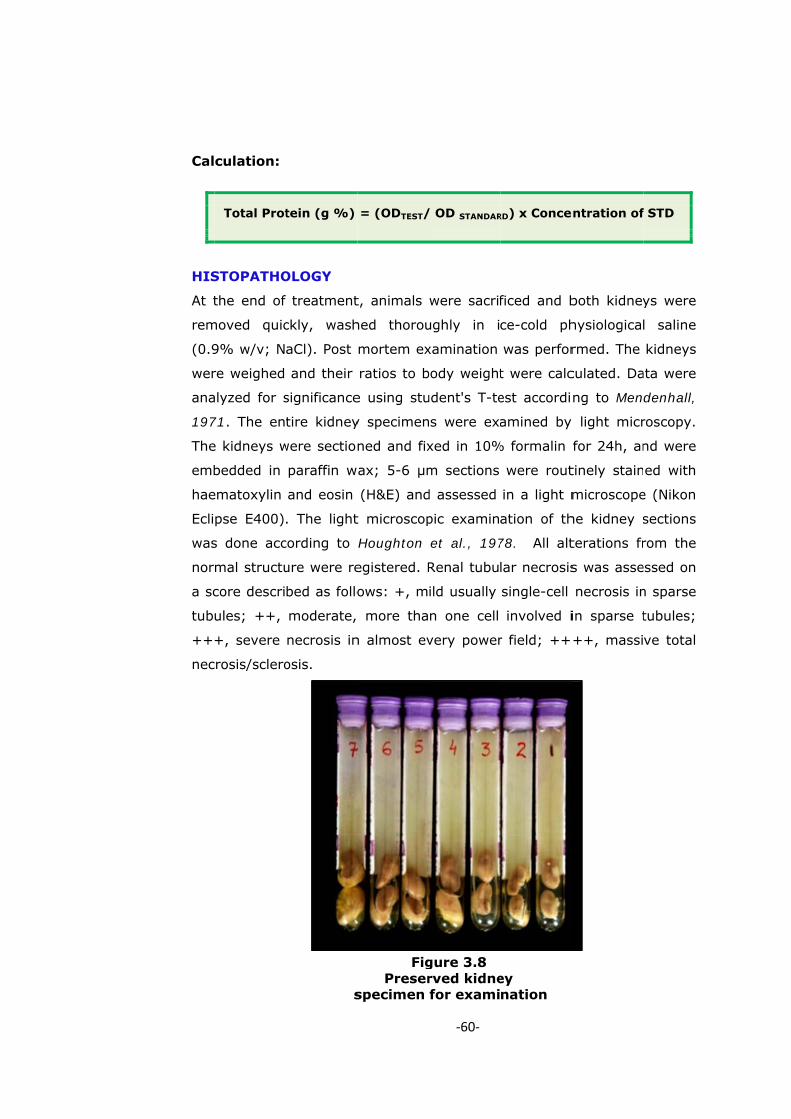
At t
rem
(0.9
wer
ana
197
The
emb
hae
Ecli
was
nor
a sc
tub
++
nec
culation:
Total Prot
STOPATHO
the end of
moved quic
9% w/v; N
re weighed
alyzed for s
71. The en
e kidneys w
bedded in
ematoxylin
pse E400)
s done acc
mal structu
core descri
ules; ++,
+, severe
crosis/sclero
s
tein (g %)
OLOGY
treatment
ckly, wash
aCl). Post
and their
significance
tire kidney
were section
paraffin w
and eosin
. The light
cording to
ure were re
bed as follo
moderate,
necrosis in
osis.
FigPreser
specimen
= (ODTEST/
t, animals
hed thorou
mortem ex
ratios to b
e using stu
y specimen
ned and fix
ax; 5-6 µm
(H&E) and
t microscop
Houghton
egistered. R
ows: +, m
more tha
n almost ev
‐60‐
gure 3.8 rved kidnefor exami
/ OD STANDAR
were sacrif
ughly in i
xamination
body weight
udent's T-te
s were exa
xed in 10%
m sections
d assessed
pic examin
et al., 197
Renal tubu
ild usually
n one cell
very power
ey nation
RD) x Conce
ficed and b
ce-cold ph
was perfor
t were calc
est accordi
amined by
% formalin
were rout
in a light m
ation of th
78. All alt
lar necrosis
single-cell
involved i
r field; ++
ntration of
both kidney
hysiological
rmed. The
culated. Da
ng to Men
y light micr
for 24h, an
tinely stain
microscope
he kidney s
terations fr
s was asse
necrosis in
in sparse t
++, massi
f STD
ys were
l saline
kidneys
ata were
denhall,
roscopy.
nd were
ed with
e (Nikon
sections
rom the
essed on
n sparse
tubules;
ve total

BIO
Eac
nal
nep
to
adm
stre
sus
adm
(ve
atta
wer
exp
pote
pote
nep
(Sc
OASSAY (R
ch extract w
failure m
phrotoxicity
a dose
ministered
eptozotocin
pension w
ministration
hicle) only.
ached to a
re perform
perimental p
ent extract
ent agains
phropathy,
heme 3.2).
O
RENAL PRO
was examin
models viz
y and diabe
of 200
to the anim
induced
was thorou
n. Control
. The extra
syringe), i
med as sh
protocol (S
t was trace
st gentam
and therefo
.
FOral admin
usin
OTECTIVE
ned for the
z. male a
etic nephro
mg/kg a
mals with
d diabetic
ughly mixe
groups o
acts were f
nserted do
hown in t
Scheme 3.1
ed. Methan
micin induc
ore selected
‐61‐
Figure 3.9nistration g oral gav
E ACTIVITY
renoprotec
albino rats
opathy. Tes
nd 250
gentamicin
c nephro
ed to ens
of animals
fed, using a
own the eso
the schem
). Based o
olic root ex
ced acute
d for the de
9 of extractvage
Y) OF THE
ctive activit
s with ge
st extracts
mg/kg bo
n induced n
pathy, re
ure homo
received
an oral gav
ophagus. A
matic repre
n the obse
xtract was
renal fail
etailed phy
t
E EXTRACT
ty against b
entamicin
(1 ml) eq
ody weigh
nephrotoxic
espectively
ogeneity, p
1% gum
vage (narro
All the expe
esentation
rvations, th
found to b
ure and
ytochemical
TS
both re-
induced
uivalent
ht was
city and
y. The
prior to
acacia
ow tube
eriments
of the
he most
be most
diabetic
studies

‐62‐
Scheme 3.1
IDENTIFICATION OF THE MOST EFFECTIVE SOLVENT EXTRACT
EXHIBITING RENOPROTECTIVE ACTIVITY
PLANT MATERIAL
Separation
AERIAL PARTS ROOTS
Shade Dried and Powdered
SOXHALATION
Separately/Successively
Pt. ETHER ARIAL EXTRACT Pt. ETHER ROOT EXTRACT
CHCl3 AERIAL EXTRACT CHCl3 ROOT EXTRACT
MeOH AERIAL EXTRACT MeOH ROOT EXTRACT
RENOPROTECTIVE ACTIVITY AGAINST GENTAMICIN
INDUCED ACUTE RENAL FAILURE
RENOPROTECTIVE ACTIVITY AGAINST STREPTOZOTOCIN
INDUCED DIABETIC NEPHOPATHY
Bioassay of each extract separately
Identification of most effective extract
MeOH ROOT EXTRACT
Evaluation of Renal Profile

‐63‐
Scheme 3.2
ACTIVITY GUIDED CHROMATOGRAPHIC FRACTIONATION AND
CHARACTERIZATION OF BIOACTIVE PRINCIPLES
MeOH ROOT EXTRACT
Column Chromatographic Separation MeOH: CHCl3 (84:16)
RECOVERED SUBSTANCE (BROWN MASS)
Re-Column Chromatographic Separation
MeOH: CHCl3 [GRADIENT ELUTION]
FMEA FMEB FMEC FMED
Trace Amount
RENOPROTECTIVE BIOASSAY
MOST EFFECTIVE FMEB + FMEC
STRUCTURAL ELUCIDATION
FMEB QUALITATIVE TESTS: Diterpene Lactone
QUALITATIVE TESTS: Diterpene Glycoside
ELEMENTAL ANALYSIS IR
NMR MASS
GLUCOSE DERIVATIVE OF ANDROGRAPHOLIDE ANDROGRAPHOLIDE
BIOACTIVE PRINCIPLES
FMEC
ACT
ROO
Met
sub
usin
4.0
mes
(84
brow
mon
(TLC
[CH
chro
dev
brow
diam
elut
RE-
Diff
and
ens
(CH
6:2
valu
FME
Rf =
frac
(thi
Rf =
MeO
Rem
(FM
trac
TIVITY GU
OT EXTRA
thanolic r
bjected to
ng a colum
cm, statio
sh; 125 g)
:16). After
wn semi-s
nitored by
C) us
HCl3:Toluen
omatogram
veloped in
wn mass
meter: 3.0
ted with CH
-COLUMN
ferent fract
d subjected
ure thei
HCl3):Toluen
.5:1.5) sol
ues, fractio
EA (first
= 0.78125
ctions 78-1
rd fracti
= 0.9678 la
OH ex
moval of so
MEC) and pa
ce amount
UIDED CH
ACT
oot extra
chromat
mn (length:
onary phas
) and elute
r the remov
solid mass
y thin lay
sing
e:MeOH
m showed
an iodine
was re-ch
cm, statio
HCl3:MeOH
CHOMATO
tions of 25
d to thin l
ir purity
ne (C6H5C
vent syste
on 29-47;
fraction),
labeled as
106, Rf = 0
on) and
abeled as F
xtract
olvent furn
ale yellow (
and not con
HOMATOGR
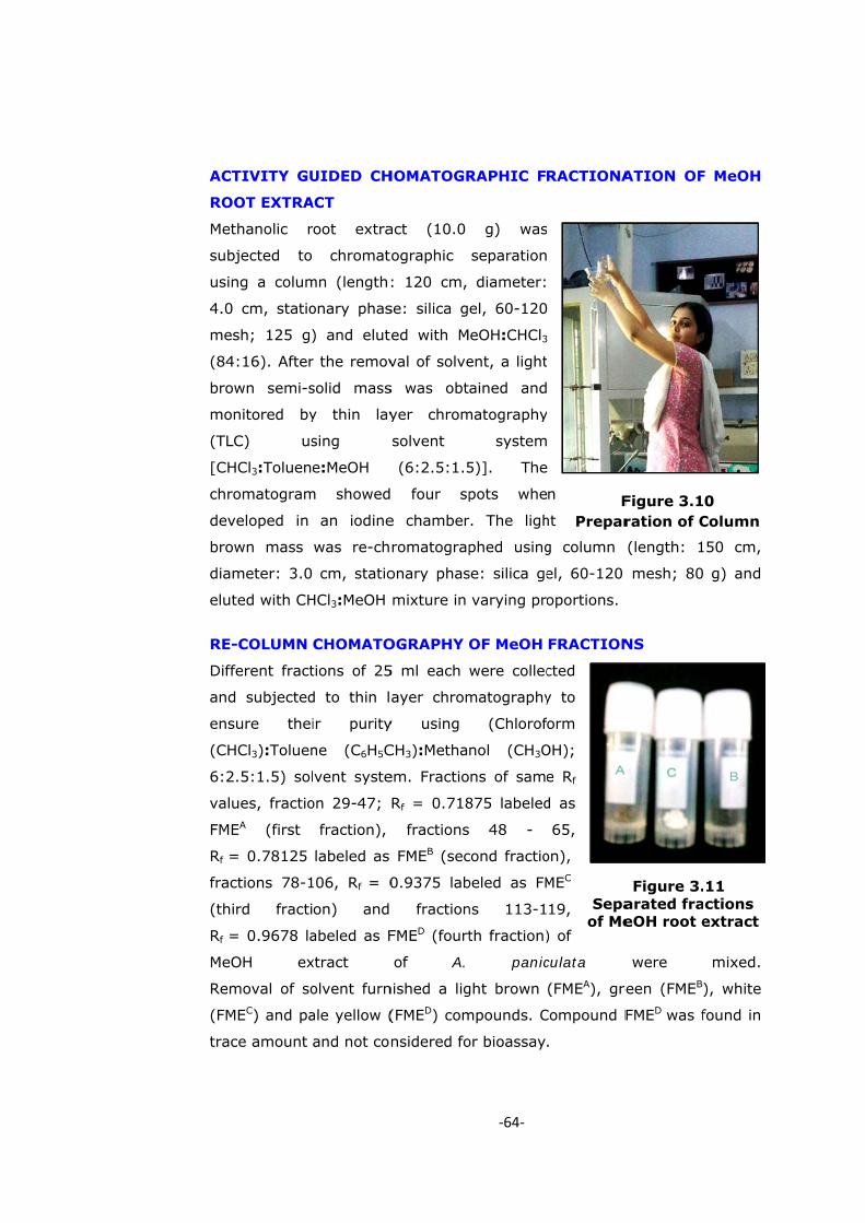
act (10.0
ographic
: 120 cm,
se: silica g
ed with M
val of solve
s was obt
yer chrom
solvent
(6:2.5:1.5
four sp
e chamber
romatograp
onary phas
mixture in
OGRAPHY
5 ml each
ayer chrom
y using
CH3):Metha
em. Fractio
Rf = 0.718
fractions
FMEB (sec
0.9375 lab
fraction
FMED (fourt
of A.
nished a lig
(FMED) com
nsidered fo
‐64‐
RAPHIC F
g) was
separation
diameter:
el, 60-120
eOH:CHCl3ent, a light
ained and
matography
system
5)]. The
pots whe
r. The ligh
phed using
se: silica ge
varying pro
OF MeOH
were collec
matography
(Chlorof
nol (CH3O
ns of same
875 labeled
48 -
cond fractio
beled as FM
ns 113-1
th fraction)
panic
ght brown
mpounds. C
or bioassay
Sepaof Me
FPrepar
RACTIONA
n
ht
g column
el, 60-120
oportions.
FRACTION
cted
y to
orm
OH);
e Rf
d as
65,
on),
MEC
19,
) of
culata
(FMEA), gre
Compound F
.
Figure 3.arated fraeOH root e
Figure 3.1ration of C
ATION OF
(length: 1
mesh; 80
NS
were
reen (FMEB)
FMED was f
11 ctions extract
0 Column
F MeOH
150 cm,
g) and
mixed.
), white
found in

‐65‐
RENAL PROTECTIVE BIOASSAY OF MeOH FRACTIONS [FMEA, FMEB
and FMEC]
The compounds (FMEA, FMEB and FMEC) recovered from methanol extract
were screened for renoprotective activity against gentamicin induced ne-
phrotoxicity and diabetic nephropathy. The different fractions were
taken in concentrations ranging from 5-100 µg/ml. No noticeable activity
could be observed in the compound labeled as FMEA at any concentration.
Compounds FMEB and FMEC showed significant renoprotective activity at a
concentration of 20 µg/ml, in gentamicin induced rats and 90 µg/ml in
streptozotocin induced rats, and therefore, subjected for their chemical
characterization.
STATISTICAL ANALYSES
All results are expressed as mean ± standard error (SE). Statistical
differences between correlated samples were evaluated using Student’s -t
test and noted to be significantly different where p < 0.05. Student t test
calculations were assessed on the online scientific calculators (GraphPad
Quick Calcs, 2009). Composite treatments were compared using
one-way analysis of variances (ANOVA) and considered significantly
different where probability values were found to be equal to or less than
0.05. All ANOVA tests, as well as mean and standard error of mean
calculations, were performed using GraphPad Prism (GraphPad Software,
Inc., San Diego, USA).
STRUCTURAL ELUCIDATION OF BIOACTIVE PRINCIPLE
The compounds FMEB and FMEC being bioactive in nature were considered
for structure elucidation using combination of various techniques including
Infrared, Mass, Nuclear Magnetic Resonance Spectrometry.


![Chapter 2 Aerodynamics Model - Virginia TechCHAPTER 2. AERODYNAMICS MODEL 21 Center [44] were also costly, requiring a 12 block grid with 876 ;912 cells. It is abun-dantly clear that](https://img.pdfslide.us/doc/110x75/5e9444e58e6edd48de184c57/chapter-2-aerodynamics-model-virginia-tech-chapter-2-aerodynamics-model-21-center.jpg)

![79: ' # '6& *#7 & 8other hand, Hirota et al. [15] present an experime ntal work in turbulent heat transfer in square ducts; they show details of turbulent flows and temperature fields](https://img.pdfslide.us/doc/110x75/6086dfba43a9704a56295074/79-6-7-8-other-hand-hirota-et-al-15-present-an-experime-ntal.jpg)