Embed Size (px)
Citation preview

Visual Field Analyzers Expanding your field of vision
3
// PERIMETRY MADE BY CARL ZEISS
Offering the industry’s broadest selection of perimetry products, Carl Zeiss continues to set the gold standard for quality, precision and innovation worldwide.
It is a standard that reflects our shared commitment to the enhancement and preservation of vision. A standard that expands the potential of perimetry with new technologies that offer unique insights to support you in glaucoma clinical detection, diagnosis and ongoing management.
Every perimetry product from Carl Zeiss is designed to provide optimized workflow, better patient comfort, and superb value not only today but also far into the future.
Take a moment to find out more about the perimetry solutions from Carl Zeiss for confident early diagnosis and comprehensive disease management.
And see where vision takes you.
Visual Field AnalyzersHumphrey® Field Analyzer/HFA™ II-i Series
Humphrey Matrix®
Humphrey FDT®
Perimeter SoftwareGuided Progression Analysis™ (GPA™)
SITA-SWAP™
Connectivity SoftwareFORUM® Eye Care Data Management
DICOM Gateway SoftwareHFA-NET Pro™
Vision in Focus

5
HFA II-i The gold standard in perimetry to aid in glaucoma diagnosis and management
Humphrey Field Analyzer – HFA II-i SeriesValidated by more than 25 years of research, design and clinical experience, the HFA is the accepted standard of care in glaucoma diagnosis and management. With over 65,000 installed units worldwide, the Humphrey Field Analyzer is the premier automated visual field perimeter.
Complete portfolio of HFA II-i perimeters
Humphrey 750i Visual Field Analyzer The ultimate in practice efficiency, advanced features and long-term value.
Humphrey 745i Visual Field Analyzer All the features of the 740i plus SITA-SWAP software for early detection.
Humphrey 740i Visual Field Analyzer The basic model in automated visual field testing for comprehensive care.
Humphrey 720i Visual Field Analyzer All purpose model for low volume practices.
// GOLD STANDARD MADE BY CARL ZEISS

6 7
HFA II-i A legacy of perimetry innovation
Advanced analysis
The HFA is the only perimeter with progression
analysis validated in the Early Manifest
Glaucoma Trial.¹
• EnhancedGuidedProgressionAnalysis(GPA)
software identifies statistically significant
progression automatically, and presents
“at a glance” visual field progression analysis
on a single page report.
• VisualFieldIndex™ (VFI™) is a simple and
intuitive global index to determine percentage
of field loss on every visual field.²,³
• PatternDeviationPlotsidentifylocalizedfield
loss, minimizing ocular media effects such
as cataracts.
• STATPAC,thelanguageofperimetry,compares
results to proprietary age-normative and
glaucoma databases.
Early glaucoma detection
• SITA-SWAPsoftwarereducesblue-yellow
threshold test time to just 4–6 minutes,
providing a clinically practical tool for early
detection of glaucoma.4,5
Enhanced exam reliability
• Patentedsystemautomaticallytracksandaligns
head and eye position.
• Kinetic,CustomandSocialSecurityDisability
testing provide a wide range of special purpose
testing protocols.
Practice and patient friendly
• DICOMGatewayoptionsupportsconnectivity
in DICOM environments such as FORUM® or
the U.S. Veterans Administration Hospitals.
(Check for availability.)
• HFA-NETProwithEasyConnect™ RCT provides
plug-n-play connectivity solutions to connect to
an office network.
• Touch-screenandmenu-driveninterface
simplifies operation.
• Ergonomicdesignpromotesmaximumcomfort,
access and versatility.
GPA – Advancing the Science of Progression Analysis
HFA Guided Progression Analysis (GPA) software accurately differentiates statistically significant progression of visual
field loss from random variability, providing an advanced, proven method to enhance the management of glaucoma.
The analysis is based upon detailed empirical knowledge of the variability found at various stages of glaucomatous
visual field loss through information acquired in extensive multi-center clinical trials worldwide.
GPA Summary Report
// HUMPHREY FIELD ANALYZER
MADE BY CARL ZEISS
1
2
3 4
5
6
7
Baseline Exams Establish initial visual field status.
VFI Value A summary measurement of the patient’s visual field status, expressed as a percent of a normal age-adjusted visual field.
2
1
VFI Rate of Progression Analysis Trend analysis of the patient’s overall visual field history.6
VFI Plot Regression analysis of VFI values and 3 to 5 year projection.
VFI Bar A graphical depiction of the patient’s remaining useful vision at the current VFI value along with a 3 to 5 year projection of the VFI regression line if the current trend continues.
5
4
3
Current Visual Field Summary Complete report of current visual field including VFI, MD, PSD, the Progression Analysis Plot and the GPA alert.
GPA Alert A message that indicates whether statistically significant deterioration was noted in consecutive tests.
7
6

8 9
Improved GPA design
• Presents ``at a glance” visual field progression
analysis on a single page report.
• Quantifies rate of progression with new global
index VFI, optimized for progression analysis.
• Displays rate of vision loss relative to patient age
for individualized patient care.
• Projects current rate of progression forward up
to 5 years to help assess risk of future vision loss
if current trend continues.
• Combines Full Threshold and SITA strategies.
• Automates removal of tests with poor reliability.
• Streamlines clinical interpretation and simplifies
patient education.
HFA II-i Keynewfeaturesavailablewiththelatestsystemsoftware
* FORUM can also connect to networked devices without DICOM.
Every HFA ships with the ability to connect to an office network through the FORUM Eye Care Data Management system. FORUM provides seamless connectivity between all ZEISS instruments, and any device using DICOM, the medical standard data protocol*.
HFA II-i and FORUM
HFA connectivity with FORUM delivers centralized
data storage, management and retrieval to make
your glaucoma patient data instantly available –
right at your fingertips.
• View the simultaneous display of reports
from multiple instruments such as HFA,
Cirrus™ HD-OCT, GDx™ and fundus cameras.
• Share raw data between HFAs through FORUM.
• Correlate structure and function at a glance with
the HFA-Cirrus Combined Report.
• Have a truly seamless workflow by connecting
FORUM to an EHR.
HFA-EHR integration with FORUM
HFA connectivity to an EHR through FORUM
powerfully extends practice efficiency.
FORUM-powered, closed-loop workflow
HFA integration to an EHR through FORUM uses
closed-loop workflow. Patient demographics
originate in the lead system, the EHR, and are
pulled into instruments connected to the EHR
(through FORUM) in a standardized format using
the FORUM Modality Worklist feature. This closed-
loop workflow avoids patient record mismatches.
For legacy patient records, FORUM offers
FORUM ASSIST match, an easy way to find and
merge multiple patient records using a variety of
match criteria.
HFA II-i and FORUM Powerful connectivity. Simple integration.
Improved workflow
• Connect to your EHR, office network or any
device using DICOM connectivity with the
FORUM Eye Care Data Management system.
• Provides VFI as a simple and intuitive new
global index to determine the percentage of
visual field loss on every test.
• Prints to virtually any network printer with
HFA-NET Pro and EasyConnect.
• Allows non-IT specialists to set up networking
with EasyConnect RCT.
• Improves database performance – with
Archive/Retrieve up to 60X faster.
With or without an EHR, FORUM offers immediate
efficiencies in patient record management. For a
practice planning a EHR purchase, FORUM can ease
the transition to paperless electronic workflow.
10
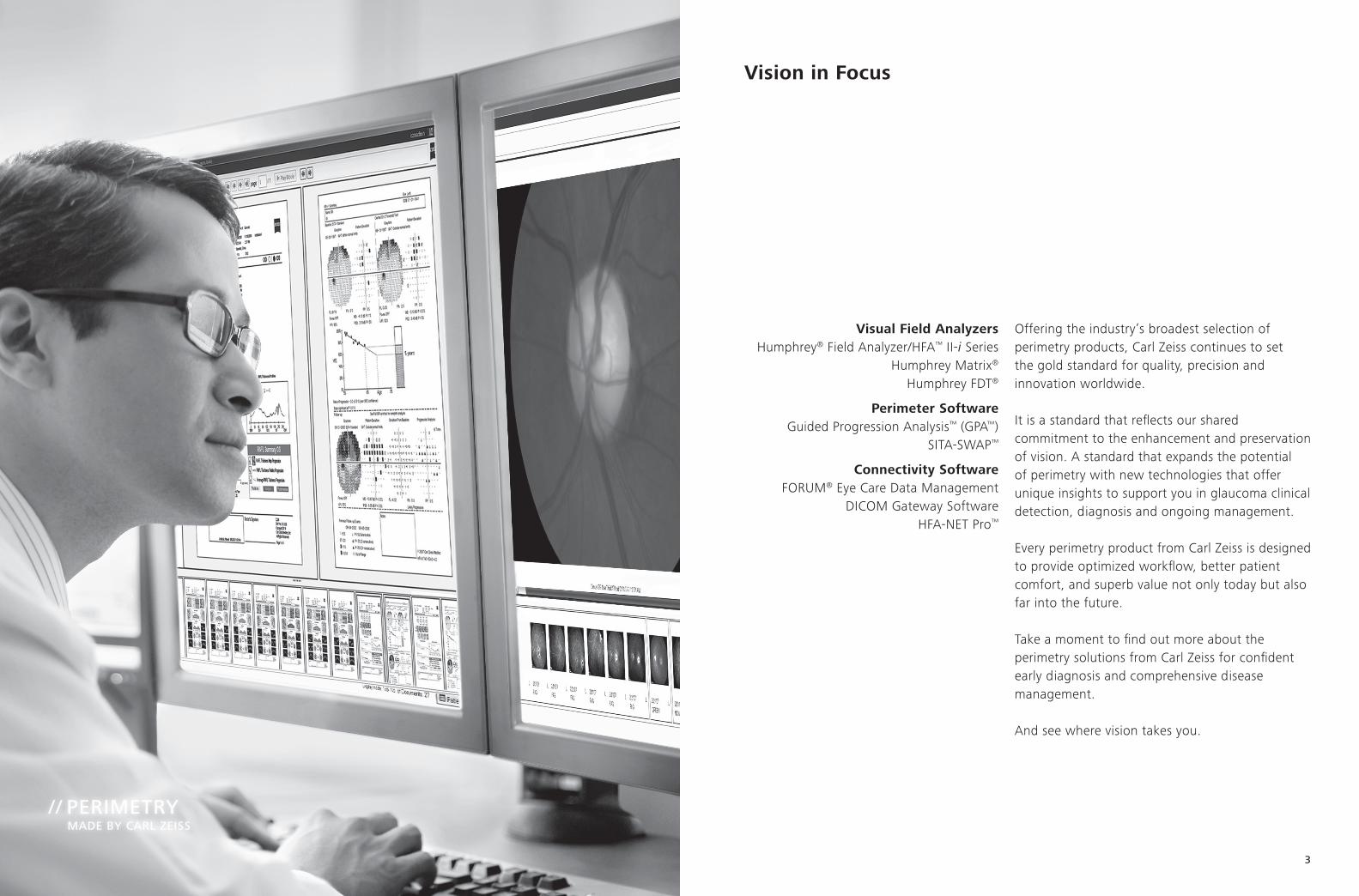
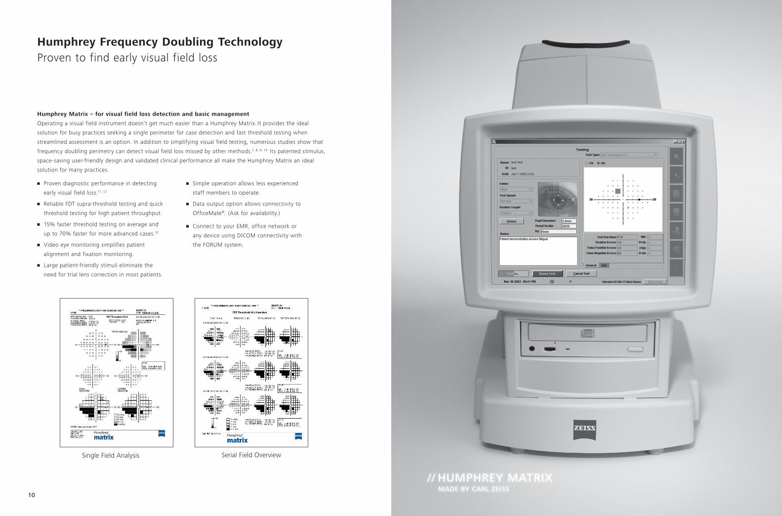
Humphrey Frequency Doubling Technology Proven to find early visual field loss
Humphrey Matrix – for visual field loss detection and basic management
Operating a visual field instrument doesn’t get much easier than a Humphrey Matrix. It provides the ideal
solution for busy practices seeking a single perimeter for case detection and fast threshold testing when
streamlined assessment is an option. In addition to simplifying visual field testing, numerous studies show that
frequency doubling perimetry can detect visual field loss missed by other methods.7, 8, 9, 10 Its patented stimulus,
space-saving user-friendly design and validated clinical performance all make the Humphrey Matrix an ideal
solution for many practices.
Single Field Analysis Serial Field Overview
// HUMPHREY MATRIX MADE BY CARL ZEISS
• Proven diagnostic performance in detecting
early visual field loss.11, 12
• ReliableFDTsupra-thresholdtestingandquick
threshold testing for high patient throughput.
• 15%fasterthresholdtestingonaverageand
upto70%fasterformoreadvancedcases.13
• Videoeyemonitoringsimplifiespatient
alignment and fixation monitoring.
• Largepatient-friendlystimulieliminatethe
need for trial lens correction in most patients.
• Simpleoperationallowslessexperienced
staff members to operate.
• Dataoutputoptionallowsconnectivityto
OfficeMate®. (Ask for availability.)
• ConnecttoyourEMR,officenetworkor
any device using DICOM connectivity with
the FORUM system.

12
Humphrey FDT – for efficient visual field loss detection
Clinically validated
Multiple studies1-15 have shown that the Humphrey FDT detects visual field loss due to a variety of ocular diseases,
including glaucoma. Thus FDT is ideal for clinics desiring to identify patients in need of ophthalmological referral.
• FDTisclinicallyvalidatedinmorethan170peer-reviewedpublications.
Proven performance on virtually all patients
Studies have found that virtually all patients can perform this fast and simple test with reliable results:
• BeijingEyeStudy:98%patientsuccess.14
• TajimiPopulationScreeningStudy:98.7%patientsuccess.15
Easy to operate and interpret
The FDT is optimized for use in non-ophthalmological settings and may be operated by healthcare workers
having little or no specialty training in ophthalmology.
• Simplifiedthreetouchoperation.
• Patientsmaybetestedusingtheirownglasses.
• Shorttest:~40secondspereye.
• Smallfootprint.
• Simplifiedinterpretationofresults.
Both the Matrix and FDT also provide:
• Large,age-relatednormativedatabase.
• Compactdesignthatfitsanywhereinyourpractice.
• Easyandintuitiveoperationforusersofanylevelofexperience.
• Norequirementfortriallensesoreyepatches.*
• Dependableperformanceinambientlight.
*Trial lenses are required beyond ± 3 diopters for the Matrix and beyond ± 7 diopters for the FDT.
Humphrey Frequency Doubling Technology Detect vision loss from ocular diseases
// HUMPHREY FDT MADE BY CARL ZEISS

14
// RELIABILITY MADE BY CARL ZEISS
Technical Data Specifications
Specifications FDT MatrixHFA II-i
720i 740i 745i 750i
Test specifications
Maximum temporal range (degrees) 30 30 89 89 89 89
Stimulus duration 300 ms 300 ms 200 ms 200 ms 200 ms 200 ms
Visual field testing distance Infinity Infinity 30 cm 30 cm 30 cm 30 cm
Backgroundillumination 100 cd/m2 100 cd/m2 31.5ASB 31.5ASB 31.5ASB 31.5ASB
Threshold test library
N-30 • •
C-20 •
24-2, 30-2, 10-2, Macula • • • • •
60-4, Nasal step • • • •
Threshold test strategies
MOBS • •
ZEST •
SITA Standard, SITA Fast, Full Threshold, FastPac • • • •
SITA-SWAP • •
Screening test library
C40, C64, C76, C80, C-Armal • • • •
C-20 •
N-30 • •
24-2 •
Peripheral test patterns • • • •
Screening test modes
Age corrected • • • • • •
Threshold related, Single intensity • • • •
Specialty test library
Social Security Disability, monocular, binocular • • • •
Superior 36, 64 • • • •
Kinetictesting Option Option •
Custom testing • • •

Selected references1.HeijlA,LeskeMC,BengsstonB,HusseinM.MeasuringvisualfieldprogressionintheEarlyManifestGlaucomaTrial.Acta Ophthalmol Scand,
2003 Jun; 81(3):286-293.2.BengtssonB,HeijlA.Avisualfieldindexforcalculationofglaucomarateofprogression;Am J Ophthalmol, 2008 Feb; 145(2): 343-53.3.LeungCK,CheungCY,WeinrebRN,QiuK,LiuS,LiH,XuG,FanN,PangCP,TseKK,LamDS.Evaluationofretinalnervefiberlayerprogression
in glaucoma: a study on optical coherence tomography guided progression analysis. Invest Ophthalmol Vis Sci., 2010 Jan; 51(1):217-22. Epub 2009 Aug 13.
4.BengsstonB,HeijlA.NormalintersubjectthresholdvariabilityandnormallimitsoftheSITASWAPandfullthresholdSWAPperimetricprograms.Invest Ophthalmol Vis Sci., 2003 Nov;44(11):5029-34.
5.BengsstonB.Anewrapidthresholdalgorithmforshort-wavelengthautomatedperimetry.Invest Ophthalmol Vis Sci., 2003 Mar; 44(3):1388-94.6.Casas-LleraP,RebolledaG,Muñoz-NegreteFJ,Arnalich-MontielF,Pérez-LópezM,Fernández-BuenagaR.Visualfieldindexrateandevent-based
glaucoma progression analysis: comparison in a glaucoma population. Br J Ophthalmol. 2009 Dec; 93(12):1576-9. Epub 2009 Jun 16.7. Albanis CV and Quinones RA. Use of Matrix Frequency Doubling Technology (FDT) to Assess Visual Field Status Following Unreliable Standard
Automated Perimetry (SAP). Invest Ophthalmol Vis Sci., 2008 Apr; 49: 1078.8.RacetteL,MedeirosFA,ZangwillLM,etal.DiagnosticaccuracyoftheMatrix24-2andoriginalN-30frequencydoublingtechnologytests
compared with standard automated perimetery. Invest Ophthalmol Vis Sci., 2008; 49: 954-960.9.SamplePA,MedeirosFA,RacetteL,etal.Identifyingglaucomatousvisionlosswithvisual-function-specificperimetryinthediagnosticinnovations
in glaucoma study. Invest Ophthalmol Vis Sci., 2006; 47: 3381-339.10.SamplePA,BosworthCF,BlumenthalEZ,GirkinC,WeinrebRN.Visualfunction-specificperimetryforindirectcomparisonofdifferentganglion
cell populations in glaucoma. Invest Ophthalmol Vis Sci., 2000; 41: 1783-1790.11.MedeirosFA,SamplePA,ZangwillLM,etal.AStatisticalApproachtotheEvaluationofCovariateEffectsontheReceiverOperatingCharacteristic
Curves of Diagnostic Tests in Glaucoma. Invest Ophthalmol Vis Sci., 2006 Jun; 47: 2520-2527.12. Giuffre I. Frequency Doubling Technology vs Standard Automated Perimetry in Ocular Hypertensive Patients. Open Ophthalmol J, 2009 Jan; 3: 6-9.13. Patel A, Wollstein G, Ishikawa H, Schuman J. Comparison of Visual Field Defects Using Matrix Perimetry and Standard Achromatic Perimetry.
Ophthalmology, 2007 Mar; 114(3): 480-487.14.JonasJB,XuL,WangYX,etal.TheBeijingEyeStudy.Acta Ophthalmol. 2009 May; 87(3): 247-61.15. Iwase A et al. Performance of frequency-doubling technology perimetry in a population-based prevalence survey of glaucoma: the Tajimi study.
Ophthalmology. 2007 Jan; 114(1): 27-32. Epub 2006 Oct 27.
Visual Field Analyzers Technical Data
14 15
Specifications FDT MatrixHFA II-i
720i 740i 745i 750i
Test specifications
Maximum temporal range (degrees) 30 30 89 89 89 89
Stimulus duration 300 ms 300 ms 200 ms 200 ms 200 ms 200 ms
Visual field testing distance Infinity Infinity 30 cm 30 cm 30 cm 30 cm
Backgroundillumination 100 cd/m2 100 cd/m2 31.5ASB 31.5ASB 31.5ASB 31.5ASB
Threshold test library
N-30 • •
C-20 •
24-2, 30-2, 10-2, Macula • • • • •
60-4, Nasal step • • • •
Threshold test strategies
MOBS • •
ZEST •
SITA Standard, SITA Fast, Full Threshold, FastPac • • • •
SITA-SWAP • •
Screening test library
C40, C64, C76, C80, C-Armal • • • •
C-20 •
N-30 • •
24-2 •
Peripheral test patterns • • • •
Screening test modes
Age corrected • • • • • •
Threshold related, Single intensity • • • •
Specialty test library
Social Security Disability, monocular, binocular • • • •
Superior 36, 64 • • • •
Kinetictesting Option Option •
Custom testing • • •
Features FDT MatrixHFA II-i
720i 740i 745i 750i
Fixation control
HeijlKrakaublindspotmonitor • • • • • •
Video eye monitor • • • • •
Gaze tracking • • •
Head tracking •
Vertex monitoring •
Remote video eye monitor capability • • • •
Operator interface
Display LCD LCD Touch-screen CRT
Keyboard • • • • •
Stimulus
Frequency doubling • •
White-on-white • • • •
Red- or blue-on-white • • •
Blue-on-yellow(SWAP) • •
General testing features
Stimulus sizes 10° 2°, 5°, 10° Goldmann III
Goldmann I-V
Goldmann I-V
Goldmann I-V
Foveal threshold testing • • •
Automatic Pupil measurement •
Test storage
User-defined • • • • •
Software features
STATPAC 2–single field analysis • • • •
Glaucoma Hemifield Test (GHT) • • • • •
Visual Field Index (VFI) • • • •
Guided Progression Analysis (GPA) • • •
Serial field overview • • • •
Networking • • • •
FORUM Connectivity • • • •
DICOM Connectivity • • • •
EasyConnect RCT / HFA-NET Pro • • • •
Printer
Thermal printer External color printer
NativegenericPCL3,PCL5andpostscript printer support for local, shared and networked printers
Data storage, retrieval and analysis
Hard drive 40GB 40GB 40GB 40GB
USB • • • •
CD-R/W drive •
Dimensions
Height 17” (43 cm) 17” (43 cm) 24” (60 cm)
Width 10” (25 cm) 11” (28 cm) 23” (58 cm)
Depth 19” (48cm) 24” (61cm) 20” (51 cm)
Weight 19 lbs (8.6 kg) 35 lbs (16 kg) 88 lbs (40 kg)
Electrical requirements
100-120 V, 50/60 Hz 230 V, 50/60 Hz
100-240 V, 50/60 Hz 100-120 V, 50/60 Hz
Standards
MeetsUL,CSAandCEstandards • • • • • •

Your local contact:
ArgentinaCarl Zeiss Argentina S.A.Calle Nahuel Huapi 4015 / 25C1430 BCO Buenos AiresArgentinaPhone: +54 11 45 45 66 [email protected]
AustraliaCarl Zeiss Pty. Ltd.Unit 13, 2 Eden Park DriveNorth Ryde, New South Wales 2113AustraliaPhone: +61 2 9020 [email protected]
AustriaCarl Zeiss GmbHLaxenburger Str. 21100 ViennaAustriaPhone: +43 1 79 51 [email protected]
BelgiumCarl Zeiss NV-SAIkaroslaan 491930 ZaventemBelgiumPhone: + 32 2 719 39 [email protected]
BrazilCarl Zeiss do Brasil Ltda.Av. Naçoes Unidas, 21711CEP04795-100 São PauloBrazilPhone: +55 11 5693 [email protected]
CanadaCarl Zeiss Canada Ltd.45 Valleybrook DriveToronto, ON M3B 2S6CanadaPhone: +1 800 387 [email protected]
ChinaCarl Zeiss Shanghai Co. Ltd.1/f., Ke Yuan Building11 Ri Yin Nan RoadWaigaoqiao Free Trade Zone2005 Yang Gao Bei RoadShanghai 200131ChinaPhone: +86 21 5048 17 [email protected]
Czech RepublicCarl Zeiss spol. s.r.o.Radlická 14/3201150 00 Prague 5Czech RepublicPhone: +420 233 101 [email protected]
FranceCarl Zeiss Meditec France SAS60, route de Sartrouville78230 Le PecqFrancePhone: +33 1 34 80 21 [email protected]
GermanyCarl Zeiss Meditec VG mbHCarl-Zeiss-Strasse 2273446 OberkochenGermanyPhone: +49 7364 20 [email protected] Ophthalmology:Phone: +49 800 470 50 [email protected]
Hong KongCarl Zeiss Far East Co. Ltd.Units 11-12. 25/FTower 2, Ever Gain PlazaNo. 88 Container Port RoadKwai ChungHong KongPhone: +852 2332 [email protected]
IndiaCarl Zeiss India Pvt. Ltd.22. Kensington RoadUlsoorBangalore 560 008IndiaPhone: +91 80 2557 88 [email protected]
ItalyCarl Zeiss S.p.A.Viale delle Industrie 2020020 Arese (Milan)ItalyPhone: +39 02 93773 [email protected]
JapanCarl Zeiss Meditec Japan Co. Ltd.Shinjuku KuTokyo 160-000322 Honchio-ChoJapanOphthalmic instruments:Phone: +81 3 33 55 [email protected] instruments:Phone: +81 3 33 55 [email protected]
MalaysiaCarl Zeiss Sdn Bhd.Lot2, Jalan 243/51 A46100 Petaling JayaSelangor Darul EhsanMalaysiaPhone: +60 3 7877 50 [email protected]
MexicoCarl Zeiss de México S.A. de C.V.Avenida Miguel Angel de Quevedo 49604010 Mexico CityMexicoPhone: +52 55 59 99 [email protected]
NetherlandsCarl Zeiss B.V.Trapezium 300Postbus 3103364 DL SliedrechtNetherlandsPhone: +31 184 43 34 [email protected]
New ZealandCarl Zeiss (N.Z.) Ltd.15B Paramount DriveP.O. Box 121 - 1001Henderson, Auckland 0650New ZealandPhone: +64 9 838 [email protected]
PolandCarl Zeiss sp. Z o.o.ul. Lopuszanska 3202-220 WarsawPolandPhone: +48 22 858 [email protected]
SingaporeCarl Zeiss Ptd. Ltd.50 Kaki Bukit PlaceSingapore 415926SingaporePhone: +65 6741 [email protected]
South AfricaCarl Zeiss (Pty.) Ltd.363 Oak AvenueFerndaleRandburg 2194South AfricaPhone: +27 11 886 [email protected]
South KoreaCarl Zeiss Co. Ltd.Seoul 121-828Mapo-gu141-1, Sangsu-dong2F, BR Elitel Bldg.South KoreaPhone: +82 2 3140 [email protected]
SpainCarl Zeiss Meditec Iberia S.A.Ronda de Poniente, 15Tres Cantos28760 MadridSpainPhone: +34 91 203 37 [email protected]
SwedenCarl Zeiss ABTegeluddsvaegen 7610254 StockholmSwedenPhone: +46 84 59 25 [email protected]
SwitzerlandCarl Zeiss AGFeldbachstrasse 818714 FeldbachSwitzerlandPhone: +41 55 254 [email protected]
ThailandCarl Zeiss ThailandFloor 8, Thosapol Land Building 2230 Ratchadapisek RoadHuaykwang, Bangkok 10310ThailandPhone: +66 2 2 74 06 [email protected]
United KingdomCarl Zeiss Ltd.15-20 Woodfield RoadWelwyn Garden CityHertfordshire, AL7 1JQUnited KingdomPhone: +44 1707 [email protected]
United States of AmericaCarl Zeiss Meditec, Inc.5160 Hacienda DriveDublin, CA 94568USAPhone: +1 925 557 [email protected]
Carl Zeiss Meditec Inc.5160 Hacienda DriveDublin, CA 94568USAwww.meditec.zeiss.com/hfa
Carl Zeiss Meditec AGGoeschwitzer Str. 51-5207745 JenaGermany www.meditec.zeiss.com/hfa
0297
Carl Zeiss Meditec AGGoeschwitzer Str. 51-5207745 JenaGermany www.meditec.zeiss.com/forum
HFA 0297 FORUM
SAP
00
00
00
-19
76
-24
1 P
ER.4
00
4
The
cont
ents
of t
he b
roch
ure
may
diff
er fr
om th
e cu
rrent
sta
tus
of a
ppro
val o
f the
pro
duct
in y
our c
ount
ry. P
leas
e co
ntac
t our
regi
onal
repr
esen
tativ
e fo
r mor
e in
form
atio
n. S
ubje
ct to
cha
nge
in
des
ign
and
scop
e of
del
iver
y an
d as
a re
sult
of o
ngoi
ng te
chni
cal d
evel
opm
ent.
Hum
phre
y, HF
A, M
atrix
, FDT
, Gui
ded
Prog
ress
ion
Anal
ysis,
GPA
, HFA
-NET
Pro
, SIT
A-SW
AP, E
asyC
onne
ct,
Visu
al F
ield
Inde
x, V
FI, C
irrus
, GDx
and
FO
RUM
are
eith
er tr
adem
arks
or r
egist
ered
trad
emar
ks o
f Car
l Zei
ss M
edite
c, In
c. in
the
Unite
d St
ates
and
/or o
ther
cou
ntrie
s. A
ll ot
her b
rand
refe
renc
es
are
trade
mar
ks o
r reg
ister
ed tr
adem
arks
of t
heir
resp
ectiv
e co
mpa
nies
in th
e Un
ited
Stat
es a
nd/o
r oth
er c
ount
ries.
© 2
011
by C
arl Z
eiss
Med
itec,
Inc.
All
copy
right
s re
serv
ed. P
rinte
d in
USA
. 101
1