Embed Size (px)
Citation preview
Exon ligation is proofread by the DExD/H-boxATPase Prp22pRabiah M Mayas1, Hiroshi Maita2,3 & Jonathan P Staley2
To produce messenger RNA, the spliceosome excises introns from precursor (pre)-mRNA and splices the flanking exons. Toestablish fidelity, the spliceosome discriminates against aberrant introns, but current understanding of such fidelity mechanismsis limited. Here we show that an ATP-dependent activity represses formation of mRNA from aberrant intermediates havingmutations in any of the intronic consensus sequences. This proofreading activity is disabled by mutations that impair the ATPaseor RNA unwindase activity of Prp22p, a conserved spliceosomal DExD/H-box ATPase. Further, cold-sensitive prp22 mutantspermit aberrant mRNA formation from a mutated 3¢ splice-site intermediate in vivo. We conclude that Prp22p generally repressessplicing of aberrant intermediates, in addition to its known ATP-dependent role in promoting release of genuine mRNA. This dualfunction for Prp22p validates a general model in which fidelity can be enhanced by a DExD/H-box ATPase.
Nuclear pre-mRNA introns are defined by conserved sequences at the5¢ splice site, the branch site and the 3¢ splice site1. These sequencesdetermine the sites of the two chemical reactions in splicing1. First, the2¢ hydroxyl of the branch-site adenosine attacks the 5¢ splice site,forming a lariat intermediate and liberating the 5¢ exon. Second, the3¢ hydroxyl of the 5¢ exon attacks the 3¢ splice site, excising the intronand ligating the exons. The consensus sequences also enable fidelitymechanisms that discriminate against incorrect splice sites, whichdiffer from correct ones in position, sequence or both.
Splicing is catalyzed by the spliceosome1, a ribonucleoproteinmachine composed of more than 100 proteins and five small nuclearRNAs (snRNAs). The snRNAs recognize intronic consensussequences1 and may participate directly in catalysis2. These functionsrequire snRNA rearrangements3 that are promoted by members of theDExD/H-box ATPase family4, which bind ATP to associate with RNAand/or hydrolyze ATP to dissociate RNA-RNA interactions, RNA-protein interactions or both.
A pioneering genetic study5 discovered that the fidelity of branch-site recognition is promoted by the DExD/H-box ATPase Prp16p,which also promotes rearrangement of the spliceosome after 5¢ splice-site cleavage6. prp16 mutants increase the abundance of aberrantbranch-site intermediates, thereby increasing the abundance of aber-rant mRNA. Because these mutants hydrolyze ATP inefficiently5,it was proposed5,7 that Prp16p enhances the fidelity of branch-site recognition by a kinetic proofreading mechanism8,9 in whichPrp16p- and ATP-dependent rejection competes with splicing. Sub-sequent genetic studies have identified additional factors that promotethe fidelity of intron recognition10–19. The activity of these factors canbe understood within a general thermodynamic framework in which
an aberrant substrate equilibrates between the two catalytic conforma-tions of the spliceosome and a fidelity factor favors the conformationthat is inactive for the aberrant substrate species18,20.
These genetic advances in understanding fidelity have raisedimportant questions. Given that Prp16p proofreads 5¢ splice-sitecleavage5,18,19, does an ATPase similarly proofread exon ligation?Given the generality of the thermodynamic framework for under-standing fidelity funcitons20, do additional mechanisms serve fidelity?
Progress has been constrained by the lack of an in vitro assay for thefidelity of splice-site sequence recognition. To investigate fidelityin vitro, we focused on 3¢ splice-site recognition, because it is requiredfor exon ligation (for example, see ref. 21) but dispensable for 5¢splice-site cleavage in budding yeast22 and in some mammalianintrons23. Mutations of the 3¢ splice-site consensus (C/UAG) partiallyimpair exon ligation (for example, see ref. 24), and mutations of theterminal AG severely inhibit exon ligation (for example, see ref. 12).Many genetic studies10–19,25–27 and one biochemical study28 haveimplicated numerous factors in the fidelity of 3¢ splice-site recogni-tion. To date, however, no factor has been shown to enhance fidelitydirectly at the second catalytic stage of splicing by repressing exonligation at 3¢ splice sites that deviate from the consensus.
Using a novel in vitro fidelity assay in Saccharomyces cerevisiae, wehave discovered a role for ATP in repressing exon ligation at mutated3¢ splice sites. We have assigned this role to the conserved DExD/H-box ATPase Prp22p, which is required to release spliced mRNA fromthe spliceosome29,30. Validating these findings, a prp22 mutant sup-presses the exon-ligation defect of a 3¢ splice-site mutant in vivo. Wehave also found in vitro that Prp22p represses exon ligation ofintermediates having an aberrant 5¢ splice site or branch site. Our
Received 18 November 2005; accepted 11 April 2006; published online 7 May 2006; doi:10.1038/nsmb1093
1Department of Biochemistry and Molecular Biology and 2Department of Molecular Genetics and Cell Biology, The University of Chicago, Cummings Life Science Center817, 920 E. 58th Street, Chicago, Illinois 60637, USA. 3Present address: Graduate School of Pharmaceutical Sciences, Hokkaido University, Kita-ku, Sapporo060-0812, Japan. Correspondence should be addressed to J.P.S. ([email protected]).
48 2 VOLUME 13 NUMBER 6 JUNE 2006 NATURE STRUCTURAL & MOLECULAR BIOLOGY
ART IC L E S©
2006
Nat
ure
Pub
lishi
ng G
roup
ht
tp://
ww
w.n
atur
e.co
m/n
smb
work suggests that Prp22p generally enhances the fidelity of exonligation by a proofreading mechanism7–9 in which Prp22p-mediatedrejection of an intermediate competes with exon ligation. Further, ourdata underscore the utility of energy in permitting discriminationbetween correct and nearly-correct substrates8.
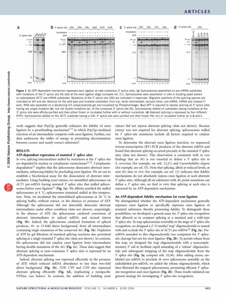
RESULTSATP-dependent repression of mutated 3¢ splice sitesIn vivo, splicing intermediates stalled by mutations at the 3¢ splice siteare degraded by nuclear or cytoplasmic exonucleases31,32. Cytoplasmicdegradation32 implies that the spliceosome dissociates aberrant inter-mediates, enhancing fidelity by precluding exon ligation. We set out toestablish a biochemical assay for the dissociation of aberrant inter-mediates. Using budding yeast extract, we assembled spliceosomes onACT1 pre-mRNA having mutated 3¢ splice sites that stalled spliceo-somes before exon ligation21 (Fig. 1a). We affinity-purified the stalledspliceosomes at 4 1C; spliceosomes remained stalled at this tempera-ture. Next, we incubated the immobilized spliceosomes at 20 1C insplicing buffer, without extract, in the absence or presence of ATP.Although the spliceosome did not detectably dissociate aberrantintermediates under either condition (data not shown), surprisingly,in the absence of ATP, the spliceosome catalyzed conversion ofaberrant intermediates to spliced mRNA and excised intron(Fig. 1b). Indeed, the spliceosome catalyzed formation of splicedproducts, 10- to 15-fold above background, from all intermediatescontaining single mutations of the conserved AG (Fig. 1b). Depletionof ATP by gel filtration of standard splicing reactions also permittedsplicing at a singly mutated 3¢ splice site (data not shown). In contrast,the spliceosome did not catalyze exon ligation from intermediateshaving double mutations of the AG (Fig. 1c). These data suggest thataberrant splicing at near-consensus 3¢ splice sites is repressed by anATP-dependent mechanism.
Indeed, aberrant splicing was repressed efficiently in the presenceof ATP, which reduced mRNA abundance to less than two-foldabove background (Fig. 1b). GTP, CTP and UTP also repressedaberrant splicing efficiently (Fig. 1d), implicating a nonspecificNTPase (see below). In contrast, the addition of budding yeast
extract did not repress aberrant splicing (data not shown). Becauseextract was not required for aberrant splicing, spliceosomes stalledby 3¢ splice-site mutations include all factors required to catalyzeexon ligation.
To determine the aberrant exon ligation junction, we sequencedreverse-transcription (RT)-PCR products of the aberrant mRNA andfound that aberrant splicing occurred precisely at the mutated 3¢ splicesites (data not shown). This observation is consistent with in vivofindings that an AG is not essential to define a 3¢ splice site inS. cerevisiae (for example, see refs. 12,21) and Caenorhabditis elegans(for example, see ref. 33). Note that splicing, albeit at reduced levels, atnon-AG sites in vivo (for example, see ref. 12) indicates that fidelitymechanisms do not absolutely repress exon ligation at such aberrant3¢ splice sites. Although all six substrates with AG point mutations candefine a 3¢ splice site, we find in vitro that splicing at such sites isrepressed by an ATP-dependent mechanism.
An ATP-dependent fidelity mechanism for exon ligationWe distinguished whether the ATP-dependent mechanism generallyrepresses exon ligation or specifically represses exon ligation ofmutated substrates, thereby promoting fidelity. To distinguish thesepossibilities, we developed a general assay for 3¢ splice-site recognitionthat allowed us to compare splicing at a mutated and a wild-type3¢ splice site. To trap spliceosomes reversibly at the stage of 3¢ splice-siterecognition, we designed a 2¢-O-methyl ‘trap’ oligonucleotide to annealwith and occlude the 3¢ splice site of ACT1 pre-mRNA34 (Fig. 2a). Pre-mRNA annealed to this oligonucleotide was competent for 5¢ splice-site cleavage but not for exon ligation (Fig. 2b). To permit release fromthis trap, we designed the trap oligonucleotide with a noncomple-mentary 5¢ tail to facilitate rapid annealing of a ‘release’ oligonucleo-tide and subsequent stripping of the trap oligonucleotide from the3¢ splice site (Fig. 2a; compare refs. 35,36). After adding excess, un-labeled pre-mRNA to preclude de novo spliceosome assembly on theradiolabeled pre-mRNA, we added the release oligonucleotide, whichthen liberated the trapped spliceosomes, promoting efficient 3¢ splice-site recognition and exon ligation (Fig. 2b). These results validated ourgeneral strategy for investigating 3¢ splice-site recognition.
a c db
3′ splice site
3′ splice site 3′ splice site 3′ splice site
UAGUAG
UcGUAc
UAc UAa UAu UgG UcG UuG UAc Ugc Ucu Ugu UAc
IncubationATP
– + + + + + ++ + + +
+ + ++
++++– –
Incubation
ATP
– + +
+– –
– + +
+– –
– + +
+– –
– + +
+– –– – – – –– –
– – ––
––––
1 2 3 41 2 3 4 5 6 7 8 9 10 11 12 1 2 3 4 5 6 1 2 3 4 5 67 8 9 10 11 1213 14 15 16 17 18
UT
PC
TP
GT
PAT
P
No
inc
–
Lane Lane LaneLane
Figure 1 An ATP-dependent mechanism represses exon ligation at near-consensus 3¢ splice sites. (a) Spliceosomes assembled on pre-mRNA substrates
with mutations of the 3¢ splice site AG stall at the exon-ligation stage (compare ref. 21). Spliceosomes were assembled in vitro in budding yeast extract
on radiolabeled ACT1 pre-mRNA substrates. Mutations of the 3¢ splice site UAG are indicated in lowercase. Migration positions of the splicing species are
indicated at left and are identical for the wild-type and mutated substrates: from top, lariat intermediate, excised intron, pre-mRNA, mRNA and cleaved 5¢exon. RNA was separated on a denaturing 6% polyacrylamide gel and visualized by PhosphorImager. (b,c) ATP is required to repress splicing at 3¢ splice sites
having any single mutation (b), but not double mutations (c), of the conserved 3¢ splice site AG. Spliceosomes stalled on substrates having mutations at the
3¢ splice site were affinity-purified and then either frozen or incubated further with or without nucleotide. (d) Aberrant splicing is repressed by four different
NTPs. Spliceosomes stalled on the ACT1 substrate having a UAc 3¢ splice site were purified and then frozen (No inc) or incubated further as in b and c.
ART IC L E S
NATURE STRUCTURAL & MOLECULAR BIOLOGY VOLUME 13 NUMBER 6 JUNE 2006 4 8 3
©20
06 N
atur
e P
ublis
hing
Gro
up
http
://w
ww
.nat
ure.
com
/nsm
b
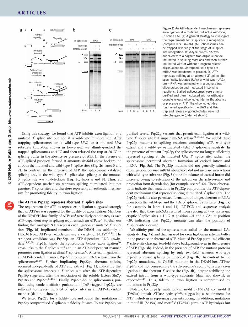
Using this strategy, we found that ATP inhibits exon ligation at amutated 3¢ splice site but not at a wild-type 3¢ splice site. Aftertrapping spliceosomes on a wild-type UAG or a mutated UAcsubstrate (mutation shown in lowercase), we affinity-purified thetrapped spliceosomes at 4 1C and then released the trap at 20 1C insplicing buffer in the absence or presence of ATP. In the absence ofATP, spliced products formed at amounts six-fold above backgroundat both the mutated and wild-type 3¢ splice sites (Fig. 2c, lanes 3 and7). In contrast, in the presence of ATP, the spliceosome catalyzedsplicing only at the wild-type 3¢ splice site; splicing at the mutated3¢ splice site was undetectable (Fig. 2c, lanes 4 and 8). Thus, anATP-dependent mechanism represses splicing at mutated, but notgenuine, 3¢ splice sites and therefore represents an authentic mechan-ism for promoting fidelity in exon ligation.
The ATPase Prp22p represses aberrant 3¢ splice sitesThe requirement for ATP to repress exon ligation suggested stronglythat an ATPase was required for the fidelity of exon ligation. Membersof the DExD/H-box family of ATPases4 were likely candidates, as eachATP-dependent step in splicing requires such an ATPase3. Further, ourfinding that multiple NTPs repress exon ligation at mutated 3¢ splicesites (Fig. 1d) implicated members of the DEAH-box subfamily ofDExD/H-box ATPases, which can use a variety of NTPs6,37,38. Thestrongest candidate was Prp22p, an ATP-dependent RNA unwin-dase29,30,38. Prp22p binds the spliceosome before exon ligation30,cross-links to the 3¢ splice site39 and, in an ATP-independent manner,promotes exon ligation at distal 3¢ splice sites30. After exon ligation, inan ATP-dependent manner, Prp22p promotes mRNA release from thespliceosome29,30. Further implicating Prp22p, aberrant splicingoccurred independently of ATP and extract (Fig. 1), indicating thatthe spliceosome inspects a 3¢ splice site after the ATP-dependentPrp16p stage and after the association of the soluble factors Slu7p,Prp18p and Prp22p30,40,41. Finally, Prp22p-bound spliceosomes, pur-ified using tandem affinity purification (TAP)-tagged Prp22p, aresufficient to repress mutated 3¢ splice sites in an ATP-dependentmanner (data not shown).
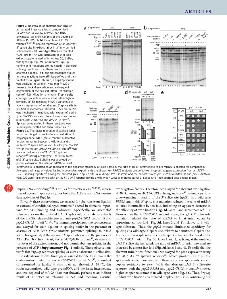
We tested Prp22p for a fidelity role and found that mutations inPrp22p compromised 3¢ splice-site fidelity in vitro. To test Prp22p, we
purified several Prp22p variants that permit exon ligation at a wild-type 3¢ splice site but impair mRNA release30,42–44. We added thesePrp22p mutants to splicing reactions containing ATP, wild-typeextract and a wild-type or mutated (UAc) 3¢ splice-site substrate. Inthe presence of mutant Prp22p, the spliceosome no longer efficientlyrepressed splicing at the mutated UAc 3¢ splice site; rather, thespliceosome permitted aberrant formation of excised intron andmRNA (Fig. 3a). The Prp22p mutants did not generally stimulateexon ligation, because mRNA abundance did not increase in reactionswith wild-type substrate (Fig. 3a); the abundance of excised intron didincrease, owing to retention of the intron on the spliceosome andprotection from degradation (for example, see ref. 42). These observa-tions indicate that mutations in Prp22p compromise the ATP-depen-dent mechanism that represses splicing at mutated 3¢ splice sites. ThePrp22p variants also permitted formation of longer, aberrant mRNAsfrom both the wild-type and the UAc 3¢ splice-site substrates (Fig. 3a;for example, in lanes 4 and 11). RT-PCR and sequence analysisrevealed that these mRNAs resulted from splicing at two upstream,cryptic 3¢ splice sites, a UuG at position –21 and a CAu at position–29, indicating that Prp22p mutants can alter the position of3¢ splice-site cleavage.
We affinity-purified the spliceosomes stalled on the mutated UAcsubstrate (Fig. 3a) and then assayed for exon ligation in splicing bufferin the presence or absence of ATP. Mutated Prp22p permitted efficient3¢ splice-site cleavage, ten-fold above background, even in the presenceof ATP (Fig. 3b). Indeed, in the presence of ATP, the mutant proteinsrepressed aberrant splicing by only two-fold, whereas wild-typePrp22p repressed splicing by nine-fold (Fig. 3b). In contrast to thePrp22p mutations, the Q423E mutation in the DEAH-box ATPasePrp43p did not compromise the spliceosome’s ability to repress exonligation at the aberrant 3¢ splice site (Fig. 3b), despite stabilizing theexcised intron from a wild-type substrate (data not shown), asexpected45,46. Thus, fidelity in exon ligation is compromised bymutations in Prp22p.
Notably, the Prp22p mutations in motif I (K512A) and motif II(D603A) impair ATPase activity30,43, suggesting a requirement forNTP hydrolysis in repressing aberrant splicing. In addition, mutationsin motif III (S635A) and motif V (T765A) permit ATP hydrolysis but
UAG3′ splice site
5′
5′
5′
A
A
A
A
Incubatein exact
Add excessunlabeled
pre-mRNA
Add releaseoligo
Incubate± ATP
Releaseoligo
3′
3′3′
3′5′
5′
5′5′3′
3′
5′
5′
3′A
Annealtrap oligo Trap oligo
UAG
UAG
UAG
UAG
UAG
3′
3′
Trap oligoRelease oligo
3′ splice site
ATP
Release oligo– + – +
– +
UAc UAG
– +
– +
– + – ++
– – +
1 2 3 1 2 3 4 5 6 7 8
a b c
Lane Lane
Figure 2 An ATP-dependent mechanism represses
exon ligation at a mutated, but not a wild-type,
3¢ splice site. (a) A general strategy to investigate
the requirements for 3¢ splice-site recognition
(compare refs. 34–36). (b) Spliceosomes can
be trapped reversibly at the stage of 3¢ splice-
site recognition. Wild-type pre-mRNA was
annealed with a cognate trap oligonucleotide,
incubated in splicing reactions and then further
incubated with or without a cognate release
oligonucleotide. Untrapped, wild-type pre-
mRNA was incubated in parallel. (c) ATP
represses splicing at an aberrant 3¢ splice site
specifically. Mutated (UAc) or wild-type (UAG)
pre-mRNA was annealed with a cognate trapoligonucleotide and incubated in splicing
reactions. Stalled spliceosomes were affinity-
purified and then incubated with or without a
cognate release oligonucleotide, in the absence
or presence of ATP. The oligonucleotides
functioned specifically; the UAG and UAc
trap and release oligonucleotides were not
interchangeable (data not shown).
ART IC L E S
48 4 VOLUME 13 NUMBER 6 JUNE 2006 NATURE STRUCTURAL & MOLECULAR BIOLOGY
©20
06 N
atur
e P
ublis
hing
Gro
up
http
://w
ww
.nat
ure.
com
/nsm
b
impair RNA unwinding42,44. Thus, as for mRNA release29,30,42, repres-sion of aberrant splicing requires both the ATPase and RNA unwin-dase activities of Prp22p.
To verify these observations, we assayed for aberrant exon ligationin extracts of conditional prp22 mutants30 altered in domains impor-tant for ATP binding and hydrolysis4. Specifically, we assembledspliceosomes on the mutated UAc 3¢ splice-site substrate in extractsof the mRNA release–defective mutants prp22-H606A (motif II) andprp22-G810A (motif VI)42. We immunoprecipitated the spliceosomesand assayed for exon ligation in splicing buffer in the presence orabsence of ATP. Both prp22 mutants permitted splicing, four-foldabove background, at the aberrant 3¢ splice site even in the presence ofATP (Fig. 3c). In contrast, the prp43-Q423N mutant47, defective inturnover of the excised intron, did not permit aberrant splicing in thepresence of ATP (Supplementary Fig. 1 online). These observationsverify that Prp22p represses splicing in vitro at aberrant 3¢ splice sites.
To validate our in vitro findings, we assayed for fidelity in vivo in thecold-sensitive mutant strain prp22-R805A (motif VI)42, a mutantcompromised for fidelity in vitro (data not shown). At 16 1C, thisstrain accumulated wild-type pre-mRNA and the lariat intermediateand was depleted of mRNA (data not shown), perhaps as an indirectresult of a defect in releasing mRNA and recycling essential
exon-ligation factors. Therefore, we assayed for aberrant exon ligationat 30 1C, using an ACT1-CUP1 splicing substrate48 having a pyrimi-dine-guanine mutation of the 3¢ splice site (gAG). In a wild-typePRP22 strain, this 3¢ splice-site mutation reduced the ratio of mRNAto lariat intermediate by ten-fold, indicating an apparent decrease inthe efficiency of exon ligation (Fig. 3d, lanes 1 and 3; compare ref. 13).However, in the prp22-R805A mutant strain, the gAG 3¢ splice-sitemutation reduced the ratio of mRNA to lariat intermediate byapproximately two-fold (Fig. 3d, lanes 2 and 4), relative to a wild-type substrate. Thus, the prp22 mutant diminished specificity forsplicing at a wild-type 3¢ splice site, relative to a mutated 3¢ splice site.Further, whereas splicing at the wild-type 3¢ splice site decreased in theprp22-R805A mutant (Fig. 3d, lanes 1 and 2), splicing at the mutatedgAG 3¢ splice site increased; the ratio of mRNA to lariat intermediateincreased by almost five-fold (Fig. 3d, lanes 3 and 4). To verify that theaberrant mRNA was functional, we assayed for gene expression usingthe ACT1-CUP1 splicing reporter48, which produces Cup1p in asplicing-dependent manner and thereby confers splicing-dependentcopper resistance to yeast. With the aberrant gAG 3¢ splice-sitereporter, both the prp22-R805A and prp22-G810A mutants42 showedhigher copper resistance than wild-type yeast (Fig. 3e). Thus, Prp22pinhibits exon ligation at a mutated 3¢ splice site in vivo, confirming our
Figure 3 Repression of aberrant exon ligation
at mutated 3¢ splice sites is compromised
in vitro and in vivo by ATPase- and RNA
unwindase–deficient variants of the DEAH-box
ATPase Prp22p. (a,b) Recombinant Prp22p
variants30,42–44 abolish repression of an aberrant
3¢ splice site in extract (a) or in affinity-purified
spliceosomes (b). Wild-type (UAG) or mutated
(UAc) pre-mRNA was incubated in wild-type
extract supplemented with nothing (–), buffer,
wild-type Prp22p (WT) or mutated Prp22p
(amino acid mutations are indicated) in standard
splicing reactions. In a, these reactions were
analyzed directly; in b, the spliceosomes stalled
in these reactions were affinity-purified and thentreated as in Figure 1b. In b, a Prp43p variant
was analyzed in parallel. Note that Prp22p
variants block dissociation and subsequent
degradation of the excised intron (for example,
see ref. 42). Migration of cryptic 3¢ splice-site
cleavage products is indicated at left as lighter
symbols. (c) Endogenous Prp22p variants also
abolish repression of an aberrant 3¢ splice site in
purified spliceosomes. Mutated (UAc) pre-mRNA
was incubated in reactions with extract of a wild-
type PRP22 strain and the cold-sensitive mutant
strains prp22-H606A and prp22-G810A42.
Spliceosomes stalled in these reactions were
immunoprecipitated and then treated as in
Figure 1b. The faster migration of excised lariat
intron in this gel is due to the concentration of
polyacrylamide. (d) A prp22 mutant is defective
in discriminating between a wild-type and amutated 3¢ splice site in vivo. A wild-type PRP22
(W) or the mutant prp22-R805A (R) strain42 was
transformed with an ACT1-CUP1 splicing
reporter48 having a wild-type UAG or mutated
gAG 3¢ splice site. Splicing was analyzed by
primer extension. The ratio of mRNA to lariat
intermediate is charted as an indicator of the apparent efficiency of exon ligation; the ratio of lariat intermediate to pre-mRNA is charted for comparison.
Averages and range of values for two independent experiments are shown. (e) PRP22 mutants are defective in repressing gene expression from an ACT1-
CUP1 splicing reporter48 having the mutated gAG 3¢ splice site. A wild-type PRP22 strain and the mutant strains prp22-R805A (R805A) and prp22-G810A
(G810A) were transformed with an ACT1-CUP1 reporter having a wild-type (UAG) or mutated (gAG) 3¢ splice site, then spotted onto copper plates.
Prp22p
Prp22p
UAG
UAG
W0
2.0
4.0
0
1.0
2.0
3.0
1
ATPIncubation
SplicingW
TH60
6A
G810A
UAc
extract
3′ splice site
ATPIncubation
Recombinantprotein Buf
fer
WT
UAc
K512A
D603A
S635A
T765A Prp
43-
Q423E
3′ splice site3′ splice site
2 3 4 5 6 7 8 9
1 2 3 4 5 6 7 8 9 10 1112 13 141
RecombinantPrp22p
Lane
Lane
Lane
Lane
UAG UAc
I Buf
fer
WT
K51
2AD
603A
S63
5AT
765A
I Buf
fer
WT
K51
2AD
603A
S63
5AT
765A
2 3 4 5 6 7 8 9 10 111213 14151617 18 19 20 21
1 2 3 4
6.00
1.2
0.8
0.4
0
2.0
4.0
6.0
R
W R W R
W R
gAG
gAG
3′ splice site
3′ splice site
Cu2+
(mM)Prp22
WT
WT
0.0
0.2
0.0
0.2
0.8
UAG gAG
pACT1-CUP1
0.4 0.8 0.4
WT
R805A
G810A
WT
G810A
A600
R805A
– + +– – + – – + – – +
– + + – + +
– + +– – +
– + +– – +
– + +– – +
– + +– – +
– + +– – +
– + +– – +
– + +– – +
a
c d e
b
ART IC L E S
NATURE STRUCTURAL & MOLECULAR BIOLOGY VOLUME 13 NUMBER 6 JUNE 2006 4 8 5
©20
06 N
atur
e P
ublis
hing
Gro
up
http
://w
ww
.nat
ure.
com
/nsm
b
finding of a novel role for Prp22p in repressing exon ligation ofaberrant 3¢ splice-site intermediates.
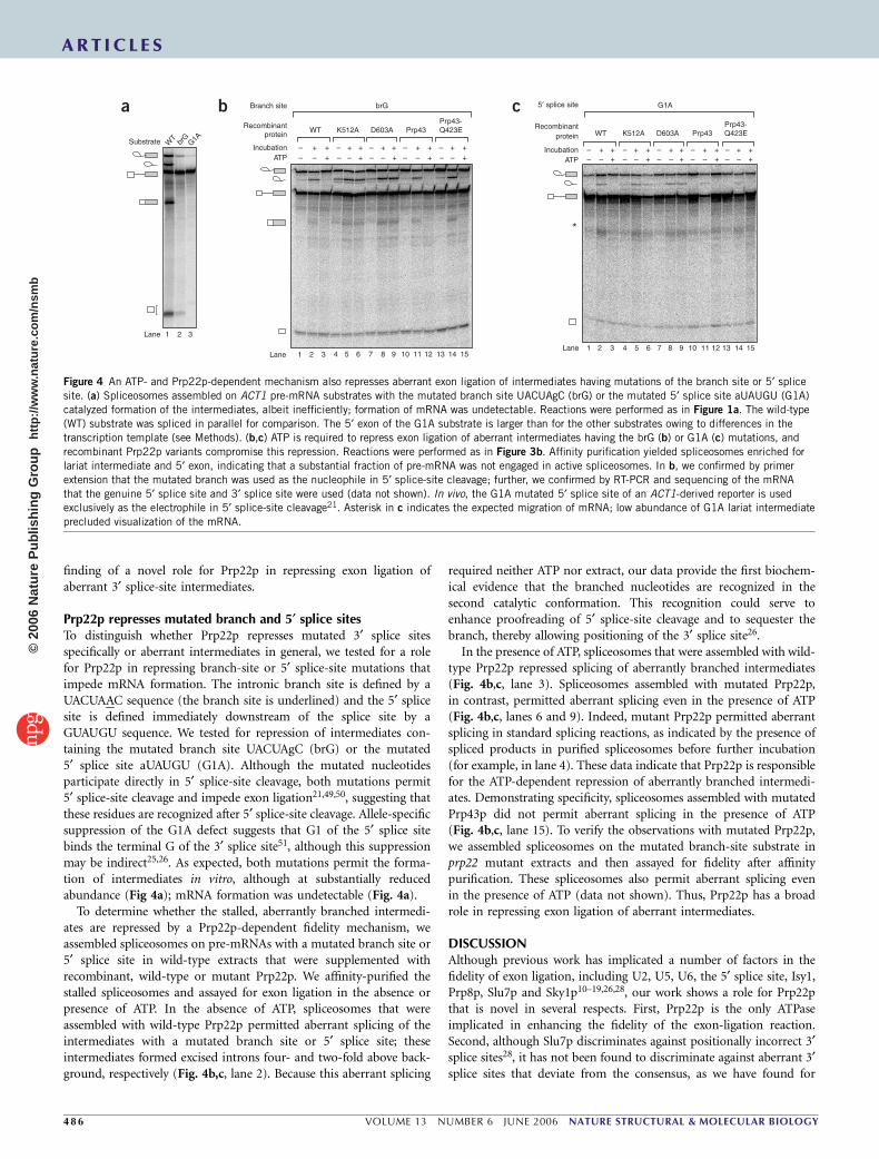
Prp22p represses mutated branch and 5¢ splice sitesTo distinguish whether Prp22p represses mutated 3¢ splice sitesspecifically or aberrant intermediates in general, we tested for a rolefor Prp22p in repressing branch-site or 5¢ splice-site mutations thatimpede mRNA formation. The intronic branch site is defined by aUACUAAC sequence (the branch site is underlined) and the 5¢ splicesite is defined immediately downstream of the splice site by aGUAUGU sequence. We tested for repression of intermediates con-taining the mutated branch site UACUAgC (brG) or the mutated5¢ splice site aUAUGU (G1A). Although the mutated nucleotidesparticipate directly in 5¢ splice-site cleavage, both mutations permit5¢ splice-site cleavage and impede exon ligation21,49,50, suggesting thatthese residues are recognized after 5¢ splice-site cleavage. Allele-specificsuppression of the G1A defect suggests that G1 of the 5¢ splice sitebinds the terminal G of the 3¢ splice site51, although this suppressionmay be indirect25,26. As expected, both mutations permit the forma-tion of intermediates in vitro, although at substantially reducedabundance (Fig 4a); mRNA formation was undetectable (Fig. 4a).
To determine whether the stalled, aberrantly branched intermedi-ates are repressed by a Prp22p-dependent fidelity mechanism, weassembled spliceosomes on pre-mRNAs with a mutated branch site or5¢ splice site in wild-type extracts that were supplemented withrecombinant, wild-type or mutant Prp22p. We affinity-purified thestalled spliceosomes and assayed for exon ligation in the absence orpresence of ATP. In the absence of ATP, spliceosomes that wereassembled with wild-type Prp22p permitted aberrant splicing of theintermediates with a mutated branch site or 5¢ splice site; theseintermediates formed excised introns four- and two-fold above back-ground, respectively (Fig. 4b,c, lane 2). Because this aberrant splicing
required neither ATP nor extract, our data provide the first biochem-ical evidence that the branched nucleotides are recognized in thesecond catalytic conformation. This recognition could serve toenhance proofreading of 5¢ splice-site cleavage and to sequester thebranch, thereby allowing positioning of the 3¢ splice site26.
In the presence of ATP, spliceosomes that were assembled with wild-type Prp22p repressed splicing of aberrantly branched intermediates(Fig. 4b,c, lane 3). Spliceosomes assembled with mutated Prp22p,in contrast, permitted aberrant splicing even in the presence of ATP(Fig. 4b,c, lanes 6 and 9). Indeed, mutant Prp22p permitted aberrantsplicing in standard splicing reactions, as indicated by the presence ofspliced products in purified spliceosomes before further incubation(for example, in lane 4). These data indicate that Prp22p is responsiblefor the ATP-dependent repression of aberrantly branched intermedi-ates. Demonstrating specificity, spliceosomes assembled with mutatedPrp43p did not permit aberrant splicing in the presence of ATP(Fig. 4b,c, lane 15). To verify the observations with mutated Prp22p,we assembled spliceosomes on the mutated branch-site substrate inprp22 mutant extracts and then assayed for fidelity after affinitypurification. These spliceosomes also permit aberrant splicing evenin the presence of ATP (data not shown). Thus, Prp22p has a broadrole in repressing exon ligation of aberrant intermediates.
DISCUSSIONAlthough previous work has implicated a number of factors in thefidelity of exon ligation, including U2, U5, U6, the 5¢ splice site, Isy1,Prp8p, Slu7p and Sky1p10–19,26,28, our work shows a role for Prp22pthat is novel in several respects. First, Prp22p is the only ATPaseimplicated in enhancing the fidelity of the exon-ligation reaction.Second, although Slu7p discriminates against positionally incorrect 3¢splice sites28, it has not been found to discriminate against aberrant 3¢splice sites that deviate from the consensus, as we have found for
– – –+ +– – + – – + – – + – – + – – +
+ + – + ++ + – + +
5′ splice site
Lane 1 2 3 4 5 6 7 8 9 10 11 12 13 14 15
Recombinant
IncubationATP
proteinSubstrate W
T brG
G1A
Lane 1 2 3
WT
– – –+ +– – + – – + – – + – – + – – +
+ + – + ++ – + +
K512APrp43-Q423ED603A Prp43 WT K512A
Prp43-Q423ED603A Prp43
Branch site
Recombinantprotein
IncubationATP
Lane 1 2 3 4 5 6 7 8 9 10 11 12 13 14 15
+
G1A
*
brGa b c
Figure 4 An ATP- and Prp22p-dependent mechanism also represses aberrant exon ligation of intermediates having mutations of the branch site or 5¢ splice
site. (a) Spliceosomes assembled on ACT1 pre-mRNA substrates with the mutated branch site UACUAgC (brG) or the mutated 5¢ splice site aUAUGU (G1A)
catalyzed formation of the intermediates, albeit inefficiently; formation of mRNA was undetectable. Reactions were performed as in Figure 1a. The wild-type
(WT) substrate was spliced in parallel for comparison. The 5¢ exon of the G1A substrate is larger than for the other substrates owing to differences in the
transcription template (see Methods). (b,c) ATP is required to repress exon ligation of aberrant intermediates having the brG (b) or G1A (c) mutations, and
recombinant Prp22p variants compromise this repression. Reactions were performed as in Figure 3b. Affinity purification yielded spliceosomes enriched for
lariat intermediate and 5¢ exon, indicating that a substantial fraction of pre-mRNA was not engaged in active spliceosomes. In b, we confirmed by primer
extension that the mutated branch was used as the nucleophile in 5¢ splice-site cleavage; further, we confirmed by RT-PCR and sequencing of the mRNA
that the genuine 5¢ splice site and 3¢ splice site were used (data not shown). In vivo, the G1A mutated 5¢ splice site of an ACT1-derived reporter is usedexclusively as the electrophile in 5¢ splice-site cleavage21. Asterisk in c indicates the expected migration of mRNA; low abundance of G1A lariat intermediate
precluded visualization of the mRNA.
ART IC L E S
48 6 VOLUME 13 NUMBER 6 JUNE 2006 NATURE STRUCTURAL & MOLECULAR BIOLOGY
©20
06 N
atur
e P
ublis
hing
Gro
up
http
://w
ww
.nat
ure.
com
/nsm
b
Prp22p (Figs. 1–3). Third, although mutations in the invariant loop Iof U5 snRNA suppress exon ligation of aberrant intermediates10, thesemutations establish complementarity between the loop and the firsttwo bases of the 3¢ exon, a complementarity that is not conserved.Thus, it is unclear whether wild-type U5 has a role in the fidelity ofexon ligation. In contrast to the U5 suppressor mutations, the prp22suppressor mutations represent general, loss-of-function muta-tions30,42,52, indicating that wild-type Prp22p does have a role infidelity. Fourth, U2 and sky1 suppressors12,15,17 have not been thor-oughly explored. Finally, U6, 5¢ splice site, prp8 and isy1 suppressorsseem to act at an earlier stage than prp22 suppressors18,19,26.
Whereas U6, 5¢ splice site, prp8 and isy1 mutants suppress exon-ligation defects indirectly at the first catalytic stage of splicing18,19,26,prp22 mutants suppress such defects directly at the second catalyticstage, according to three lines of evidence. First, suppression of exon-ligation defects by the former mutants correlates with defects in 5¢splice-site cleavage18,19,26; in contrast, suppression of such defects byprp22 mutants (Figs. 3 and 4) correlates with defects in mRNArelease42–44,52. Second, prp22 mutants suppress exon-ligation defectsin purified spliceosomes in the absence of budding yeast extract(Figs. 3 and 4), indicating that prp22 mutants act after the associationof exon-ligation factors and after the earlier Prp16p-dependentrearrangement6,30,40,41. Third, prp22 mutants can suppress an aberrantsubstrate that stalls at the exon-ligation stage, because the aberrant,near-consensus UAc 3¢ splice-site intermediate stalls at this stage6.Prp22p does not seem to act in fidelity after exon ligation, as it does inmRNA release, because ATP does not reverse the formation ofaberrant mRNA observed in Figure 1b (data not shown). Thus,
Prp22p performs a previously undescribed ATP-dependent proof-reading function in splicing at the stage of exon ligation. Prp22pand its role in mRNA release are conserved in humans53, suggestingthat its role in fidelity is also conserved.
Although the role of Prp22p is unlike those of other fidelity factors,the prp22 suppressors could function similarly to the U6, 5¢ splice site,prp8 and isy1 suppressors. According to a general model for suppres-sion of substrate mutations18, the U6, 5¢ splice site, prp8 and isy1mutants suppress exon-ligation defects by altering an equilibriumbetween the first and second catalytic conformations of the spliceo-some18,19,26. Specifically, these suppressors destabilize the first catalyticconformation of the spliceosome, thereby suppressing exon-ligationdefects by favoring the second catalytic conformation. According tothe model, prp22 suppressors could also alter the equilibrium, but bystabilizing the second conformation. In this view, wild-type Prp22pdestabilizes the second catalytic conformation. Indeed, Prp22p may berequired to establish the equilibrium, promoting retrograde transitionfrom the second catalytic conformation to the first (Fig. 5), counter-acting Prp16p. Prp22p interacts with Prp16p in a two-hybrid analy-sis54, suggesting that these two factors may cooperate to promote theequilibrium. Consistent with a reversible role for Prp22p, rejection ofan aberrant intermediate by Prp22p does not commit the intermediateto discard, because aberrant splicing can be derepressed by depletingATP (Figs. 1 and 4, and data not shown). Supporting a general role forPrp22p in destabilizing the second catalytic conformation, Prp22puses ATP to repress exon ligation for a broad range of aberrantintermediates (Figs. 3 and 4).
In the simplest form of the general equilibrium model, specificity ofthe spliceosome for a genuine substrate over an aberrant substratewould be determined by the differences in the relative, inherentstabilities of the first and second catalytic conformations. However,a simple equilibrium is not likely to account fully for fidelity insplicing. In other processes, energy is required to discriminate betweengenuine substrates and similar but incorrect substrates8. In splicing,genetic studies have implicated ATP in fidelity5. Our biochemicalstudies have demonstrated directly for the first time a requirement for
Spliceosomeassembly
and activation
First catalyticconformation
Second catalyticconformation
5′ splice sitecleavage
ATP
ADP
Prp22p
Prp22pPrp22p
Kexon ligation
Krejection
Prp16p
ATP
ADPPrp22p
ATP
ADPPrp22p
Prp22p Prp22p
Discard
1
2
3
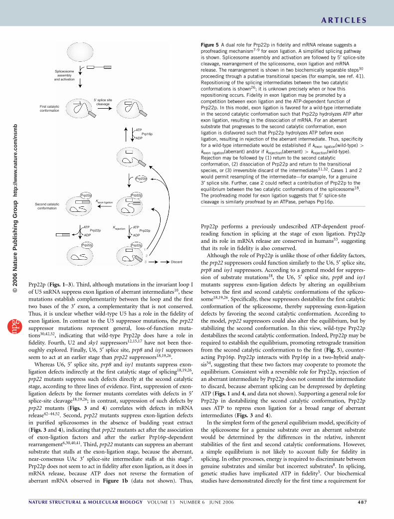
Figure 5 A dual role for Prp22p in fidelity and mRNA release suggests a
proofreading mechanism7–9 for exon ligation. A simplified splicing pathway
is shown. Spliceosome assembly and activation are followed by 5¢ splice-site
cleavage, rearrangement of the spliceosome, exon ligation and mRNA
release. The rearrangement is shown in two biochemically separable steps30
proceeding through a putative transitional species (for example, see ref. 41).
Repositioning of the splicing intermediates between the two catalytic
conformations is shown26; it is unknown precisely when or how this
repositioning occurs. Fidelity in exon ligation may be promoted by a
competition between exon ligation and the ATP-dependent function of
Prp22p. In this model, exon ligation is favored for a wild-type intermediate
in the second catalytic conformation such that Prp22p hydrolyzes ATP after
exon ligation, resulting in the dissociation of mRNA. For an aberrant
substrate that progresses to the second catalytic conformation, exon
ligation is disfavored such that Prp22p hydrolyzes ATP before exonligation, resulting in rejection of the aberrant intermediate. Thus, specificity
for a wild-type intermediate would be established if kexon ligation(wild-type) 4kexon ligation(aberrant) and/or if krejection(aberrant) 4 krejection(wild-type).
Rejection may be followed by (1) return to the second catalytic
conformation, (2) dissociation of Prp22p and return to the transitional
species, or (3) irreversible discard of the intermediates31,32. Cases 1 and 2
would permit resampling of the intermediate—for example, for a genuine
3¢ splice site. Further, case 2 could reflect a contribution of Prp22p to the
equilibrium between the two catalytic conformations of the spliceosome18.
The proofreading model for exon ligation suggests that 5¢ splice-site
cleavage is similarly proofread by an ATPase, perhaps Prp16p.
ART IC L E S
NATURE STRUCTURAL & MOLECULAR BIOLOGY VOLUME 13 NUMBER 6 JUNE 2006 4 8 7
©20
06 N
atur
e P
ublis
hing
Gro
up
http
://w
ww
.nat
ure.
com
/nsm
b
ATP in repressing aberrant splicing (Figs. 1, 2 and 4). Furthermore,our studies have revealed that ATP is required to repress splicing ofnear-consensus 3¢ splice sites but not nonconsensus 3¢ splice sites(Fig. 1). We propose that Prp22p uses ATP to repress specifically thoseaberrant intermediates that are competent for catalysis and thereforerequire an additional inspection beyond binding.
We envision two distinct models by which Prp22p could use ATP toenhance fidelity. In either model, Prp22p-mediated rejection couldlead either to discard31,32 or to resampling of the substrate (Fig. 5). Inthe first model, a thermodynamic model, both an aberrant anda genuine substrate are subjected to equilibration between the firstand second catalytic conformations (Fig. 5, krejection 4 kexon ligation). Inthis model, Prp22p could use ATP to destabilize the spliceosomein the second catalytic conformation faster when the spliceosomewas bound to aberrant intermediates, thereby enhancing thethermodynamic differences between genuine and aberrant inter-mediates (compare ref. 9).
In the second model, a kinetic model, Prp22p functions as agatekeeper that rejects or accepts a substrate (compare refs. 7,9). Inthis model, Prp22p subjects only aberrant substrates to equilibration;Prp22p permits genuine substrates to splice under kinetic control, justas the ribosome permits cognate aminoacyl-tRNA substrates to reactunder kinetic control55. Consistent with kinetic control of a genuinesubstrate, our own data have revealed no evidence that splicing of agenuine substrate is repressed to any extent by Prp22p (Figs. 2 and 3).In this kinetic model, the ATP-dependent function of Prp22p com-petes with exon ligation. In the case of a genuine intermediate, thespliceosome would catalyze exon ligation faster than Prp22p per-formed its ATP-dependent function, thereby promoting splicing(Fig. 5, kexon ligation 4 krejection). Conversely, in the case of an aberrantintermediate, Prp22p would function faster than the spliceosome cata-lyzed exon ligation, thereby repressing splicing (Fig. 5, krejection 4kexon ligation). Thus, Prp22p could promote specificity for a genuinesubstrate if a genuine substrate undergoes exon ligation faster than theaberrant substrate and/or if the aberrant substrate is rejected fasterthan the genuine substrate.
If a genuine intermediate undergoes exon ligation faster than anaberrant intermediate, then Prp22p could promote specificity byfunctioning as an ATP-dependent timer that limits the time forchemistry (compare ref. 7). By hydrolyzing ATP and rejecting anintermediate after a certain time, Prp22p would allow only the fastestsubstrates to splice. For a wild-type intermediate, Prp22p wouldhydrolyze ATP after exon ligation and thereby promote release ofthe genuine mRNA. If an aberrant intermediate is rejected faster thana wild-type intermediate, then Prp22p could promote specificity byfunctioning as a physical sensor for substrates. For example, aberrantintermediates bound improperly to the spliceosome could triggerPrp22p-mediated rejection (compare ref. 56). In addition, genuineintermediates bound properly to the spliceosome could repressPrp22p until after exon ligation, when the altered constitution ofthe substrate might trigger Prp22p-mediated mRNA release. Thus, as asensor of time or fit, Prp22p could act generally on the second catalyticconformation to promote either fidelity or mRNA release, dependingon whether the substrate is at the intermediate or product stage,respectively (Fig. 5).
Prp22p could promote fidelity and mRNA release as a translocase.The ATP-dependent, 3¢-5¢ translocase activity of Prp22p38, which isrequired for both fidelity (Figs. 3 and 4) and mRNA release29,30, maypromote both processes by similar mechanisms. For example, Prp22pmay generally translocate along the substrate from the 3¢ exonupstream, disrupting spliceosome interactions with the 3¢ splice site
before exon ligation or with the 5¢ exon after exon ligation. Such anactivity would lead to rejection of the intermediate or release of themRNA product, respectively.
Alternatively, Prp22p could function as an ATP-regulated RNA-binding protein. DExD/H-box ATPases bind RNA in a manner that ispromoted by ATP4. Moreover, the DEAD-box ATPase eIF4AIII, acomponent of the exon-junction complex (EJC), mediates both ATP-dependent assembly of an EJC on RNA and ATP hydrolysis–depen-dent disassembly of the complex57. Further, an EJC component,MAGOH-Y14, inhibits ATP hydrolysis, thereby stabilizing the com-plex57. In analogy to eIF4AIII, Prp22p may facilitate both associationof a 3¢ splice site with the spliceosome and, upon ATP hydrolysis,dissociation of the 3¢ splice site, thereby repressing aberrant exonligation and promoting release of genuine mRNA. Indeed, Pp22pcross-links to the intron just upstream of the 3¢ splice site39 andrecruits distal 3¢ splice sites30. In analogy to MAGOH-Y14 (ref. 57),other fidelity factors, such as Slu7, Prp8 or U5, may regulate the ATP-dependent activity of Prp22p, mediating discrimination betweengenuine and aberrant intermediates. Slu7p is a strong candidate forsuch a cofactor, given that (i) Slu7p interacts with Prp22p in two-hybrid analysis54, (ii) Slu7p, like Prp22p, promotes use of distal 3¢splice sites27,58 and (iii) human SLU7 is required to select a position-ally correct 3¢ splice site28.
The dual role of Prp22p in splicing and fidelity parallels a dual rolefor the DEAH-box ATPase Prp16p in splicing and fidelity5,6. Inparticular, both Prp22 and Prp16p are thought to repress splicing ofaberrant substrates at an earlier step in splicing than when theypromote splicing of genuine substrates. To account for the loss offidelity in prp16 mutants, it has been reasoned that Prp16p promotesfidelity of branch-site recognition at a stage earlier than when Prp16ppromotes spliceosome rearrangements5. Through a biochemical ana-lysis of fidelity, we have been able to demonstrate directly that Prp22pdiscriminates against aberrant substrates before the second transester-ification reaction (Fig. 5), a stage earlier than when Prp22p promotesmRNA release. Thus, by simply acting earlier on aberrant substratesthan on genuine substrates, DExD/H-box ATPases in general mayproofread the myriad RNA-dependent processes in the cell.
METHODSOligonucleotides, plasmids and strains. See Supplementary Methods online.
In vitro splicing. Pre-mRNA substrates were prepared as described59. Splicing
extracts of S. cerevisiae were prepared using the liquid nitrogen method as
described59, except that frozen cells were disrupted in a ball mill (MM301,
Retsch) for 3 min at 10 Hz for 5 cycles. Splicing reactions59 containing splicing
buffer (3% (w/v) PEG 8000, 2.5 mM MgCl2, 60 mM potassium phosphate (pH
7.0)), 40% (v/v) budding yeast extract, 2 mM ATP and 0.4 nM substrate were
incubated at 20 1C for 25–30 min. Spliceosomes from TAP-tagged PRP19
extracts were affinity-purified from 200-ml reactions by adding 20 ml of a 50%
(w/v) slurry of IgG Sepharose beads (Amersham) that had been washed twice
with 1 ml IPP150 (10 mM Tris (pH 8.0), 0.1% (v/v) NP40, 150 mM NaCl)60.
Alternatively, spliceosomes from untagged, wild-type PRP22 or mutant prp22
extracts were immunoprecipitated from 100-ml reactions by adding 10 ml of a
50% (w/v) slurry of protein A–Sepharose beads (Sigma) that had been
preincubated with 5 ml anti-Ntc20 serum in IPP500 for 1 h at room temperature
and washed twice with 1 ml IPP150. In both cases, the reactions were rotated
with the bead slurry for 1.5–2 h at 4 1C, washed twice at 4 1C with 1 ml splicing
buffer lacking MgCl2, then resuspended in 600 ml of the same buffer at 4 1C.
Aliquots (50 ml) of immobilized spliceosomes were incubated at 20 1C for
45–60 min with 0.5 mM MgCl2 with or without 2 mM ATP-MgCl2, then
stopped and processed as described59. NTPs were from Amersham. Exon-
ligation efficiency was calculated as the ratio of mRNA to the sum of mRNA
and lariat intermediate; background splicing was calculated from the ‘no
ART IC L E S
48 8 VOLUME 13 NUMBER 6 JUNE 2006 NATURE STRUCTURAL & MOLECULAR BIOLOGY
©20
06 N
atur
e P
ublis
hing
Gro
up
http
://w
ww
.nat
ure.
com
/nsm
b
incubation’ lanes. To trap spliceosomes, 250 nM trap oligonucleotide was
annealed with 2 nM labeled pre-mRNA in splicing buffer lacking ATP by
heating to 65 1C for 3 min in a thermocycler (MJ Research), cooling 1 1C per
min to 30 1C and incubating for 5 min at room temperature and then 5 min
on ice. One ml of annealed substrate was incubated per 5 ml of splicing reaction
for 25–30 min at 20 1C. Then, 50 mM unlabeled substrate was added and
incubated for 5 min. Finally, the trap was released by adding 10 mM release
oligonucleotide and incubating for 45 min at 20 1C. For reactions containing
recombinant protein, TAP-tagged PRP19 extract was supplemented with
2–10 ng of recombinant protein per 5 ml of splicing reaction; splicing reactions
and affinity purifications were performed with this supplemented extract
as described above.
RT-PCR. See Supplementary Methods.
Primer extension. Wild-type PRP22 and prp22-R805A42 strains transformed
with wild-type or mutated pACT1-CUP1 plasmids were grown in SD-Ura
liquid medium at 30 1C to an A600 of 0.8–1.0 and then total RNA was isolated59.
RNA (15–50 mg) was analyzed by primer extension59 using 32P-radiolabeled
ACT1-CUP1 3¢ exon primer oJPS233 (Supplementary Methods) and AMV
reverse transcriptase (USB). Reactions were treated with 0.25 M NaOH for
3 min at 90 1C to degrade the RNA, then extracted with phenol/chloroform/
isoamyl alcohol (25:24:1) and ethanol-precipitated. Products were separated on
a 6% denaturing polyacrylamide gel and visualized by PhosphorImager
(Molecular Dynamics).
Recombinant protein. Recombinant, His6-tagged Prp22p and His6-
tagged Prp43p were expressed and purified as described44,45; aliquots of peak
glycerol-gradient fractions were used in splicing reactions.
Copper growth assay. Wild-type PRP22, prp22-R805A and prp22-G810A cells
were transformed with pACT1-CUP1 plasmids, grown at 30 1C in SD-Ura
liquid medium to an A600 of 1.0–1.5 and then diluted. A B25-ml aliquot of
each dilution was spotted onto plates containing 0.0 or 0.2 mM CuSO4 and
grown for 3 d at 33 1C (prp22-R805A) or 30 1C (prp22-G810A)48.
Note: Supplementary information is available on the Nature Structural & MolecularBiology website.
ACKNOWLEDGMENTSWe thank S.-C. Cheng (Academia Sinica) for the gift of antibodies to Ntc20;D. Bishop, L. Cochella, B. Glick, R. Green, J. Piccirilli, H. Singh, E. Sontheimerand members of the Staley laboratory for critical reading of the manuscript; andC. Jordan, V. Shaw and M. Norman for technical assistance. This research wassupported by a predoctoral fellowship from the Ford Foundation to R.M.M. andby grants from the US National Institutes of Health and the Packard Foundationto J.P.S.
COMPETING INTERESTS STATEMENTThe authors declare that they have no competing financial interests.
Published online at http://www.nature.com/nsmb/
Reprints and permissions information is available online at http://npg.nature.com/
reprintsandpermissions/
1. Will, C.L. & Luhrmann, R. Spliceosome structure and function. in The RNA World 3rdedn. (eds. Gesteland, R.F., Cech, T.R. & Atkins, J.F.) 369–400 (Cold Spring HarborLaboratory Press, New York, 2006).
2. Valadkhan, S. snRNAs as the catalysts of pre-mRNA splicing. Curr. Opin. Chem. Biol.9, 603–608 (2005).
3. Staley, J.P. & Guthrie, C. Mechanical devices of the spliceosome: motors, clocks,springs, and things. Cell 92, 315–326 (1998).
4. Cordin, O., Banroques, J., Tanner, N.K. & Linder, P. The DEAD-box protein family ofRNA helicases. Gene 367, 17–37 (2005).
5. Burgess, S.M. & Guthrie, C. A mechanism to enhance mRNA splicing fidelity: the RNA-dependent ATPase Prp16 governs usage of a discard pathway for aberrant lariatintermediates. Cell 73, 1377–1391 (1993).
6. Schwer, B. & Guthrie, C. A conformational rearrangement in the spliceosomeis dependent on PRP16 and ATP hydrolysis. EMBO J. 11, 5033–5039 (1992).
7. Burgess, S.M. & Guthrie, C. Beat the clock: paradigms for NTPases in the maintenanceof biological fidelity. Trends Biochem. Sci. 18, 381–384 (1993).
8. Hopfield, J.J. Kinetic proofreading: a new mechanism for reducing errors in biosyn-thetic processes requiring high specificity. Proc. Natl. Acad. Sci. USA 71, 4135–4139(1974).
9. Yarus, M. Proofreading, NTPases and translation: constraints on accurate biochemistry.Trends Biochem. Sci. 17, 130–133 (1992).
10. Newman, A.J. & Norman, C. U5 snRNA interacts with exon sequences at 5¢ and 3¢splice sites. Cell 68, 743–754 (1992).
11. Lesser, C.F. & Guthrie, C. Mutations in U6 snRNA that alter splice site specificity:implications for the active site. Science 262, 1982–1988 (1993).
12. Madhani, H.D. & Guthrie, C. Randomization-selection analysis of snRNAs in vivo:evidence for a tertiary interaction in the spliceosome. Genes Dev. 8, 1071–1086(1994).
13. Umen, J.G. & Guthrie, C. Mutagenesis of the yeast gene PRP8 reveals domainsgoverning the specificity and fidelity of 3¢ splice site selection. Genetics 143, 723–739 (1996).
14. Collins, C.A. & Guthrie, C. Allele-specific genetic interactions between Prp8 and RNAactive site residues suggest a function for Prp8 at the catalytic core of the spliceosome.Genes Dev. 13, 1970–1982 (1999).
15. Chang, J.S. & McPheeters, D.S. Identification of a U2/U6 helix la mutantthat influences 3¢ splice site selection during nuclear pre-mRNA splicing. RNA 6,1120–1130 (2000).
16. Ben-Yehuda, S., Russell, C.S., Dix, I., Beggs, J.D. & Kupiec, M. Extensive geneticinteractions between PRP8 and PRP17/CDC40, two yeast genes involved in pre-mRNAsplicing and cell cycle progression. Genetics 154, 61–71 (2000).
17. Dagher, S.F. & Fu, X.-D. Evidence for a role of Sky1p-mediated phosphorylation in 3¢splice site recognition involving both Prp8 and Prp17/Slu4. RNA 7, 1284–1297(2001).
18. Query, C.C. & Konarska, M.M. Suppression of multiple substrate mutations byspliceosomal prp8 alleles suggests functional correlations with ribosomal ambiguitymutants. Mol. Cell 14, 343–354 (2004).
19. Villa, T. & Guthrie, C. The Isy1p component of the NineTeen Complex interacts withthe ATPase Prp16p to regulate the fidelity of pre-mRNA splicing. Genes Dev. 19,1894–1904 (2005).
20. Konarska, M.M. & Query, C.C. Insights into the mechanisms of splicing: more lessonsfrom the ribosome. Genes Dev. 19, 2255–2260 (2005).
21. Vijayraghavan, U. et al. Mutations in conserved intron sequences affect multiple stepsin the yeast splicing pathway, particularly assembly of the spliceosome. EMBO J. 5,1683–1695 (1986).
22. Rymond, B.C. & Rosbash, M. Cleavage of 5¢ splice site and lariat formation areindependent of 3¢ splice site in yeast mRNA splicing. Nature 317, 735–737 (1985).
23. Reed, R. The organization of 3¢ splice-site sequences in mammalian introns. GenesDev. 3, 2113–2123 (1989).
24. Fouser, L.A. & Friesen, J.D. Effects on mRNA splicing of mutations in the 3¢ region ofthe Saccharomyces cerevisiae actin intron. Mol. Cell. Biol. 7, 225–230 (1987).
25. Luukkonen, B.G. & Seraphin, B. The role of branchpoint-3¢ splice site spacing andinteraction between intron terminal nucleotides in 3¢ splice site selection in Sacchar-omyces cerevisiae. EMBO J. 16, 779–792 (1997).
26. Konarska, M.M., Vilardell, J. & Query, C.C. Repositioning of the reaction inter-mediate within the catalytic center of the spliceosome. Mol. Cell 21, 543–553(2006).
27. Frank, D. & Guthrie, C. An essential splicing factor, SLU7, mediates 3¢ splice sitechoice in yeast. Genes Dev. 6, 2112–2124 (1992).
28. Chua, K. & Reed, R. The RNA splicing factor hSlu7 is required for correct 3¢ splice-sitechoice. Nature 402, 207–210 (1999).
29. Wagner, J.D., Jankowsky, E., Company, M., Pyle, A.M. & Abelson, J.N. The DEAH-boxprotein PRP22 is an ATPase that mediates ATP-dependent mRNA release from thespliceosome and unwinds RNA duplexes. EMBO J. 17, 2926–2937 (1998).
30. Schwer, B. & Gross, C.H. Prp22, a DExH-box RNA helicase, plays two distinct roles inyeast pre-mRNA splicing. EMBO J. 17, 2086–2094 (1998).
31. Bousquet-Antonelli, C., Presutti, C. & Tollervey, D. Identification of a regulatedpathway for nuclear pre-mRNA turnover. Cell 102, 765–775 (2000).
32. Hilleren, P.J. & Parker, R. Cytoplasmic degradation of splice-defective pre-mRNAs andintermediates. Mol. Cell 12, 1453–1465 (2003).
33. Aroian, R.V. et al. Splicing in Caenorhabditis elegans does not require an AG at the 3¢splice acceptor site. Mol. Cell. Biol. 13, 626–637 (1993).
34. Dominski, Z. & Kole, R. Identification and characterization by antisense oligonucleo-tides of exon and intron sequences required for splicing. Mol. Cell. Biol. 14, 7445–7454 (1994).
35. Frilander, M.J. & Steitz, J.A. Dynamic exchanges of RNA interactions leading tocatalytic core formation in the U12-dependent spliceosome. Mol. Cell 7, 217–226(2001).
36. Lingner, J. & Cech, T.R. Purification of telomerase from Euplotes aediculatus:requirement of a primer 3¢ overhang. Proc. Natl. Acad. Sci. USA 93, 10712–10717(1996).
37. Kim, S.H., Smith, J., Claude, A. & Lin, R.-J. The purified yeast pre-mRNA splicingfactor PRP2 is an RNA-dependent NTPase. EMBO J. 11, 2319–2326 (1992).
38. Tanaka, N. & Schwer, B. Characterization of the NTPase, RNA-binding, and RNAhelicase activities of the DEAH-box splicing factor Prp22. Biochemistry 44, 9795–9803 (2005).
39. McPheeters, D.S. & Muhlenkamp, P. Spatial organization of protein-RNA interactionsin the branch site-3¢ splice site region during pre-mRNA splicing in yeast. Mol. Cell.Biol. 23, 4174–4186 (2003).
40. Horowitz, D.S. & Abelson, J. Stages in the second reaction of pre-mRNA splicing: thefinal step is ATP independent. Genes Dev. 7, 320–329 (1993).
41. James, S.A., Turner, W. & Schwer, B. How Slu7 and Prp18 cooperate in the secondstep of yeast pre-mRNA splicing. RNA 8, 1068–1077 (2002).
42. Schwer, B. & Meszaros, T. RNA helicase dynamics in pre-mRNA splicing. EMBO J. 19,6582–6591 (2000).
ART IC L E S
NATURE STRUCTURAL & MOLECULAR BIOLOGY VOLUME 13 NUMBER 6 JUNE 2006 4 8 9
©20
06 N
atur
e P
ublis
hing
Gro
up
http
://w
ww
.nat
ure.
com
/nsm
b
43. Schneider, S., Hotz, H.R. & Schwer, B. Characterization of dominant-negative mutantsof the DEAH-box splicing factors Prp22 and Prp16. J. Biol. Chem. 277, 15452–15458 (2002).
44. Schneider, S., Campodonico, E. & Schwer, B. Motifs IV and V in the DEAH box splicingfactor Prp22 are important for RNA unwinding, and helicase-defective Prp22 mutantsare suppressed by Prp8. J. Biol. Chem. 279, 8617–8626 (2004).
45. Martin, A., Schneider, S. & Schwer, B. Prp43 is an essential RNA-dependent ATPaserequired for release of lariat-intron from the spliceosome. J. Biol. Chem. 277, 17743–17750 (2002).
46. Arenas, J.E. & Abelson, J.N. Prp43: An RNA helicase-like factor involved in splice-osome disassembly. Proc. Natl. Acad. Sci. USA 94, 11798–11802 (1997).
47. Leeds, N.B., Small, E.C., Hiley, S.L., Hughes, T.R. & Staley, J.P. The splicing factorPrp43p, a DEAH box ATPase, functions in ribosome biogenesis. Mol. Cell. Biol. 26,513–522 (2006).
48. Lesser, C.F. & Guthrie, C. Mutational analysis of pre-mRNA splicing in Saccharomycescerevisiae using a sensitive new reporter gene, CUP1. Genetics 133, 851–863 (1993).
49. Fouser, L.A. & Friesen, J.D. Mutations in a yeast intron demonstrate the importance ofspecific conserved nucleotides for the two stages of nuclear mRNA splicing. Cell 45,81–93 (1986).
50. Query, C.C., Strobel, S.A. & Sharp, P.A. Three recognition events at the branch-siteadenine. EMBO J. 15, 1392–1402 (1996).
51. Parker, R. & Siliciano, P.G. Evidence for an essential non-Watson-Crick interactionbetween the first and last nucleotides of a nuclear pre-mRNA intron. Nature 361,660–662 (1993).
52. Campodonico, E. & Schwer, B. ATP-dependent remodeling of the spliceosome:intragenic suppressors of release-defective mutants of Saccharomyces cerevisiaePrp22. Genetics 160, 407–415 (2002).
53. Ohno, M. & Shimura, Y. A human RNA helicase-like protein, HRH1, facilitates nuclearexport of spliced mRNA by releasing the RNA from the spliceosome. Genes Dev. 10,997–1007 (1996).
54. van Nues, R.W. & Beggs, J.D. Functional contacts with a range of splicing pro-teins suggest a central role for Brr2p in the dynamic control of the order of eventsin spliceosomes of Saccharomyces cerevisiae. Genetics 157, 1451–1467(2001).
55. Cochella, L. & Green, R. Fidelity in protein synthesis. Curr. Biol. 15, R536–R540(2005).
56. Mohr, S., Stryker, J.M. & Lambowitz, A.M. A DEAD-box protein functions as anATP-dependent RNA chaperone in group I intron splicing. Cell 109, 769–779(2002).
57. Ballut, L. et al. The exon junction core complex is locked onto RNA by inhibition ofeIF4AIII ATPase activity. Nat. Struct. Mol. Biol. 12, 861–869 (2005).
58. Brys, A. & Schwer, B. Requirement for SLU7 in yeast pre-mRNA splicing is dictatedby the distance between the branchpoint and the 3¢ splice site. RNA 2, 707–717(1996).
59. Stevens, S.W. & Abelson, J. Yeast pre-mRNA splicing: methods, mechanisms, andmachinery. Methods Enzymol. 351, 200–220 (2002).
60. Rigaut, G. et al. A generic protein purification method for protein complex character-ization and proteome exploration. Nat. Biotechnol. 17, 1030–1032 (1999).
ART IC L E S
49 0 VOLUME 13 NUMBER 6 JUNE 2006 NATURE STRUCTURAL & MOLECULAR BIOLOGY
©20
06 N
atur
e P
ublis
hing
Gro
up
http
://w
ww
.nat
ure.
com
/nsm
b