Embed Size (px)
Citation preview

Effect of position and alteration in synergist muscle forcecontribution on hip forces when performing hip strengtheningexercises
Cara L. Lewis, PT, PhD1, Shirley A. Sahrmann, PT, PhD, FAPTA2, and Daniel W. Moran,PhD3
1 Post-doctoral Fellow, Human Neuromechanics Laboratory, Division of Kinesiology, University ofMichigan, Ann Arbor, MI, USA
2 Professor, Physical Therapy, Neurology, Cell Biology and Physiology, Washington University in St. Louis,St. Louis, MO, USA
3 Assistant Professor, Biomedical Engineering, Neurobiology, and Physical Therapy, Washington Universityin St. Louis, St. Louis, MO, USA
AbstractBackground—Understanding the magnitude and direction of joint forces generated by hipstrengthening exercises is essential for appropriate prescription and modification of these exercises.The purpose of this study was to evaluate hip joint forces created across a range of hip flexion andextension angles during two hip strengthening exercises: prone hip extension and supine hip flexion.
Methods—A musculoskeletal model was used to estimate hip joint forces during simulated pronehip extension and supine hip flexion under a control condition and two altered synergist muscle forceconditions. Decreased strength or activation of specific muscle groups was simulated by decreasingthe modeled maximum force values by 50%. For prone hip extension, the gluteal muscle strengthwas decreased in one condition and the hamstring muscle strength in the second condition. For supinehip flexion, the strength of the iliacus and psoas muscles was decreased in one condition, and therectus femoris, tensor fascia lata, and sartorius muscles in the second condition.
Findings—The hip joint forces were affected by hip joint position and partially by alterations inmuscle force contribution. For prone hip extension, the highest net resultant force occurred with thehip in extension and the gluteal muscles weakened. For supine hip flexion, the highest resultant forcesoccurred with the hip in extension and the iliacus and psoas muscles weakened.
Interpretation—Clinicians can use this information to select exercises to provide appropriateprescription and pathology-specific modification of exercise.
Address correspondence to Cara Lewis, Human Neuromechanics Laboratory, Division of Kinesiology, University of Michigan, 401Washtenaw Avenue, Ann Arbor, MI 48109-2214, USA. E-mail: E-mail: [email protected] of interest statementThe authors affirm that they have no financial affiliation (including research funding) or involvement with any commercial organizationthat has a direct financial interest in any matter included in this manuscript.Publisher's Disclaimer: This is a PDF file of an unedited manuscript that has been accepted for publication. As a service to our customerswe are providing this early version of the manuscript. The manuscript will undergo copyediting, typesetting, and review of the resultingproof before it is published in its final citable form. Please note that during the production process errors may be discovered which couldaffect the content, and all legal disclaimers that apply to the journal pertain.
NIH Public AccessAuthor ManuscriptClin Biomech (Bristol, Avon). Author manuscript; available in PMC 2010 January 1.
Published in final edited form as:Clin Biomech (Bristol, Avon). 2009 January ; 24(1): 35–42. doi:10.1016/j.clinbiomech.2008.09.006.
NIH
-PA Author Manuscript
NIH
-PA Author Manuscript
NIH
-PA Author Manuscript
Keywordship joint force; hip pain; prone hip extension; straight leg raising
1. INTRODUCTIONHip rehabilitation exercises are commonly prescribed to patients with joint pain and muscleimbalance or weakness. Knowledge of the magnitude and direction of joint forces generatedduring these exercises is essential for appropriate exercise prescription. For example, high jointforces are associated with the development of hip osteoarthritis (Mavcic et al., 2004).Therefore, clinicians should modify exercises to reduce the magnitude of the joint force inpatients with or at risk for hip osteoarthritis. Modification may include changing the positionin which the exercise is performed. Furthermore, it has been suggested that the musculoskeletalsystem is finely optimized to minimize stresses in bones and muscles and that any alterationin this system, such as muscle imbalance or weakness, may significantly increase the jointforces (Bergmann et al., 2004). Therefore, it is important to investigate how joint forces areaffected by muscle weaknesses, especially during performance of common strengtheningexercises. An improved understanding of these joint forces, including the direction of the force,is essential for appropriate prescription and pathology-specific modification of exercise, andmay improve rehabilitation outcomes (Heller et al., 2001).
The purpose of this study was to use a musculoskeletal model to evaluate the hip joint forcescreated across a range of hip flexion and extension angles during two standard hip strengtheningexercises: active hip extension in prone and active hip flexion in supine. As these exercises arenormally strengthening exercises and alterations in muscle balance may affect joint forces, wealso artificially induced weakness in our model by reducing the strength of synergist musclesto investigate the effect of changes in muscle force contribution on the hip joint forces.
2. METHODS2.1. Musculoskeletal model
We used a 3-dimensional musculoskeletal model to estimate the hip joint force. Amusculoskeletal model is a mathematical representation of bone and muscle, and illustrateshow external forces (i.e. ground reaction force, gravity) and internal forces (i.e. musclecontraction, joint reaction forces) affect joint movement. Using a model allows us to artificiallymanipulate components of the model to test hypotheses. In this study, we manipulated the hipjoint position in the sagittal plane and the maximum isometric force of specific muscles inorder to test the effect of hip position and muscle force contribution on hip joint forces.
The musculoskeletal model we used was based on a bilateral model developed by Carhart tostudy the feasibility of utilizing functional neuromuscular stimulation to effect single-stepcompensatory movements in paraplegics (Carhart, 2003). This model does not take intoaccount properties of the muscle-tendon unit nor forces due to passive response of the musculartissue. As in another study (Lewis et al., 2007), we simplified Carhart’s bilateral model toinclude only 4 segments: the pelvis, thigh, shank and foot of the right leg. The model contains6 degrees of freedom (DOF) to represent the primary motions at the hip, knee and ankle asfollows: (i) 3 DOF at the hip to model adduction-abduction, internal-external rotation andflexion-extension, (ii) 1 DOF at the knee to model flexion-extension, and (iii) 2 DOF at theankle to model inversion-eversion and dorsiflexion-plantar flexion (Carhart, 2003). Thedefinition of the kinematics of each joint was based on work by Delp (Delp, 1990).
Lewis et al. Page 2
Clin Biomech (Bristol, Avon). Author manuscript; available in PMC 2010 January 1.
NIH
-PA Author Manuscript
NIH
-PA Author Manuscript
NIH
-PA Author Manuscript
Musculoskeletal parameters, including muscle path and maximum isometric force, wereadapted from work by Delp (Delp, 1990) for the 43 muscle units included in the model. Delpsubdivided large or complex muscles such as the gluteal muscles into multiple muscle units tomore accurately represent their muscle paths and functions than would single muscle units.We modified the path of the iliacus and psoas muscles via an iterative process to be moreconsistent with the muscle moment arms as determined in a recent magnetic resonance imaging(MRI) study of their architecture (Arnold et al., 2000). We compared the muscle moment armscalculated by our model and found them to be in agreement with those calculated by SIMM(MusculoGraphics, Inc, Santa Rosa, CA, USA) for the published models (Arnold et al.,2000;Delp et al., 1990) from which the muscle data was obtained. The published models werevalidated previously by comparing the calculated muscle moment arms from their model withmoment arms measured on magnetic resonance images (Arnold et al., 2000) and from cadaversand cross-sectional anatomy texts (Delp et al., 1990). We used Kane’s Method (Kane andLevinson, 1985) and AUTOLEV 3.1 (OnLine Dynamics, Inc., Sunnyvale, CA, USA) togenerate the equations of motion. In this study, because we were interested in the hip jointforce only when the limb was held in a hip flexed or hip extended position, we simplified theequations of motion to include only the torques due to muscle force and gravity. Thus, the setof equations was simplified to:
(1)
In this equation, the position of the limb is defined by Q⃗, which is a column vector of the sixangles, one for each degree of freedom at each modeled joint. Similarly, T⃗ is comprised of thenet joint torques generated by the muscles at each degree of freedom. G⃗ is comprised of thetorques due to gravity at each degree of freedom, and is also affected by the position of thelimb. Equation 1 indicates that the net torques due to muscle across all joints have to be equaland opposite the torques due to gravity. The torques due to gravity were estimated based onlimb position, anthropometric parameters, and gravity (9.81 m/s2). In a method similar toYamaguchi and colleagues (Yamaguchi et al., 1995), we used an optimization routine(fmincon in MATLAB 6.5.1 (The MathWorks, Inc, Natick, MA, USA)) to solve for thepercentage of maximal force contribution (PForce) from each muscle to generate net muscletorques which were equal and opposite to the torques due to gravity. PForce represents the levelof force that the muscle is contributing as a percentage of the muscle’s maximal force, and wasconstrained between 0% (no force) and 100% (maximal force). These constraints ensured thata muscle could not push (have a negative PForce) nor exceed its maximum isometric force(PForce greater than 100%). The optimization routine minimized the sum of the squaredPForce of the system. This routine is a scaled equivalent of minimizing muscle stress, whichhas the goal of maximum muscle endurance (Crowninshield and Brand, 1981).
In this study, we manipulated the maximum muscle force values for selected muscles in orderto test the effect of decreased muscle strength. Manipulating the maximal muscle force alsoallows us to indirectly test the effect of decreased muscle activation.
Once the optimized PForce for each muscle was solved simultaneously across all joints, themodel estimated the total resulting force in the hip joint due to the muscles at their percentagesof force. This net resultant force was also resolved into its three force components in the pelvicreference frame. The pelvic reference frame was defined by a vertical (superior/inferior) axisin line with the trunk when in a standing posture, a sagittal (anterior/posterior) axisperpendicular to the vertical axis and in line with movement in the anterior direction, and atransverse (lateral/medial) axis defined as the cross product of the other two axes. Forces werealways calculated with regard to the pelvic reference frame, and from the perspective of theforce of the femur on the acetabulum. For example, an “anterior force” indicates a force which
Lewis et al. Page 3
Clin Biomech (Bristol, Avon). Author manuscript; available in PMC 2010 January 1.
NIH
-PA Author Manuscript
NIH
-PA Author Manuscript
NIH
-PA Author Manuscript
is imparted from the femur onto the acetabulum, and is in the anterior direction without regardfor the position of the femur.
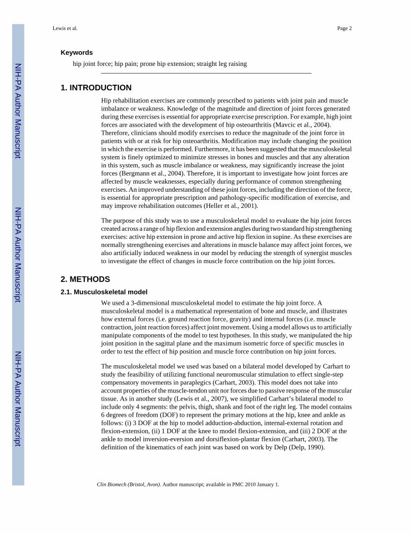
2.2. ExercisesThe hip joint forces generated during the simulation of two hip exercises were evaluated. Weselected these exercises as they both are often used as a rehabilitation exercise for patients witha variety of conditions (Hall and Brody, 2005;Moffat, 2006;Prentice and Voight, 2001) Thefirst exercise simulated was prone hip extension. For the prone hip extension simulatedexercise, gravity was specified as acting from posterior to anterior in line with the pelvicreference frame (Figure 1). The knee joint angle and ankle joint angles were set at zero so thatboth joints were in the neutral position and had to be maintained in neutral through a balanceof muscle forces. The hip joint adduction/abduction and internal/external rotation angles werealso set and maintained at zero. The hip joint angle was increased in one degree incrementsfrom 10° of hip flexion to 20° of hip extension. The hip joint angle range started at 10° of hipflexion because we recommend starting patients in 10° of hip flexion when performing pronehip extension in order to avoid hip hyperextension (Sahrmann, 2002).
The second exercise simulated was hip flexion in the supine position, or straight leg raising.For the supine hip flexion simulated exercise, gravity was specified as acting from anterior toposterior to simulate the supine position (Figure 1). Again, the knee and ankle joint angles aswell as the hip adduction/abduction and internal/external rotation angles were set to zero. Thehip joint angle was increased in one degree increments from 10° of hip extension to 30° of hipflexion. The range of simulated hip joint angles started at 10° of hip extension because thisposition is the presumed position of the hip when the lumbar spine is against the mat (Kendallet al., 1993) and is the typical starting position for a straight leg raise.
2.3. ConditionsWe simulated 3 different conditions for each exercise to estimate the hip joint force when themaximum muscle force value for selected muscles was reduced. The first condition (NormalCondition) served as a control condition to which the other conditions were compared. For thiscondition, the hip joint force due to muscle was estimated using the maximum muscle forcevalues specified in the original model (Delp, 1990). For the two altered conditions, themaximum muscle force value for selected muscles were reduced by 50%. This level ofreduction has been used by other researchers (Goldberg and Neptune, 2007) and has been notedclinically in runners with overuse injuries (Niemuth et al., 2005). We induced weakness in themodel to simulate the conditions under which these strengthening exercises are performed asit has been suggested that alterations in muscle force balance may increases joint forces(Bergmann et al., 2004).
For the prone hip extension exercise simulation, one altered condition was the Gluteal ReducedCondition in which the maximum force values of the gluteal muscles (gluteus maximus,medius, and minimus) were decreased by 50%. The second altered condition was the HamstringReduced Condition in which the maximum force values of the hamstring muscles(semimembranosis, semitendinosis, and biceps femoris) were decreased. We altered themaximum muscle force of the gluteal and hamstring muscle groups because both groups aremajor contributors to hip extensor torque and are strengthened by performing prone hipextension.
For the supine hip flexion exercise simulation, one altered condition was the Iliopsoas ReducedCondition, in which the maximum muscle force values for the iliacus and psoas muscles weredecreased by 50%. These muscles were selected as they are primary hip flexor muscles andare strengthened by performing supine hip flexion. In the second condition, the Rectus Reduced
Lewis et al. Page 4
Clin Biomech (Bristol, Avon). Author manuscript; available in PMC 2010 January 1.
NIH
-PA Author Manuscript
NIH
-PA Author Manuscript
NIH
-PA Author Manuscript
Condition, the maximum force values for the rectus femoris, tensor fascia lata (TFL) andsartorius muscles were decreased. These three muscles were selected because they contributeto hip flexion torque and may compensate for weakness of the iliacus and psoas muscles. Foreach altered condition, the optimization routine recalculated the optimal set of muscle forceswith the altered constraint of the reduced maximum muscle force values.
2.4. Sensitivity AnalysisWe also conducted an analysis to determine how sensitive the results of the study were to theminimization parameter used. We tested two additional minimization routines, one whichminimized the sum of PForce to the third power, and one which minimized the sum of PForceto the fourth power.
2.5. Experimental Model ValidationWe collected surface electromyographic (EMG) data from five subjects who provided writteninformed consent as approved by the University of Michigan Medical School InstitutionalReview Board. Surface electrodes (1.1 cm diameter) with an inter-electrode distance of 2.5 cmwere placed over the muscle bellies of the medial hamstrings, lateral hamstring, gluteusmaximus, gluteus medius, tensor fascia lata, rectus femoris, and iliopsoas muscles perguidelines (Konrad, 2005). The iliopsoas electrode was placed lateral to the femoral pulse,medial to the rectus femoris, and inferior to the inguinal ligament (Gottschall and Kram,2005) and verified with movement tests (Cram and Kasman, 1998). Data was collected at 1200Hz while the subject held the leg at the midpoint of the range of motion for each exercise asdetermined by a physical therapist. The midpoint for prone hip extension was approximately5 degrees of hip extension while 10 degrees of flexion was used for supine hip flexion. Themidpoint was used to avoid the influence of joint limitation or muscle length limitations at theend range of motion. Data were collected while each position was held for at least two secondsand repeated three times. Data were also collected during maximal voluntary contractions instandard manual muscle testing positions (Kendall et al., 1993). All emg data were high passfiltered with a 10 Hz zero-phase lag Butterworth filter, rectified and low pass filtered at 500Hz. The root mean square (RMS) of the emg data was calculated using a window length of12.5 msec. For each repetition of each exercise, we calculated the average RMS emg while theleg was held stationary. We then normalized the data using the peak RMS value during themaximum voluntary contraction for each muscle. Finally, we averaged the data across subjectsfor each muscle in each position to compare with the value estimated by the model.
2.6. Data analysisThe two focuses of this study were the effect of hip position and the effect of muscle forcecontribution on hip joint forces. To evaluate the effect of hip position, we calculated the netresultant force due to muscle at their level of PForce throughout the range of hip angles for eachsimulated exercise. We also divided the resultant force into each force component to determinethe effect of hip angle on the force components in each direction.
In order to evaluate the effect of altering muscle force contribution on hip joint forces, wecalculated the joint forces for all three conditions. We also evaluated the change in PForce foreach muscle between the normal condition and the altered conditions to determine how thePForce of each muscle in the model would be modified to compensate for the altered muscles.As a measure of the overall efficiency of the system of muscles, we calculated the sum of thePForce values across all muscles which cross the hip or knee joints. The change in the PForceand the sum of the PForce values were all calculated at the neutral position (0°) of hi p flexion/extension to allow comparisons between simulated exercises.
Lewis et al. Page 5
Clin Biomech (Bristol, Avon). Author manuscript; available in PMC 2010 January 1.
NIH
-PA Author Manuscript
NIH
-PA Author Manuscript
NIH
-PA Author Manuscript
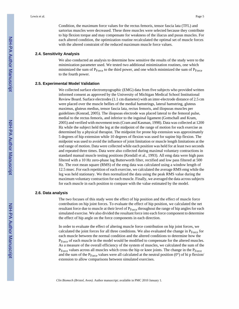
3. RESULTS3.1. Prone hip extension
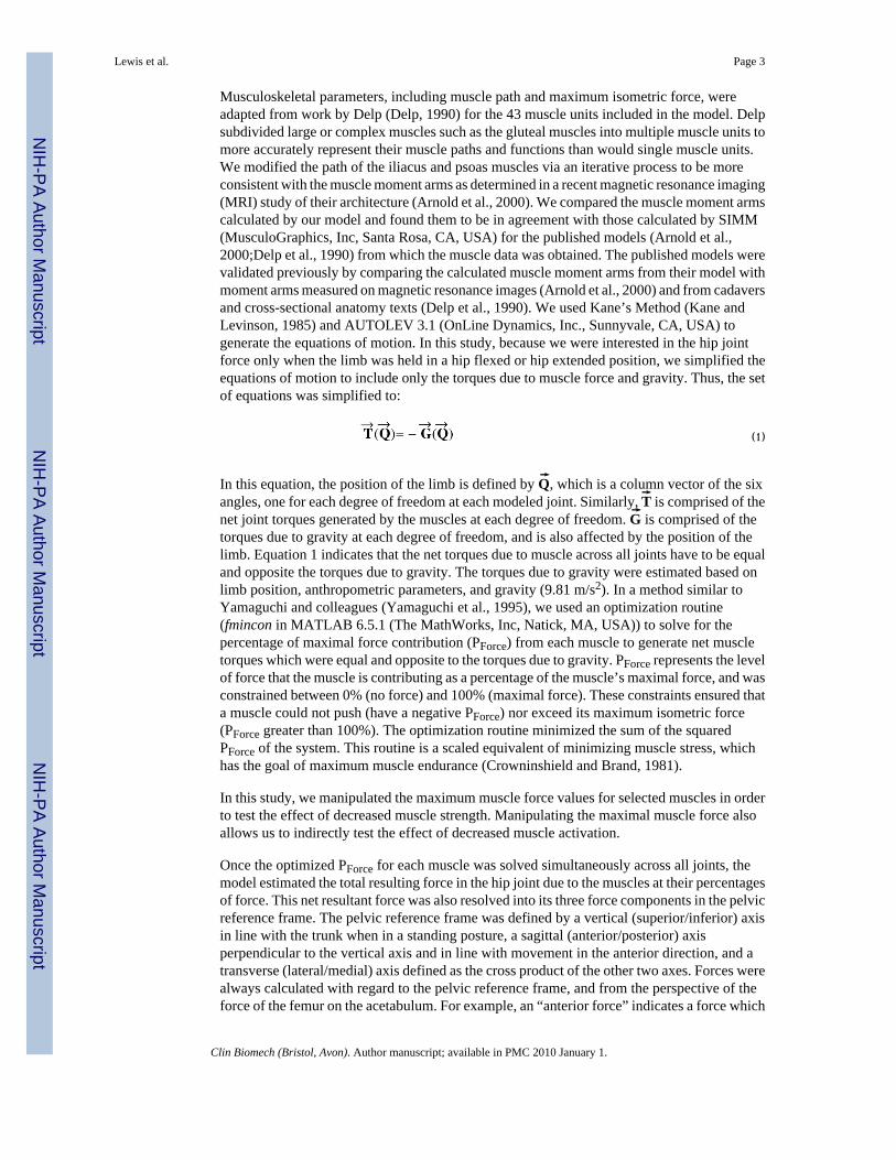
Effect of position—The net resultant hip force increases with increasing hip extension angleduring simulated prone hip extension (Figure 2). The resultant force increased 181 N (22.7%body weight) with a 30° change in hip angle (10° of hip flexion to 20° of hip extension). Thevertical force is the lar gest component of the hip force, followed by the force in the sagittalplane, and then in the transverse plane.
Effect of muscle force alteration—Decreased force contribution from the gluteal muscleswhen performing prone hip extension resulted in an increase in the net resultant and verticalhip joint forces compared to the normal condition. The magnitude of the sagittal force increasedat the end ranges of hip motion. Compared to the normal condition, the hip force in the anteriordirection was higher when in hip extension while the posterior hip force was higher when inhip flexion. Decreased force contribution from the gluteal muscles also decreased thetransverse force component.
The overall muscle efficiency of the system was decreased when the maximum muscle forcevalues for the gluteal muscles were decreased by 50%. At neutral hip flexion, the sum of thePForce was 123, an increase of 47.2% over the normal condition which had a PForce of 83.8.
Decreased force contribution from the hamstring muscles when performing prone hip extensionresulted in a decrease in the resultant and vertical hip forces along with a decrease in themagnitude of the sagittal force compared to the normal condition. The transverse force wasalso slightly decreased.
When the maximum muscle force values for the hamstring muscles were decreased, the muscleefficiency of the system was also decreased compared to the normal condition. The sum of thePForce was 122, an increase of 45.5% over the normal condition when in neutral hip extension.
In the prone hip extension simulation, when the gluteal muscle strength was reduced, the modelincreased the PForce of the semimembranosis, gluteus medius (posterior fibers), TFL, and vastimuscles (Supplemental Figure 1). When the hamstring muscle strength was decreased, thegluteus maximus (all fibers), and the anterior portions of the gluteus medius and minimusdemonstrated increased PForce.
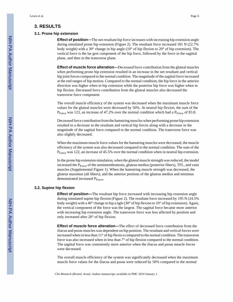
3.2. Supine hip flexionEffect of position—The resultant hip force increased with increasing hip extension angleduring simulated supine hip flexion (Figure 2). The resultant force increased by 195 N (24.5%body weight) with a 40° change in hip a ngle (30° of hip flexion to 10° of hip extension). Again,the vertical component of the force was the largest. The sagittal force became more anteriorwith increasing hip extension angle. The transverse force was less affected by position andonly increased after 20° of hip flexion.
Effect of muscle force alteration—The effect of decreased force contribution from theiliacus and psoas muscles was dependent on hip position. The resultant and vertical forces wereincreased when in less than 11° of hip flexio n compared to the normal condition. The transverseforce was also increased when in less than 7° of hip flexion compared to the normal condition.The sagittal force was consistently more anterior when the iliacus and psoas muscle forceswere decreased.
The overall muscle efficiency of the system was significantly decreased when the maximummuscle force values for the iliacus and psoas were reduced by 50% compared to the normal
Lewis et al. Page 6
Clin Biomech (Bristol, Avon). Author manuscript; available in PMC 2010 January 1.
NIH
-PA Author Manuscript
NIH
-PA Author Manuscript
NIH
-PA Author Manuscript
condition. At neutral hip extension, the sum of the PForce was 382, an increase of 58.1% overnormal which had a PForce of 242.
Decreased force contribution from the rectus femoris, TFL, and sartorius muscles whenperforming supine hip flexion consistently resulted in an increase in the resultant, vertical andtransverse forces when compared to the normal condition. The sagittal force was slightly moreposterior in the Rectus Reduced Condition. The overall muscle efficiency was only slightlyaffected by reduced force contribution from the rectus femoris, TFL and sartorius muscles,increasing the sum of the PForce only 6.8% above the normal condition.
In the supine hip flexion simulation, when the maximum force values for the iliacus and psoasmuscles were reduced, the model increased the PForce of a number of muscles: gluteus minimus(all fibers), adductor longus, pectineus, sartorius, TFL, and rectus femoris (SupplementalFigure 2). When the maximum force value of the rectus femoris, TFL, and sartorius werereduced, the model increased the PForce of the gluteus minimus (all fibers), gluteus medius(posterior fibers), piriformis, iliacus and psoas muscles.
3.3. Sensitivity AnalysisIn addition to the sum of the squared PForce of the system, we tested two other minimizationparameters (PForce to the third and to the fourth power) to determine the sensitivity of the modelto the optimization used. Although the different minimization parameters did result in slightchanges in individual muscle stress and joint force values, the conclusions of the study werenot affected by the minimization parameter used.
3.4 Experimental Model ValidationTo validate the model, we compared the calculated PForce from the model to the measuredEMG data from our subjects. We found that the measured muscle activation during prone hipextension and supine hip flexion follows the pattern of the muscle force estimated by the model.For prone hip extension, the hamstring muscles are activated at the highest percentage of theirmaximum, with the gluteus maximus and medius activated at approximately half the activationof the hamstrings. The tensor fascia lata has the smallest activation of the muscles measured.The musculoskeletal model predicts this same pattern of activation (hamstring greater thangluteal and gluteal greater than tensor fascia lata). Similarly, both the experimental data andthe model estimations during supine hip flexion display a pattern of greater activation of theiliopsoas than the rectus femoris or tensor fascia lata.
4. DISCUSSIONThe results of this study indicate that hip joint forces are affected both by position and byalterations in synergist muscle force contribution, with position having a much greater effect.The highest forces are when the hip is in extension, and are greater during hip flexion in supinethan hip extension in prone. The sensitivity analysis demonstrated that the conclusions of thisstudy are not affected by the minimization parameters tested. Experimental data follow therelative contributions of muscles during prone hip extension and supine hip flexion.
4.1 Effect of positionThe effect of hip joint position was varied in this study to determine if modifying the positionof the exercise would significantly change the forces at the hip. We found that the net resultanthip force increased with increasing hip extension angle whether performing supine hip flexionor prone hip extension. Similar to the net resultant force, the vertical force was higher when inhip extension. These findings have implications for the prescription and modification ofstrengthening exercises for certain patient populations. For example, high joint forces may
Lewis et al. Page 7
Clin Biomech (Bristol, Avon). Author manuscript; available in PMC 2010 January 1.
NIH
-PA Author Manuscript
NIH
-PA Author Manuscript
NIH
-PA Author Manuscript
contribute to the development of cartilage degeneration and osteoarthritis (Mavcic et al.,2004); therefore, when prescribing prone hip extension exercises for a patient with hiposteoarthritis, the hip should start in flexion and be limited to neutral extension to decrease thecompressive force on the joint.
The direction of the sagittal force was also dependent on the position of the hip. The force wasdirected anteriorly when in hip extension, while it was directed posteriorly when in hip flexion.Again, this finding can be used to appropriately modify exercises. For a patient with anteriorhip pain that is aggravated by anterior hip forces, supine hip flexion should not be initiatedwith the hip in extension.
4.2. Effect of alteration of muscle force contributionAlthough the joint position affects the magnitude of the hip force more than alterations inmuscle force contribution, the effect of muscle weakness or decreased activation is importantto understand, especially for strengthening exercises. If the gluteal muscles are weak, it maybe even more important to strengthen in neutral hip extension rather than full hip extension asweakness of the gluteal muscles increases the resultant force by 46.3 N at 20° degrees of hipextension compared to the normal condition. Limiting the total force may be important for apatient with osteoarthritis (Mavcic et al., 2004). Similarly, when prescribing supine hip flexionfor a person with weakness of the iliacus and psoas muscles, the exercise should be initiatedin greater hip flexion than if the muscles were not weak in order to reduce the anterior hipforce.
Evaluating the effect of altered muscle force contribution also provides insight into thespecificity of muscle function and the compensations for certain muscle weaknesses. Reductionof the maximum muscle strength of the gluteal muscles results in increased activation of thesemimembranosis over the other hamstring (semitendinosis and biceps femoris) muscles. Thesemimembranosis muscle has the smallest rotational and adduction moment arms relative tothe extension moment arm, making it a more efficient hip extensor than the other hamstringmuscles (Supplemental Figure 3). The hip rotation and adduction moment arms of thesemimembranosis muscle are 2.4% and 28.2% respectively of its hip extension moment arm.For the semitendinosis and biceps femoris muscles, the hip rotation moment arms are 0.3%and 9.9% and hip adduction moment arms are 43.8% and 38.8% of their hip extension momentarms.
In both prone hip extension and supine hip flexion, the increased PForce of one muscle tocompensate for weakness of another muscle results in extraneous torques or torques not directlyrelated to the desired task. For example, during supine hip flexion, the TFL compensates forthe hip flexion torque lost by the weakened iliacus and psoas muscles; however, it also createsextraneous abduction torque (Supplemental Figure 3). The extraneous abduction torque is149% of its hip flexion torque. In other words, for each 100 units of hip flexion torque the TFLproduces, it produces 149 units of abduction torque, making it a more efficient hip abductorthan hip flexor. Other muscles, therefore, must contribute more force. To compensate for theextraneous abduction torque from the TFL, the PForce of the adductor longer muscle isincreased. The additional muscle forces result in an overall increase in the sum of the PForceof the system of muscles.
Weakness of certain muscles affects the system differently. When the maximum musclestrength of the gluteal muscle group was reduced, the sum of the PForce increased by 39.5 unitsover the normal condition. When the maximum muscle strength of the hamstring muscles werereduced, the sum increased by 38.2 units. It seems reasonable that the change in the overallPForce would be similar for these two alterations as the total hip extension torque for the glutealmuscle group is similar to the hip extension torque for the hamstring muscle group (111.7 Nm
Lewis et al. Page 8
Clin Biomech (Bristol, Avon). Author manuscript; available in PMC 2010 January 1.
NIH
-PA Author Manuscript
NIH
-PA Author Manuscript
NIH
-PA Author Manuscript
and 121.0 Nm respectively). However, during supine hip flexion, reduction of the maximalforce the iliacus and psoas muscles resulted in an increase of 140.4 units over the normalcondition while reduction of the rectus femoris, TFL and sartorius muscles resulted in only a16.4 unit increase despite similar total hip flexion torques (28.0 Nm for iliacus and psoascombined and 38.8 Nm for rectus femoris, TFL and sartorius combined.) This extreme effectof weakness of the iliacus and psoas muscles emphases the unique role these muscles play instraight plane hip flexion.
Although other studies have investigated hip joint forces during gait and stairs (Bergmann etal., 2001;Heller et al., 2001;Stansfield and Nicol, 2002), and contact pressures during exercises(Tackson et al., 1997), our study is the first to evaluate the force components in each directionduring performance of hip strengthening exercises. In this study, the net hip joint forces weredivided into the component forces in the vertical, sagittal, and transverse directions relative tothe pelvis. The individual forces in each direction are important when determining appropriateinterventions for patients with particular pathologies. For example, abnormal or excessiveforces in the anterior direction have been recognized as a potential cause of anterior hip painand subtle hip instability (Shindle et al., 2006) and may lead to a tear of the anterior acetabularlabrum even in the absence of a traumatic event (Mason, 2001;McCarthy et al., 2001;Shindleet al., 2006). Therefore, in patients with anterior hip pain and an anterior labral tear, it may bemore important to limit anterior force than to limit the net resultant force.
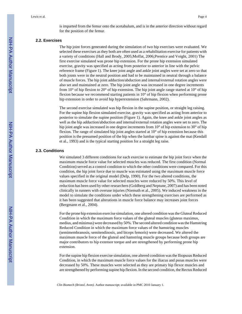
As with all musculoskeletal models, there are limitations inherent in attempting to modelcomplex human movement with simplified lines of action for muscles and computerizedoptimization routines for motor control. One major limitation specific to this study was ourinability to accurately model the stabilizing effect, if any, of the iliacus and psoas muscles asthey pass over the front of the femoral head. The iliacus and psoas muscles may be similar tothe rotator cuff muscles of the shoulder, applying forces to the femur not only through theirmuscle insertion, but also through their muscle bellies and tendons as they pass over theunderlying structures (Figure 3). It has been suggested that the psoas muscle tendon addsstrength to the anterior hip joint capsule when in hip extension (Philippon, 2001), especiallyin the presence of capsular laxity (Shindle et al., 2006). While wrapping of the iliopsoas maybe more accurately modeled in software such as OpenSim (https://simtk.org/home/opensim),our model uses via points to modify the path of the muscle around the femoral head.
The optimization criterion used to determine PForce is another limitation. We used anoptimization routine which minimized the sum of the squared PForce over the system ofmuscles. Theoretically, this optimization function captures the physiological properties ofmuscle (muscle moment arm and maximum muscle strength), as well as the goal of maximummuscle endurance and produces solutions with significant temporal agreement betweenpredicted muscle forces and recorded muscle electromyography (Crowninshield and Brand,1981). A similar optimization routine has been used with this model in the prediction of muscleforces during gait and have resulted in temporally consistent and “intuitively reasonable”solutions (Carhart, 2003). We also tested two other minimization parameters (PForce to the thirdand to the fourth power) and found that the conclusions of the study were not affected. This isin agreement with the study by Crowninshield and Brand (Crowninshield and Brand, 1981),which demonstrated that calculated muscle stress using minimization of the sum of musclestresses to the nth power was not sensitive to small changes in n. Furthermore, the pattern ofmeasured muscle activation was similar to the muscle forces predicted by the model. We alsomodeled only static behavior. Because both of the exercises used in this study are typicallyperformed at a slow velocity (30° per second) and within the normal range of motion, we didnot include torques from inertia, passive joint structures or viscoelastic damping.
Lewis et al. Page 9
Clin Biomech (Bristol, Avon). Author manuscript; available in PMC 2010 January 1.
NIH
-PA Author Manuscript
NIH
-PA Author Manuscript
NIH
-PA Author Manuscript
Another limitation is in our choice of exercises. Although prone hip extension and supine hipflexion are not functional tasks, they are commonly prescribed exercises. The joint forcescreated when performing these exercises should be understood so that appropriatemodifications can be made. Furthermore, analysis of these simple exercises may provideinsight into the forces that muscles create when active in other tasks such as walking.
5. CONCLUSIONIn this study, the musculoskeletal model predicted that hip joint forces are greatly affected bythe angle of hip flexion or extension and partially by alterations in muscle force contribution.During prone hip extension, the highest net resultant forces occurred when the hip was inextension and when the gluteal muscles were weakened. During supine hip flexion, the highestnet resultant forces occurred when the hip was in extension and when the iliacus and psoasmuscles were weakened. Clinicians can use this information to modify exercises to providethe most appropriate intervention for a patient based on the specific location of hip jointpathology.
Supplementary MaterialRefer to Web version on PubMed Central for supplementary material.
AcknowledgementsThis work was supported in part by the National Institutes of Health (HD07434 and HD007422). We would also liketo thank Michael Carhart for assistance with the musculoskeletal model.
This research was conducted at Washington University in St. Louis and University of Michigan, and was supportedin part by the National Institutes of Health (HD07434 and HD007422).
ReferencesArnold AS, Salinas S, Asakawa DJ, Delp SL. Accuracy of muscle moment arms estimated from MRI-
based musculoskeletal models of the lower extremity. Comput Aided Surg 2000;5(2):108–119.[PubMed: 10862133]
Bergmann G, Deuretzbacher G, Heller M, Graichen F, Rohlmann A, Strauss J, Duda GN. Hip contactforces and gait patterns from routine activities. J Biomech 2001;34(7):859–871. [PubMed: 11410170]
Bergmann G, Graichen F, Rohlmann A. Hip joint contact forces during stumbling. Langenbecks ArchSurg 2004;389(1):53–59. [PubMed: 14625775]
Carhart, MR. Biomechanical analysis of compensatory stepping: implications for paraplegics standingvia FNS. Doctor of Philosophy, Arizona State University; 2003.
Cram, JR.; Kasman, GS. Electrode Placement. Aspen, Gaithersburg, MD: 1998.Crowninshield RD, Brand RA. A physiologically based criterion of muscle force prediction in
locomotion. J Biomech 1981;14(11):793–801. [PubMed: 7334039]Delp, SL. Parameters for a model of the lower limb. http://www.isbweb.org/data/delp/. 1990.
http://www.isbweb.org/data/delp/. 12-1-2005.Delp SL, Loan JP, Hoy MG, Zajac FE, Topp EL, Rosen JM. An interactive graphics-based model of the
lower extremity to study orthopaedic surgical procedures. IEEE Trans Biomed Eng 1990;37(8):757–767. [PubMed: 2210784]
Goldberg EJ, Neptune RR. Compensatory strategies during normal walking in response to muscleweakness and increased hip joint stiffness. Gait Posture 2007;25(3):360–367. [PubMed: 16720095]
Gottschall JS, Kram R. Energy cost and muscular activity required for leg swing during walking. J ApplPhysiol 2005;99(1):23–30. [PubMed: 16036902]
Hall, CM.; Brody, LT. Therapeutic Exercise: Moving toward function. Vol. 2. Lippincott Williams &Wilkins; Baltimore: 2005.
Lewis et al. Page 10
Clin Biomech (Bristol, Avon). Author manuscript; available in PMC 2010 January 1.
NIH
-PA Author Manuscript
NIH
-PA Author Manuscript
NIH
-PA Author Manuscript
Heller MO, Bergmann G, Deuretzbacher G, Durselen L, Pohl M, Claes L, Haas NP, Duda GN. Musculo-skeletal loading conditions at the hip during walking and stair climbing. J Biomech 2001;34(7):883–893. [PubMed: 11410172]
Kane, TR.; Levinson, DA. Dynamics: theory and application. McGraw-Hill; New York, NY: 1985.Kendall, FP.; McCreary, EK.; Provance, PG. Muscles Testing and Function. Vol. 4. Williams & Wilkins;
Baltimore, MD: 1993.Konrad, P. The ABC of EMG - A practical introduction to kinesiological electromyography. Vol. 1.
Noraxon INC; Scottsdale: 2005.Lewis CL, Sahrmann SA, Moran DW. Anterior hip joint force increases with hip extension, decreased
gluteal force, or decreased iliopsoas force. J Biomech 2007;40(16):3725–3731. [PubMed: 17707385]Mason JB. Acetabular labral tears in the athlete. Clin Sports Med 2001;20(4):779–790. [PubMed:
11675886]Mavcic B, Slivnik T, Antolic V, Iglic A, Kralj-Iglic V. High contact hip stress is related to the development
of hip pathology with increasing age. Clin Biomech(Bristol, Avon) 2004;19(9):939–943.McCarthy JC, Noble PC, Schuck MR, Wright J, Lee J. The Otto E. Aufranc Award: The role of labral
lesions to development of early degenerative hip disease. Clin Orthop 2001;(393):25–37. [PubMed:11764355]
Moffat, M. Musculoskeletal Essentials: Applying the Preferred Physical Therapist Practice Patterns.SLACK, Inc; Thorofare: 2006.
Niemuth PE, Johnson RJ, Myers MJ, Thieman TJ. Hip muscle weakness and overuse injuries inrecreational runners. Clin J Sport Med 2005;15(1):14–21. [PubMed: 15654186]
Philippon MJ. The role of arthroscopic thermal capsulorrhaphy in the hip. Clin Sports Med 2001;20(4):817–829. [PubMed: 11675889]
Prentice, WE.; Voight, ML. Techniques in Musculoskeletal Rehabilitation. McGraw-Hill; Columbus:2001.
Sahrmann, SA. Diagnosis and treatment of movement impairment syndromes. Mosby, Inc; St. Louis,MO: 2002.
Shindle MK, Ranawat AS, Kelly BT. Diagnosis and management of traumatic and atraumatic hipinstability in the athletic patient. Clin Sports Med 2006;25(2):309–326. [PubMed: 16638494]
Stansfield BW, Nicol AC. Hip joint contact forces in normal subjects and subjects with total hipprostheses: walking and stair and ramp negotiation. Clin Biomech(Bristol, Avon) 2002;17(2):130–139.
Tackson SJ, Krebs DE, Harris BA. Acetabular pressures during hip arthritis exercises. Arthritis Care Res1997;10(5):308–319. [PubMed: 9362597]
Yamaguchi GT, Moran DW, Si J. A computationally efficient method for solving the redundant problemin biomechanics. J Biomech 1995;28(8):999–1005. [PubMed: 7673268]
Lewis et al. Page 11
Clin Biomech (Bristol, Avon). Author manuscript; available in PMC 2010 January 1.
NIH
-PA Author Manuscript
NIH
-PA Author Manuscript
NIH
-PA Author Manuscript

FIGURE 1.Exercises simulated using the musculoskeletal model. Red lines indicate all the musclesincluded in the model. For hip extension in prone, gravity was specified as acting from posteriorto anterior in line with the pelvic reference frame. The hip started in 10° of hip flexion andmoved through 30° to a final position of 20° of hip extension. For hip flexion in supine, gravitywas specified as acting from anterior to posterior in line with the pelvic reference frame. Thehip started in 10° of hip extension and was moved through 40° to a final position of 30° of hipflexion. (Image created in SIMM (MusculoGraphics, Inc, Santa Rosa, CA, USA))
Lewis et al. Page 12
Clin Biomech (Bristol, Avon). Author manuscript; available in PMC 2010 January 1.
NIH
-PA Author Manuscript
NIH
-PA Author Manuscript
NIH
-PA Author Manuscript
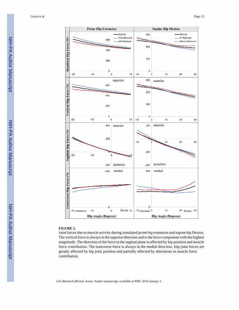
FIGURE 2.Joint forces due to muscle activity during simulated prone hip extension and supine hip flexion.The vertical force is always in the superior direction and is the force component with the highestmagnitude. The direction of the force in the sagittal plane is affected by hip position and muscleforce contribution. The transverse force is always in the medial direction. Hip joint forces aregreatly affected by hip joint position and partially affected by alterations in muscle forcecontribution.
Lewis et al. Page 13
Clin Biomech (Bristol, Avon). Author manuscript; available in PMC 2010 January 1.
NIH
-PA Author Manuscript
NIH
-PA Author Manuscript
NIH
-PA Author Manuscript

FIGURE 3.Stabilizing effect of iliacus and psoas muscles crossing the hip joint. The iliacus and psoasmuscles (thick red lines) may apply forces (black arrows) to the femoral head through theirmuscle bellies as they pass over the underlying anterior hip joint structure. These forces mayhelp maintain the femoral head positioning within the acetabulum similar to the rotator cuffmuscles of the shoulder. This effect has been suggested to add strength to the anterior hip jointcapsule when in hip extension (Philippon, 2001).
Lewis et al. Page 14
Clin Biomech (Bristol, Avon). Author manuscript; available in PMC 2010 January 1.
NIH
-PA Author Manuscript
NIH
-PA Author Manuscript
NIH
-PA Author Manuscript


![Electromyographic Evaluation of Hip Exercises[1]](https://img.pdfslide.us/doc/110x75/563dbb3b550346aa9aab62bd/electromyographic-evaluation-of-hip-exercises1.jpg)