Embed Size (px)
Citation preview
Gravitational and Space Biology 18(2) June 2005 39
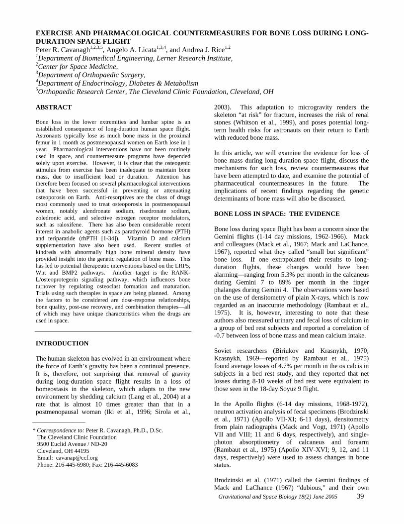
EXERCISE AND PHARMACOLOGICAL COUNTERMEASURES FOR BONE LOSS DURING LONG-DURATION SPACE FLIGHT Peter R. Cavanagh1,2,3,5, Angelo A. Licata1,3,4, and Andrea J. Rice1,2 1Department of Biomedical Engineering, Lerner Research Institute, 2Center for Space Medicine, 3Department of Orthopaedic Surgery, 4Department of Endocrinology, Diabetes & Metabolism 5Orthopaedic Research Center, The Cleveland Clinic Foundation, Cleveland, OH ABSTRACT Bone loss in the lower extremities and lumbar spine is an established consequence of long-duration human space flight. Astronauts typically lose as much bone mass in the proximal femur in 1 month as postmenopausal women on Earth lose in 1 year. Pharmacological interventions have not been routinely used in space, and countermeasure programs have depended solely upon exercise. However, it is clear that the osteogenic stimulus from exercise has been inadequate to maintain bone mass, due to insufficient load or duration. Attention has therefore been focused on several pharmacological interventions that have been successful in preventing or attenuating osteoporosis on Earth. Anti-resorptives are the class of drugs most commonly used to treat osteoporosis in postmenopausal women, notably alendronate sodium, risedronate sodium, zoledronic acid, and selective estrogen receptor modulators, such as raloxifene. There has also been considerable recent interest in anabolic agents such as parathyroid hormone (PTH) and teriparatide (rhPTH [1-34]). Vitamin D and calcium supplementation have also been used. Recent studies of kindreds with abnormally high bone mineral density have provided insight into the genetic regulation of bone mass. This has led to potential therapeutic interventions based on the LRP5, Wnt and BMP2 pathways. Another target is the RANK-L/osteoprotegerin signaling pathway, which influences bone turnover by regulating osteoclast formation and maturation. Trials using such therapies in space are being planned. Among the factors to be considered are dose-response relationships, bone quality, post-use recovery, and combination therapies—all of which may have unique characteristics when the drugs are used in space. INTRODUCTION The human skeleton has evolved in an environment where the force of Earth’s gravity has been a continual presence. It is, therefore, not surprising that removal of gravity during long-duration space flight results in a loss of homeostasis in the skeleton, which adapts to the new environment by shedding calcium (Lang et al., 2004) at a rate that is almost 10 times greater than that in a postmenopausal woman (Iki et al., 1996; Sirola et al.,
2003). This adaptation to microgravity renders the skeleton “at risk” for fracture, increases the risk of renal stones (Whitson et al., 1999), and poses potential long-term health risks for astronauts on their return to Earth with reduced bone mass. In this article, we will examine the evidence for loss of bone mass during long-duration space flight, discuss the mechanisms for such loss, review countermeasures that have been attempted to date, and examine the potential of pharmaceutical countermeasures in the future. The implications of recent findings regarding the genetic determinants of bone mass will also be discussed. BONE LOSS IN SPACE: THE EVIDENCE Bone loss during space flight has been a concern since the Gemini flights (1-14 day missions, 1962-1966). Mack and colleagues (Mack et al., 1967; Mack and LaChance, 1967), reported what they called “small but significant” bone loss. If one extrapolated their results to long-duration flights, these changes would have been alarming—ranging from 5.3% per month in the calcaneus during Gemini 7 to 89% per month in the finger phalanges during Gemini 4. The observations were based on the use of densitometry of plain X-rays, which is now regarded as an inaccurate methodology (Rambaut et al., 1975). It is, however, interesting to note that these authors also measured urinary and fecal loss of calcium in a group of bed rest subjects and reported a correlation of -0.7 between loss of bone mass and mean calcium intake. Soviet researchers (Biriukov and Krasnykh, 1970; Krasnykh, 1969—reported by Rambaut et al., 1975) found average losses of 4.7% per month in the os calcis in subjects in a bed rest study, and they reported that net losses during 8-10 weeks of bed rest were equivalent to those seen in the 18-day Soyuz 9 flight. In the Apollo flights (6-14 day missions, 1968-1972), neutron activation analysis of fecal specimens (Brodzinski et al., 1971) (Apollo VII-XI; 6-11 days), densitometry from plain radiographs (Mack and Vogt, 1971) (Apollo VII and VIII; 11 and 6 days, respectively), and single-photon absorptiometry of calcaneus and forearm (Rambaut et al., 1975) (Apollo XIV-XVI; 9, 12, and 11 days, respectively) were used to assess changes in bone status. Brodzinski et al. (1971) called the Gemini findings of Mack and LaChance (1967) “dubious,” and their own
____________________
* Correspondence to: Peter R. Cavanagh, Ph.D., D.Sc. The Cleveland Clinic Foundation 9500 Euclid Avenue / ND-20 Cleveland, OH 44195 Email: [email protected] Phone: 216-445-6980; Fax: 216-445-6083

P.R. Cavanagh - Preventing Bone Loss in Space
40 Gravitational and Space Biology 18(2) June 2005
measurements of calcium loss in Apollo VII-XI crewmembers suggested less substantial changes. They estimated that loss of total body calcium could be as little as 7.5% per year of space flight, but they suggested that a calcium balance experiment should be conducted on Skylab, and this was in fact accomplished (see below). Using similar methodology to their earlier studies, Mack and Vogt (1971) reported average losses of 11.6% per month in the lower extremity and 22.6% losses in the upper extremity of six Apollo VII and VIII crew members. As discussed above, in retrospect, these changes in “bone density” measured from plain radiographs were clearly erroneous; control subjects on Earth did not show such large changes. The single gamma photon absorptiometry of Rambaut et al. (1975) found a loss of 5.1% per month in the lower extremity, a gain of 0.6% per month in the radius, and a loss of 4.6% per month in the ulnae of nine Apollo XIV-XVI crew members. One of the most complete series of calcium balance studies in space was conducted during the Skylab missions (Skylab 2, 28 days, 1973; Skylab 3, 59 days, 1973; Skylab 4, 84 days, 1973-1974—Rambaut and Johnston, 1979; Smith et al., 1977; Smith et al., 1998; Smith et al., 1999; Tilton et al., 1980; Whedon et al., 1977). Commencing 21 days prior to flight, during flight, and for 18 days post flight, the intake of 30 nutrients were monitored, 24-hour pooled urine collections were made, and fecal samples were vacuum dried for analysis. Weekly plasma samples were also taken. A 56-day ground-based control experiment was also conducted. During the Skylab 4 mission, average negative calcium balances of -100 (+25), -180 (+36), -229 (+60), -223 (+42), -88 (+52) were reported for the pre-flight, flight days 1-24, 25-56, 57-84, and post-flight measurements, respectively. In addition, using single-photon absorp-tiometry, Smith et al. (1977) indicated that a mean loss of 0.4% per month occurred in the calcanei of all nine Skylab 2-4 crew members, while the investigators detected negligible losses in the radii (0.06% per month) and a gain in the ulnae (0.4% per month). Rambaut et al. (1979) pointed out that “the chain of events leading ultimately to bone loss inflight remains elusive.” In an article almost 20 years later, in which the urine of Skylab crew members was re-analyzed, Smith et al. (1998) shed light on this mechanism by demonstrating that urinary excretion of collagen breakdown products during the Skylab 4 mission was 40-45% higher than pre-flight values, indicating that space flight is associated with increased bone resorption. Using computer tomography (CT), Oganov et al. (1990) measured mineral density of lumbar vertebrae in four Salyut-7 crew members before and after extended flights (5-7 months’ duration). These authors reported that bone mineral density (BMD) diminished only in some of the test subjects and emphasized that the magnitude of
changes was not correlated with flight time, presumably due to individual differences in rates of bone loss. McCarthy et al. (2000) used three techniques (dual energy X-ray absorptiometry [DXA], ultrasonic measurements of velocity [SOS], and broadband attenuation [BUA] of the calcaneus) to evaluate changes in bone during two missions, of 180 and 20 days, to the Mir space station, involving three subjects. DXA measurements resulted in significant variation between different sites in the body for changes in BMD, with the greatest losses occurring in the lumbar spine and proximal femur.
-2.5
-2
-1.5
-1
-0.5
0
Del
ta B
MD
/Mon
th (%
)
Spine Neck Trochanter Pelvis Arm
Space Flight
Bedrest
Figure 1a: Change in bone mineral density in different anatomical regions (in percent change per month; negative values represent loss) during Mir missions and bed rest. Data adapted from LeBlanc et al. (2000). The application of modern imaging techniques to bone changes during space flight was first accomplished by LeBlanc and colleagues (LeBlanc et al., 1996; LeBlanc et al., 1998; LeBlanc et al., 2000). In 1989, they installed a Hologic 1000W dual X-ray absorptiometry (DXA) scanner at the cosmonaut training center in Star City, Moscow, in the former USSR. Between 1990 and 1995, they studied 18 cosmonauts who had flown for between 126 and 438 days (LeBlanc et al., 1996; LeBlanc et al., 2000). These measurements showed regional losses during flight of between 1.06% and 1.56% per month in the spine, pelvis and proximal femur, but no significant changes in the upper extremities (Figure 1a). Losses were parallel, but smaller, during bed rest, except in the arms, where losses were greater than during flight. These data showed, for the first time, a pattern of lower-extremity loss and upper-extremity preservation during flight. The authors concluded that the in-flight exercise programs were not sufficient to completely ameliorate bone loss during flight (no countermeasures were used during bed rest). Lang et al. (2004) provided data from DXA, volumetric quantitative computer tomography (vQCT), and quantitative ultrasound (QUS) on crew members from the Expeditions 2-6 to the International Space Station (ISS; 2001-2003, 130-197 days). The authors’ data confirmed

P.R. Cavanagh - Preventing Bone Loss in Space
Gravitational and Space Biology 18(2) June 2005 41
that little progress had been made in preventing loss of bone mineral in the 30 years since Skylab. Notably, vQCT allowed an examination of the loss in both trabecular and cortical fractions of bone and also estimates of the volumetric BMD (vBMD) as well as the conventional areal BMD (aBMD). These data confirmed the large losses in the spine and proximal femur (Figure 1b), and indicated that the rate of loss of bone mineral content (BMC) in trabecular bone in the proximal femur was approximately twice that of the cortical loss. Since trabecular bone cannot be replaced after loss of trabecular continuity (Langton et al., 2000), this later finding is of particular concern. The authors also found that calcaneal estimates are not good surrogates for central or upper extremity skeletal measures and concluded that there was a continuing need to improve countermeasures to bone loss, as it has become clear that current efforts are inadequate.
-2.5
-2
-1.5
-1
-0.5
0
Del
ta B
MD
/mon
th (%
)
Vertebraltrabecular
Vertebralposteriorelement
Lumbarspine
Femoralneck
Total femur Trabecularproximal
femur
Corticalproximal
femur
Figure 1b: Data showing change in regional bone (in percent change per month; negative values represent loss) from 13 crew members on the International Space Station. Data adapted from Lang et al. (2004). It is interesting to note that long-duration space flight continues to be a male bastion, and thus we do not have adequate data on gender differences in bone loss in space. For the 32 subjects for whom DXA data are available, there are only two women: one in the LeBlanc et al. (2000) series, who was reported to have similar responses to the mean of the group, and one in the Lang et al. (2004) series, whose data were not uniquely identifiable. Presumably, privacy issues prevented this disclosure, but one would hope that all crew members would make such data available in the future in the interest of science.
PRIOR COUNTERMEASURES The only countermeasure that has so far been used in space for bone loss, albeit unsuccessfully, is exercise. Astronaut-physician William E. Thornton was a tireless proponent of exercise countermeasures, and his accounts of exercise countermeasures and devices (Thornton, 1989a; Thornton, 1989b; Thornton, 1989c) are required reading in order to understand the history of use of this modality. There is also a good description of exercise and other countermeasures in Nicogossian et al. (1994). The countermeasure tradition began in the confined space of the Gemini capsule, where a bungee cord held by a loop to the feet was pulled to exercise the arms and legs (Dietlein, 1965). There is no record of its efficacy, although measurements of heart rate, blood pressure, and respiration rate were taken during exercise to record cardiac response to exercise in space (Dietlein and Rapp, 1966). The Soyuz 9 flight (18 days, 1970), on which bungee and expanders were also used for exercise (Nicogossian et al., 1994), highlighted the need for more effective countermeasures to combat the general loss of conditioning (Yegorov et al., 1972). Subsequently, some of the Salyut Space orbital stations (Salut 1, 1971 to Salyut 6, 1985) were equipped with a passive treadmill, a bicycle ergometer, and a gravity simulation suit for long wear (Gazenko et al., 1976). The efficacy of this “Penguin Suit” (Nicogossian et al., 1994) has not been confirmed. The Skylab astronauts used several on-board exercise devices, including a bicycle ergometer and a Teflon® plate, not available until Skylab 4, on which they performed an unusual form of tethered locomotion (Figure 2a; Thornton and Rummel, 1977; Thornton, 1989a). They also had a Mini Gym exercise device, which allowed concentric muscular exercise to be performed, primarily benefiting the arms and trunk. Although this device probably transmitted higher forces to the legs than those from the bicycle ergometer, the force levels were still considered inadequate (Thornton and Rummel, 1977). No systematic record of the use of these devices by Skylab crew members is available in the literature, although it is likely that such records were kept.

P.R. Cavanagh - Preventing Bone Loss in Space
42 Gravitational and Space Biology 18(2) June 2005
Figure 2a: A Teflon® plate on which Skylab astronauts exercised in an unusual form of locomotion (Thornton and Rummel, 1977; Thornton, 1989a). Artwork courtesy of NASA. Thornton is thought to be the first man to run around the world in low Earth orbit. This feat was performed during one complete orbit of STS-8 (1983) on a treadmill that Thornton helped to design. Because the mid-deck of the Space Shuttle was not particularly spacious, the passive treadmill had to be stowable in a locker, a fact that severely limited its belt length (Figure 2b). The subject was tethered by bungee cords, which applied an unknown tension to return the crew member to the treadmill surface. Kinematic analysis of on-orbit film taken during running on the treadmill (Thornton et al., 1998) indicated that there was restricted range of motion at the lower-extremity joints and a plantar-flexed “tip-toe” gait. No measurements of the foot forces were made.
Figure 2b: The passive Shuttle treadmill designed by Astronaut-Physician William Thornton (Thornton, 1989c). Artwork courtesy of NASA.
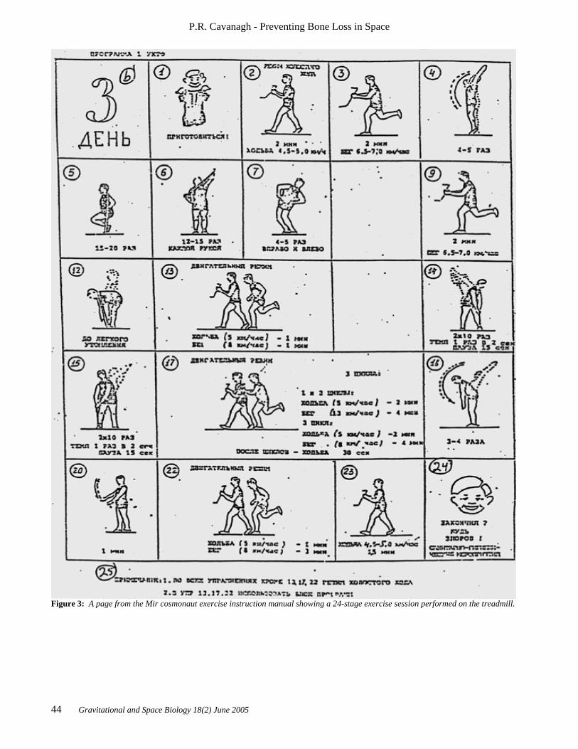
Cosmonauts on Mir have been said to perform exercise “up to 3 hours per day” (Nicogossian et al., 1994), while others believe that the exercise was 2-3 hours on 3 of 4 days (LeBlanc et al., 2000). The passive treadmill was considered the “stadium” from which exercise was performed. While the subject was tethered in place using bungees, he not only walked and ran, but also performed calisthenics and upper-body exercises using additional bungee cords for resistance (Figure 3). The data from LeBlanc et al. (2000) showed clearly that this protocol, even if faithfully performed, is not an effective countermeasure for bone loss. The exercise facilities available on the ISS through Expedition 12 consist of a Treadmill Vibration Isolation and Stabilization System (TVIS; Figure 2c; McCrory et al., 1999), a cycle ergometer with vibration isolation (CEVIS; Figure 2d), and the Interim Resistive Exercise Device (iRED; Figure 2e; Schneider et al., 2003). There is also a bicycle ergometer available in the Russian segment. None of these devices has a force measurement capability, and there is very little published information about their performance characteristics. When running on the treadmill, a subject must be tethered using a subject load device (SLD) to restrain him on the treadmill surface, and, optionally, a subject position device (SPD) is used to keep the subject in an area of the treadmill where a pitch oscillation of the treadmill will not be initiated. Each crew member is assigned a period of 2.25-2.5 hours every day for exercise—including set-up and break-down time, which can consume more than 50% of the assigned period. The work of Lang et al. (2004) showed that these devices as they are presently used are not effective as a countermeasure for bone loss during long-duration flights. As we shall discuss below, prolonged bed rest is considered to be a viable analog of space flight. Shackelford et al. (2004) conducted a program of vigorous resistance training (averaging 74% of one repetition maximum) in nine individuals during a 17-week confinement. The exercise was found to have a beneficial effect on BMD during bed rest compared to controls, specifically in the lumbar spine (+3% vs. -1%), total hip (+1% vs. -3%), heel (+1% vs. -3%), total body (0% vs. -1%), and pelvis (-0.5% vs. -3%). However, the high levels of load imposed on the muscle groups studied have never been achieved in space, and it is unlikely that in-flight exercise devices currently in use will permit such loads to be achieved.
P.R. Cavanagh - Preventing Bone Loss in Space
Gravitational and Space Biology 18(2) June 2005 43
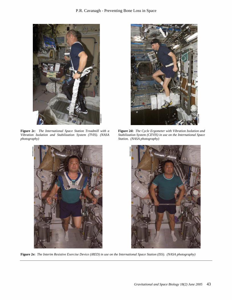
Figure 2c: The International Space Station Treadmill with a Vibration Isolation and Stabilization System (TVIS). (NASA photography)
Figure 2d: The Cycle Ergometer with Vibration Isolation and Stabilization System (CEVIS) in use on the International Space Station. (NASA photography)
Figure 2e: The Interim Resistive Exercise Device (iRED) in use on the International Space Station (ISS). (NASA photography)
P.R. Cavanagh - Preventing Bone Loss in Space
44 Gravitational and Space Biology 18(2) June 2005
Figure 3: A page from the Mir cosmonaut exercise instruction manual showing a 24-stage exercise session performed on the treadmill.
P.R. Cavanagh - Preventing Bone Loss in Space
Gravitational and Space Biology 18(2) June 2005 45
WHY HAVE EXERCISE COUNTERMEASURES IN SPACE NOT BEEN EFFECTIVE? Since exercise has been the only countermeasure to bone loss so far attempted in space, and since considerable bone loss has occurred on all flights to date, it would be tempting to conclude that exercise is not an appropriate countermeasure. There are, however, several reasons why such a conclusion may be premature: (1) There has never been a controlled study of exercise, either in space or during bed rest. The lack of such a study in the more than 40 years that this problem has been recognized is highly perplexing to the current authors and perhaps reflects the fact that NASA has traditionally been an engineering rather than a science agency; (2) The loads applied to the body by any piece of exercise equipment were not measured prior to 2003, so it is not known whether or not equipment exerted 1-g-like loads; (3) Exercise adherence may have been less than optimal and, contrary to common belief, the ISS program was the first time a mandatory exercise program was instituted as part of a flight plan; (4) It is not known if a single daily concentrated “dose” of exercise in 0-g can effectively replace a “dose” that in 1-g is distributed throughout the day; (5) The duration of exercise programmed to date may not have been adequate to achieve the desired result; (6) There is considerable debate in the scientific community about the optimal loading strategy that will provide an osteogenic stimulus to bone (Turner, 1998; Turner and Pavalko, 1998). Evidence in the literature ranges from a few intermittent large loads per day (Lanyon, 1996) to 18,000 small-amplitude vibrations in a 10-minute period (Rubin et al., 2002a; Rubin et al., 2002b). There is also debate regarding the relative role of force and rate of change of force (Cullen et al., 2001; Linde et al., 1991; Mosley and Lanyon, 1998). Our own experiments using force-measuring insoles during exercise on the ISS (Rice et al., 2004) have suggested that neither the load nor the duration of treadmill exercise in the current ISS exercise program is adequate to replace 1-g exercise. Only when all six issues noted above have been carefully examined can the role of exercise as a countermeasure to in-flight bone loss be determined. Until such time, it is reasonable that the flight medicine community is looking to explore the use in space of pharmacological options that are being used on Earth to prevent postmenopausal osteoporosis. The remainder of this review will examine the cellular and molecular targets for such therapy, present the currently available options, and discuss the limitations of knowledge required for the implementation of these therapies in space.
BONE REMODELING Bone is an active tissue that is constantly being remodeled, principally by the action of two cell types: osteoclasts, which resorb bone, and osteoblasts, which build new bone (Figure 4). It is estimated that all of the bone in the adult skeleton is replaced every 10 years (Marx, 2004). Homeostasis of bone is only maintained if the opposing—or perhaps complementary—actions of osteoblasts and osteoclasts are balanced. A defect in either process can result in accumulation of bone (as in osteopetrosis) or in a net loss of bone (as in osteoporosis) (Helfrich, 2003; Phan et al., 2004). The mineral phase of bone is of primary importance to density, and therefore BMD has been used in the past as a main indicator of bone status (Kanis, 2002). However, there is now increasing interest in measures of bone “quality” that include structural as well as compositional information (Ammann and Rizzoli, 2003; Turner, 2002), and it is likely that a composite measure will eventually replace BMD as the parameter of choice. The majority of current therapeutic interventions could be classed as resorption-prevention drugs. A number of advances in understanding how osteoclasts differentiate, mature, and are activated have recently been made (Boyle et al., 2003; Marx, 2004). Prevention of osteoclast formation (osteoclastogenesis) and development has been a prime target through a number of different pathways (see below). On the formation side of the equation, preventing osteoblast cell death (apoptosis) is also of interest, and a number of other “anabolic” or bone-building drugs with uncertain mechanisms are also being explored (Bisello et al., 2004; Deal and Gideon, 2003). SUPPLEMENTATION Traditionally, supplementation of daily intake of vitamin D and calcium (current recommended daily allowances [RDAs] 400 IUs and 1500 mg, respectively) have been considered mainstays of osteoporosis prevention. Adequate calcium is needed for mineralization, and vitamin D plays a role in the regulation of calcium deposition for bone mineralization. Both of these agents have weak antiresorptive properties (compared, for example, to bisphosphonates [Reginster, 2004]—see below), but combined therapy for 18 months (1200 mg calcium plus 800 IU vitamin D3 [cholecalciferol]) has been shown to be effective in reducing hip fracture in elderly women who were Vitamin D deficient (Chapuy et al., 1992). The bioavailability of the various forms of calcium used in supplementation (calcium carbonate, citrate, phosphate, lactate, and formate) have been shown to be different (Hanzlik et al., 2005). Since it is not always clear whether or not dietary intake of these agents is adequate, most drug trials routinely include calcium and vitamin D supplementation in control, placebo, and treatment arms. Astronaut diets can

P.R. Cavanagh - Preventing Bone Loss in Space
46 Gravitational and Space Biology 18(2) June 2005
Figure 4: Schematic of a bone multi-cellular unit (BMU). Osteoclasts resorb a cavity that is later occupied by osteoblasts that lay down new bone in the form of osteoid that subsequently undergoes mineralization (Deal and Gideon, 2003). Reprinted with the permission of The Cleveland Clinic Foundation.
be closely controlled, so inclusion of RDA and supplementation in diet can be easily accomplished. HORMONE REPLACEMENT THERAPY Estrogen, in the form of 17β-estradiol, has a complex agonistic action on estrogen receptors (ERs) in the nucleus of osteoblastsic cells (Riggs and Hartmann, 2003), which in turn affect estrogen receptor elements (EREs) in target genes. In estrogen deficiency, resorption outpaces formation, resulting in net bone loss. Estrogen also stimulates breast epithelial cell production and has been implicated in breast cancer risk (Riggs and Hartmann, 2003). Hormone replacement therapy (HT) using estrogen (unopposed HT) or estrogen-progestin (opposed HT) was widely recommended for postmenopausal women until the landmark Women’s Health Initiative (WHI) study (Rossouw et al., 2002) demonstrated a number of adverse responses (increased risk of coronary artery disease, stroke, thromboembolism, and breast cancer) in subjects using opposed HT. Riggs and Hartmann (2003) stated that estrogen was the most widely prescribed drug in the world and that it was taken by 38% of postmenopausal women in the United States. In addition to reducing the risk for nonvertebral fractures (Torgerson and Bell-Syer, 2001), HT also had the added advantage of relieving a number of perimenopausal symptoms. Because of the increased risk of adverse side effects, HT is no longer recommended for prevention or treatment of osteoporosis (Kessel, 2004). Estrogen also has significant effects on skeletal metabolism in men. Traditionally, estrogen was
considered the regulator of the female skeleton and testosterone the male regulator. The discovery of mutations in the aromatase gene in men and concurrent abnormalities in skeletal metabolism (osteopenia and unfused epiphyses) have focused attention on the importance of estrogen physiology. Aberrations in osteoclast acivity due to deficiency of inhibitors may attend the loss of estrogen with aging in both men and women and cause increased bone turnover (Carani et al., 1997; Khosla et al., 2002; Khosla et al., 2004). SELECTIVE ESTROGEN RECEPTOR MODULATORS (SERMS) Because of the side effects of HT, there has been increased interest in this class of nonhormonal drugs that target the ER. SERMs can have both agonist and antagonist effects in different tissues (e.g., tamoxifen [Tamofen], used in the treatment of ER-positive breast cancer, is an antagonist that slows the proliferation of tumor cells, whereas raloxifene [Evista] is a bone agonist that has an antiresorptive effect) (Riggs and Hartmann, 2003). Different SERMs that have similar effects on bone (e.g., raloxifene and idoxifene [investigational]) appear to have their modes of action through different molecular pathways (Nuttall et al., 2000). Because raloxifene, which is administered orally once per day, has a preferential effect on vertebral fracture risk reduction (Ettinger et al., 1999), it is possible that there are differences between the action of SERMs on trabecular vs. cortical bone. There are some indications that raloxifene therapy decreases cardiovascular events in women with risk factors at baseline (Barrett-Connor et al., 2002) but carries

P.R. Cavanagh - Preventing Bone Loss in Space
Gravitational and Space Biology 18(2) June 2005 47
with it a small increase in the risk for thromboembolism (Daly et al., 1996). SERMs do not appear to alleviate postmenopausal symptoms (National Osteoporosis Foundation, 2002; Cranney et al., 2002). The common risk for both therapies is that of deep vein thrombosis, especially in conditions of clotting abnormalities. This risk is small but nonetheless present statistically. In other situations the drugs have divergent risks. Breast hyperplasia and breast cancer are not found with the SERM agents as they are with estrogen. Moreover, the SERM drugs do not cause cervical endometrial hyperplasia, menstrual bleeding, or cervical cancer. The SERM drugs may offer an option for treatment of prostate cancer. In the presence of decreasing androgens with aging, estrogen induces prostatic hyperplasia and neoplasia. Antiestrogens and SERMs suppress prostate carcinogenesis. Some preliminary studies suggest that SERMs may not be useful as a general treatment for male osteoporosis, but there are some male patients, small in number, with the requisite balance of estrogen and testosterone for whom SERMs may be beneficial (Steiner and Raghow, 2003; Doran et al., 2001).
ANTIRESORPTIVE DRUGS The largest class of antiresorptive drugs is the bisphosphonates (such as alendronate [Fosamax], etidronate [Didronel], ibandronate [Boniva], pamidronate [Aredia], risedronate [Actonel], zoledronate [Zometa], and tiludronate (Skelid)]. The drugs are distinguished by their potency, which is usually positively affected by the presence of a nitrogen atom (e.g., etidronate [low] to zoledronate [high]), by their mode of delivery (e.g., intravenously for pamidronate and zoledronate, orally for alendronate, orally, intravenously, or by injection for ibandronate), and by the frequency and size of dosing (e.g., 5 or 10 mg daily or weekly for alendronate, 2.5 mg daily for ibandronate, 10-90 mg annually for zoledronate). An excellent review of these drugs is provided by Reginster (2004). Bisphosphonates are powerful and specific inhibitors of osteoclasts (Figure 5). They were originally thought to exert their action via incorporation in the skeleton by mimicking pyrophosphate and binding to the hydroxy-apatite crystals in the bone matrix (Licata, 2005), especially at sites of remodeling, the bone multicellular units (BMUs) (Russell et al., 1999).
Figure 5: Schematic of bisphosphonate action (Rodan and Fleisch, 1996). Where bisphosphonate (BP in the diagram) has been incorporated into the bone matrix, osteoclastic resorption of bone cannot occur. Reprinted with permission.
Their actions have since been shown to be complex, however. The amino bisphosphonates inhibit osteoclastic cholesterol synthesis and membrane function and increase cellular apoptosis. The non-amino bisphosphonates produce ineffective ATP analogs and inhibit osteoclast
function by “energy starving” the cell. Once incorporated, bisphosphonates remain bound at the bone surface and exhibit extremely low serum concentrations, thus limiting side effects. In general, the third generation (N2 containing) bisphosphonates have shown approximately

P.R. Cavanagh - Preventing Bone Loss in Space
48 Gravitational and Space Biology 18(2) June 2005
40-50% reduction in the risk of vertebral and nonvertebral fractures compared with placebo in postmenopausal women (Black et al., 1996; Chesnut et al., 2004; Harris et al., 1999) and have also resulted in increased BMD in the lumbar spine, total hip, and trochanter in women with and without osteoporosis (Cooper et al., 2003; Mortensen et al., 1998; Ravn et al., 1999). There are two bed rest studies involving bisphosphonates that are relevant to the space program (LeBlanc et al., 2002; Watanabe et al., 2004). LeBlanc et al. (2002) administered 10 mg of alendronate daily to eight male subjects undergoing 17 weeks of horizontal bed rest. Compared with concurrent and historical controls, BMD loss was significantly attenuated (or eliminated) in the alendronate treatment group in the lumbar spine, femoral neck, trochanter, and pelvis (but not calcaneus). Most markers of bone collagen breakdown and resorption (cross-linked N-teleopeptide of type I collagen [NTX], pyridinium [Pyd], and deoxypyridinium [D-Pyd]) in-creased in both groups, but significantly less so in the treated group than in controls. Markers of bone formation (alkaline phosphatase, bone-specific alkaline phosphatase, and osteocalcin) were unchanged in controls, but were decreased in the treated group because of the reduced bone turnover. These results demonstrate that the drug does not ablate the bone loss totally, thus the observed clinical effects may require simultaneous mechanical stress. Watanabe et al. (2004) administered 60 mg of pamid-ronate to seven male subjects 14 days before 90 days of 6-degree head-down bed rest. These authors also showed that alendronates, in addition to their osteoprotective properties, decrease the risk of renal stones. Compared with sedentary and resistance training controls, the pamidronate-treated subjects not only maintained significantly more bone in the proximal femur and lumbar spine, but also showed no evidence of urolithiasis (stones in the urinary tract). In the other groups, six subjects were found to have radiographic evidence of stone formation during bed rest. All but one of these stone-forming subjects had baseline hypercalciuria (>250 mg per day). Such patterns of stone formation may be a feature of all bed rest studies, and perhaps of long-duration space flight, that has been previously overlooked. However, it is extremely unusual for healthy patients with no prior stone risk to become “at-risk” in such a short time, and these results, although cautionary, need to be replicated. Shapiro et al. (personal communication), in an as yet unpublished study, showed a reduction of bone loss in the lower extremities of patients with spinal cord injuries who had been administered intravenous zoledronate. The paradigm of spinal cord injury has been suggested to be another analog of space flight, although the absence of muscular action may tend to make it even more severe from a disuse point of view. The side effects of bisphosphonate therapy from the major study series have generally been mild (adverse effects in
the upper gastrointestinal (GI) tract, constipation, flatulence, hypocalcemia, and diarrhea), but severe esophageal reactions have been reported with alendronate (Schnitzer et al., 2000). Consequently, its use is not recommended for patients with a history of upper GI complaints. There is some concern that bone formed during the administration of bisphosphonates may not have the same “quality” as normal bone, thus negatively affecting on the mechanical integrity of the skeleton. Animal studies with bisphosphonates have shown a delay in fracture healing in rats and rabbits and an increase in the presence and persistence of microcracks and reduced remodeling, suggesting a potential change in biomechanical factors (Li et al., 1999; Li et al., 2001; Mashiba et al., 2001; Lehman et al., 2004). In addition, it is notable that Ruggiero et al. (2004) have identified a cluster of patients on chronic bisphosphonate therapy that had an associated risk of osteonecrosis of the jaw. This condition is also seen in the myeloma patients treated with i.v. zoledronic acid monthly (Lugassy et al., 2004), which is not the way it is used for treating osteoporosis. It is possible that such patients may have immune compromise supporting local dental infection and subsequent bone destruction. If sequential combination therapy of different drugs is planned, Gasser et al. (2000) showed in studies of rat bone that the response to an anabolic drug (see below) was delayed in animals pretreated with bisphosphonates. However, in clinical studies, long-term use of alendronate and risedronate for 7-10 years shows no similar findings. Both drugs still suppress fractures, which argues against the adverse effects seen in animal models. Furthermore, histomorphometry shows no abnormal characteristics in patients after 3 or more years of use. ANABOLIC DRUGS Drugs in this class exert their mode of action by increasing bone formation rather than by inhibiting resorption. The important role of parathyroid hormone (PTH) in regulating bone and mineral metabolism has been known for more than 70 years (Bisello et al., 2004), but classical teaching identifies PTH as a powerful mobilizer of skeletal calcium into the serum in the presence of hypocalcemia (i.e., a state of secondary hyperparathyroidism). Evidence from animal exper-iments has shown that daily injection of PTH had anabolic effects on bone, and recent work has resolved these apparently paradoxical effects by showing a dependency on the pattern of exposure. Chronic elevation of PTH (as in primary hyperparathyroidism) leads to increased bone resorption, whereas intermittent elevation (as in once-daily injections with a short half-life) leads to increased formation. The mechanism of action of PTH appears to be the stimulation of existing osteoblasts via surface PTH receptors and interaction with RANK-L (NF-κB; see below) (Deal and Gideon, 2003). It is known that the amino-terminal region of PTH (the first 34 amino acids) is necessary and sufficient for full activity,

P.R. Cavanagh - Preventing Bone Loss in Space
Gravitational and Space Biology 18(2) June 2005 49
and the only anabolic agent that is currently Food and Drug Administration (FDA) approved for use in the treatment of osteoporosis is recombinant teriparatide (rhPTH [1-34] [Forteo]). Administration of teriparatide (daily subcutaneous injection 20 µg or 40 µg for 19 months) to women with low bone mass and a history of prior fracture resulted in an almost 10% increase in vertebral BMD; treatment reduced the risk of a second vertebral fracture by approximately 65% and that of a nonvertebral fracture by approximately 50% compared with placebo. There is some concern that high doses of teriparatide (up to 60 times greater than approved human doses) have caused osteosarcoma in rats, but not monkeys. No similar complications have been observed in human studies, but an initially promising trial conducted in men (Orwoll et al., 2003) was terminated because of concern regarding the animal results. It is also known that PTH-related peptide (PTHrP), a protein with some homology to PTH that is produced by tumors and leads to hypercalcemia, shares many of the actions of PTH but has receptors that are much more widely distributed (Bisello et al., 2004). The authors have initial evidence from human studies that PTHrP has the potential to be a powerful anabolic agent, and clinical trials to explore this possibility are ongoing. It is possible that the mechanism for the differential effects of intermittent vs. continuous levels of PTH is in the modulation of the osteoprotegerin (OPG)/receptor
activator of nuclear factor-κB ligand (RANK-L) ratio (see below) (Locklin et al., 2001). RANK-L/OPG In 1997, a new pathway regulating bone resorption was identified (serendipitously) by a group looking for novel genes in the rat intestine (Simonet et al., 1997). The transgenic mouse overexpressing one particular gene was found to have ostopetrosis and a deficiency of osteoclasts (Khosla, 2001) and the responsible protein was called osteoprotegerin for its protective role in maintaining bone mass. Simultaneously, Yasuda et al. (1998) found the same protein in a targeted search for the signaling link that had been previously hypothesized to exist between osteoclasts and ostoblasts (Rodan and Martin, 1982). The pathway that has been identified as a result of these and subsequent studies is shown in Figure 6. OPG is secreted as a soluble protein from bone marrow stromal cells and appears to be a decoy receptor, which binds to RANK-L. Since RANK-L is a major factor in osteoclast differ-entiation, activation, and apoptosis inhibition, it follows that the binding of RANK-L to OPG, rather than to its target RANK on the osteoclast precursor cell, will prevent bone resorption. Because RANK-knockout mice also exhibited osteopetrosis and absence of osteclasts (Li et al., 2000), the existence of a new OPG/RANK/RANK-L pathway in the control of bone resorption was confirmed. Several genetic mutations of this pathway are associated with bone diseases such as the family of hyperphos-phatasias, Paget’s disease, and possible bone loss in inflammatory arthritis (Boyle et al., 2003; Khosla, 2001).
Figure 6: Schematic of the OPG/RANK-L pathway (Khosla, 2001). Note that OPG acts as a decoy receptor preventing RANK from attaching to its ligand RANK-L and therefore inhibiting osteoclast differentiation. Copyright 2001, The Endocrine Society. Reprinted with permission.

P.R. Cavanagh - Preventing Bone Loss in Space
50 Gravitational and Space Biology 18(2) June 2005
OPG was an obvious choice as a clinical therapeutic agent to prevent osteoporosis, and indeed two forms of the protein were examined by Amgen in clinical trials of osteoporosis (Bekker et al., 2001) and multiple myeloma and breast carcinoma (Body et al., 2003). A presumed combination of concerns regarding efficacy, safety, treatment duration and manufacturing factors has resulted in OPG’s no longer being examined for clinical use. OPG does, however, continue to be explored for the treatment of bone tumors (Wittrant et al., 2004). A fully human monoclonal antibody for RANK-L, AMG 162, is being developed as an osteoporosis treatment instead (Bekker et al., 2004; McClung et al., 2004). Phase III clinical trials were initiated in late 2004 for AMG 162. CALCITONIN The peptide calcitonin exerts a complex inhibitory action on osteoclast function (Kajiya et al., 2003). It has been used in trials of both men and women with low bone mass and has been shown to stabilize (or prevent) bone loss (Toth et al., 2005) and, in women, to decrease vertebral fracture rate (Munoz-Torres et al., 2004). One attractive
feature of calcitonin is that it can be administered by many routes, including nasally in the form of a daily (or intermittently administered [Tekeoglu et al., 2005]) spray. BONE GENETICS In the last 5 years, early insights into some of the genetic determinants of bone mass have been obtained. Ralston (2003) and Recker (2004) have recently reviewed the status of present knowledge in this area. Johnson et al. (2004) have commented regarding “how little we really know about the genes that control bone mass.” The genetic basis for diseases caused by a defect in osteoclasts is discussed by Helfrich (2003). Gong et al. (2001) found that that the LRP5 gene, which encodes the low-density lipoprotein receptor-related protein 5, is important in bone mass accrual. They reported that loss-of-function mutations in LRP5 caused the autosomal recessive disorder osteoporosis-pseudoglioma syndrome and that Wnt-mediated signaling via LRP5 affects bone accrual during growth and peak bone mass (Figure 7).
Figure 7: The Wnt signaling pathway that has been discovered through genetic studies of patients with high bone mass (Johnson et al., 2004). Adapted and reproduced from J Bone Miner Res 2004;19:1749-1757 with permission of the American Society for Bone and Mineral Research. Subsequently, mutations in the same gene were also found to be associated with diseases in which there was high bone mass (Boyden et al., 2002; Little et al., 2002). The lack of inhibitory action of the protein Dkk-1 on the Wnt signaling pathway suggested this protein as a potential therapeutic target for modulating bone mass. A review of LRP5 and Wnt signaling is presented by
Johnson et al. (2004). Genes regulating lipoxygenase are also believed to influence bone mass (Klein et al., 2004). There are indications that the Wnt signaling pathway is activated in response to mechanical loading (Johnson, 2004), and this may be a key element in the elusive mechanotransduction that has long been hypothesized to exist.
P.R. Cavanagh - Preventing Bone Loss in Space
Gravitational and Space Biology 18(2) June 2005 51
An alternative approach to the human linkage and association studies described above is the use of mouse models in quantitative trait locus (QTL) analysis (Liu et al., 2003; Rosen et al., 2001). QTL is basically a statistical analysis, sometimes of the entire genome, to identify which regions of the genome contain loci that influence the phenotype of interest. The genes encoding type I collagen (COLIA1 and COLIA2) are mutated in osteogenesis imperfecta and may be useful markers of other osteoporotic phenotypes (Mottes et al., 1998). The estrogen receptor gene may regulate some aspects of bone density since the discovery of a male patient with a gene mutation and osteoporosis (Gennari et al., 2005).
Finally, the early discovery of the vitamin D receptor gene helped introduce the notion that bone mass had a genetic basis (Eisman, 1995). The complexity of BMD as a trait and the importance of gene-environment interactions have been emphasized in a study of risk factors for low spine and hip BMD involving 12 candidate gene loci and lifestyle factors by Lau et al. (2005). While these various studies of genetic influence on bone mass are in their early stages, there is a high likelihood that they will eventually identify new therapeutic targets.
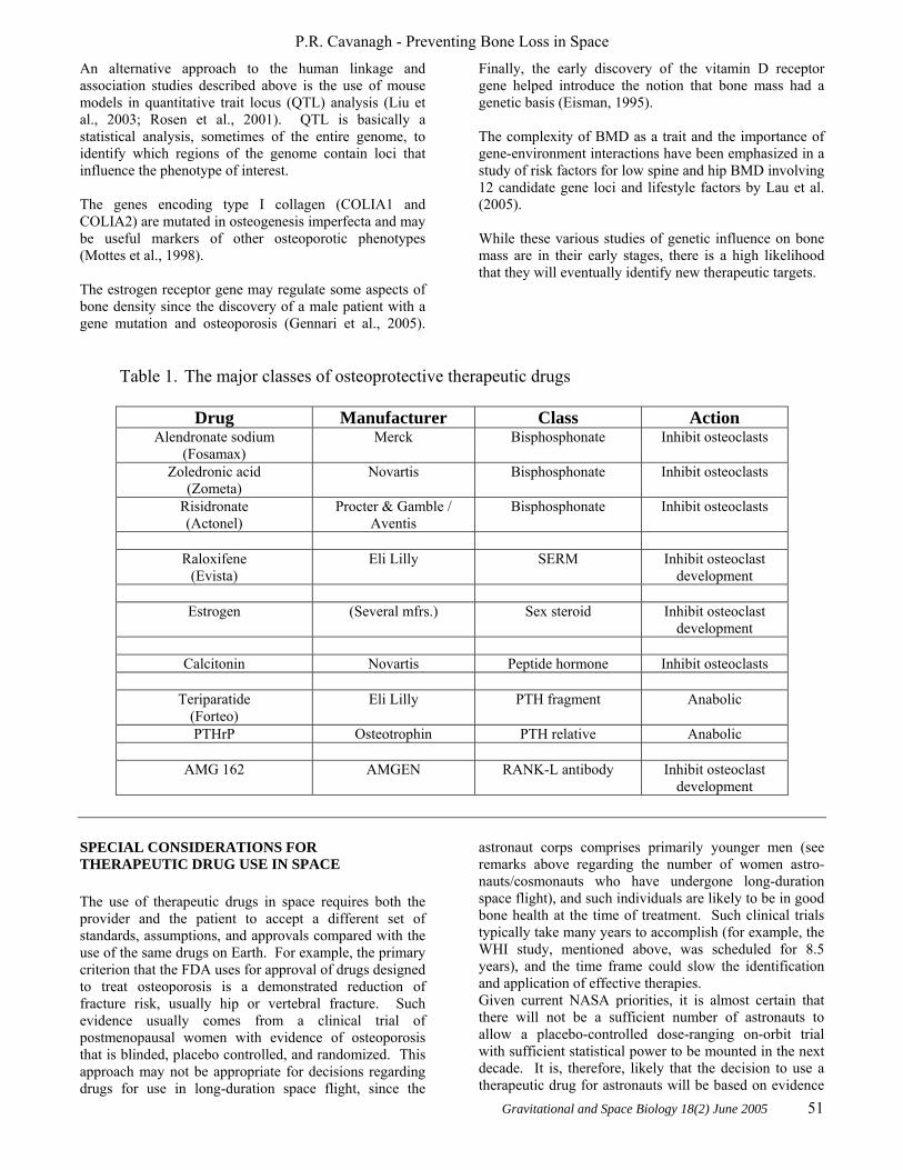
Table 1. The major classes of osteoprotective therapeutic drugs
Drug Manufacturer Class Action Alendronate sodium
(Fosamax) Merck Bisphosphonate Inhibit osteoclasts
Zoledronic acid (Zometa)
Novartis Bisphosphonate Inhibit osteoclasts
Risidronate (Actonel)
Procter & Gamble / Aventis
Bisphosphonate Inhibit osteoclasts
Raloxifene
(Evista) Eli Lilly SERM Inhibit osteoclast
development
Estrogen (Several mfrs.) Sex steroid Inhibit osteoclast development
Calcitonin Novartis Peptide hormone Inhibit osteoclasts
Teriparatide
(Forteo) Eli Lilly PTH fragment Anabolic
PTHrP Osteotrophin PTH relative Anabolic
AMG 162 AMGEN RANK-L antibody Inhibit osteoclast development
SPECIAL CONSIDERATIONS FOR THERAPEUTIC DRUG USE IN SPACE The use of therapeutic drugs in space requires both the provider and the patient to accept a different set of standards, assumptions, and approvals compared with the use of the same drugs on Earth. For example, the primary criterion that the FDA uses for approval of drugs designed to treat osteoporosis is a demonstrated reduction of fracture risk, usually hip or vertebral fracture. Such evidence usually comes from a clinical trial of postmenopausal women with evidence of osteoporosis that is blinded, placebo controlled, and randomized. This approach may not be appropriate for decisions regarding drugs for use in long-duration space flight, since the
astronaut corps comprises primarily younger men (see remarks above regarding the number of women astro-nauts/cosmonauts who have undergone long-duration space flight), and such individuals are likely to be in good bone health at the time of treatment. Such clinical trials typically take many years to accomplish (for example, the WHI study, mentioned above, was scheduled for 8.5 years), and the time frame could slow the identification and application of effective therapies. Given current NASA priorities, it is almost certain that there will not be a sufficient number of astronauts to allow a placebo-controlled dose-ranging on-orbit trial with sufficient statistical power to be mounted in the next decade. It is, therefore, likely that the decision to use a therapeutic drug for astronauts will be based on evidence

P.R. Cavanagh - Preventing Bone Loss in Space
52 Gravitational and Space Biology 18(2) June 2005
from a bed rest study supported by experience in a few individual volunteers who will take the drugs prior to and/or during space flight. Among the questions that will need to be answered in these human trials are: (1) What is the bioavailability of the various drug therapies in 0-g? (2) Are the dose-response curves similar in 0-g to those established in 1-g? (3) What are the post-flight consequences for bone health of taking osteoprotective drugs? (4) If drugs need to be taken on-orbit, how should they be stored for maximum effectiveness? (5) How will a drug’s effectiveness be determined on-orbit so that doses can be modulated? (6) What is the best combination of drug and exercise countermeasures? SUMMARY AND CONCLUSIONS This review has defined the current status of exercise and therapeutic drug countermeasures for bone loss during long-duration space flight. The available data indicate that exercise countermeasures to date have not been effective and crew members continue to lose significant bone mass in the lower extremities and lumbar spine. Better-designed studies are needed to determine if the entire distributed daily dose of exercise that occurs in 1-g can be successfully replaced by short periods of high-intensity exercise on-orbit. Exercise dose on-orbit must also be quantified. Drug therapeutics for bone have not yet been used in space, and, given the considerable experience using several classes of osteoprotective drugs on Earth (mostly in postmenopausal women with low bone mass), it seems wise to explore such interventions for use during space flight. However, the many differences between the 1-g clinical studies and the 0-g individual prescription must be carefully considered. Many new therapies can be expected in the future as investigators achieve a better understanding of the genetic regulation of bone mass, and genetic screening may offer a means of selecting crew members with a low susceptibility to bone loss. ACKNOWLEDGEMENTS Supported by National Space Biomedical Research Institute grants BL00401 and BL00402 through NASA NCC 9-58. The assistance of Ted Bateman, Ph.D., was appreciated. REFERENCES Ammann, P., and Rizzoli, R. 2003. Bone strength and its determinants. Osteoporosis International 14 Suppl 3:S13-8 Barrett-Connor, E., Grady, D., Sashegyi, A., Anderson, P.W. , Cox, D.A., Hoszowski, K., Rautaharju, P., and Harper, K.D. 2002. Raloxifene and cardiovascular events in osteoporotic postmenopausal women: four-year results from the MORE (Multiple Outcomes of Raloxifene
Evaluation) randomized trial. Journal of the American Medical Association 287(7):847-57 Bekker, P.J., Holloway, D., Nakanishi, A., Arrighi, M., Leese, P.T., and Dunstan, C.R. 2001. The effect of a single dose of osteoprotegerin in postmenopausal women. Journal of Bone and Mineral Research 16(2):348-60 Bekker, P.J., Holloway, D.L., Rasmussen, A.S., Murphy, R., Martin, S.W., Leese, P.T., Holmes, G.B., Dunstan, C.R., and DePaoli, A.M. 2004. A single-dose placebo-controlled study of AMG 162, a fully human monoclonal antibody to RANKL, in postmenopausal women. Journal of Bone and Mineral Research 19(7):1059-66 Biriukov, E.N., and Krasnykh, I.G. 1970. Changes in the Optical Density of Bone Tissue an din teh Calcium Metabolism of the Astronauts. In: Kosmicheskaia Biologiia i Meditsina. (Nikivaev, A.G., and Sevastianov, V.I. Eds) Moscow: pp. 42-45. Bisello, A., Horwitz, M.J., and Stewart, A.F. 2004. Parathyroid hormone-related protein: an essential physiological regulator of adult bone mass. Endocrinology 145(8):3551-3 Black, D.M., Cummings, S.R., Karpf, D.B., Cauley, J.A., Thompson, D.E., Nevitt, M.C., Bauer, D.C., Genant, H.K., Haskell, W.L., Marcus, R., Ott, S.M., Torner, J.C., Quandt, S.A., Reiss, T.F., and Ensrud, K.E. 1996. Randomised trial of effect of alendronate on risk of fracture in women with existing vertebral fractures. Fracture Intervention Trial Research Group. Lancet 348(9041):1535-41 Body, J.J., Greipp, P., Coleman, R.E., Facon, T., Geurs, F., Fermand, J.P., Harousseau, J.L., Lipton, A., Mariette, X., Williams, C.D., Nakanishi, A., Holloway, D., Martin, S.W., Dunstan, C.R., and Bekker, P.J. 2003. A phase I study of AMGN-0007, a recombinant osteoprotegerin construct, in patients with multiple myeloma or breast carcinoma related bone metastases. Cancer 97(3 Suppl):887-92
Boyden, L.M., Mao, J., Belsky, J., Mitzner, L., Farhi, A., Mitnick, M.A., Wu, D., Insogna, K., and Lifton, R.P. 2002. High bone density due to a mutation in LDL-receptor-related protein 5. New England Journal of Medicine 346(20):1513-21 Boyle, W.J., Simonet, W.S., and Lacey, D.L. 2003. Osteoclast differentiation and activation. Nature 423(6937):337-42 Brodzinski, R.L., Rancitelli, L.A., Haller, W.A., and Dewey, L.S. 1971. Calcium, potassium, and iron loss by Apollo VII, VIII, IX, X and XI astronauts. Aerospace Medicine 42(6):621-26 Carani, C., Qin, K., Simoni, M., Faustini-Fustini, M., Serpente, S., Boyd, J., Korach, K.S., and Simpson, E.R.

P.R. Cavanagh - Preventing Bone Loss in Space
Gravitational and Space Biology 18(2) June 2005 53
1997. Effect of testosterone and estradiol in a man with aromatase deficiency. New England Journal of Medicine 337(2):91-95 Chapuy, M.C., Arlot, M.E., Duboeuf, F., Brun, J., Crouzet, B., Arnaud, S., Delmas, P.D., and Meuntier, P.J. 1992. Vitamin D3 and Calcium to prevent hip fractures in elderly women. New England Journal of Medicine 327:1637-42 Chesnut, C.H., III, Skag, A., Christiansen, C., Recker, R., Stakkestad, J.A., Hoiseth, A., Felsenberg, D., Huss, H., Gilbride, J., Schimmer, R.C., and Delmas, P.D. 2004. Effects of oral ibandronate administered daily or intermittently on fracture risk in postmenopausal osteoporosis. Journal of Bone and Mineral Research 19(8):1241-9 Cooper, C., Emkey, R.D., McDonald, R.H., Hawker, G., Bianchi, G., Wilson, K., and Schimmer, R.C. 2003. Efficacy and safety of oral weekly ibandronate in the treatment of postmenopausal osteoporosis. Journal of Clinical Endocrinology and Metabolism 88(10):4609-15 Cranney, A., Tugwell, P., Zytaruk, N., Robinson, V., Weaver, B., Adachi, J., Wells, G., Shea, B., and Guyatt, G. 2002. Meta-analyses of therapies for postmenopausal osteoporosis. IV. Meta-analysis of raloxifene for the prevention and treatment of postmenopausal osteoporosis. Endocrine Reviews 23(4):524-8 Cullen, D.M. , Smith, R.T., and Akhter, M.P. 2001. Bone-loading response varies with strain magnitude and cycle number. Journal of Applied Physiology 91(5):1971-6 Daly, E., Vessey, M.P., Hawkins, M.M., Carson, J.L., Gough, P., and Marsh, S. 1996. Risk of venous thromboembolism in users of hormone replacement therapy. Lancet 348(9033):977-80 Deal, C., and Gideon, J. 2003. Recombinant human PTH 1-34 (Forteo): an anabolic drug for osteoporosis. Cleveland Clinic Journal of Medicine 70(7):585-6, 589-90, 592-4 passim Dietlein, L.F. 1965. Experiment M-3, inflight exerciser on Gemini IV. In: Manned Space Flight Experiments Symposium. Gemini missions III and IV. Washington DC: National Aeronautics and Space Administration, Dietlein, L.F., and Rapp, R.M. 1966. Experiment M-3, inflight exercise work tolerance. In: Gemini Midprogram Conference Including Experiment Results.NASA Special Publication, NASA-SP-121., pp. 393-96. Doran, P.M., Riggs, B.L., Atkinson, E.J., and Khosla, S. 2001. Effects of raloxifene, a selective estrogen receptor modulator, on bone turnover markers and serum sex steroid and lipid levels in elderly men. Journal of Bone and Mineral Research 16(11):2118-25
Eisman, J.A. 1995. Vitamin D receptor gene alleles and osteoporosis: An affirmative view. Journal of Bone and Mineral Research 10(9):1289-93 Ettinger, B. , Black, D.M., Mitlak, B.H., Knickerbocker, R.K., Nickelsen, T., Genant, H.K., Christiansen, C., Delmas, P.D., Zanchetta, J.R., Stakkestad, J., Gluer, C.C., Krueger, K., Cohen, F.J., Eckert, S., Ensrud, K.E., Avioli, L.V., Lips, P., and Cummings, S.R. 1999. Reduction of vertebral fracture risk in postmenopausal women with osteoporosis treated with raloxifene: results from a 3-year randomized clinical trial. Multiple Outcomes of Raloxifene Evaluation (MORE) Investigators. Journal of the American Medical Association 282(7):637-45 Gasser, J.A., Kneissel, M., Thomsen, J.S., and Mosekilde, L. 2000. PTH and interactions with bisphosphonates. Journal of Musculoskeletal and Neuronal Interactions 1(1):53-56 Gazenko, O.G., Gurovsky, N.N., Genin, A.M., Bryanov, I.I., Eryomin, A.V., and Egorov, A.D. 1976. Results of medical investigations carried out on board the Salyut orbital stations. Life Sciences and Space Research 14:145-52 Gennari, L., Merlotti, D., De Paola, V., Calabro, A., Becherini, L., Martini, G., and Nuti, R. 2005. Estrogen receptor gene polymorphisms and the genetics of osteoporosis: a HuGE review. American Journal of Epidemiology 161(4):307-20 Gong, Y., Slee, R.B., Fukai, N., Rawadi, G., Roman-Roman, S., Reginato, A.M., Wang, H., Cundy, T., Glorieux, F.H., Lev, D., Zacharin, M., Oexle, K., Marcelino, J., Suwairi, W., Heeger, S., Sabatakos, G., Apte, S., Adkins, W.N., Allgrove, J., Arslan-Kirchner, M., Batch, J.A., Beighton, P., Black, G.C., Boles, R.G., Boon, L.M., Borrone, C., Brunner, H.G., Carle, G.F., Dallapiccola, B., De Paepe, A., Floege, B., Halfhide, M.L., Hall, B., Hennekam, R.C., Hirose, T., Jans, A., Juppner, H., Kim, C.A., Keppler-Noreuil, K., Kohlschuetter, A., LaCombe, D., Lambert, M., Lemyre, E., Letteboer, T., Peltonen, L., Ramesar, R.S., Romanengo, M., Somer, H., Steichen-Gersdorf, E., Steinmann, B., Sullivan, B., Superti-Furga, A., Swoboda, W., van den Boogaard, M.J., Van Hul, W., Vikkula, M., Votruba, M., Zabel, B., Garcia, T., Baron, R., Olsen, B.R., and Warman, M.L. 2001. LDL receptor-related protein 5 (LRP5) affects bone accrual and eye development. Cell 107(4):513-23 Hanzlik, R.P., Fowler, S.C., and Eells, J.T. 2005. Absorption and elimination of formate following oral administration of calcium formate in female human subjects. Drug Metabolism and Disposition 33(2):282-86 Harris, S.T. , Watts, N.B., Genant, H.K., McKeever, C.D., Hangartner, T., Keller, M., Chesnut, C.H. 3rd, Brown, J., Eriksen, E.F., Hoseyni, M.S., Axelrod, D.W., and Miller, P.D. 1999. Effects of risedronate treatment on vertebral and nonvertebral fractures in women with

P.R. Cavanagh - Preventing Bone Loss in Space
54 Gravitational and Space Biology 18(2) June 2005
postmenopausal osteoporosis: a randomized controlled trial. Vertebral Efficacy With Risedronate Therapy (VERT) Study Group. Journal of the American Medical Association 282(14):1344-52 Helfrich, M.H. 2003. Osteoclast diseases. Microscopy Research and Technique 61(6):514-32 Iki, M., Kajita, E., Dohi, Y., Nishino, H., Kusaka, Y., Tsuchida, C., Yamamoto, K., and Ishii, Y. 1996. Age, menopause, bone turnover markers and lumbar bone loss in healthy Japanese women. Maturitas 25(1):59-67 Johnson, M.L. 2004. The high bone mass family--the role of Wnt/Lrp5 signaling in the regulation of bone mass. Journal of Musculoskeletal and Neuronal Interactions 4(2):135-38 Johnson, M.L., Harnish, K., Nusse, R., and Van Hul, W. 2004. LRP5 and Wnt signaling: a union made for bone. Journal of Bone and Mineral Research 19(11):1749-57 Kajiya, H., Okamoto, F., Fukushima, H., and Okabe, K. 2003. Calcitonin inhibits proton extrusion in resorbing rat osteoclasts via protein kinase A. Pflugers Archiv 445(6):651-8 Kanis, J.A. 2002. Diagnosis of osteoporosis and assessment of fracture risk. Lancet 359(9321):1929-36 Kessel, B. 2004. Hip fracture prevention in postmenopausal women. Obstetrical and Gynecological Survey 59(6):446-55; quiz 485 Khosla, S. 2001. Minireview: the OPG/RANKL/RANK system. Endocrinology 142(12):5050-5 Khosla, S., Melton, L.J. 3rd, and Riggs, B.L. 2002. Clinical review 144: Estrogen and the male skeleton. Journal of Clinical Endocrinology and Metabolism 87(4):1443-50 Khosla, S., Riggs, B.L., Atkinson, E.J., Oberg, A.L., Mavilia, C., Del Monte, F., Melton, L.J. 3rd, and Brandi, M.L. 2004. Relationship of estrogen receptor genotypes to bone mineral density and to rates of bone loss in men. Journal of Clinical Endocrinology and Metabolism 89(4):1808-16 Klein, R.F., Allard, J., Avnur, Z., Nikolcheva, T., Rotstein, D., Carlos, A.S., Shea, M., Waters, R.V., Belknap, J.K., Peltz, G., and Orwoll, E.S. 2004. Regulation of bone mass in mice by the lipoxygenase gene Alox15. Science 303(5655):229-32 Krasnykh, I.G. 1969. Mineral saturation of bone tissue under conditions of prolonged hypodynamia. NASA TT F-639 Lang, T., LeBlanc, A., Evans, H., Lu, Y., Genant, H., and
Yu, A. 2004. Cortical and trabecular bone mineral loss from the spine and hip in long-duration spaceflight. Journal of Bone and Mineral Research 19(6):1006-12 Langton, C.M., Haire, T.J., Ganney, P.S., Dobson, C.A., Fagan, M.J., Sisias, G., and Phillips, R. 2000. Stochastically simulated assessment of anabolic treatment following varying degrees of cancellous bone resorption. Bone 27(1):111-8 Lanyon, L.E. 1996. Using functional loading to influence bone mass and architecture: objectives, mechanisms, and relationship with estrogen of the mechanically adaptive process in bone. Bone 18(1 Suppl 1):37S-43S Lau, H.H., Ng, M.Y., Ho, A.Y., Luk, K.D., and Kung, A.W. 2005. Genetic and environmental determinants of bone mineral density in Chinese women. Bone [Available online via doi:10.1016/j.bone.2005.01.014.] LeBlanc, A., Schneider, V., Shackelford, L., West, S., Oganov, V., Bakulin, A., and Voronin, L. 2000. Bone mineral and lean tissue loss after long duration space flight. Journal of Musculoskeletal and Neuronal Interactions 1(2):157-60 LeBlanc, A., Shackelford, L., and Schneider, V. 1998. Future human bone research in space. Bone 22(5 Suppl):113S-6S LeBlanc, A.D., Driscol, T.B., Shackelford, L.C., Evans, H.J., Rianon, N.J., Smith, S.M., Feeback, D.L., and Lai, D. 2002. Alendronate as an effective countermeasure to disuse induced bone loss. Journal of Musculoskeletal and Neuronal Interactions 2(4):335-43 LeBlanc, A.D., Schneider, V., Shackelford, L., West, S., Oganov, V., Bakulin, A., and Veronin, L. 1996. Bone mineral and lean tissue loss after long duration space flight (Abstract). Journal of Bone and Mineral Research 11:S323 Lehman, R.A. Jr, Kuklo, T.R., Freedman, B.A., Cowart, J.R., Mense, M.G., and Riew, K.D. 2004. The effect of alendronate sodium on spinal fusion: a rabbit model. Spine Journal 4(1):36-43 Li, J., Mashiba, T., and Burr, D.B. 2001. Bisphosphonate Treatment Suppresses Not Only Stochastic Remodeling but Also the Targeted Repair of Microdamage. Calcified Tissue International 69(5):281-6 Li, J., Mori, S., Kaji, Y., Mashiba, T., Kawanishi, J., and Norimatsu, H. 1999. Effect of bisphosphonate (incadronate) on fracture healing of long bones in rats. Journal of Bone and Mineral Research 14(6):969-79 Li, J., Sarosi, I., Yan, X.Q., Morony, S., Capparelli, C., Tan, H.L., McCabe, S., Elliott, R., Scully, S., Van, G., Kaufman, S., Juan, S.C., Sun, Y., Tarpley, J., Martin, L., Christensen, K., McCabe, J., Kostenuik, P., Hsu, H., Fletcher, F., Dunstan, C.R., Lacey, D.L., and Boyle, W.J.
P.R. Cavanagh - Preventing Bone Loss in Space
Gravitational and Space Biology 18(2) June 2005 55
2000. RANK is the intrinsic hematopoietic cell surface receptor that controls osteoclastogenesis and regulation of bone mass and calcium metabolism. Proceedings of the National Academy of Sciences of the United States of America 97(4):1566-71 Licata, A.A. 2005. Discovery, clinical development, and therapeutic uses of bisphosphonates. Annals of Pharma-cotherapy 39(4):668-77 Linde, F., Norgaard, P., Hvid, I., Odgaard, A., and Soballe, K. 1991. Mechanical properties of trabecular bone: dependency on strain rate. Journal of Biomechanics 24(9):803-9 Little, R.D. , Carulli, J.P., Del Mastro, R.G., Dupuis, J., Osborne, M., Folz, C., Manning, S.P., Swain, P.M., Zhao, S., Eustace, B., Lappe, M.M., Spitzer, L., Zweier, S., Braunschweigner, K., Benchekroun, Y., Hu, X., Adair, R., Chee, L., Fitzgerald, G., McGuiere, S., Nogues, X., Gong, G., Allen, K.M., Tulig, C., Caruso, A., Tzellas, N., Bawa, A., Franklin, B., Anisowicz, A., Morales, A.J., Lomedico, P.T., Recker, S.M., Van Eedewegh, P., Recker, R.R., and Johnson, M.L. 2002. A mutation in the LDL receptor-related protein 5 gene results in the autosomal dominant high-bone-mass trait. American Journal of Human Genetics 70(1):11-19 Liu, Y.Z., Liu, Y.J., Recker, R.R., and Deng, H.W. 2003. Molecular studies of identification of genes for osteoporosis: the 2002 update. Journal of Endocrinology 177(2):147-96 Locklin, R.M., Khosla, S., and Riggs, B.L. 2001. Mechanisms of biphasic anabolic and catabolic effects of parathyroid hormone (PTH) on bone cells. Bone 28(Suppl):S80 Lugassy, G., Shaham, R., Nemets, A., Ben-Dor, D., and Nahlieli, O. 2004. Severe osteomyelitis of the jaw in long-term survivors of multiple myeloma: a new clinical entity. American Journal of Medicine 117(6):440-441 Mack, P.B., LaChance, P.A., Vose, G.P., and Vogt, F.B. 1967. Bone demineralization of foot and hand of Gemini-Titan IV, V and VII astronautis during orbital flight. American Journal of Roentgenology, Radium Therapy and Nuclear Medicine 100(3):503-11 Mack, P.B., and LaChance, P.L. 1967. Effects of recumbency and space flight on bone density. American Journal of Clinical Nutrition 20(11):1194-205 Mack, P.B., and Vogt, F.B. 1971. Roentgenographic bone density changes in astronauts during representative Apollo space flight. American Journal of Roentgenology, Radium Therapy and Nuclear Medicine 113(4):621-33 Marx, J. 2004. Coming to grips with bone loss. Science 305(5689):1420-2
Mashiba, T., Turner, C.H., Hirano, T., Forwood, M.R., Johnston, C.C., and Burr, D.B. 2001. Effects of suppressed bone turnover by bisphosphonates on microdamage accumulation and biomechanical properties in clinically relevant skeletal sites in beagles. Bone 28(5):524-31 McCarthy, I., Goodship, A., Herzog, R., Oganov, V., Stussi, E., and Vahlensieck, M. 2000. Investigation of bone changes in microgravity during long and short duration space flight: comparison of techniques. European Journal of Clinical Investigation 30(12):1044-54 McClung, M.R., Lewiecki, E.M., Bolognese, M.A., Woodson, G., Moffell, A., Peacock, M., Miller, P.D., Lederman, S., Chesnut, C.H., Murphy, R., Holloway, D.L., and Bekker, P.J. 2004. AMG 162 increases bone mineral density (BMD) within 1 month in postmenopausal women with low BMD (Abstract). Journal of Bone and Mineral Research 19(Suppl 1):S20 McCrory, J.L., Lemmon, D.R., Sommer, H.J., Prout, B., Smith, D., Korth, D.W., Lucero, J., Greenisen, M., Moore, J., Kozlovskaya, I., Pestov, I., Stapansov, V., Miyakinchenko, Y., Cavanagh, P.R., and (The TVIS Study Group). 1999. Evaluation of a treadmill with vibration isolation and stabilization (TVIS) for use on the International Space Station. Journal of Applied Biomechanics 15:292-302 Mortensen, L., Charles, P., Bekker, P.J., Digennaro, J., and Johnston, C.C. Jr. 1998. Risedronate increases bone mass in an early postmenopausal population: two years of treatment plus one year of follow-up. Journal of Clinical Endocrinology and Metabolism 83(2):396-402 Mosley, J.R., and Lanyon, L.E. 1998. Strain rate as a controlling influence on adaptive modeling in response to dynamic loading of the ulna in growing male rats. Bone 23(4):313-18 Mottes, M., Gomez Lira, M., Zolezzi, F., Valli, M., Lisi, V., and Freising, P. 1998. Four new cases of lethal osteogenesis imperfecta due to glycine substitutions in COL1A1 and genes. Mutations in brief no. 152. Online. Human Mutation 12(1):71-72 Munoz-Torres, M., Alonso, G., and Raya, M.P. 2004. Calcitonin therapy in osteoporosis. Treatments in Endocrinology 3(2):117-32 National Osteoporosis Foundation. 2002. Physician's Guide 2002 (online version; text can be accessed at http://www.nof.org/physguide/index.htm) Nicogossian, A.E., Sawin, C.F., and Crigoriev, A.I. 1994. Countermeasures to space deconditioning. In: Space Physiology and Medicine. (Nicogossian, A.E., Huntoon, C.L., and Pool, S.L. Eds) Philadelphia: Lea & Febiger, pp. 447-67.
P.R. Cavanagh - Preventing Bone Loss in Space
56 Gravitational and Space Biology 18(2) June 2005
Nuttall, M.E., Stroup, G.B., Fisher, P.W., Nadeau, D.P., Gowen, M., and Suva, L.J. 2000. Distinct mechanisms of action of selective estrogen receptor modulators in breast and osteoblastic cells. American Journal of Physiology: Cell Physiology 279(5):C1550-7 Oganov, V.S. , Cann, C., Rakhmanov, A.S., and Ternovoi, S.K. 1990. [Study of the Musculoskeletal System of the Spine in Humans After Long-Term Space Flights by the Method of Computerized Tomography]. [Russian]. Kosmicheskaia Biologiia i Aviakosmicheskaia Meditsina 24(4):20-1 Orwoll, E.S. , Scheele, W.H., Paul, S., Adami, S., Syversen, U., Diez-Perez, A., Kaufman, J.M., Clancy, A.D., and Gaich, G.A. 2003. The effect of teriparatide human parathyroid hormone (1-34). Journal of Bone and Mineral Research 18(1):9-17 Phan, T.C., Xu, J., and Zheng, M.H. 2004. Interaction between osteoblast and osteoclast: impact in bone disease. Histology and Histopathology 19(4):1325-44 Ralston, S.H. 2003. Genetic determinants of susceptibility to osteoporosis. Current Opinion in Pharmacology 3(3):286-90 Rambaut, P.C., and Johnston, R.S. 1979. Prolonged weightlessness and calcium loss in man. Acta Astronautica 6:1113-22 Rambaut, P.C., Leach, C.S., and Whedon, G.D. 1979. A study of metabolic balance in crew members of Skylab IV. Acta Astronautica 6:1313-22 Rambaut, P.C., Smith, M.C., Mack, P.B., and Vogel, J.M. 1975. Skeletal response. In: Biomedical Results of Apollo. (Johnson, R.S., Dietlein, L.F., and Berry, C.A. Eds) pp. 303-22. Ravn, P., Bidstrup, M., Wasnich, R.D., Davis, J.W., McClung, M.R., Balske, A., Coupland, C., Sahota, O., Kaur, A., Daley, M., and Cizza, G. 1999. Alendronate and estrogen-progestin in the long-term prevention of bone loss: four-year results from the early postmenopausal intervention cohort study. A randomized, controlled trial. Annals of Internal Medicine 131(12):935-42 Recker, R.R. 2004. Genetic research in osteoporosis: Where are we? Where should we go next? Journal of Musculoskeletal and Neuronal Interactions 4(1):86-90 Reginster, J.Y. 2004. Prevention of postmenopausal osteoporosis with pharmacological therapy: practice and possibilities. Journal of Internal Medicine 255(6):615-28 Rice, A.J., Maender, C.C., Genc, K.O., Ochia, R.S., Snedeker, J.G., and Cavanagh, P.R. 2004. Bone Loss and Lower Extremity Loading During Long-Duration Space Flight (Abstract). Journal of Bone and Mineral Research 19(Suppl 1):S93
Riggs, B.L., and Hartmann, L.C. 2003. Selective estrogen-receptor modulators -- mechanisms of action and application to clinical practice. New England Journal of Medicine 348(7):618-29 Rodan, G.A., and Fleisch, H.A. 1996. Bisphosphonates: mechanisms of action. Journal of Clinical Investigation 97(12):2692-96 Rodan, G.A., and Martin, T.J. 1982 . Role of osteoblasts in hormonal control of bone resorption - a hypothesis. Calcified Tissue International 34(3):311 Rosen, C.J., Beamer, W.G., and Donahue, L.R. 2001. Defining the genetics of osteoporosis: using the mouse to understand man. Osteoporosis International 12(10):803-10 Rossouw, J.E., Anderson, G.L., Prentice, R.L., LaCroix, A.Z., Kooperberg, C., Stefanick, M.L., Jackson, R.D., Beresford, S.A., Howard, B.V., Johnson, K.C., Kotchen, J.M., and Ockene, J. 2002. Risks and benefits of estrogen plus progestin in healthy postmenopausal women: principal results From the Women's Health Initiative randomized controlled trial. Journal of the American Medical Association 288(3):321-33 Rubin, C., Turner, A.S., Mallinckrodt, C., Jerome, C., McLeod, K., and Bain, S. 2002a. Mechanical Strain, Induced Noninvasively in the High-Frequency Domain, Is Anabolic to Cancellous Bone, but Not Cortical Bone. Bone 30(3):445-52 Rubin, C., Turner, A.S., Muller, R., Mittra, E., Mcleod, K., Lin, W., and Qin, Y. 2002b. Quantity and quality of trabecular bone in the femur are enhanced by a strongly anabolic, noninvasive mechanical intervention. Journal of Bone and Mineral Research 17( 2):349-57 Ruggiero, S.L., Mehrotra, B., Rosenberg, T.J., and Engroff, S.L. 2004. Osteonecrosis of the Jaws Associated With the Use of Bisphosphonates: a Review of 63 Cases. Journal of Oral and Maxillofacial Surgery 62(5):527-34 Russell, R.G., Croucher, P.I., and Rogers, M.J. 1999. Bisphosphonates: pharmacology, mechanisms of action and clinical uses. Osteoporosis International 9 Suppl 2:S66-80 Schneider, S.M., Amonette, W.E., Blazine, K., Bentley, J., Lee, S.M., Loehr, J.A., Moore, A.D. Jr. , Rapley, M., Mulder, E.R., and Smith, S.M. 2003. Training with the International Space Station interim resistive exercise device. Medicine and Science in Sports and Exercise 35(11):1935-45 Schnitzer, T., Bone, H.G., Crepaldi, G., Adami, S., McClung, M., Kiel, D., Felsenberg, D., Recker, R.R., Tonino, R.P., Roux, C., Pinchera, A., Foldes, A.J., Greenspan, S.L., Levine, M.A., Emkey, R., Santora, A.C. 2nd, Kaur, A., Thompson, D.E., Yates, J., and Orloff, J.J.

P.R. Cavanagh - Preventing Bone Loss in Space
Gravitational and Space Biology 18(2) June 2005 57
2000. Therapeutic equivalence of alendronate 70 mg once-weekly and alendronate 10 mg daily in the treatment of osteoporosis. Alendronate Once-Weekly Study Group. Aging (Milano) 12(1):1-12 Shackelford, L.C., LeBlanc, A.D., Driscoll, T.B., Evans, H.J., Rianon, N.J., Smith, S.M., Spector, E., Feeback, D.L., and Lai, D. 2004. Resistance exercise as a countermeasure to disuse-induced bone loss. Journal of Applied Physiology 97(1):119-29 Simonet, W.S., Lacey, D.L., Dunstan, C.R., Kelley, M., Chang, M.S., Luthy, R., Nguyen, H.Q., Wooden, S., Bennett, L., Boone, T., Shimamoto, G., DeRose, M., Elliott, R., Colombero, A., Tan, H.L., Trail, G., Sullivan, J., Davy, E., Bucay, N., Renshaw-Gegg, L., Hughes, T.M., Hill, D., Pattison, W., Campbell, P., Boyle, W.J., and et, a.l. 1997. Osteoprotegerin: a novel secreted protein involved in the regulation of bone density. Cell 89(2):309-19 Sirola, J., Kroger, H., Honkanen, R., Jurvelin, J.S., Sandini, L., Tuppurainen, M.T., and Saarikoski, S. 2003. Factors affecting bone loss around menopause in women without HRT: a prospective study. Maturitas 45(3):159-67 Smith, M.C. , Rambaut, P.C., Vogel, J.M., and Whittle, M.W. 1977. Bone mineral measurement - experiment M078. In: Biomedical Results from Skylab. (Johnston, R.S., and Dietlein, L.F. Eds) Washington, DC: NASA, pp. 183-90. Smith, S.M. , Nillen, J.L., Leblanc, A., Lipton, A., Demers, L.M., Lane, H.W., and Leach, C.S. 1998. Collagen cross-link excretion during space flight and bed rest. Journal of Clinical Endocrinology and Metabolism 83(10):3584-91 Smith, S.M. , Wastney, M.E., Morukov, B.V., Larina, I.M., Nyquist, L.E., Abrams, S.A., Taran, E.N., Shih, C.Y., Nillen, J.L., Davis-Street, J.E., Rice, B.L., and Lane, H.W. 1999. Calcium metabolism before, during, and after a 3-mo spaceflight: kinetic and biochemical changes. American Journal of Physiology 277(1 Pt 2):R1-R10 Steiner, M.S., and Raghow, S. 2003. Antiestrogens and selective estrogen receptor modulators reduce prostate cancer risk. World Journal of Urology 21(1):31-36 Tekeoglu, I., Adak, B., Budancamanak, M., Demirel, A. , and Ediz, L. 2005. Comparison of cyclic and continuous calcitonin regimens in the treatment of postmenopausal osteoporosis. Rheumatology International Thornton, W. 1989a. Work, exercise and space flight. I. Operations, environment, and effects of spaceflight. In: Proceedings of the 1986 Workshop on Exercise Prescription for Long-Duration Space Flight. (Harris, B.A., Jr., and Stewart, D.F. Eds.) NASA Office of
Management, Scientific and Technical Information Division, pp. 23-30. Thornton, W. 1989b. Work, exercise and space flight. II. Modification of adaptation by exercise (Exercise Prescription). In: NASA CP 3051 Workshop on Exercise Prescription for Long-Duration Space Flight. (Harris, B.A., Jr., and Stewart, D.F., Eds.) NASA Office of Management, Scientific and Technical Information Division, pp. 107-15. Thornton, W. 1989c. Work, exercise and space flight. III. Exercise devices and protocols. In: Proceedings of the 1986 Workshop on Exercise Prescription for Long-Duration Space Flight. (Harris, B.A., Jr., and Stewart, D.F., Eds.) NASA Office of Management, Scientific and Technical Information Division, pp. 31-42. Thornton, W.E., Cavanagh, P.R., Buczek, F.L., Milliron, M.J., and Davis, B.L. 1998. The kinematics of treadmill locomotion in space. In: Three-Dimensional Analysis of Human Locomotion. (Allard, P., Cappozzo, A., Lundberg, A., and Vaughan, C.L., Eds.) Chichester: John Wiley & Sons, pp. 375-88. Thornton, W.E., and Rummel, J.A. 1977. Muscular deconditioning and its prevention in space flight. In: Biomedical Results From Skylab. (Johnson, R.S., and Dietlein, L.F., Eds.) Houston: National Aeronautics and Space Administration, pp. 191-97. Tilton, F.E., Degioanni, J.J.C., and Schneider, V.S. 1980. Long-term follow-up of Skylab bone demineralization. Aviation, Space, and Environmental Medicine 51(11):1209-13 Torgerson, D.J., and Bell-Syer, S.E. 2001. Hormone replacement therapy and prevention of nonvertebral fractures: a meta-analysis of randomized trials. Journal of the American Medical Association 285(22):2891-7 Toth, E., Csupor, E., Meszaros, S., Ferencz, V., Nemeth, L., McCloskey, E.V., and Horvath, C. 2005. The effect of intranasal salmon calcitonin therapy on bone mineral density in idiopathic male osteoporosis without vertebral fractures--an open label study. Bone 36(1):47-51 Turner, C.H. 1998. Three rules for bone adaptation to mechanical stimuli. Bone 23(5):399-407 Turner, C.H. 2002. Biomechanics of bone: determinants of skeletal fragility and bone quality. Osteoporosis International 13(2):97-104 Turner, C.H., and Pavalko, F.M. 1998. Mechanotransduction and functional response of the skeleton to physical stress: the mechanisms and mechanics of bone adaptation. Journal of Orthopaedic Science 3(6):346-55 Watanabe, Y., Ohshima, H., Mizuno, K., Sekiguchi, C.,

P.R. Cavanagh - Preventing Bone Loss in Space
58 Gravitational and Space Biology 18(2) June 2005
Fukunaga, M., Kohri, K., Rittweger, J., Felsenberg, D., Matsumoto, T., and Nakamura, T. 2004. Intravenous pamidronate prevents femoral bone loss and renal stone formation during 90-day bed rest. Journal of Bone and Mineral Research 19(11):1771-78 Whedon, G.D., Lutwak, L., Rambaut, P., Whittle, M., Leach, C., Reid, J., and Smith, M. 1977. Effect of weightlessness on mineral metabolism; metabolic studies on skylab orbital space flights. Calcified Tissue Research 21(Suppl):423-30 Whitson, P.A., Pietrzyk, R.A., and Sams, C.F. 1999. Space flight and the risk of renal stones. Journal of Gravitational Physiology 6(1):P87-P88 Wittrant, Y., Theoleyre, S., Chipoy, C., Padrines, M. ,
Blanchard, F., Heymann, D., and Redini, F. 2004. RANKL/RANK/OPG: New Therapeutic Targets in Bone Tumours and Associated Osteolysis. Biochimica Et Biophysica Acta 1704(2):49-57 Yasuda, H., Shima, N., Nakagawa, N., Mochizuki, S.I., Yano, K., Fujise, N., Sato, Y., Goto, M., Yamaguchi, K., Kuriyama, M., Kanno, T., Murakami, A., Tsuda, E., Morinaga, T., and Higashio, K. 1998. Identity of osteoclastogenesis inhibitory factor (OCIF) and osteoprotegerin (OPG): a mechanism by which OPG/OCIF inhibits osteoclastogenesis in vitro. Endocrinology 139(3):1329-37 Yegorov, A.D., Kakurin, L.I., and Nefyodov, Y.G. 1972. Effects of an 18-day flight on the human body. Life Sciences and Space Research 10:57-60