Embed Size (px)
Citation preview

Excisional Wound Healing Is Delayed in a Murine Modelof Chronic Kidney DiseaseAkhil K. Seth, Mauricio De la Garza, Robert C. Fang, Seok J. Hong, Robert D. Galiano*
Laboratory for Wound Repair and Regenerative Medicine, Division of Plastic and Reconstructive Surgery, Feinberg School of Medicine, Northwestern University, Chicago,
Illinois, United States of America
Abstract
Background: Approximately 15% of the United States population suffers from chronic kidney disease (CKD), oftendemonstrating an associated impairment in wound healing. This study outlines the development of a surgical murine modelof CKD in order to investigate the mechanisms underlying this impairment.
Methods: CKD was induced in mice by partial cauterization of one kidney cortex and contralateral nephrectomy, modifyinga previously published technique. After a minimum of 6-weeks, splinted, dorsal excisional wounds were created to permitassessment of wound healing parameters. Wounds were harvested on postoperative days (POD) 0, 3, 7, and 14 forhistological, immunofluorescent, and quantitative PCR (qPCR).
Results: CKD mice exhibited deranged blood chemistry and hematology profiles, including profound uremia and anemia.Significant decreases in re-epithelialization and granulation tissue deposition rates were found in uremic mice woundsrelative to controls. On immunofluorescent analysis, uremic mice demonstrated significant reductions in cellularproliferation (BrdU) and angiogenesis (CD31), with a concurrent increase in inflammation (CD45) as compared to controls.CKD mice also displayed differential expression of wound healing-related genes (VEGF, IL-1b, eNOS, iNOS) on qPCR.
Conclusions: These findings represent the first reported investigation of cutaneous healing in a CKD animal model. Ongoingstudies of this significantly delayed wound healing phenotype include the establishment of renal failure model in diabeticstrains to study the combined effects of CKD and diabetes.
Citation: Seth AK, De la Garza M, Fang RC, Hong SJ, Galiano RD (2013) Excisional Wound Healing Is Delayed in a Murine Model of Chronic Kidney Disease. PLoSONE 8(3): e59979. doi:10.1371/journal.pone.0059979
Editor: Utpal Sen, University of Louisville, United States of America
Received September 2, 2012; Accepted February 19, 2013; Published March 25, 2013
Copyright: � 2013 Seth et al. This is an open-access article distributed under the terms of the Creative Commons Attribution License, which permits unrestricteduse, distribution, and reproduction in any medium, provided the original author and source are credited.
Funding: This work was supported by internal funds within the Division of Plastic Surgery at Northwestern University Feinberg School of Medicine. None of theauthors have any financial disclosures or commercial associations that may pose a conflict of interest with any information presented in this manuscript. Thefunders were involved in study design, data collection and analysis, decision to publish, and preparation of the manuscript.
Competing Interests: The authors have declared that no competing interests exist.
* E-mail: [email protected]
Introduction
The impact of chronic kidney disease (CKD) on health care
costs has received increased attention over recent years [1–7]. In
fact of 1.2% of Medicare patients in the United States were found
to have CKD in 2005, which represented a disproportionate share
of total Medicare costs at 6.4% [7]. According to the most recent
United States Renal Data System Annual Report, approximately
14–16% of the U.S. population suffers from CKD (United States
Renal Data System, 2011 Atlas of CKD, http://www.usrds.org/
atlas.aspx). Unfortunately, this number continues to rise, due in
part to widened access to dialysis, along with a concurrent increase
in the prevalence of hypertension and diabetes [6].
As part of its systemic impact, CKD leads to pleiotropic changes
in the skin, including dryness, rashes, microangiopathy, and even
calciphylaxis, for which a direct correlation exists with the severity
and duration of the CKD state [8,9]. This is further complicated
by the association between CKD and other pervasive, chronic co-
morbidities that impact wound healing, such as peripheral
vascular disease and diabetes [10–12]. Given this complexity,
impaired wound healing in this population represents a challenge
to clinicians, with difficult to treat pathologies such as chronic
open wounds, venous ulcers, and critical limb ischemia [13–15].
Consequently, patients’ prognoses are often poor, with many
suffering significant morbidity including extremity amputation
[16].
With CKD and wound healing both being multi-cellular and
multi-organ processes that involve the vascular system, immune
system, skin, and growth factors, there is no ideal way to study
their intricate interaction outside of an animal model. Therefore,
the refinement of animal models has remained an essential
component to the research surrounding both of these processes
[17–26]. To date, a variety of models have been developed to
simulate human CKD pathophysiology. Non-surgical approaches
include the administration of pharmacological substances, such as
uranium nitrate in dogs [17] or cisplatin in rats [18]. In contrast,
surgically-based methods of inducing renal failure include injuring
the kidneys, resecting a kidney, or a combination of both
techniques [19–23]. Similarly, in vivo models of wound healing
have been integral to advancing our understanding of normal
wound healing processes and the pathologies that impact their
natural progression [24–26]. In particular, the senior author has
PLOS ONE | www.plosone.org 1 March 2013 | Volume 8 | Issue 3 | e59979

previously refined an excisional, murine model of wound healing
through the use of cutaneous splints, which allows for wound
reepithelialization by minimizing the contraction typically seen in
rodent wounds [25]. This allows for more clinically translatable
wound healing research while taking advantage of the assortment
of genetic and molecular tools available for murine-based research.
Despite an established knowledge base surrounding both CKD
and wound healing, only a paucity of literature exists addressing
the mechanistic link between CKD, and the phenotypic impair-
ment of wound healing [17,27–31]. In particular, current in vivo
models of these two concurrent processes are lacking, with the
majority developed more than 15–40 years ago without any
further refinement [17,28–31]. This lack of research progress
stands in contrast to the growing burden of chronic wounds and
CKD on patient quality of life [32–34]. In an effort to address this
growing need for continued research, we have developed a robust
and consistent murine model of chronic wound healing in a CKD
background. By modifying a previously described method for renal
failure induction by Gagnon et al [22], within our splinted,
cutaneous wound healing model [25], we aimed to understand the
extent to which CKD-based uremia impairs normal wound
healing. Furthermore, we performed initial studies into the
mechanisms underlying these impairments, providing a founda-
tion for future therapeutic development and testing.
Methods
AnimalsCKD-inducing surgical procedures were performed in 8–10
weeks old male C57BL/6 inbred mice obtained from Jackson
Laboratories (Bar Harbor, ME, USA). The animals were
acclimatized to their environment for at least 1-week before the
initial procedure. Throughout the totality of the experiment all
animals were fed with the same standard mouse pellet diet,
received water ad libitum, and were maintained in a temperature-
controlled animal facility with a 12-hour light/dark cycle. All mice
were administered pre-operative analgesia 30-minutes prior to the
surgical procedures via a subcutaneous injection of buprenorphine
(0.5 mg/kg). Anesthesia was achieved by means of 1.5–2.5%
inhaled isofluorane at a flow rate of 1–2 liters per minute.
Experimental groups for both the chronic kidney disease and
control mice consisted of n = 6–8 mice for each time point. CKD
mice were defined as C57BL/6 mice that underwent CKD-
induction surgery, while control mice were defined as equivalently
old C57BL/6 mice with uninjured kidneys.
Each mouse had two dorsal splinted excisional wounds, as
previously described [25], allowing for a total of 12–16 wounds for
analysis per time point. Their ability to thrive throughout the
entire experiment was assessed by survival, growth rate and
general subjective well-being. Mice were housed up to 5-animals
per cage before and after the partial nephrectomy. CKD mice
were continuously monitored during progression into their CKD
state for at least 6-weeks before the initiation of wound healing
studies. Mice were then caged alone after the splinted excisional
wounds were created on the dorsum to permit quantification of
wound healing parameters each individual mouse. Wounds were
harvested on days 0, 3, 7, and 14 for histologic, immunofluores-
cent, RNA, and protein analyse (Figure 1A). All animal
experimentation described in the manuscript was conducted in
accordance with the National Institutes of Health Guide for the
Care and Use of Laboratory Animals using protocols approved by
the Institutional Animal Care and Use Committee of Northwest-
ern University School of Medicine. The animals utilized in this
experiment all received humane care.
Induction of Chronic Kidney DiseaseCKD was induced using a modification of the partial
nephrectomy technique previously described by Gagnon et al
[22]. Our primary modification was to perform both the
nephrectomy and contralateral injury concurrently during one
procedure. We attribute the feasibility of the surgical model mainly
to the use of inhaled anesthesia, which decreases the physiologic
burden on the animal as compared to intraperitoneal anesthesia.
The result is a reduction in peri-operative morbidity to almost null.
Briefly, through a small flank incision, the left renal hilum was
surgically exposed and tied with a 5-0 Vicryl suture proximal to
the kidney. The kidney was then removed while maintaining
hemostasis. Immediately following removal, the right kidney was
exposed in a similar fashion. However, following clearance of the
renal capsule, the renal pedicle was briefly clamped with
subsequent electrical cauterization of 50–75% of the renal cortex
so that at least 1-mm around the hilum was left free of injury.
During both procedures, special care was taken not to manipulate
the ureters. Incisions were closed using a standard two-layer
approach, including closure of the peritoneum and body wall
muscle with a 6-0 Vicryl suture and then skin approximation with
6-0 nylon sutures. All mice received a 1-ml bolus of intraperitoneal
phosphate-buffered saline (PBS) immediately postoperatively.
After completing the procedure, the animals were placed on a
warming pad and monitored until full recovery for 5–10 minutes.
The total duration of the surgery never exceeded 20 minutes.
Renal function was routinely monitored until sacrifice day through
analysis tail vein blood for quantitative colorimetric urea using a
Quantichrom Urea Assay (DIUR-500, Bioassay Systems, Hay-
ward, CA, USA) and a mQuant microplate spectrophotometer
(Bio-tek Instruments, Winooski, VT, USA). Body weight was
recorded routinely using a conventional Triple Beam Balance
scale (OHAUS, Florham Park, NJ, USA). Hemoglobin levels were
also measured intermittently using a Hemoglobin Colorimetric
Assay kit (Quantichrom from Bioassay Systems, Hayward, CA,
USA).
Experimental Wound ModelAfter a minimum of 6-weeks post-partial nephrectomy, mice
underwent splinted excisional wound creation while being
maintained in a chronic renal failure state. As previously described
by Galiano et al [25], this model has the advantage of minimizing
rodent wound contraction, ensuring that healing occurs by
granulation tissue deposition and epithelialization. The resultant
wounds allow for improved interrogation of the processes
important in human chronic wound healing (Figure 1B). Briefly,
a sterile 6-mm biopsy punch was dipped in marking ink and
pressed against the shaved skin in the dorsum to mark the line of
incision. A sharp iris scissors was used to cut out a 6-mm circular
wound from the skin, including the panniculus carnosus layer. A
second wound was made in a similar fashion on the opposite side
of the dorsal midline. An 8-mm in inner diameter circular silicone
splint was fixed to the skin using a silicone immediate-bonding
adhesive (NO TAPE, Vapon Inc, Fairfield, NJ, USA) and then
sutured onto the skin just beyond the wound periphery using
interrupted 6-0 nylon sutures in a horizontal mattress fashion. The
wounds were then covered with a small piece of semi-occlusive
dressing (TegaDerm, 3 M, St. Paul, MN, USA). A thin protective
dressing (Wet-Pruf, Kendall, Mansfield, MA, USA) was then
placed around the surgical site to prevent the mouse from marring
either the semi-occlusive dressing or the silicone ring with their
normal grooming behaviors. Procedure length is approximately
20 minutes, followed by a 5–10 minute anesthetic recovery time.
Chronic Kidney Disease Delays Wound Healing
PLOS ONE | www.plosone.org 2 March 2013 | Volume 8 | Issue 3 | e59979

Animals were then caged separately until they reached their
respective time point for wound analysis.
Kidney Glomerular Tuft AreaAfter at least 6-weeks of being in a CKD state, 6–8 mice were
sacrificed for kidney glomerular tuft area analysis. For pathohis-
tological evaluation, kidneys were cut longitudinally at the time of
animal sacrifice and fixed with 10% buffered formalin phosphate
(Fisher Scientific, Pittsburg, PA, USA) overnight. Sections were
embedded in paraffin, cut into section 6-microns in thickness, and
stained with hematoxylin and eosin (H&E). Glomerular images
were digitalized using a color video camera attached to a
microscope (NIS-Elements BR 2.30, Nikon Instruments Inc.,
Melville, NY) at 4006 magnification. After capture, the glomer-
ular tuft was traced digitally, and the areas were calculated using
computer image analysis software (NIS-Elements BR 2.30, Nikon
Instruments Inc., Melville, NY). Indirect signs of mesangial
expansion were assessed by glomerular hypertrophy, quantified
by measuring the glomerular tuft cross-sectional area from
glomeruli in which the vascular pole was evident (using at least
20 glomeruli per section). This was performed to reduce the
probability of including tangentially cut glomeruli.
Histology and Wound AnalysisWounds were harvested on days 0, 3, 7 and 14, for histological
analysis. At the time of sacrifice, a full thickness circular excision
was made around each wound, including a surrounding 1 mm of
healthy non-wounded normal skin. Wounds were bisected, with
fixation of one semicircular wound section in 10% buffered
formalin phosphate (Fisher Scientific, Pittsburg, PA, USA) for
6 hours, followed by embedding in paraffin. The remaining half of
the wound was used for immunofluorescence analysis, as described
below. Paraffin embedded tissues were cut using a Microm HM
315 histological microtome (Thermo Fisher Scientific Inc.,
Kalamazoo, MI) at 6-microns in thickness, followed by placement
on positively charged Superfrost-Plus micro slides (VWR Interna-
tional, West Chester, PA). Histological wound healing parameters
including epithelial gap closure and granulation tissue deposition
area were measured after staining with H&E from digital images at
206 magnification with an Eclipse Phase 50i upright digital
microscope (Nikon Instruments Inc., Melville, NY). The percent-
age of epithelial gap closure and granulation tissue area were
quantified using NIS-Elements BR 2.30 computer image analysis
system (Nikon Instruments Inc., Melville, NY). Epithelial gap was
defined as the distance between the advancing edges of
keratinocyte migration across the wound and calculated as a
percentage of the total epithelial closure of the wound. A wound
was considered completely reepithelialized when the epithelial gap
was equal to zero. Because the wound edges are kept at a constant
length with the splint, these were used to normalize the epithelial
closure. Area of granulation tissue was calculated by digitally
outlining the regions of granulation tissue and then calculating
pixel area. The total area of granulation tissue was the sum of
these regions.
Figure 1. Outline of experimental protocol with dates of procedures indicated and splinted excisional wound model. A) RF = RenalFailure, Hd = Harvest day. B) The splinted excisional wound model minimizes the role of wound contraction during rodent wound healing, allowingfor a finer analysis of parameters such as epithelial gap closure and granulation tissue deposition.doi:10.1371/journal.pone.0059979.g001
Chronic Kidney Disease Delays Wound Healing
PLOS ONE | www.plosone.org 3 March 2013 | Volume 8 | Issue 3 | e59979

ImmunofluorescenceIn addition to H&E processing, 6–8 wounds per group were
harvested on days 0, 3, 7 and 14, for immunofluorescence analysis.
Following the aforementioned excision protocol, half of the wound
was immediately embedded with OCT compound (Sakura Finetek
USA Inc., Torrance, CA) in a 15 mm615 mm65 mm plastic
cryomold (Sakura Finetek USA Inc., Torrance, CA) and frozen
with liquid nitrogen. Frozen tissue cross-sections were then cut to
5-microns in thickness from the mid-portion of each wound using
a CM1850 cryostat (Leica Microsystems Inc., Bannockburn, IL),
and placed on positively charged glass slides. The frozen sectioned
tissue samples were then individually stained with a M20107S
sheep anti-BrdU primary antibody (Meridian Life Science Inc.,
Saco, ME) at a 1:125 dilution to quantify cell proliferation,
PECAM-1 553370 rat anti-mouse CD31 primary antibody (BD
Biosciences, San Jose, CA) at a 1:50 dilution to assess vessel
density, and with 550539 rat anti-mouse CD45 primary antibody
(BD Biosciences, San Jose, CA) at a 1:50 dilution to quantify
inflammatory cells. All slides were co-stained with P-36931 anti-
fade DNA selective nuclei DAPI staining compound (Invitrogen,
Carlsbad, CA) for cell identification reference.
Tissue Gnostics quantification and statistical analysisQuantification of BrdU, CD31 and CD45 positive cells was
performed using TissueFAXS (Tissue Gnostics, Los Angeles, CA),
a computerized, high profile, multi-channel microscopic system
that scans immunofluorescence stained whole sections and
performs quantitative analysis of staining intensities, allowing for
automated whole single cell detection. Microscopic thresholds
were set so that each immunofluorescent marker-positive cell was
identified only when co-stained with a DAPI-positive cell. Utilizing
TissueQuest (Tissue Gnostics, Los Angeles, CA) analysis software,
the total marker-positive individual cells were quantified in whole
wounded tissues. Results were presented as mean value 6
standard deviation, with statistical analyses performed using an
unpaired, student’s t-test for continuous variable comparisons. A p
value of less than 0.05 was considered statistically significant.
Quantitative Reverse-transcription PCRTotal RNA was also isolated from wound bed tissues for RNA
extraction and quantitative reverse-transcription PCR (qRT-PCR)
analysis. Wound samples were obtained from 4 CKD mice and 4
control mice per group. Samples were harvested on days 0, 3, and
7 following dorsal splinted wound creation. Wound samples were
homogenized using a Mini-bead beater-8 equipment (Biospec
Products Inc, Bartlesville, OK) using the Zirconia beads (2.0 mm
diameter, Biospec Products Inc) in the presence of Trizol Reagent
(Sigma-Aldrich, St. Louis, MO). Total RNA was isolated
according to the manufacturer’s protocol. Contaminating genomic
DNA during RNA preparation was removed using the Turbo
DNA-free kit (Ambion, Austin, TX). cDNA was generated from 5-
mg of total RNA using superscript II (Invitrogen, Carlsbad, CA)
with 100-ng of random primers (Invitrogen). For quantitative
analysis of the expression level of mRNAs, real-time PCR analyses
using SYBR green I were performed using an ABI prism 7000
sequence detection system (Applied Biosystems, Foster City, CA).
PCR primers were designed using the Primer 3 program (http://
frodo.wi.mit.edu/) or selected from the PrimerBank database
(http://pga.mgh.harvard.edu/primerbank/). The amplifications
were performed in 25-ml vials containing 0.2 mM each primer, 0.5
X SYBR Green I (Molecular Probes, Eugene, OR), and 1.5 ml of 5
fold diluted cDNA. Expression of each gene was normalized to the
level of GAPDH (glyceraldehyde 3-phosphate dehydrogenase) to
obtain a DCt. The 2-DDCt method was used to calculate gene
expression difference in the wounded skin of CKD and control
mice, with subsequent days (days 3 and 7) being than expressed as
the fold difference over day 0 of the control wound. Expression of
genes was detected by PCR with the following oligonucleotides:
GAPDH: 59- TGACATCAAGAAGGTGGTGAAGC-39 and 59-
CCCTGTTGCTGTAGCCGTATTC-39, VEGF: 59-GTACCT-
CCACCATGCCAAGT-39 and 59- ACACAGGACGGCTT-
GAAGAT-39, PDGF-b: 59-GATCTCTCGGAACCTCATCG-
39 and 59-GGCTTCTTTCGCACAATCTC-39, eNOS: 59-GA-
CCCTCACCGCTACAACAT-39 and 59-CTGGCCTTCTG-
CTCATTTTC-39, iNOS: 59-GTTCTCAGCCCAACAATACA-
AGA-39 and 59-GTGGACGGGTCGATGTCAC-39, IL-1b: 59-
TGTGAAATGCCACCTTTTGA-39 and 59- TGTCCTCAT-
CCTGGAAGGTC-39, TNF-a: 59-CGGACTCCGCAAAGTC-
TAAG-39 and 59-CGTCAGCCGATTTGCTATCT-39.
Results
Single-stage renal injury and contralateral nephrectomyis a viable surgical model that produces consistent CKDin mice
Our modification of the surgical technique originally described
by Gagnon et al [22], which combined the stages of nephrectomy
and contralateral renal injury, did not lead to an unacceptable
mortality rate. Furthermore, we successfully created moderate and
severe renal failure groups by controlling the degree of electro-
cautery applied to the preserved kidney. The moderate renal
failure group had a mean BUN level of 80620 mg/dl (52–
104 mg/dl), while the severe renal failure group had a mean BUN
level of 130620 mg/dl (105–155 mg/dl) (Figure 2A). Our surgical
technique was free of local complications, and we did not have any
peri-operative deaths secondary to inhaled anesthesia.
Despite carefully limiting the degree of electrocautery for the
severe CKD group, BUN levels above 155 mg/dl were found in
20% of the animals. These mice displayed protracted lethargy,
poor hair grooming, and aversion to food only 2–3 days after the
partial nephrectomy. Since death appeared imminent for these
mice, they were properly euthanized and excluded from analysis.
Death was ascribed to the inability of the remaining kidney to
satisfy the animal’s metabolic demands. Autopsy performed in all
instances revealed no gross intra-peritoneal abnormalities, no signs
of bleeding, intestinal obstruction or peritonitis. In all CKD mice,
the flank wounds were completely healed and with no signs of
dehiscence, inflammation, infection or erythema detectable. All
80% of the operated mice that survived the CKD producing
surgery with inclusive severe chronic kidney disease BUN levels
demonstrated normal behaviors, level of activity, and an overall
appearance comparable to control mice throughout the remainder
of the experimental protocol. The only exception to this was a
reduction in hair growth rates at the site of surgical flank shaving.
The effects of CKD on growth and body weight were readily
observed, with serial measurements shown in Figure 2B. CKD
mice showed significant growth retardation throughout the study.
Along with their profound uremia, these mice also exhibited
deranged hematological profiles. Gagnon et al sequentially
evaluated CKD mice from the 1st to the 15th week of renal
failure, observing that anemia was established by the 2nd week
after surgery [35]. This CKD anemia is comparable to that of
humans, in that it is normocytic, normochromic, with the mice
possessing normal serum iron levels and stores. In order to
corroborate the anemia in our model, we serially measured the
hemoglobin concentration of both groups every 2-weeks. Mean
hemoglobin concentration in control mice was 13.960.9 g/dl,
while CKD mice showed a significant reduction in the concen-
Chronic Kidney Disease Delays Wound Healing
PLOS ONE | www.plosone.org 4 March 2013 | Volume 8 | Issue 3 | e59979

tration of hemoglobin to 10.461.5 g/dl at 2 weeks post CKD-
inducing surgery. This level was maintained for the remainder of
the experimental period (data not shown).
CKD kidneys exhibit an increased glomerular tuft areaGross examination of the peritoneal cavity of CKD mice
revealed enlarged right kidneys surrounded by firm adhesions
along the flanks, none of which appeared to obstruct intestinal flow
or interfere with mouse intestinal physiology. No other abnormal-
ities were noted outside of the kidneys. Post-mortem histological
sections of kidney glomeruli stained with H&E after 6-weeks of
CKD, and their representative control, are shown in Figures 3, A
and B, respectively. Kidneys from the CKD group exhibited a
significant increase in glomerular tuft area when compared to
control kidneys (Figure 3C). Differences in the mean glomerular
tuft area were statistically significant across all kidneys (p,0.001),
with an increased mean glomerular tuft area of 8,7126671 mm2,
as compared to 3,1736100 mm2 in control kidneys. This
suggested indirect signs of mesangial expansion secondary to
glomerular injury.
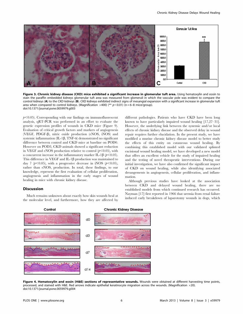
Chronic Kidney Disease mice exhibit severe woundhealing impairments
Cross-sectional analysis (Figure 4) of histological wound sections
allowed for quantification of the maximal distance between the
advancing edges of keratinocyte migration across the wound
(epithelial gap), and the digital calculation of all the regions of
granulation tissue (granulation tissue area) present in wounds at
time of harvest. Control wounds displayed normal reepithelializa-
tion kinetics consistent with previously described studies [25,36].
However, CKD wounds demonstrated statistically significant
disruption of normal reepithelialization kinetics and granulation
tissue deposition rates. As shown in Figure 5A, the mean
percentage of epithelial gap relative to their original size was
significantly larger in CKD wounds (day 7 epithelial gap:
37.867.0% vs. 3.161.9%, p,0.05; day 14: 27.369.2% vs 0%,
p,0.05). In addition, although not statistically significant, these
CKD affected wounds also exhibited decreased granulation tissue
formation (Figure 5B). Animals did not demonstrate any local signs
of wound infection or disruption, suggesting that the effects of
CKD in these wounds were systemically based. This was further
supported by the finding that contralateral wounds in the same
animal were found to heal at the same delayed rate. No post-
wound splinting fatalities were encountered in any animal.
CKD wounds exhibit significantly less cellularproliferation and angiogenesis, with concurrentderangements in inflammation, during the early stagesof wound healing
Cellular proliferation, angiogenesis and inflammation were
assessed at days 3, 7 and 14 by measuring immunofluorescent
BrdU, CD31 and CD45-positive cells, respectively, within the
entire sampled wounded tissues. Representative images for control
(Figure 6A) and CKD (Figure 6B) wounds at day 7 illustrate the
differences in BrdU-positive cells within the full thickness wound
tissue near the wound edge. Wounds in CKD mice showed
significantly less proliferating cells as compared to control wounds
in mice with normal renal function (day 3: 355640 vs. 6526100,
p,0.001; day 7: 599681 vs. 12056117, p,0.001; day 14:
414630 vs. 456615, p,0.05) (Figure 6C). CKD mice also
demonstrated a significant reduction in angiogenesis in wound
healing tissue when compared to control mice (day 3: 40.5626.1
vs. 3498.66114.5, p,0.001; day 7: 69.75653.6 vs. 586.76191.4,
p,0.001; day 14: 169.26140.1 vs. 438.66203.2, p,0.05
(Figure 7C). A digital representation of the sizeable difference in
the presence of CD31-positive cells is shown in Figures 7A and 7B
for the control and CKD mice at day 7, respectively. Intravascular
injection of 8 mice with a fluorescein labeled tetrameric
glycoprotein lectin II (FL 1211, Vector Lab, Burlingame, CA)
allowed for the visualization and assessment of the presence of
vascular endothelium between groups. These results were also
consistent with those found following CD31 staining (data not
shown).
Interestingly, CKD mice exhibited an increased and maintained
inflammatory state during the early stages of wound healing.
Digital acquisition of representative control and CKD splinted
wounded samples obtained at day 7 are shown in Figures 8A and
8B. CKD wounds revealed a trend towards increased CD45-
positive cells at day 3 (478561293 vs 457861450) (Figure 8C),
which by day 7 represented a significant increase in inflammation
(42126456 vs 2940696, p,0.05). This difference was maintained
throughout the experiment to day 14 (346161617 vs 14346541,
Figure 2. Mice exhibited different degrees of chronic kidney disease. A) By controlling the amount of cautery to the injured kidney,moderate and severe degrees of chronic kidney disease were produced. Only mice with a severe degree of chronic kidney disease were wounded forexperimental purposes. (n = 6–8 mice/group) B) These animals exhibited metabolic derangements analogous to those in human chronic kidneydisease and a notable growth retardation.doi:10.1371/journal.pone.0059979.g002
Chronic Kidney Disease Delays Wound Healing
PLOS ONE | www.plosone.org 5 March 2013 | Volume 8 | Issue 3 | e59979
p,0.05). Corresponding with our findings on immunofluorescent
analysis, qRT-PCR was performed in an effort to evaluate the
genetic expression profiles of wounds in CKD mice (Figure 9).
Evaluation of critical growth factors and markers of angiogenesis
(VEGF, PDGF-b), nitric oxide production (eNOS, iNOS) and
systemic inflammation (IL-1b, TNF-a) demonstrated no significant
difference between control and CKD mice at baseline on POD0.
However on POD3, CKD animals showed a significant reduction
in VEGF and eNOS production relative to control (p,0.05), with
a concurrent increase in the inflammatory marker IL-1b (p,0.05).
This difference in VEGF and IL-1b production was maintained to
day 7 (p,0.05), with a progressive decrease in iNOS (p,0.05),
rather than eNOS, production. In total, these findings, to our
knowledge, represent the first evaluation of cellular proliferation,
angiogenesis and inflammation in the early stages of wound
healing in mice with chronic kidney disease.
Discussion
Much remains unknown about exactly how skin wounds heal at
the molecular level, and furthermore, how they are affected by
different pathologies. Patients who have CKD have been long
known to have particularly impaired wound healing [17,27–31].
However, the underlying link between the systemic and/or local
effects of chronic kidney disease and the observed delay in wound
repair requires further elucidation. In the present study, we have
modified a murine chronic kidney disease model to better study
the effects of this entity on cutaneous wound healing. By
combining this established model with our validated splinted
excisional wound healing model, we have developed a new model
that offers an excellent vehicle for the study of impaired healing
and the testing of novel therapeutic interventions. During our
initial investigation, we have also confirmed the significant impact
of CKD on wound healing, while also identifying associated
derangements in angiogenesis, cellular proliferation, and inflam-
mation.
Although previous studies have looked at the association
between CKD and delayed wound healing, there are no
established models from which continued research has occurred.
Nayman [17] first reported in 1966 that uremia from renal failure
induced early breakdown of laparotomy wounds in dogs, which
Figure 3. Chronic kidney disease (CKD) mice exhibited a significant increase in glomerular tuft area. Using hematoxylin and eosin tostain the paraffin embedded kidneys glomerular tuft area was measured from glomeruli in which the vascular pole was evident to compare thecontrol kidneys (A) to the CKD kidneys (B). CKD kidneys exhibited indirect signs of mesangial expansion with a significant increase in glomerular tuftarea when compared to control kidneys. (Magnification 6400) (** p,0.01) (n = 6–8 mice/group).doi:10.1371/journal.pone.0059979.g003
Figure 4. Hematoxylin and eosin (H&E) sections of representative wounds. Wounds were obtained at different harvesting time points,processed, and stained with H&E. Red arrows indicate epithelial keratinocyte migration across the wounds. (Magnification 620).doi:10.1371/journal.pone.0059979.g004
Chronic Kidney Disease Delays Wound Healing
PLOS ONE | www.plosone.org 6 March 2013 | Volume 8 | Issue 3 | e59979

was prevented by early and frequent hemodialyses [17]. Mean-
while, Kursh et al [28] demonstrated that uremia had a significant
effect on wound healing by measuring wound tensile strength and
collagen formation in subcutaneously implanted polyvinyl sponges.
However, given the strong correlation between their findings and
final body weights, they concluded nutritional factors might be the
primary link between uremia and wound healing impairment.
Colin et al [29] reported similar results for both abdominal
wounds and intestinal anastomoses, although no measurements of
the extent of wound healing were performed. Meanwhile, work
Shindo et al [30] and Yue et al [31] in uremic rats indicated that
the control of surgical infections and diabetes, respectively, might
be the most important factors to consider for appropriate wound
healing in CKD patients. Given that the most recent of these
studies was performed 15 years ago, we believe that the validation
of an updated model, which allows for the incorporation of
different comorbidities and the study of several different endpoints,
is important. Although we have attempted to investigate the
mechanistic link between CKD and wound healing, we intend to
continue to utilize this model in an attempt to address the growing
need for in vivo CKD research.
The model we have presented possesses several advantages for
the study of CKD-impaired wound healing in vivo. In particular,
the model’s technical ease, along with a lack of local complications,
makes it ideal for even the relatively inexperienced investigator to
use. The model requires only one surgery to induce CKD,
shortening the total experimental time, and more importantly the
burden to the animal. Unlike the conventional intraperitoneal
anesthetic used for in Gagnon’s original model [22], the
introduction of inhaled anesthesia allows the mice to better
respond to the combination of left nephrectomy and right kidney
electro-coagulation in a single procedure, including an almost null
mortality rate and a much faster recovery (5–10 minutes v. 30–
60 minutes) time. In addition to this, this CKD-inducing surgical
model can be tested in any mouse strain, obviating the use of
expensive breeds and allowing for a wider horizon of experimen-
tation. However, it is important to note that the degree of uremia
that develops in this model is directly proportional to the area of
electrocoagulated kidney cortex, which can be subject to technical
variability. We attempted to minimize differences in technical
performance for this study by having only one of the authors
perform all of the surgical procedures.
The incorporation of a reliable, and inexpensive, wound healing
model represents another unique advantage of the model that we
have presented. In particular, previous work has shown the
reproducibility of our excisional wound model with 90% splint
Figure 5. Epithelial gap closure and granulation tissuedeposition measurements. A) Epithelial gap closure was signifi-cantly delayed in the chronic kidney disease model at days 7 and 14. B)Granulation tissue deposition exhibited a trend towards beingdecreased in the chronic kidney disease group. (* p,0.05) (n = 6–8mice/group).doi:10.1371/journal.pone.0059979.g005
Figure 6. Chronic kidney disease demonstrated significantly less cellular proliferation in the early stages of wound healing.Representative immunofluorescent sections of wound edges stained with BrdU and DAPI in A) normal mice and B) chronic kidney disease mice atday 7. C) Utilizing Tissue Gnostics quantification technology, chronic kidney disease wounds demonstrated significantly less cellular proliferation inthe early stages of wound healing. (Arrows indicate wound edges, white dots indicate epidermis, and brackets indicate dermis) (* p,0.05, ** p,0.01)(Magnification 6200) (n = 6–8 mice/group).doi:10.1371/journal.pone.0059979.g006
Chronic Kidney Disease Delays Wound Healing
PLOS ONE | www.plosone.org 7 March 2013 | Volume 8 | Issue 3 | e59979

maintenance in C57bl/6J mice [25]. This low failure rate was
explained by the model utilizing a thin protective dressing around
the surgical site, preventing the mice from disrupting the silicone
ring splint with their normal grooming behaviors. Consequently,
the splints are maintained such that wound contraction is
minimized, allowing for an appropriate analysis of epithelial and
granulation tissue in-growth. The wrap was also fit to the mouse
such that it was ergonomic and did not otherwise affect its normal
behavior. With a robust method for evaluating wound healing,
intentional modifications to the model can be evaluated and
compared in a consistent manner, emphasizing its flexibility for in
vivo experimentation. Furthermore, the histopathological increase
in glomerular tuft area, moderately increased BUN levels, growth
retardation, and anemia present in our model are comparable to
changes that are seen clinically with CKD, underscoring the
model’s translatability. The resultant delay in normal wound
healing, evidenced by an increased epithelial gap and a decrease in
granulation tissue, appropriately models the detrimental, systemic
effects of CKD on human wounds, making it a useful model for
future wound healing studies.
The consistency of our model was further established by our
immunofluorescence and qRT-PCR findings. With a delay in
wound healing, an associated decrease in cellular proliferation
(BrdU) and angiogenesis (CD31) was to be expected. A concurrent
significant decrease in VEGF on qRT-PCR further emphasized
the detrimental impact of CKD on angiogenesis. Meanwhile, the
distinct change in host inflammatory response was a surprising
finding. Highlighted by a significant increase in CD45-positive
cells and IL-1b that was maintained over time, we believe that
these findings are indicative of the potential importance of
inflammatory mediators in the systemic effects of CKD on wound
healing. Interestingly, the expression of systemic markes IL-1b and
TNF-a did not correlate, despite both being markers of systemic
inflammation. Rather than being an unclear discrepancy, this may
represent a specific inflammatory pathyway that is associated with
CKD, warranting further investigation. Furthermore, IL-1b is
expressed by a larger variety of cells beyond macrophages,
including fibroblasts, and has been seen to have a different
expression pattern than TNF-a in other wound-related settings,
such as with bacterial biofilm [37]. Taken individually, perturba-
tions in angiogenesis and inflammation can have significant
systemic effects given the ubiquity of these processes within the
human body. In particular, their importance to normal wound
healing cannot be emphasized enough. Therefore, with CKD
Figure 7. Chronic kidney disease demonstrated a significant reduction in angiogenesis in wound healing tissue. Representativeimmunofluorescent sections of wound edges stained with CD31 and DAPI in A) normal mice and B) chronic kidney disease mice at day 7. C) UtilizingTissue Gnostics quantification technology, chronic kidney disease wounds demonstrated significantly less angiogenesis in the early stages of woundhealing. (Arrows indicate wound edges, white dots indicate epidermis, and brackets indicate dermis) (* p,0.05, ** p,0.01) (Magnification 6200)(n = 6–8 mice/group).doi:10.1371/journal.pone.0059979.g007
Figure 8. Chronic kidney disease demonstrated significantly increased and maintained inflammation in the early stages of woundhealing. Representative immunofluorescent sections of wound edges stained with CD45 and DAPI in A) normal mice and B) chronic kidney diseasemice at day 7. C) Utilizing Tissue Gnostics quantification technology, chronic kidney disease wounds demonstrated a significantly increased andmaintained inflammation in the early stages of wound healing. (Arrows indicate wound edges, white dots indicate epidermis, and brackets indicatedermis) (* p,0.05, ** p,0.01) (Magnification 6200) (n = 6–8 mice/group).doi:10.1371/journal.pone.0059979.g008
Chronic Kidney Disease Delays Wound Healing
PLOS ONE | www.plosone.org 8 March 2013 | Volume 8 | Issue 3 | e59979

having a demonstrable impact on both of these systems, our
findings validate its importance to the pathophysiology of non-
healing wounds. Further studies aimed at elucidating the
molecular pathways responsible for these changes may identify
potential avenues for therapeutic intervention, which can then be
quickly translated into the clinical arena.
Despite a rigorous, scientific approach, we acknowledge the
limitations of our study. In particular, although we were
consistently able to induce a moderate CKD phenotype, we did
not evaluate its individual impact on our different endpoints,
including wound healing. Given the spectrum of severity seen
clinically, the ‘dose-dependent’ impact decreasing renal function in
CKD would be of interest for future studies. Also, given our goal of
establishing and validating an initial model, we did not extend our
experiments to other strains of mice. As previously mentioned, the
inherent flexibility of our model provides a platform for studying
CKD-impaired wound healing against different genetic back-
grounds. For example, utilizing a diabetic mouse strain would
allow for further characterization of the complex interplay
between CKD, diabetes and wounds, the three of which are
commonly found within a single patient. Meanwhile, the
introduction of bacteria into our wounds, specifically biofilm,
would help to address another essential pillar of chronic wound
pathogenesis. Given this potential for extensive experimentation,
we chose to limit our study to the findings that have been
presented. It is our hope that the information presented has
provided greater insight into the relationship between CKD and
wound healing, while establishing a foundation from which
investigators can continue answer questions related to wound
and CKD pathophysiology.
Acknowledgments
We thank Clara Barrios from the Nephrology Research Laboratory of
Northwestern University for her assistance on glomerular tuft analysis.
Figure 9. Quantitative reverse-transcription PCR (qRT-PCR) of growth factor, nitric oxide, and inflammatory genese. Genetic analysisof wounds by qRT-PCR exhibited differences in expression of important wound healing related genes due chronic kidney disease. At post-operativeday (POD) 3, CKD wounds demonstrate significantly reduced expression of VEGF and eNOS, with an increase in IL-1b. These trends were maintainedat POD7, except for a significant decrease in iNOS instead of eNOS. Rows show POD when the tissue was collected, POD0, 3, and 7 at top, middle andbottom, respectively. Genes analyzed are represented by columns: (A) growth factor genes, (B) nitric oxide genes and (C) cytokine genes. (* p,0.05)(n = 4 wounds/group).doi:10.1371/journal.pone.0059979.g009
Chronic Kidney Disease Delays Wound Healing
PLOS ONE | www.plosone.org 9 March 2013 | Volume 8 | Issue 3 | e59979

Author Contributions
Conceived and designed the experiments: AKS MD RCF SJH RDG.
Performed the experiments: MD RCF SJH. Analyzed the data: AKS MD
RCF SJH. Contributed reagents/materials/analysis tools: SJH. Wrote the
paper: AKS MD.
References
1. Schieppati A, Remuzzi G (2005) Chronic renal diseases as a public health
problem: epidemiology, social, and economic implications. Kidney Int Suppl 98:S7–S10.
2. Weiner DE (2007) Causes and consequences of chronic kidney disease:implications for managed health care. J Manag Care Pharm 3 Suppl: S1–S9.
3. Obrador GT, Pereira BJ, Kausz AT (2002) Chronic kidney diease in the United
States: an underrecognized problem. Semin Nephrol 22: 441–448.4. Coresh J, Byrd-Holt D, Astor BC, Briggs JP, Eggers PW, et al. (2005) Chronic
kidney disease awareness, prevalence, and trends among U.S. adults, 1999 to2000. J Am Soc Nephrol 16: 180–188.
5. Owen WF Jr (2003) Patterns of care for patients with chronic kidney disease in
the United States: dying for improvement. J Am Soc Nephrol 14: S76–S80.6. Coresh J, Selvin E, Stevens LA, Manzi J, Kusek JW, et al. (2007) Prevalance of
chronic kidney disease in the United States. JAMA 298: 2038–2047.7. St Peter WL (2007) Chronic kidney disease: A Burgeoning health epidemic.
J Manag Care Pharm 13: S2–S5.8. Lundin AP, Fani K, Berlyne GM, Friedman EA (1995) Dermal angiopathy in
hemodialysis patients: the effect of time. Kidney Int 47: 1775–1780.
9. Gilchrest BA, Rowe JW, Mihm MC Jr (1980) Clinical and histological skinchanges in chronic renal failure: evidence for a dialysis-resistant, transplant-
responsive microangiopathy. Lancet 2(8207): 1271–1275.10. Martens CR, Edwards DG (2011) Peripheral vascular dysfunction in chronic
kidney disease. Cardiol Res Pract 2011:267257. Epub 2011 May 24.
11. Alssema M, Newson RS, Bakker SJ, Stehouwer CD, Heymans MW, et al. (2012)One risk assessment tool for cardiovascular disease, type 2 diabetes, and chronic
kidney disease. Diabetes Care 35: 741–748.12. Shurraw S, Hemmelgarn B, Lin M, Majumdar SR, Klarenbach S, et al. (2011)
Association between glycemic control and adverse outcomes in people with
diabetes mellitus and chronic kidney disease: a population-based cohort study.Arch Intern Med 171: 1920–1927.
13. Werdin F, Tennenhaus M, Schaller HE, Rennekampff HO (2009) Evidence-based management strategies for treatment of chronic wounds. Eplasty 9: e19.
14. Mustoe TA, O’Shaughnessy K, Kloeters O (2006) Chronic wound pathogenesisand current treatment strategies: a unifying hypothesis. Plast Reconstr Surg 117:
35–41.
15. Menke NB, Ward KR, Witten TM, Bonchev DG, Diegelmann RF (2007)Impaired wound healing. Clin Dermatol 25: 19–25.
16. Valabhji J (2012) Foot problems in patients with diabetes and chronic kidneydisease. J Ren Care 38 Suppl 1: 99–108.
17. Nayman J (1966) Effect of renal failure on wound healing in dogs. Response to
hemodialysis following uremia induced by uranium nitrate. Ann Surg 164: 227–235.
18. Mylonas AI, Massoulas GB, Nicolatou O, Dontas IA, Nakopoulou L, et al.(2000) Progress of ossification and epithelialization of wounds after simple or
surgical extractions of teeth in rats with chronic renal failure: an experimentalstudy. Br J Oral Maxillofac Surg 38: 35–43.
19. Zotta E, Ochoa F, Tironi Farinati C, Damiano A, Silberstein C, et al. (2008)
UT-A expression in pars recta from a rat model of chronic renal failure.J Nephrol 21: 947–958.
20. Wang SP, West MW, Dresner LS, Fleishhacker JF, Distant DA, et al. (1996)
Effects of diabetes and uremia on mesenteric vascular reactivity. Surgery
120:328–335.
21. Gagnon RF, Duguid WP (1983) A reproducible model for chronic renal failure
in the mouse. Urol Res 11: 11–14.
22. Gagnon RF, Gallimore B (1988) Characterization of a mouse model of chronic
uremia. Urol Res 16: 119–126.
23. Kennedy DJ, Elkareh J, Shidyak A, Shapiro AP, Smaili S, et al. (2008) Partial
nephrectomy as a model for uremic cardiomyopathy in the mouse. Am J Physiol
Renal Physiol 294: F450–F454.
24. Ahn ST, Mustoe TA (1990) Effects of ischemia on ulcer wound healing: a new
model in the rabbit ear. Ann Plast Surg 24: 17–23.
25. Galiano RD, Michaels J 5th, Dobryansky M, Levine JP, Gurtner GC (2004)
Quantitative and reproducible murine model of excisional wound healing.
Wound Repair Regen 12: 485–492.
26. Gottrup F, Agren MS, Karlsmark T (2000) Models for use in wound healing
research: a survey focusing on in vitro and in vivo adult soft tissue. Wound
Repair Regen 8: 83–96.
27. Cheung AH, Wong LM (2001) Surgical infections in patients with chronic renal
failure. Infect Dis Clin North Am 15: 775–796.
28. Kursh ED, Klein L, Schmitt J, Kayal S, Persky L (1977) The effect of uremia on
wound tensile strength and collagen formation. J Surg Res 23: 37–42.
29. Colin JF, Elliot P, Ellis H (1979) The effect of uraemia upon wound healing: an
experimental study. Br J Surg 66: 793–797.
30. Shindo K, Kosaki G (1982) Effects of chronic renal failure on wound healing in
rats. II. Microscopic study and hydroxyproline assay. Jpn J Surg 12: 46–51.
31. Yue DK, McLennan S, Marsh M, Mai YW, Spaliviero J, et al. (1987) Effects of
experimental diabetes, uremia, and malnutrition on wound healing. Diabetes 36:
295–299.
32. Phillips T, Stanton B, Provan A, Lew R (1994) A study of the impact of leg ulcers
on quality of life: financial, social, and psychological implications. J Am Acad
Dermatol 31: 49–53.
33. Krasner D. Painful venous ulcers (1998) Themes and stories about their impact
on quality of life. Ostomy Wound Management 44: 38–49.
34. Perlman RL, Finkelstein FO, Liu L, Roys E, Kiser M, et al. (2005) Quality of life
in chronic kidney disease (CKD): a cross-sectional analysis in the Renal
Research Institute-CKD study. Am J Kidney Dis 45: 658–666.
35. Gagnon RF, Ansari M (1990) Development and progression of uremic changes
in the mouse with surgically induced renal failure. Nephron 54: 70–76.
36. Michaels J 5th, Churgin SS, Blechman KM, Greives MR, Aarabi S, et al. (2007)
db/db mice exhibit severe wound-healing impairments compared with other
murine diabetic strains in a silicone-splinted excisional wound model. Wound
Repair Regen 15: 665–670.
37. Gurjala AN, Geringer MR, Seth AS, Hong SJ, Smeltzer MS, et al. (2011)
Development of a novel, highly quantitative in vivo model for the study of
biofilm-impaired cutaneous wound healing. Wound Repair Regen 19: 400–410.
Chronic Kidney Disease Delays Wound Healing
PLOS ONE | www.plosone.org 10 March 2013 | Volume 8 | Issue 3 | e59979