Embed Size (px)
Citation preview

Examination of the inflammatory nature of different
placental syncytiotrophoblast microparticles (STBM)
preparations on human monocytes
Inauguraldissertation
zur
Erlangung der Würde eines Doktors der Philosophie
vorgelegt der
Philosophisch-Naturwissenschaftlichen Fakultät
der Universität Basel
von
Marianne Simone Messerli
aus Kaufdorf BE
Basel, 2009
Originaldokument gespeichert auf dem Dokumentenserver der Universität Basel edoc.unibas.ch
Dieses Werk ist unter dem Vertrag „Creative Commons Namensnennung-Keine kommerzielle Nutzung-Keine Bearbeitung 2.5 Schweiz“ lizenziert. Die vollständige Lizenz kann unter
creativecommons.org/licences/by-nc-nd/2.5/ch eingesehen werden.

Genehmigt von der Philosophisch-Naturwissenschftlichen Fakultät
auf Antrag von Prof. Dr. Antonius Rolink
Fakultätsverantwortlicher
Prof. Dr. Sinuhe Hahn
Dissertationsleiter
Prof. Dr. Ian Sargent
Koreferent
Basel, den 15.September 2009
Prof. Dr. Eberhard Parlow
Dekan

Table of content 1
Table of content
ABBRIVIETATIONS ................................................................................................................ 4
SUMMARY ............................................................................................................................... 6
INTRODUCTION ...................................................................................................................... 9
1. HUMAN PLACENTATION ................................................................................................................. 9
1.1. Implantation .......................................................................................................................................... 9
1.2. Haemochorial placentation .................................................................................................................. 10
1.3. Physiological hypoxia in early placentation ........................................................................................ 13
1.4. Physiological placental oxidative burst at the end of the first trimester .............................................. 14
2. IMMUNOLOGY OF HUMAN PREGNANCY ....................................................................................... 15
2.1. Immunological interfaces .................................................................................................................... 15
2.2. Immunological interactions ................................................................................................................. 17
2.2.1. Predominant Th2 immunity .......................................................................................................................... 17
2.2.2. Absent trophoblast expression of MHC class II and classical polymorphic MHC class I molecules ........... 18
2.2.3. Decidual NK (dNK) cells and their interaction with trophoblast HLA-E, -G, -C ......................................... 18
2.2.4. Decidual macrophages .................................................................................................................................. 19
2.2.5. Regulatory T-cells (Treg) ............................................................................................................................... 20
2.2.6. B7 family: ..................................................................................................................................................... 21
2.2.7. Indoleamine-2,3-dioxygenase (IDO): ........................................................................................................... 21
2.2.8. Fas-Fas ligand system: .................................................................................................................................. 22
2.2.9. Complement regulatory proteins: ................................................................................................................. 22
2.2.10. Deportation of syncytiotrophoblast debris into maternal blood .................................................................... 22
2.3. Systemic maternal inflammation ......................................................................................................... 24
2.3.1. Placental cytokines ....................................................................................................................................... 25
2.3.2. Placental angiogenic factors ......................................................................................................................... 25
3. PREECLAMPSIA ............................................................................................................................ 27
3.1. Epidemiology ...................................................................................................................................... 27
3.2. Pathophysiology .................................................................................................................................. 29
3.2.1. Preclinical stage: shallow trophoblast invasion and incomplete vascular remodelling ................................. 29
3.2.1.1. Placental oxidative stress ...................................................................................................................... 30
3.2.1.2. Maternal-paternal immune maladaptation ............................................................................................ 31
3.2.2. Clinical stage: excessive maternal systemic inflammation and placental factors .......................................... 32
3.3. Microparticles ..................................................................................................................................... 35
3.3.1. Microparticles in general .............................................................................................................................. 35
3.3.2. Microparticles in inflammation..................................................................................................................... 36
3.3.3. Syncytiotrophoblast microparticles (STBM) ................................................................................................ 36
3.3.4. STBM and the maternal inflammation.......................................................................................................... 37
RESEARCH OBJECTIVE ....................................................................................................... 40
Table of content 2
MATERIALS AND METHODS ............................................................................................. 41
1. PREPARATION OF SYNCYTIOTROPHOBLAST MICROPARTICLES (STBM) ..................................... 41
1.1. In vitro explant cultures (eS20 and eS3) ............................................................................................... 41
1.2. Mechanical dissection (mS) ................................................................................................................ 42
1.3. Placental dual perfusion (pS) .............................................................................................................. 42
1.4. STBM quantification ........................................................................................................................... 43
1.5. Caspases activity assay........................................................................................................................ 43
1.6. Lactate dehydrogenase (LDH) cytotoxicity assay ............................................................................... 43
2. CELL CULTURE ............................................................................................................................. 45
2.1. Cell line: Mono Mac 6 ........................................................................................................................ 45
2.2. Human blood monocytes ..................................................................................................................... 45
2.3. Co-culture of monocytes and STBM ................................................................................................... 46
3. FUNCTIONAL ANALYSIS .............................................................................................................. 47
3.1. Cell viability (WST-1 assay) ............................................................................................................... 47
3.2. Flow cytometry ................................................................................................................................... 47
3.3. Enzyme-linked immunosorbent assay (ELISA) .................................................................................. 48
3.3.1. IL-1β, IL-6, IL-8 and CD54 ELISA .............................................................................................................. 48
3.3.2. IL-10 ELISA ................................................................................................................................................. 49
3.3.3. Placental alkaline phosphatase (PLAP) ELISA ............................................................................................ 49
3.4. RNA extraction ................................................................................................................................... 50
3.5. GAPDH real-time PCR with mRNA ................................................................................................... 51
3.6. Reverse transcription reaction ............................................................................................................. 51
3.7. Human NF-κB Signalling Pathway RT2 Profiler PCR Array .............................................................. 52
3.8. Fluorescence microscopy .................................................................................................................... 52
3.9. Statistical analysis ............................................................................................................................... 53
RESULTS ................................................................................................................................. 54
1. STBM PREPARATIONS ................................................................................................................. 54
1.1. All microparticle preparations contain the syncytiotrophoblast specific placental alkaline phosphatase
(PLAP) ................................................................................................................................................ 55
1.2. STBM associated cytokines ................................................................................................................ 56
1.3. Conclusion .......................................................................................................................................... 57
2. STBM PREPARED FROM NORMAL TERM PLACENTAS DO NOT ACTIVATE THE MONOCYTIC CELL
LINE MONO MAC 6....................................................................................................................... 58
2.2. STBM only marginally alter the expression and median fluorescent intensity of cell surface markers
on Mono Mac 6 cells ........................................................................................................................... 58
2.3. STBM do not trigger the secretion of pro-inflammatory cytokines in Mono Mac 6 cells .................. 61
2.4. STBM do not induce an anti-inflammatory response in Mono Mac 6 cells ........................................ 62
2.5. Conclusion .......................................................................................................................................... 62

Table of content 3
3. STBM PREPARED FROM NORMAL TERM PLACENTAS ACTIVATE HUMAN PERIPHERAL BLOOD
MONOCYTES ................................................................................................................................. 63
3.1. STBM do not affect the viability of human monocytes ...................................................................... 63
3.2. STBM differently alter the expression of cell surface markers on human primary monocytes .......... 63
3.3. eS20 and pS induce the secretion of proinflammatory cytokines by human monocytes....................... 65
3.4. STBM-induced cytokine secretion is dose- and time-dependent ........................................................ 66
3.5. IL-8 is not responsible for eS20 mediated cytokine secretion .............................................................. 67
3.6. Conclusion .......................................................................................................................................... 68
4.1. eS20 prepared from term preeclamptic placentas modify the expression of cell surface molecules .... 69
4.2. eS20 generated from normal and preeclamptic placentas similarly trigger the secretion of
proinflammatory cytokines ................................................................................................................. 71
4.3. Conclusion .......................................................................................................................................... 71
5. STBM INDUCE SECRETION OF PROINFLAMMATORY MEDIATORS BY PRIMARY HUMAN
MONOCYTES IN A NF-ΚB-DEPENDENT MANNER .......................................................................... 72
5.1. The transcription of proinflammatory mediators is induced by eS20 ................................................... 72
5.2. eS20 decrease the gene expression of pro-apoptotic mediators, pattern recognition receptors and their
adaptor molecules ................................................................................................................................ 74
5.3. Inhibitors of NF- κB reduce cytokine secretion mediated by eS20 ...................................................... 74
5.4. Conclusion .......................................................................................................................................... 75
6. INTERACTION OF STBM AND PRIMARY HUMAN MONOCYTES .................................................... 76
6.1. eS20 attach to the monocytic cell surface ............................................................................................. 76
6.2. eS20 mediated secretion of proinflammatory mediators is independent of phagocytosis by human
monocytes ........................................................................................................................................... 77
6.3. STBM contain CD54 ........................................................................................................................... 78
6.4. STBM-associated CD54 is not responsible for eS20 mediated cytokine secretion .............................. 79
6.5. Basal expression of cell surface marker is different in Mono Mac 6 cells and peripheral blood
monocytes ........................................................................................................................................... 79
6.6. MyD88 inhibitor peptide reduces eS20 mediated secretion of proinflammatory mediators ................. 82
6.7. Conclusion .......................................................................................................................................... 83
DISCUSSION .......................................................................................................................... 84
ACKNOWLEDGEMENTS ..................................................................................................... 94
REFERENCES ......................................................................................................................... 95
CURRICULUM VITAE ........................................................................................................ 117
ABBRIVIETATIONS 4
ABBRIVIETATIONS
APC Antigen-presenting cells
AP-1 Activator protein 1
6AQ 6-Amino-4-(4-phenoxyphenylethylamino)quinazoline
BCL3 B-cell CCL/lymphoma 3
CCL2 Chemokine (C-C-motif) ligand 2
CD Cluster of differentiation
CRP C-reactive protein
CSF Colony stimulating factor
DAF Decay accelerating factor
DMSO Dimethyl sulfoxide
dNK cell Decidual natural killer cell
EGR1 Epidermal growth factor 1
Eng Endoglin
eS3 Villous explant STBM prepared at 3% O2
eS20 Villous explant STBM prepared at 20% O2
EVT Extravillous trophoblast
FADD Fas-Associated protein with Death Domain
FasL Fas ligand
Fc Fragment, crystallizable
FcR Fc receptor
FFA Free fatty acids
Flt-1 Fms-like tyrosine kinase-1 receptor
GM-CSF Granulocyte-macrophage colony-stimulating factor
HELLP Hemolysis, elevated liver enzymes, and low platelet counts
HLA Human leukocyte antigen
IDO Indoleamine-2,3-dioxygenase
IFN Interferon
IL Interleukin
IL-1R1 IL-1 receptor type 1
IP-10 Interferon-inducible-protein-10
IUGR Intrauterine growth restriction
KIR killer cell immunoglobulin receptor
LDL Low-density lipoproteins
LFA-1 Lymphocyte function-associated antigen 1
LIR leukocyte Ig-like receptor

ABBRIVIETATIONS 5
LPS Lipopolysaccharide
LTA Lymphotoxin α
MAC Membrane attack complex
MCP Membrane co-factor protein
MCP-1 Monocyte chemotactic protein-1
MHC Major histocompatibility complex
mS STBM prepared by mechanical dissection
MyD88 Myeloid differentiation primary response gene 88
NETs Neutrophil extracellular traps
NF-κB Nuclear factor kappa-light-chain-enhancer of activated B cells
nIUGR Normotensive intrauterine growth restriction
NK cell Natural killer cell
NOD-1 Nucleotide-binding oligomerization domain 1
OFR Oxygen free radical
PA Perillyl alcohol
PBMC Peripheral blood mononuclear cells
PD-1 Programmed death 1
PHA Phytohemagglutinin
PLAP Placental alkaline phosphatase
PlGF Placental growth factor
PMA Phorbol 12-myristate 13-acetate
PMN polymorphonuclear leukocytes
pS STBM prepared by dual perfusion of a placental cotyledon
RT Room temperature
sEng Soluble endoglin
sFlt-1 Soluble fms-like tyrosine kinase-1 receptor
STBM Syncytiotrophoblast microparticle
TCR T-cell receptor
TGF Transformation growth factor
Th T helper
TLR Toll like receptor
TNF Tumor necrosis factor
TNFR Tumor necrosis factor receptor
Treg Regulatory T-cells
TREM-1 Triggering receptor expressed on myeloid cells 1
VEGF Vascular endothelial growth factor

SUMMARY 6
SUMMARY
Background: Healthy human pregnancies are accompanied by a mild systemic maternal
inflammatory response, which includes activation of peripheral blood monocytes. This
generalized inflammation is exaggerated in preeclampsia, a placenta-dependent disorder
specific to human pregnancies. It has been proposed that placental syncytiotrophoblast
membrane microparticles (STBM), which are released into the peripheral blood, might
contribute to the maternal response in normal pregnancy and preeclampsia.
Aim: The aim of this work was to assess the inflammatory properties of STBM generated in
vitro from human term placentas by four different approaches which should mimic
physiologic or patho-physiologic conditions, and their mode of action on human monocytes in
vitro.
Methods: STBM were prepared by: (1) placental explant cultures of villous tissue incubated
at 20% O2/5% CO2 (air) at 37°C for 72 hours (eS20); (2) perfusion of the maternal side of a
placental cotyledon (pS); (3) placental explant cultures of villous tissue incubated at 3%
O2/5% CO2 (hypoxia) at 37°C for 72 hours (eS3); and (4) mechanical dissection of villous
tissue (mS). In all approaches, STBM were isolated by serial high-speed centrifugation.
STBM were co-incubated with either the human monocytic cell line Mono Mac 6 or human
peripheral blood monocytes. In some cases, agents which inhibit cellular functions or
signalling pathways were used. Analysis of viability, phenotype and function were performed
by real-time PCR, flow cytometry, ELISA and fluorescence microscopy.
Results: Viability of Mono Mac 6 cells was not impaired following treatment with STBM.
However, STBM only induced a marginal response in Mono Mac 6. None of the STBM
population affected the viability of primary monocytes. eS3 and mS decreased the expression
of CD54 on peripheral blood monocytes, but did not induce the secretion of IL-1β, IL-6 and
IL-8. However, pS and eS20 up-regulated the cell surface expression of CD54 on primary
monocytes and stimulated the secretion of IL-1β, IL-6 and IL-8 in a dose- and time-dependent

SUMMARY 7
manner. Interestingly, eS20 derived from normal and preeclamptic placentas stimulated
monocyte activation to similar degrees.
eS20 induced the transcription of several NF-κB responsive genes, including IL-6 and IL-8,
and the secretion of IL-6 and IL-8 was reduced upon treatment with NF-κB inhibitors.
eS20 was located at the monocytic cell surface and a phagocytosis inhibitor did not reverse the
eS20 induced production of IL-6 and IL-8.
Primary monocytes and the non-responding Mono Mac 6 cells expressed toll like receptors
(TLRs) differently. Pre-incubation of primary monocytes with an inhibitor of intracellular
TLR signalling reduced the inflammatory response triggered by eS20.
Conclusions: STBM populations evoked neither a proinflammatory nor an anti-inflammatory
phenotype in Mono Mac 6 cells. However, STBM prepared at conditions which are believed
to mimic the physiologic situation in human pregnancy (eS20 and pS) triggered the secretion
of IL-1β, IL-6 and IL-8 and up-regulated the expression of the adhesion molecule CD54 on
peripheral blood monocytes. These findings indicate that Mono Mac 6 cells are not the
appropriate cells to study the interaction between monocytes and STBM.
STBM prepared at non-physiologic (mS) and hypoxic (eS3) conditions, which are thought to
mimic the patho-physiologic situation in preeclampsia, did not induce an inflammatory
response in peripheral blood monocytes. In addition, the observation that eS20 derived from
normal as well as from preeclamptic placentas triggered an equally strong and dose-dependent
inflammatory response in primary monocytes, suggests that there are no or only minor
qualitative differences between the microparticles. These findings presume that the overt
maternal inflammation associated with preeclampsia may be due to the higher concentration
of circulating STBM, rather than to a qualitative difference between microparticles released
from normal and patho-physiologic placentas.
The results also suggest that the inflammatory reaction in monocytes may be initiated by the
attachment of STBM to the cell surface and the activation of TLRs. In turn, NF-κB mediates

SUMMARY 8
transcription of proinflammatory genes, including IL-1β, IL-6, IL-8 and CD54. The altered
expression of CD54 may modulate the adhesion properties of monocytes, whereas the
secretion of IL-1β, IL-6 and IL-8 could recruit further immune cells, leading to generalized
inflammation.

INTRODUCTION 9
INTRODUCTION
1. Human placentation
Although reproduction is a fundamental feature of life, in humans it is paradoxically a quite
inefficient process. The probability of conception per menstrual cycle just accounts for 30%
and only 60% of human pregnancies are progressing beyond week 20 of gestation [1, 2].
Successful human pregnancy relies on well coordinated complex processes, including
implantation of the fertilized egg into the hormonally primed uterus, followed by placentation
to ensure fetal supply with oxygen and nutrients derived from the maternal circulation.
Furthermore, the fetal and placental oxygen demand and the interactions between maternal
and feto-placental cells are changing throughout gestation. Any disturbance in this well
controlled development, can lead to pregnancy disorders or even pregnancy loss.
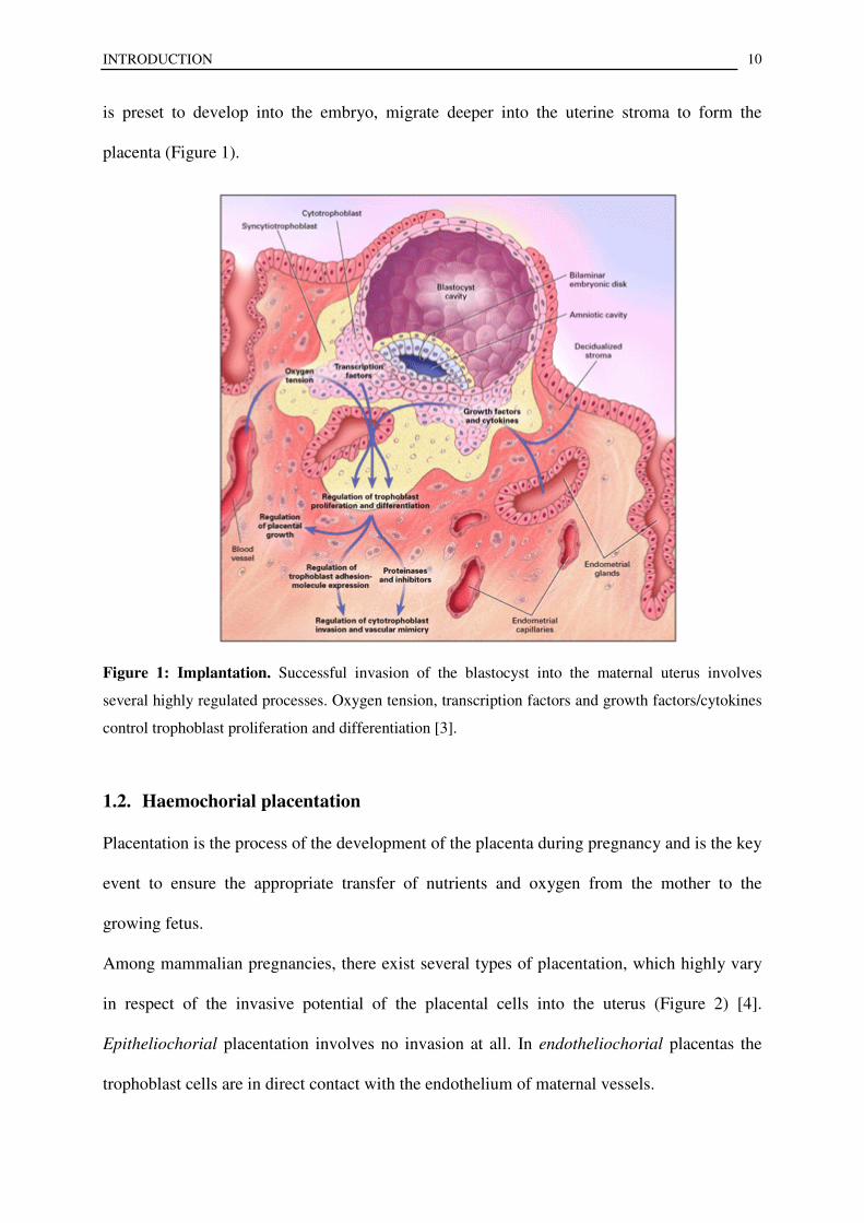
1.1. Implantation
During the passage of the fallopian tube, the fertilized ovum (zygote) divides to form the
morula (12 to 16 cells). Six to seven days following conception, the 128 cell stage mass
(blastocyst) starts to implant into the uterine wall (Figure 1). Human implantation can be
divided in three steps. The first unstable adhesion of the blastocyst is called apposition and is
characterized by the contact between microvilli of the trophoblast cells and the large and
smooth projections (pinopodes) of the uterine epithelium. During the stable adhesion these
interactions are increasing, leading to the final step of implantation, namely the invasion of
the blastocyst into the uterus. As soon as the blastocyst had penetrated the uterine wall, the
trophoblast cells, which form the extra-embryonic tissue covering the inner cell mass, which
INTRODUCTION 10
is preset to develop into the embryo, migrate deeper into the uterine stroma to form the
placenta (Figure 1).
Figure 1: Implantation. Successful invasion of the blastocyst into the maternal uterus involves
several highly regulated processes. Oxygen tension, transcription factors and growth factors/cytokines
control trophoblast proliferation and differentiation [3].
1.2. Haemochorial placentation
Placentation is the process of the development of the placenta during pregnancy and is the key
event to ensure the appropriate transfer of nutrients and oxygen from the mother to the
growing fetus.
Among mammalian pregnancies, there exist several types of placentation, which highly vary
in respect of the invasive potential of the placental cells into the uterus (Figure 2) [4].
Epitheliochorial placentation involves no invasion at all. In endotheliochorial placentas the
trophoblast cells are in direct contact with the endothelium of maternal vessels.

INTRODUCTION 11
Several mammals, including rodents, monkeys and humans, have the invasive haemochorial
placentation. However, rodents and monkeys are not complete models for human
placentation, since the intermixture of fetal and maternal cells is highest in humans.
Figure 2: The three major types of placentation. Depending on the mode of placentation, the
relationship between maternal blood cells and fetal trophoblast cells is different. (a) Epitheliochorial
placentation is characterised by the apposition of the uterine epithelium and the trophoblast cell layer,
in the absence of trophoblast invasion into the maternal tissue and vessels. (b) In endotheliochorial
placentation the uterine epithelial cell layer is ruptured and trophoblasts are in direct tangency with the
maternal endothelium. (c) The most invasive type of placentation is the haemochorial one, where
trophobalst cells are penetrating maternal vessels and the syncytiotophoblast is in direct contact with
maternal blood in the intervillous space [4].
During human placentation, there are three main changes in the uterus. First, the uterine
mucosa (endometrium) differentiates into a dense cellular matrix (decidua) by a process
called decidualization [5]. Second, trophoblast cells are invading the decidua and the
underlying myometrium [5]. Third, the uterine spiral arteries are transformed into widened,
low resistance vessels, to direct an increased maternal blood flow into the placenta [5]. This
enlargement is mainly done by extravillous trophoblast cells (EVTs), comprising interstitial
and endovascular EVTs, which penetrate maternal vessels, disrupt and replace the
INTRODUCTION 12
endothelium and some parts of the muscle coat, resulting in a pseudo-endothelium being half
fetal and half maternal [6].
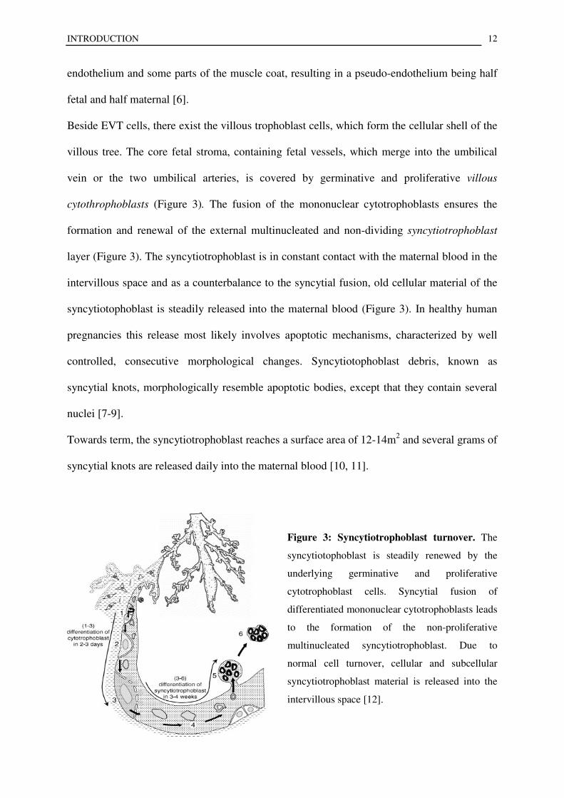
Beside EVT cells, there exist the villous trophoblast cells, which form the cellular shell of the
villous tree. The core fetal stroma, containing fetal vessels, which merge into the umbilical
vein or the two umbilical arteries, is covered by germinative and proliferative villous
cytothrophoblasts (Figure 3). The fusion of the mononuclear cytotrophoblasts ensures the
formation and renewal of the external multinucleated and non-dividing syncytiotrophoblast
layer (Figure 3). The syncytiotrophoblast is in constant contact with the maternal blood in the
intervillous space and as a counterbalance to the syncytial fusion, old cellular material of the
syncytiotophoblast is steadily released into the maternal blood (Figure 3). In healthy human
pregnancies this release most likely involves apoptotic mechanisms, characterized by well
controlled, consecutive morphological changes. Syncytiotophoblast debris, known as
syncytial knots, morphologically resemble apoptotic bodies, except that they contain several
nuclei [7-9].
Towards term, the syncytiotrophoblast reaches a surface area of 12-14m2 and several grams of
syncytial knots are released daily into the maternal blood [10, 11].
Figure 3: Syncytiotrophoblast turnover. The
syncytiotophoblast is steadily renewed by the
underlying germinative and proliferative
cytotrophoblast cells. Syncytial fusion of
differentiated mononuclear cytotrophoblasts leads
to the formation of the non-proliferative
multinucleated syncytiotrophoblast. Due to
normal cell turnover, cellular and subcellular
syncytiotrophoblast material is released into the
intervillous space [12].

INTRODUCTION
1.3. Physiological hypoxia in early placentation
Early human placentation is not truly haemochorial. In fact, w
uterine tissue, they also obstruct the tips of the spiral arteries by forming cellular plugs
(Figure 4) [13]. On the one hand, this cell shell firmly connects placenta and maternal tissue.
On the other hand, these plugs filter maternal blood and strongly limit blood flow into the
intervillous space. In this stage, placental villi only contain few capillary vesse
low feto-placental blood flow. Only few spiral arteries in the periphery are never plugged by
EVT and allow a limited maternal blood flow into the placenta
the amniotic cavity and the placenta are spatially separated by the exocoelomic cavity (Figure
4). All these anatomical changes mediate a hypoxic environment, which is essential for the
development of the fetus. Fetal organogenesis is very susceptible to teratogenic damage by
reactive oxygen free radicals (OFRs) and hypoxia is needed to keep the full pluripotency of
stem cells [14].
Physiological hypoxia in early placentation
Early human placentation is not truly haemochorial. In fact, when EVT start to invade the
uterine tissue, they also obstruct the tips of the spiral arteries by forming cellular plugs
. On the one hand, this cell shell firmly connects placenta and maternal tissue.
On the other hand, these plugs filter maternal blood and strongly limit blood flow into the
intervillous space. In this stage, placental villi only contain few capillary vesse
placental blood flow. Only few spiral arteries in the periphery are never plugged by
EVT and allow a limited maternal blood flow into the placenta [13]. Additionally, the fetus in
the amniotic cavity and the placenta are spatially separated by the exocoelomic cavity (Figure
4). All these anatomical changes mediate a hypoxic environment, which is essential for the
fetus. Fetal organogenesis is very susceptible to teratogenic damage by
reactive oxygen free radicals (OFRs) and hypoxia is needed to keep the full pluripotency of
Figure 4: Early human pregnancy (End of 2
month). Obstruction of the tips of the spiral arteries
by extravillous trophoblast cells prevents the flow of
maternal blood into the intervillous space of the
placenta, resulting in a hypoxic environment for the
placenta and the growing fetus
arteries in the periphery are never plugged allowing a
minimal maternal blood flow into the intervillous
space (white arrows). M = myometrium, D = decidua,
P = placenta, ECC = exo-coelomic cavity, AC =
amniotic cavity, SYS = secondary yolk sac
13
hen EVT start to invade the
uterine tissue, they also obstruct the tips of the spiral arteries by forming cellular plugs
. On the one hand, this cell shell firmly connects placenta and maternal tissue.
On the other hand, these plugs filter maternal blood and strongly limit blood flow into the
intervillous space. In this stage, placental villi only contain few capillary vessels, leading to a
placental blood flow. Only few spiral arteries in the periphery are never plugged by
. Additionally, the fetus in
the amniotic cavity and the placenta are spatially separated by the exocoelomic cavity (Figure
4). All these anatomical changes mediate a hypoxic environment, which is essential for the
fetus. Fetal organogenesis is very susceptible to teratogenic damage by
reactive oxygen free radicals (OFRs) and hypoxia is needed to keep the full pluripotency of
Figure 4: Early human pregnancy (End of 2nd
Obstruction of the tips of the spiral arteries
by extravillous trophoblast cells prevents the flow of
maternal blood into the intervillous space of the
placenta, resulting in a hypoxic environment for the
placenta and the growing fetus. Only few spiral
arteries in the periphery are never plugged allowing a
minimal maternal blood flow into the intervillous
space (white arrows). M = myometrium, D = decidua,
coelomic cavity, AC =
amniotic cavity, SYS = secondary yolk sac [13].

INTRODUCTION 14
1.4. Physiological placental oxidative burst at the end of the first trimester
After week 12 of gestation the decidual partial pressure of O2 (PO2) is two to three times
higher than between week 8 and 10 [15, 16]. This rise in the intraplacental PO2 at the end of
the first trimester triggers an oxidative burst in the periphery of the early placenta [16]. This
higher local O2 concentration damages the trophoblasts and leads to villous degeneration,
inducing the formation of fetal membranes [17].
The increased placental oxygenation triggers trophoblast growth and differentiation, and
invasive EVT extensively transform the spiral arteries, including the myometrial segments
[13]. Spiral arterial rearrangement is complete by week 20 and allows an increased maternal
blood supply of the placenta.

INTRODUCTION 15
2. Immunology of human pregnancy
2.1. Immunological interfaces
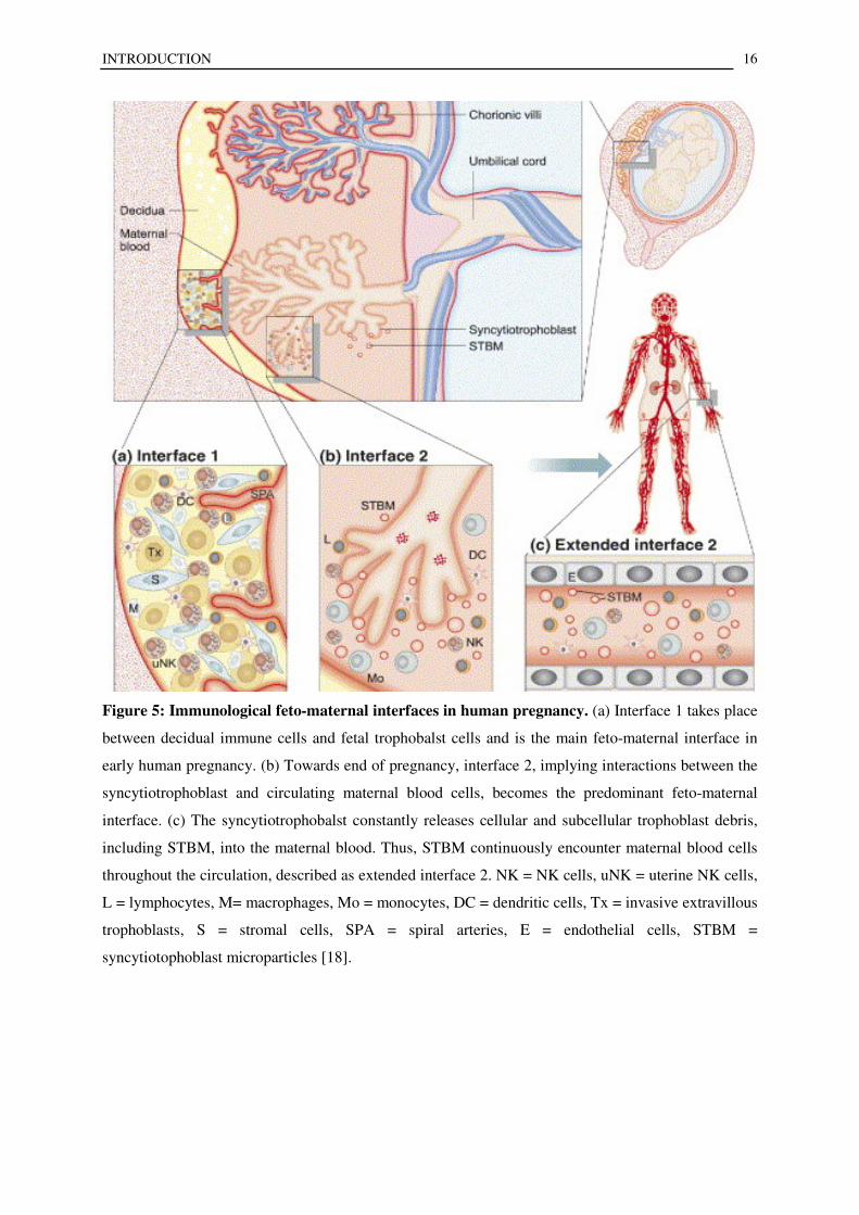
The invasive nature of haemochorial placentation implicates direct contact of maternal and
fetal cells. There are two immunological interfaces between mother and child in human
pregnancy (Figure 5). Interface 1 involves the local interactions between maternal cells and
placental trophoblasts in the decidua and is the main feto-maternal interface during early
pregnancy (Figure 5A) [18]. Interface 2 is set up towards the end of the first trimester with the
onset of maternal blood flow into the placental intervillous space and takes place between the
syncytiotrophoblast, the outer lining of the placenta, and circulating maternal blood cells
(Figure 5B) [16, 18, 19]. Due to the large size of the placenta, the second interface becomes
the main interface towards term pregnancy [18]. The syncytiotrophoblast constantly releases
placentally-produced factors, including hormones, cytokines, angiogenic factors and
trophoblast debris, into the intervillous space, from where they are carried with the maternal
blood flow into the entire circulation (extended interface 2) (Figure 5C) [18].
INTRODUCTION 16
Figure 5: Immunological feto-maternal interfaces in human pregnancy. (a) Interface 1 takes place
between decidual immune cells and fetal trophobalst cells and is the main feto-maternal interface in
early human pregnancy. (b) Towards end of pregnancy, interface 2, implying interactions between the
syncytiotrophoblast and circulating maternal blood cells, becomes the predominant feto-maternal
interface. (c) The syncytiotrophobalst constantly releases cellular and subcellular trophoblast debris,
including STBM, into the maternal blood. Thus, STBM continuously encounter maternal blood cells
throughout the circulation, described as extended interface 2. NK = NK cells, uNK = uterine NK cells,
L = lymphocytes, M= macrophages, Mo = monocytes, DC = dendritic cells, Tx = invasive extravillous
trophoblasts, S = stromal cells, SPA = spiral arteries, E = endothelial cells, STBM =
syncytiotophoblast microparticles [18].
INTRODUCTION 17
2.2. Immunological interactions
In immunological term, the growing fetus in the uterus is considered as a semi-allograft,
having half of the genes from the mother (maternal) and the other half from the father
(paternal). Interestingly, in healthy human pregnancies the fetal and maternal cells are
coexisting, without leading to the rejection of the developing fetus. This feto-maternal
tolerance is believed to be carried out by several mechanisms used by the maternal immune
system and placental trophoblast cells locally at interface 1, but also systemically in the
maternal circulation.
2.2.1. Predominant Th2 immunity
In 1993 Wegmann and co-workers suggested, that the human fetus is not rejected by the
maternal immune system thanks to a prevalent production of T helper cell 2 (Th2) cytokines,
both systemically and in the placental bed [20-22]. Th2 mediators, such as interleukin (IL)-4,
IL-5, IL-6, IL-10 and IL-13, attenuate the production of proinflammatory Th1 cytokines, such
as IL-2 and interferon (IFN)-γ. Thereby, the Th2 response interferes with the stimulation of
CD8+ cytotoxic T-cells and natural killer (NK)-cells, which may be harmful for the fetal cells
[20].
The Th2 cytokine IL-4 is continuously expressed by the syncytiotrophoblast, cytotrophoblast
cells and decidual macrophages [23, 24]. In addition, the spontaneous secretion of the Th1
mediator IL-12 by peripheral blood mononuclear cells (PBMC) from pregnant women is
significantly decreased than in non-pregnant controls [25]. However, few years ago, the
question raised, if this bias towards Th2 cytokine secretion is important for successful
pregnancy, or if it is just a secondary effect [26]. Although, in humans, a propensity to Th1
immune reactions has been reported in pregnancy complications, such as recurrent
spontaneous abortions, so far, a T-cell mediated attack of the fetus, triggered by a shift
towards Th1 immunity has only been shown in an abortion-prone mouse model [27, 28].

INTRODUCTION 18
Thereby, the abortions in the matings of CBA/J females with DBA/2 males were mediated by
the production of the Th1 cytokine TNF-α in the decidua and could be hindered by the Th2
cytokine IL-10 [28, 29].
2.2.2. Absent trophoblast expression of MHC class II and classical polymorphic MHC
class I molecules
Major histocompatibility complex (MHC) class I and class II molecules present peptides,
derived from pathogens or foreign cells, on the cell surface of antigen presenting cells (APCs)
to cytotoxic CD8+ T-cells and helper CD4
+ T-cells, respectively [30]. MHC class I and II
themselves are recognized as non-self antigens by the immune cells of a recipient of an
allogeneic organ graft [31, 32]. Thus, an analogy could be drawn between the fetus and an
allograft. However, all human trophoblast cell subsets completely lack MHC class II
expression and display an unconventional pattern of MHC class I molecules [4]. The classical
polymorphic MHC class I molecules human leukocyte antigen (HLA)–A and HLA-B, which
are driving the T-cell-dependent transplant rejection, are not expressed by any trophoblast cell
subset [18]. Intriguingly, EVT cells display the classical polymorphic HLA-C and the non-
classical, less polymorphic HLA-G, HLA-E and HLA-F [33-37].
Originally it was believed that the syncytiotrophoblast cells are deficient of MHC class I and
II expression, but lately it has been proposed that they might display non-classical HLA-E and
produce soluble HLA-G [4, 38-40].
2.2.3. Decidual NK (dNK) cells and their interaction with trophoblast HLA-E, -G, -C
70% of all leukocytes at the implantation site are made up by a particular CD56hi
CD16-
decidual natural killer (dNK) cell subset, which are recruited in great numbers in early
pregnancy and remain the most abundant maternal immune cells in the decidua until mid-
gestation, before their quantity is decreasing again towards term [41, 42].

INTRODUCTION 19
Despite their expression of perforin, granzyme A and B, and some NK-activating receptors,
and the close contact with infiltrating interstitial trophoblasts, dNK cells do not mediate
cytotoxicity against the allogeneic trophoblast cells [43, 44]. This is believed to be due to the
interaction between the non-classical, largely monomorphic MHC class I molecules HLA-G
and HLA-E displayed by EVT and its receptors expressed by dNK cells. HLA-E is suggested
to bind to the inhibitory receptor CD94/NKG2A, which is highly expressed by dNK cells
[45]. A number of inhibitory NK cell receptors, including the killer cell immunoglobulin
receptor (KIR) KIR2DL4 (CD158d) and the leukocyte Ig-like receptor (LIR-1), have been
suggested to bind to HLA-G and, thus, prevent NK-mediated killing [46, 47].
In addition to tolerance induction, the great abundance of dNK cells also indicates an essential
role in placentation. Indeed, dNK cells are suggested to actively support vascular remodelling
by secreting interferon (IFN)-γ, vascular endothelial growth factor (VEGF) and placental
growth factor (PlGF) [48, 49]. Furthermore, dNK cells produce the chemokines IL-8 and
interferon-inducible-protein-10 (IP-10), which trigger migration of invading trophoblasts [49].
It has also been proposed that specific dNK cell – EVT interactions might be more beneficial
for the transformation of the spiral arteries [50]. EVT express the classical, polymorphic
MHC class I molecule HLA-C, which interacts with specific members of the KIR family of
NK cell receptors, namely KIR2D (contain two immunoglobulin-like domains) [51].
Depending on the combination of the haplotypes, KIR2D may be more activating or more
inhibiting [51]. Particular (more activating) KIR2D-HLA-C pairs may be more favourable for
the enlargement of the spiral arteries, suggesting that activated dNK cells might be essential
for an efficient vascular remodelling [4, 50].
2.2.4. Decidual macrophages
20-30% of the maternal cells at the implantation site account for decidual macrophages and
their numbers remain high throughout pregnancy [52]. Decidual macrophages display CD14,

INTRODUCTION 20
the MHC class II molecule HLA-DR and low levels of the T-cell co-stimulatory cell surface
markers CD80 and CD86 [52-54]. In vitro, decidual macrophages spontaneously produce high
levels of the anti-inflammatory cytokine IL-10 and express the tryptophan catabolising
enzyme indoleamine dioxygenase (IDO), suggesting that decidual macrophages exhibit
immunosuppressive functions, which may be important to maintain feto-maternal tolerance
[54, 55].
Since decidual macrophages are localized in the proximity of apoptotic trophoblast cells in the
placental bed, it has been speculated that the engulfment of these dying cells by decidual
macrophages might result in a decreased synthesis of proinflammatory cytokines and an
enhanced secretion of anti-inflammatory Th2 mediators [52]. The uptake of apoptotic
trophoblast cells may prevent secondary necrosis, which is characterized by the release of
potentially proinflammatory and immunogenic intracellular material from the dying cells [52].
2.2.5. Regulatory T-cells (Treg)
The human CD4+CD25
high regulatory T-cells (Treg) are involved in tolerance induction against
self-antigens and allogeneic organ grafts [56, 57].
Levels of peripheral blood Treg almost double during pregnancy, compared to non-pregnant
controls, and drop again postpartum [58]. Treg are also localized in the decidua and during
early pregnancy they account for 22% of CD4+ cells. Spontaneous abortion is associated with
a reduction of circulating Treg to non-pregnant levels and a decrease of the
CD4+CD25
high/CD4
+ ratio to 7% in the decidua [59, 60].
These observations suggest that Treg are essential for the maintenance of the maternal
tolerance towards the growing fetus and, thus, may contribute to the successful progression of
human pregnancy.

INTRODUCTION 21
2.2.6. B7 family:
Activation of naive T-cells requires the engagement of the T cell receptor (TCR) by binding
to an antigen peptide – MHC protein complex on APCs. To complete T-cell stimulation, a
costimulatory signal is needed, which is mediated by the interaction between T-cell expressed
CD28 and members of the B7 family, displayed by the partner cell [61]. However, B7
molecules are able to trigger both, activating and inhibitory signals in lymphocytes.
The expression of at least five of the seven known B7 family members has been reported in
the human placenta [62, 63]. From all placentally expressed B7 family members, only B7-H1
has been documented to play a key role in maternal tolerance toward the fetus, since
inhibition or genetic deletion of B7-H1 resulted in an elevated rejection of the allogeneic fetus
in animal experiments [64]. In humans, placental expression of B7-H1 is found on
syncytiotrophoblasts, villous and extravillous cytotrophoblasts, and is increasing throughout
pregnancy [62]. B7-H1 interacts with the immunoinhibitory molecule programmed death
(PD)-1, which is present on activated T-cells, B-cells and monocytes [65]. The in vitro
ligation of human PD-1 blocks T-cell proliferation and cytokine production [66]. As human
decidual T-cells express PD-1, the induction of the PD-1-B7-H1 pathway may provide a
mechanism of immunological tolerance towards the fetal semi-allograft [67].
2.2.7. Indoleamine-2,3-dioxygenase (IDO):
Indoleamine-2,3-dioxygenase (IDO) is a tryptophan-degrading enzyme, which is expressed at
the feto-maternal interfaces by trophoblasts, syncytiotrophoblasts and glandular epithelial
cells [60]. By depleting tryptophan from the local microenvironment, IDO can inhibit the
activation of T-cells, which are particularly sensitive to the loss of this essential amino acid
[68, 69]. IDO may mediate T-cell suppression either by limiting the availability of tryptophan,
or by indirect effects on the biology of IDO-expressing cells [69].

INTRODUCTION 22
2.2.8. Fas-Fas ligand system:
Expression of the death-triggering receptor-ligand pair Fas (CD95) – Fas ligand
(FasL/CD95L) is found at the feto-maternal interface. FasL is expressed by EVT, villous
cytotrophoblasts, syncytiotrophoblasts and maternal decidual cells, and eliminates activated
Fas-positive maternal immune cells invading the uterus [70-72]. In vitro, human trophoblast
cells induce Fas/FasL dependent apoptosis in T-cells [73, 74]. Recently, Abrahams et al.
demonstrated that trophoblast cells from the first trimester placenta do not express cell-
surface FasL [75]. However, the first trimester trophoblast cells are able to secrete FasL in
vitro, which might protect them from the recognition by the maternal immune system [75].
2.2.9. Complement regulatory proteins:
The complement system is an essential component of the immune defence, assembled by a set
of proteins, and mediates clearance of pathogens, apoptotic cells and immune complexes [76,
77]. The net result of the complement cascade is the formation of the membrane attack
complex (MAC), leading to cellular lysis [76]. The syncytiotrophoblast, villous
cytotrophoblasts and EVT all express the three complement regulatory proteins decay
accelerating factor (DAF/CD55), membrane co-factor protein (MCP/CD46) and CD59, which
protect healthy cells from cell lysis [78-82].
2.2.10. Deportation of syncytiotrophoblast debris into maternal blood
As mentioned earlier, the old cellular material of the syncytiotrophoblast, such as syncytial
knots and microparticles, is constantly released into the intervillous space (Figure 5).
Syncytial knots are detectable in the maternal uterine veins, but not in the peripheral blood of
pregnant women, which might be due to their uptake by alveolar macrophages in the lungs
[83-85]. Engulfment of syncytial knots by a macrophage cell line induces an anti-
inflammatory response in vitro, suggesting that the shedding of apoptotic debris is not only a

INTRODUCTION 23
mechanism to dispose aged syncytiotrophoblast cells, but also a mechanism to provide
tolerogenic parental antigens to the maternal immune system [85].

INTRODUCTION 24
2.3. Systemic maternal inflammation
Healthy third-trimester pregnant women display a generalized inflammatory response, which
is in some regards as extensive as in sepsis [86]. However, this controlled systemic
inflammatory reaction does not appear to be harmful for the mother.
Markers of overall maternal inflammation include an elevated total white blood cell count
(leukocytosis) and increased levels of the proinflammatory cytokines IL-1, IL-6, TNF-α and
the acute phase protein C-reactive protein (CRP) in the serum of pregnant women compared
to non-pregnant subjects [87-90]. CRP levels are moderately raised as early as week 4 of
gestation, providing evidence that there is a low-grade maternal inflammation already during
the implantation of the conceptus [90]. Furthermore, there is an activation of peripheral blood
leukocytes, which is characterized by the significantly elevated cell surface expression of the
adhesion molecules CD11b, CD11c, CD64 and the pattern recognition receptor CD14 on
granulocytes and monocytes compared to non-pregnant controls [18, 86, 91]. Moreover,
monocytes, granulocytes and lymphocytes display significantly increased values of basal
intracellular reactive oxygen species [86].
Granulocytes from pregnant donors spontaneously produce the proinflammatory cytokine IL-
6 and the chemokine IL-8 [92]. Phorbol 12-myristate 13-acetate (PMA)/ionomycin-stimulated
third trimester granulocytes synthesise significantly more IL-8 than PMA/ionomycin-
stimulated cells from non-pregnant women, suggesting that circulating maternal granulocytes
are primed to produce inflammatory mediators [92].
Monocytes from pregnant women display an enhanced phagocytic rate with peak levels in the
third trimester and exhibit a progressive up-regulation of the cell surface adhesion molecule
CD54 [92-94]. In addition, PMA/ionomycin triggers a higher production of intracellular IL-
1β in second- and third-trimester monocytes than in the cells from pregnant women in the first
trimester and non-pregnant controls [92]. These findings suggest a continuous activation of
maternal monocytes throughout pregnancy.

INTRODUCTION 25
It has also been shown, that during pregnancy, the platelets, the complement - and the clotting
system are activated [18].
Until now the cause of this maternal inflammation is unknown. However, it is generally
accepted, that it consists in a sterile inflammatory reaction, which is stimulated by the
pregnancy itself and not by an infection. Candidate triggers include placental factors released
into maternal blood, such as cytokines and angiogenic factors [95, 96].
2.3.1. Placental cytokines
Pro-inflammatory cytokines and chemokines, including IL-6, IL-1β, IL-8 and TNF-α, are
produced by the placenta, as shown by in vitro cultures of placental villous tissue [97-100].
Beside their local action, they are also secreted into the maternal circulation, where they may
contribute to the mild systemic inflammation by attracting and stimulating maternal immune
and endothelial cells [101]. The placental expression of the different cytokines varies in
function of the gestational age, most likely reflecting a specific function at particular stages of
pregnancy [102, 103].
2.3.2. Placental angiogenic factors
Placental angiogenesis is of great importance in pregnancy to ensure appropriate blood supply
to the growing fetus. Angiogenesis is supported by placentally-produced angiogenic factors,
such as PlGF and VEGF, which interact with receptors expressed by the vascular
endothelium, including the VEGF receptor (VEGFR / Flt-1) and endoglin (Eng) [104-110]. In
addition, soluble forms of Flt-1 (sFlt-1) and Eng (sEng) with anti-angiogenic properties are
produced by the placenta [96, 111]. These factors are released into the maternal peripheral
blood, where they could disturb the maternal endothelium and explain the vascular reactivity
and mild glomerular endotheliosis found towards term [112, 113]. The expression of these
markers varies with the gestational age [96, 114].

INTRODUCTION 26
A potential role in the maternal inflammatory response is also ascribed to membrane
microparticles released from the syncytiotrophoblast into the maternal circulation, which are
discussed later, in chapter 3 of the introduction [115].

INTRODUCTION 27
3. Preeclampsia
3.1. Epidemiology
Preeclampsia is a heterogeneous placenta-dependent disorder specific to human pregnancies
and is characterized by new onset hypertension (systolic and diastolic pressure of ≥140 and
90mm Hg) and proteinuria (protein excretion of ≥300 mg in a 24 h urine collection, or a
dipstick of ≥2+) after week 20 of gestation [116, 117]. The maternal symptoms become
apparent in the second half of pregnancy and may be either early (<34 weeks of gestation) or
late (>34 weeks of gestation), and mild or severe, according to the degree of hypertension
and/or protenuria. In addition, patients may exhibit edema, reduced blood supply to several
organs, including the placenta, and can end up in organ (mainly renal or liver) failures. In
severe cases preeclamptic women may develop the HELLP syndrome (hemolysis, elevated
liver enzymes, and low platelet counts) or the disorder may evolve into eclampsia, which is
characterized by convulsions and seizures, pre- or postnatally. Preeclampsia may manifest as
a maternal syndrome only, or it can affect the fetus, through in utero growth restriction
(IUGR) or sudden fetal distress.
The reported incidence of preeclampsia is 2 to 7% among human pregnancies world wide
[117]. However, there are large differences in the frequency of incidence among populations,
which may be due to racial, geographic, social and economic distinctions. Other predisposing
factors, which make women more prone to become preeclamptic during pregnancy, include
obesity, chronic hypertension, diabetes or insulin resistance, multiple gestations and a history
of previous preeclampsia [118].
Owing to the excellent medical care in western countries, preeclampsia is less and less fatal
[119]. However, in underdeveloped countries preeclampsia remains the major cause of
maternal death. In Latin America and Caribbean over 25% of maternal deaths are attributed to

INTRODUCTION 28
pregnancy associated hypertensive disorders [120]. In Columbia the number of maternal
deaths caused by preeclampsia (42%) is ten times higher than in the United States [121].
Pregnancy may be lengthened by giving antihypertensive drugs to treat the symptoms of the
disorder [122]. To prevent imminent eclampsia magnesium sulphate, a promising
anticonvulsant drug, is applied [123-126]. However, none of these drugs prevents the onset of
preeclampsia and the only definite cure of preeclampsia is the removal of the placenta by
elective preterm delivery, either by induction or caesarean section. Though, preterm delivery
(<37 weeks of gestation) accounts for 75% of perinatal mortality and preterm babies are at
increased risk for long-term medical problems affecting the neurological, respiratory,
cardiovascular and gastrointestinal systems [127].
Although the acute maternal symptoms of preeclampsia can be cured by delivering the baby,
women have an increased risk to develop cardiovascular diseases in later life [128].
Since preeclampsia is a life-threatening disorder, it is very demanding to recognize the disease
early enough. In the past few years, research focussed in the finding of biophysical and
biochemical markers, which are differently expressed in preeclamptic women or in women,
who will develop the disease, compared to healthy pregnant controls, and could be used for
detection and prediction [129].
Several clinical studies were/are evaluating the efficiency of prophylactic agents, such as the
anti-oxidant vitamins C and E, low-dose aspirin, folic acid and calcium [117, 130-136].
However, the results of different clinical trials are either contradictory or not efficacious.
INTRODUCTION 29
3.2. Pathophysiology
Preeclampsia may be divided into two main stages. The first stage is related to the
asymptomatic preclinical phase in early pregnancy. The clinical stage appears in the second
half of pregnancy and is characterized by the maternal syndrome.
3.2.1. Preclinical stage: shallow trophoblast invasion and incomplete vascular
remodelling
Preeclampsia may develop in pregnancies without a viable fetus (hydatidiform moles), but its
onset always depends on the presence of a placenta [118]. A long lasting hypothesis has been
that the onset of preeclampsia relies on a deficient development of the early placenta and an
incomplete remodelling of the maternal spiral arteries (Figure 6). This poor placentation takes
place before week 20 of gestation [137]. The invasion of the decidua by EVT is reduced and
the transformation of spiral arteries into dilated tubes is shallow or absent.
However, reduced placental perfusion cannot be the only trigger of preeclampsia since
normotensive intrauterine growth restriction (nIUGR) is associated with an incomplete
placentation in the absence of the maternal syndrome [138].
Furthermore, the current hypothesis on the placental origin of preeclampsia has been
challenged [139]. It has been suggested that the failure of the development and the
differentiation of the trophoblast lineage at various time-points may result in preeclampsia
with late-onset appearance and mild symptoms, or IUGR.
However, women suffering from arterial diseases, hypertension, obesity and diabetes before
conception are predisposed to preeclampsia, and in this so-called maternal preeclampsia the
challenge rather lies in an inappropriate maternal response than in an abnormal placentation
[140, 141].
.

INTRODUCTION 30
Figure 6: Incomplete remodelling of spiral arteries in preeclampsia. During normal pregnancy
uterine spiral arteries are extensively remodelled. In order to increase the placental and fetal blood
supply, the spiral arteries are enlarged by replacing the vascular endothelium by infiltrating
trophoblast cells. In preeclampsia and IUGR vascular transformation is shallow resulting in a reduced
blood flow in the intervillous space [41].
3.2.1.1. Placental oxidative stress
The shallow remodelling of spiral arteries implicates maintenance of smooth muscle cells in
the placental vascular walls and persistence of up to 50% of the vascular contractibility [13].
This might result in an intermittent perfusion of the intervillous space mediating transient
hypoxic conditions [142]. In in vitro experiments, Hung and collaborators demonstrated that
oxidative stress, which is characterized by an imbalance favouring oxidant over antioxidant
forces, occurs after reoxygenation of hypoxic placental tissue [143]. Enhanced placental
oxidative stress is associated with increased tissue damage mediated by reactive oxygen free
radicals (OFRs) [13].

INTRODUCTION 31
Lipid peroxidation, induced by OFRs, enhances the incorporation of cholesterol, oxidized free
fatty acids (FFAs) and low-density lipoproteins (LDLs) into cell membranes [144].
Furthermore, in regions of spiral and myometrial arteries, where the physiologic remodelling
is missing, pathologic lesions known as acute atherosis can be found [143]. Acute atherosis
shares a lot of clinical features with atherosclerosis, namely clusters of macrophages loaded
with lipids (foam cells), fibrinoid necrosis of vascular walls, dysfunctional endothelium and
accumulations of platelets [145-148]. Markers of oxidative stress have been found in the
peripheral blood of preeclamptic women [149, 150]. Hence, it has been proposed that
oxidatively stressed placentas of preeclamptic patients release soluble factors into the
maternal bloodstream, where they might affect the maternal vascular endothelium [151].
3.2.1.2. Maternal-paternal immune maladaptation
There are multiple lines of evidence that imply a crucial role of the maternal immune system
in the onset of preeclampsia.
Preeclampsia is a disorder of first pregnancies (primigravidity) and the risk to develop
preeclampsia decreases upon an earlier healthy pregnancy [152, 153]. However, the change of
the partner reverses this natural protection generated by multiple pregnancies (multigravidity).
Thus, Robillard and co-workers described preeclampsia as a “disease of new couples”
(primipaternity), according to the observation that the length of sexual cohabitation before
conception inversely correlates with the risk of preeclampsia [154]. Though, the use of barrier
contraceptives, such as condoms, does not reduce the risk of developing preeclampsia, leading
to the hypothesis that preeclampsia might develop due to missing seminal priming [155].
Other mechanisms of tolerance induction are altered or missing in preeclampsia as well. A
reduced trophoblast FasL expression and an elevated Fas expression have been observed and
this correlates with increased apoptosis of EVTs and villous trophoblast cells [156-160].
Furthermore HLA-G expression on EVTs is reduced or even missing, and, thus, the resulting

INTRODUCTION 32
deficient interactions with dNK cells might lead to dNK-cell-mediated cytolysis of EVTs
[161, 162]. Santoso and co-workers reported a decreased expression of the T-cell inhibitor
IDO in preeclamptic placentas [163]. In addition, in pregnancy pathologies, such as
preeclampsia and recurrent pregnancy loss, an increased activation of the complement system
has been found [128, 164-166]. However, studies gave controversial results about the
frequency of peripheral blood Treg cells in women suffering from preeclampsia [167-169].
In addition, preeclampsia is associated with a predominant Th1 response in the peripheral
maternal blood, as well as in the placenta, in contrast to the Th2 bias in normal pregnancy
[170]. The spontaneous and phytohemagglutinin (PHA)-stimulated production of the Th1
cytokines TNFα, IL-2 and IFNγ by PBMC from preeclamptic patients is higher than those
from normal pregnant controls [171, 172]. Furthermore, the in vitro stimulation of PBMC
from preeclamptic women with the classical Th1 cytokine IFNγ results in an increased
production of IL-12 and IL-18 [173]. In the presence of IL-12, IL-18 does not act as a Th2
mediator anymore, but supports the IL-12 driven Th1 response [174]. Th1 cytokines are
known to trigger chronic inflammation, and IFNγ, together with the proinflammatory factors
IL-1 and TNFα, has been shown to amplify this chronic inflammatory response [170].
3.2.2. Clinical stage: excessive maternal systemic inflammation and placental factors
An excessive, generalized maternal inflammatory response including a dysfunctional maternal
endothelium is thought to be at the basis of the maternal clinical manifestations [175, 176].
This overt inflammatory response is believed to be the extreme end of the mild inflammation
found in healthy pregnancy [177]. There is a significantly increased leukocytosis and a
significantly higher concentration of the proinflammatory mediators IL-6 and IL-8 in the
peripheral blood of preeclamptic women relative to normal pregnant women [178-185].
Enhanced activation of peripheral blood leukocytes is marked by the higher basal as well as
PMA-induced production of intracellular reactive oxygen species and a significant increased

INTRODUCTION 33
cell surface expression of the integrin CD11b, in monocytes and granulocytes from
preeclamptic women compared to the monocytes and granulocytes from healthy pregnant
women [86, 91, 186].
Additionally, expression of the activation marker HLA-DR is significantly enhanced on
monocytes derived from preeclamptic women relative to the cells of normotensive pregnant
women [25]. However, the findings on the level of CD14 expression on monocytes are
contradictory [25, 91]. Furthermore, spontaneous intra-monocytic synthesis of IL-1β, IL-6
and IL-8 is higher in cells from preeclamptic women than in monocytes derived from normal
pregnant and non-pregnant subjects [187]. Monocyte-derived microparticles were also more
elevated in preeclamptic patients compared with pregnant controls, reflecting activation of
their parental cells in preeclampsia [188].
A dysfunctional endothelium is marked by the increased plasma concentrations of the
vasoconstrictive mediators’ asymmetric dimethylarginine and endothelin, and the released
integrin fibronectin in women with adjacent preeclampsia relative to women with
uncomplicated pregnancies [189-192].
Furthermore, there is an elevated activation of the complement cascade, the clotting system
and the platelets in the peripheral blood of preeclamptic women compared to normal pregnant
women [193-195].
As it is the case in normal pregnancy, candidate triggers are believed to be derived from the
placenta [196]. The expression of placental cytokines and placenta-derived angiogenic factors
is altered compared to normal pregnancy. Placental tissue from preeclamptic women produce
increased levels of the proinflammatory cytokines TNFα, IL-1, and IFN-γ relative to the
placenta of normal pregnant subjects [95]. The anti-angiogenic factors sFlt-1 and sEng, which
are secreted by the placenta, are elevated in the peripheral blood of preeclamptic women for a
long time before the onset of the disease [96, 197-200]. On the other hand, the levels of the
pro-angiogenic factor PlGF are reduced before and during onset of preeclampsia [129, 201].

INTRODUCTION 34
The rates of syncytial apoptosis and shedding of debris are significantly increased in
preeclampsia compared to normotensive pregnancy (Figure 7), consistent with the increased
placental damage and dysfunction observed in this pathological condition [160]. Other
markers for apoptosis, such as cytokeratin and cell free fetal DNA of placental origin are also
elevated in preeclampsia [202, 203]. It was suggested that the shedding of placental debris
might be exacerbated by apo-necrosis or even necrosis in this pathologic condition (Figure 7)
[204]. Among this syncytial debris, there are small membrane microparticles released from
the syncytiotrophoblast [115].
Figure 7: Release of Syncytial knots and STBM by
apoptosis, apo-necrosis and necrosis. (A) Controlled
apoptotic shedding of syncytial knots and STBM due to
normal cell turnover. (B) If the final steps of apoptosis fail
and the membranes of the apoptotic bodies break (apo-
necrotic shedding), released intracellular material may trigger
inflammation. (C) Necrotic rupture of the syncytiotrophoblast
layer leads to uncontrolled disposal of the cellular content,
inducing a maternal inflammatory response. CT =
cytotrophoblast; ST = syncytiotrophoblast [205].

INTRODUCTION 35
3.3. Microparticles
3.3.1. Microparticles in general
Microparticles are subcellular membrane-sealed fragments and exhibit a mean diameter of
100 nm. They are shed from the cell surface in both physiologic and patho-physiologic
conditions, and are generated during cell death and cellular activation [206]. The properties of
microparticles may differ according to the characteristics of the parental cell, including
membrane components, content of proteins, lipids and messenger RNA, size and antigenicity
[206].
Microparticles are components of normal peripheral blood (5-50µg microparticles/ml blood)
and are released from leukocytes, endothelium, erythrocytes and platelets [206]. Platelet-
derived microparticles are the most abundant ones in normal serum (80%), whereas
microparticles released from endothelial cells and leukocytes only account for 10% each
[206]. However, their numbers are increasing during inflammation, cell injury, infection,
thrombosis, cardiovascular diseases and platelet activation [206]. Furthermore, microparticles
are released from tumour cells in cancer patients [207-209].
Due to their interactions with cells, microparticles are essential modulators of cell to cell
communication. Hence, microparticles may affect the function or nature of the target cell by
the following means [206, 210]:
• Stimulation of the target cell by a ligand/receptor interaction
• Transfer of membrane molecules
• Transfer of cytoplasmic proteins, mRNA, lipids
• Delivery of pathogens, such as HIV, prions
Indeed, it has been assumed, that lots of cell-free receptors and molecules detected in body
fluids are in truth microparticle-associated [211-213].

INTRODUCTION 36
3.3.2. Microparticles in inflammation
During inflammation the numbers of circulating microparticles in peripheral blood are
increased, exerting various effects on cells of the immune system and the endothelium.
Microparticles shed from endothelial cells have been shown to mediate procoagulant activity
in monocytes [214]. Platelet-derived microparticles display IL-1β and have been reported to
change the expression of adhesion molecules and to trigger the production of
proinflammatory mediators in monocytes and endothelial cells [206, 215]. Microparticles,
also named ectosomes, released from monocytes display procoagulant properties [216]. Upon
activation, monocytes secrete bioactive IL-1β, which is associated with microparticles [217].
IL-1β is then released from the microparticles. On the contrary, ectosomes derived from
activated polymorphonuclear leukocytes (PMN) and erythrocytes possess immunosuppressive
activities. Gasser et al. described a PMN-ectosome dependent increase of transformation
growth factor (TGF)-β1 production by macrophages, whereas the secretion of the
proinflammatory mediators IL-8 and TNF-α was not induced [218]. Furthermore, ectosomes
derived from PMN and erythrocytes inhibited the zymosan A and LPS induced activation of
macrophages [218, 219].
3.3.3. Syncytiotrophoblast microparticles (STBM)
Next to microparticles derived from platelets, leukocytes and endothelium, in pregnant
women, unique circulating microparticles originated from placental syncytiotrophoblasts can
be found [84].
Syncytiotrophoblast microparticles (STBM) are 100-200 nm in diameter and are free of nuclei
[115, 220]. Unlike the bigger syncytial knots, STBM are not trapped in the maternal lungs and
get into the maternal peripheral circulation, where they encounter maternal immune and
endothelial cells, and, thus, may affect their function and phenotype [84].
INTRODUCTION 37
In the peripheral blood of preeclamptic women there are significantly increased
concentrations of STBM compared to normotensive subjects [84, 221].
3.3.4. STBM and the maternal inflammation
Circulating STBM have been proposed to be involved in the systemic feto-maternal tolerance,
as well as in the generalized maternal inflammation. But, as placental microparticles represent
less than 6% of the total amount of microparticles in the maternal peripheral blood in normal
pregnant women, it is very difficult to obtain appropriate numbers of pure STBM from the
maternal blood [222]. Divers ex vivo and in vitro approaches for the preparation of STBM
from term placentas have been published [220]. Some approaches may better mimic the
physiologic situation in human pregnancy, whereas others could reflect more closely non-
physiologic or the patho-physiologic conditions found in preeclampsia. Thus, depending on
the mode of preparation, STBM induce different responses in target cells.
On the one hand, STBM have been shown to affect the adaptive immune system. Both,
STBM generated by mechanical dissection of villous tissue from human term placentas as
well as STBM prepared by in vitro explant cultures of villous tissues incubated in air
significantly inhibited the proliferation of phorbol ester and Ca2+
ionophore stimulated
peripheral blood T-cells, whereas STBM isolated from the wash of the maternal side of a
dually-perfused placental lobe enhanced proliferation [223]. Gercel-Taylor and co-workers
isolated shed placental membrane fragments from the serum of pregnant women [224]. These
“naturally occurring” placental microvesicles expressed FasL and triggered Fas/FasL-
mediated apoptosis and the down-regulation of CD3-ζ on a T-cell line, suggesting a role of
placental membrane fragments in the systemic maternal immune tolerance [224]. However,
this study cannot be directly compared with the one from Gupta and co-workers, as Gercel-
Taylor et al. isolated membrane fragments, which were smaller in size (exosomes) than the in

INTRODUCTION 38
vitro prepared STBM. Furthermore, they used a T-cell line and not peripheral blood T-cells to
evaluate the effects of the exosmes [224, 225].
On the other hand, artificially generated STBM have also shown to activate innate immune
cells. STBM prepared by in vitro villous explant cultures incubated in air and STBM collected
from the maternal side of an ex vivo dually perfused placental cotyledon induced partial
inhibition of endothelial cell proliferation, but no apoptosis [220, 226]. However, in the same
experiment STBM prepared by mechanical dissection of villous tissue triggered endothelial
cell detachment from the collagen matrix, and apoptosis, supporting the proposal that STBM
prepared by the physical disruption of the villous tissue integrity are released during cellular
necrosis [220]. In addition, these STBM triggered the production of superoxide radicals in
neutrophils. This production was higher upon co-culture with STBM prepared from
preeclamptic placentas [227]. This observation correlated with the N-formyl-methionyl-
leucyl-phenylalanine (FMLP)-induced synthesis of superoxide radicals in neutrophils from
normal pregnant and preeclamptic women, suggesting that STBM might be a trigger of the
production of superoxide radicals in maternal neutrophils [227].
Furthermore, STBM generated by in vitro culture of villous tissue incubated in air
significantly increased the expression of the activation marker CD11b on peripheral blood
neutrophils [228]. The same STBM preparations mediated the formation of fibrous
extracellular lattices containing DNA, called neutrophil extracellular traps (NETs), which are
known to be generated upon an inflammatory signal, such as gram-negative and gram-positive
bacteria, IL-8 and PMA, in neutrophils [228-230]. As large numbers of NETs have been
observed in the intervillous space of preeclamptic placentas, it has been suggested that STBM
might be a key activator of maternal neutrophils and, thus, mediate the formation of NETs
[228].
Less is known about the inflammatory effects of STBM on human peripheral blood
monocytes. Monocytes belong to the mononuclear leukocytes and develop from monoblasts

INTRODUCTION 39
in the bone morrow [231]. Once entered the blood, human monocytes circulate for few days,
before they invade tissues, where they may differentiate into macrophages or dendritic cells
[232]. Two main monocyte subsets may be distinguished according to their expression of
CD14, which is part of the lipopolysaccharide receptor, and CD16 (FcγRIII): the “classical”
CD14+CD16
- monocytes, which account for 95% of all blood monocytes, and the “non-
classical” CD14lo
CD16+ monocytes [232]. The key functions of monocytes during
inflammation are the processing of antigens, the release of cytokines and thereby stimulating
other immune cells. A previous study revealed that STBM isolated ex vivo from the maternal
perfusion of a placental cotyledon induced the intra-monocytic production of the Th1
cytokines TNF-α and IL-12p70, thereby strongly suggesting that placental microparticles may
play a role in the development of the inflammatory state in pregnancy [173].

RESEARCH OBJECTIVE 40
RESEARCH OBJECTIVE
Since maternal monocytes are progressively activated during normal pregnancy and are
further activated in preeclampsia, the aim of this PhD project was to evaluate STBM as
candidate trigger for this activation [86, 91, 92, 101, 176, 187]. The inflammatory nature of
STBM was investigated by their co-incubation with monocytes in vitro.
STBM were artificially generated from healthy or preeclamptic human term placentas.
Several conditions, either mimicking the physiological situation of normal pregnancy or the
patho-physiological conditions found in preeclampsia, were used.
Functional analysis was performed by assessing the following items in STBM-treated
monocytes:
• cell viability
• gene expression
• cytokine secretion
• cell surface expression
• interaction/communication
• intracellular signalling

MATERIALS AND METHODS 41
MATERIALS AND METHODS
1. Preparation of syncytiotrophoblast microparticles (STBM)
This study was approved by the Cantonal Institutional Review Board of Basel, Switzerland,
and the Ethical Committee Review Board for studies in human subjects of Lund, Sweden, and
written informed consent was received in all cases. Human placentas were obtained from the
University Women’s Hospital Basel, or the Department of Obstetrics and Gynecology, Lund
University Hospital, immediately after elective caesarean section or vaginal delivery from
uncomplicated pregnancies or cases of preeclampsia. STBM were generated by four different
approaches, indifferently of the mode of delivery.
Preparations of STBM by in vitro cultures of placental villous fragments and by mechanical
dissection of villous tissue were performed in the laboratory of Prenatal Medicine in Basel,
Switzerland.
1.1. In vitro explant cultures (eS20 and eS3)
After the removal of the decidua, the villous tissue was cut into small pieces and washed 3
times in phosphate buffered saline (PBS) to eliminate clotted blood. The villous fragments
were dissected into smaller pieces (2-4 mm3) free of any visible vessels. Villous explants were
cultured in 10 cm-diameter culture dishes (Corning, NY, USA) in Dulbecco Modified Eagle’s
Medium (DMEM):F12 Nutrient Mixture (1:1) (Gibco, Grand Island, NY, USA) supplemented
with 10% FCS (Amimed, Allschwil, Switzerland), 1 x antibiotic/antimycotic (Gibco, Grand
Island, NY, USA), 25 IU/ml heparin (B. Braun Medical AG, Sempach, Switzerland) and 50
U/ml aprotinin (Fluka, Buchs, Switzerland) for 72 h at 37 °C. Incubation was performed in
20% O2 / 5% CO2 (air) for healthy as well as for preeclamptic placentas, or in 3% O2 / 5%

MATERIALS AND METHODS 42
CO2 (hypoxia) for normal term placentas. 25 µg/ml of vitamin C (Sigma, Saint Louis, MO,
USA) was added to the cultures incubated in 20% O2 / 5% CO2, to preserve villous tissue
from elevated programmed cell death or necrosis [233].
1.2. Mechanical dissection (mS)
The villous tissue from healthy term placentas was separately dissected using the protocol
described before in [220]. In brief, small fragments of villous tissue were extensively washed
three times in ice cold 100 mM CaCl2 to remove blood cells and stirred in 0.15 M NaCl
supplemented with 400 U/ml penicillin/streptomycin (Gibco, Grand Island, NY, USA)
overnight at 4 °C.
1.3. Placental dual perfusion (pS)
Placental dual perfusions of healthy term placentas were carried out in the laboratory of Prof.
Dr. S. Hansson at the Department of Obstetrics and Gynecology, University Hospital Lund,
Lund, Sweden.
A suitable cotyledon was set up for separate dual perfusion of the maternal (intervillous
space) and the fetal compartment (villous vasculature), without recirculation at flow rates of
approximately 12 and 6 ml/min respectively, using a well-described perfusion system [234].
Particulate matter was collected with a 30 minute wash of the maternal side of the placental
cotyledon.
In all approaches STBM were isolated from supernatants by a three step centrifugation at 4
°C: 1000 × g for 10 min, 10’000 × g for 10 min, and 60’000 × g for 90 min. The final pellets
contained STBM, were washed with PBS and resuspended in PBS containing 5% sucrose.
STBM and STBM-free supernatants were stored at -20 °C until use.

MATERIALS AND METHODS 43
1.4. STBM quantification
A surrogate assay was used to quantify STBM. Thereby, the protein content of the different
STBM preparations was measured with the Advanced protein assay reagent from
Cytoskeleton Inc. (Denver, CO, USA), according to the manufacturer’s recommendations.
The optical density was read at 595 nm with the Spectramax 250 microplate
spectrophotometer (Molecular Devices, Sunnyvale, CA, USA).
1.5. Caspases activity assay
Whole protein extracts were obtained by homogenizing the explants in 50 mM Tris pH 7.6,
150 mM NaCl, 1 x protease inhibitor cocktail, 1% Triton X-100 and 0.5mM PMSF, with a
Polytron PT 1200 E tissue homogenizer (Kinematica AG, Littau-Lucerne, Switzerland). The
protein content was quantified by the Bradford assay. An adaptation of the fluorimetric
homogeneous caspases assay (Roche Diagnostics GmbH; Mannheim, Germany) was used to
assess activated caspases 2, 3, 6, 7, 8, 9 and 10. In brief, protein extracts were diluted 1/10 in
the incubation buffer, 1 volume of the substrate working solution was added and the reaction
was incubated at 37 °C for 24 h. The cleavage of the substrate by active caspases released was
measured with an excitation filter set at 485 nm, and an emission filter set at 538 nm. The cut
off value was set at 530 nm. Relative fluorescence units (RFU) were measured on a
Spectramax Gemini spectrofluorometer (Molecular Devices, Sunnyvale, CA, USA), using the
Softmax Pro software (Molecular Devices, Sunnyvale, CA, USA). The results are presented
as RFU / µg of proteins
1.6. Lactate dehydrogenase (LDH) cytotoxicity assay
The release of LDH into the culture medium was assessed relative to a positive control, made
from a villous explant lysed in 1% Triton X-100, using an adaptation of the colorimetric
lactate dehydrogenase based toxicity assay from Sigma (Saint Louis, MO, USA). In brief,

MATERIALS AND METHODS 44
culture supernatants were diluted 1/3 in the assay mixture consisting of equal amounts of
substrate, enzyme and dye solutions. The reaction was stopped with 1 N HCl and the optical
density (OD) was measured at 490 nm in a Spectramax 250 microplate spectrophotometer
(Molecular Devices, Sunnyvale, CA, USA). Percent necrotic cell death was calculated as (OD
of sample / OD of Triton X-100-lysed explant) × 100%.
MATERIALS AND METHODS 45
2. Cell culture
2.1. Cell line: Mono Mac 6
The monocytic cell line Mono Mac 6 (kindly provided by Prof. R. Landmann, Department of
Biomedicine, University of Basel, Switzerland) was established in 1985 from the peripheral
blood of a 64-year-old man with relapsed acute monocytic leukemia (AML FAB M5) upon
myeloid metaplasia [235]. Mono Mac 6 cells were cultured in RPMI-1640 medium (Gibco,
Grand Island, NY, USA), containing 10% FCS (Amimed, Allschwil, Switzerland), 4 mM
glutamine (Gibco, Grand Island, NY, USA), 100 U/ml penicillin/streptomycin (Gibco, Grand
Island, NY, USA), 1 x non-essential amino acids (Gibco, Grand Island, NY, USA), 1 mM
sodium pyruvate (Gibco, Grand Island, NY, USA) and 9 µg/ml human insulin (Gibco, Grand
Island, NY, USA).
2.2. Human blood monocytes
40 ml of venous blood from male donors, collected in EDTA-containing tubes, were diluted
in PBS supplemented with 2 mM EDTA and peripheral blood mononuclear cells (PBMC)
were separated by density gradient centrifugation on Histopaque (Sigma, Saint Louis, MO,
USA). PBMC were washed twice with PBS / 2 mM EDTA. The remaining erythrocytes were
lysed with the red blood cell (RBC) lysis solution (Qiagen, Valencia, CA, USA). Monocytes
were isolated by negative selection using the human Monocyte Isolation Kit II and magnetic
cell separation (MACS) (Miltenyi Biotec Inc., Auburn, CA, USA), according to the
manufacturer’s protocol. Purified populations of monocytes generally contained >94% CD14+
cells, as confirmed by flow cytometry, and were resuspended in RPMI-1640 medium (Gibco,
Grand Island, NY, USA), supplemented with 10% FCS (Amimed, Allschwil, Switzerland), 4
mM glutamine (Gibco, Grand Island, NY, USA) and 100 U/ml penicillin/streptomycin
(Gibco, Grand Island, NY, USA).

MATERIALS AND METHODS 46
2.3. Co-culture of monocytes and STBM
Peripheral blood monocytes or Mono Mac 6 cells were cultured at 5 × 10⁵ cells / ml in
complete RPMI-1640 medium (Gibco, Grand Island, NY, USA). STBM were added in
different concentrations, ranging from 10 µg/ml up to 300 µg/ml, as indicated in the figure
legends. STBM preparations were tested individually on at least 2 different monocyte
populations to exclude a donor-specific response. As a positive control of the monocyte
response, cells were treated with lipopolysaccharide (LPS) from gram-negative bacteria
(Sigma, Saint Louis, MO, USA). In some experiments, STBM were pre-treated with 0.4
µg/ml or 1 µg/ml of a mouse anti-human IL-8 blocking antibody (R&D Systems Inc.,
Minneapolis, MN, USA) or 10 µg/ml of a mouse anti-human CD54 antagonistic antibody
(R&D Systems Inc., Minneapolis, MN, USA) for 15 min before co-culture with monocytes.
Alternatively, the cells were cultured with 1 ng/ml, 10 ng/ml and 100 ng/ml of recombinant
human IL-8 (Sigma, Saint Louis, MO, USA). In some cultures monocytes were pre-incubated
for 15 min with 1 µM and 10 µM of cytochalasin B (Sigma, Saint Louis, MO, USA), 10 µM
of 6-Amino-4-(4-phenoxyphenylethylamino)quinazoline (6AQ) (Calbiochem, San Diego, CA,
USA) and 10 µM of Perillyl alcohol (PA) (Sigma, Saint Louis, MO, USA), and for 24 h with
1 µM of the MyD88 homodimerization peptide (Imgenex, San Diego, CA, USA), followed by
the addition of the STBM. Cells and culture supernatant were separately harvested after 4 h or
16 h incubation at 37 °C in air / 5% CO2.
MATERIALS AND METHODS 47
3. Functional Analysis
3.1. Cell viability (WST-1 assay)
The colorimetric WST-1 assay (Roche Diagnostics GmbH; Mannheim, Germany) was used to
assess cell viability and is based on the cleavage of the tetrazolium salt WST-1 by
mitochondrial dehydrogenases in viable cells. Following co-culture, monocytes were
harvested, washed, resuspended in 100 µl complete RPMI-1640 medium and transferred into
96-well plates. 10 µl of WST-1 reagent was added to each well. As a negative control, the
same volume of culture medium and WST-1 reagent were used. The plate was incubated for 2
h at 37 °C, in air / 5% CO2 and analyzed on the Spectramax 250 microplate
spectrophotometer (Molecular Devices, Sunnyvale, CA, USA) at 450 nm, corrected by the
reference wavelength of 600 nm.
3.2. Flow cytometry
To block unspecific binding through Fc receptors (FcRs), monocytes were pre-incubated with
purified human IgG (Sigma, Saint Louis, MO, USA), diluted to a working concentration of
200 µg/ml in PBS / 2 mM EDTA / 1% FCS for 5 min at 4 °C. FACS stainings were
performed for 15 min at 4 °C with ready to use concentrations of FITC-conjugated antibodies
against CD14 (BD Pharmingen, San Jose, CA, USA) and TLR2 (eBioscience, San Diego,
CA, USA), PE-conjugated antibodies against CD54, CD11b (BD Pharmingen, San Jose, CA,
USA), TLR1 and TLR4 (eBioscience, San Diego, CA, USA), and APC-conjugated antibody
against CD11a (BD Pharmingen, San Jose, CA, USA). For the detection of TREM-1, the
supernatant of the human TREM-1-specific hybridoma 21C7 (kindly provided by Prof. Dr.
Christoph Müller, University of Berne, Switzerland) - diluted 1/10 in PBS / 2 mM EDTA /
1% FCS - and, as a second step, PE-conjugated goat anti-mouse Ig (SouthernBiotech,
Birmingham, AL, USA) - diluted 1/1000 in PBS / 2 mM EDTA / 1% FCS - were used. After
MATERIALS AND METHODS 48
washing, the stained cells were resuspended in PBS / 2 mM EDTA / 1% FCS and 10’000
events were acquired on a Dako Cyan flow cytometer (Beckman Coulter, Fullerton, CA,
USA) and analyzed with the Summit software.
3.3. Enzyme-linked immunosorbent assay (ELISA)
3.3.1. IL-1β, IL-6, IL-8 and CD54 ELISA
IL-1ß, IL-6, IL-8 and CD54 were quantified with commercial DuoSet®
ELISA Development
Kits (R&D Systems Inc., Minneapolis, MN, USA) following the manufacturer’s instructions.
Precisely, 96-well maxisorb immunoplates (Nunc, part of Thermo Fisher Scientific,
Rochester, NY, USA) were coated with 50 µl/well capture antibody, diluted 1/180 in PBS,
sealed and kept at RT overnight. Wells were rinsed once with H2O and blocked at RT with
250 µl of PBS / 0.5% BSA / 1 mM EDTA. After 1 h, plates were rinsed with H2O and 50
µl/well of samples and standard, diluted in 1x PBS / 0.5% BSA / 1 mM EDTA, were added.
Plates were kept at RT for 2 h and washed 4 times with PBS / 0.05% Tween. 50 µl of the
biotin-conjugated detection antibody, diluted in 1x PBS / 1% BSA to the requested working
concentration, were pipetted into each well and incubated at RT. After 2 h the plates were
washed 4 times with PBS / 0.05% Tween and 50 µl of streptavidin-conjugated horseradish-
peroxidase (HRP), diluted 1/200 in PBS / 1% BSA, were added into each well. Plates were
incubated for 30 min at RT and washed 4 times with PBS / 0.05% Tween. Into each well 100
µl of TMB substrate solution (Sigma, St. Louis, MO, USA) were added and kept in the dark
at RT until colour development. The colour reaction was stopped with 50 µl of 2 N H2SO4.
Optical density was read on the Spectramax 250 microplate spectrophotometer (Molecular
Devices, Sunnyvale, CA, USA) at 450 nm, adjusted by the wavelength correction of 562 nm,
using Softmax Pro software (Molecular Devices, Sunnyvale, CA, USA).

MATERIALS AND METHODS 49
Samples and standard were measured in duplicates and mean values were calculated in each
experiment.
3.3.2. IL-10 ELISA
IL-10 production was measured using the human IL-10 ELISA Ready-SET-Go kit from
eBioscience (San Diego, CA, USA), according to the manufacturer’s protocol. Plates were
coated with 100 µl/well capture antibody, diluted 1/250 in coating buffer, sealed and
incubated overnight at 4 °C. The next day, plates were washed 4 times with PBS / 0.05%
Tween and blocked with 200 µl/well of 1 x assay diluent for 1 h at RT. Wells were washed 4
times with 1x PBS / 0.05% Tween and 100 µl of the samples and the recombinant IL-10
standard, diluted in 1 x assay diluent, were added. After 2 h at RT, plates were washed 5 times
with PBS / 0.05% Tween and 100 µl of detection antibody, diluted 1/250 in 1 x assay diluent,
were pipetted into each well. Following incubation at RT for 1 h, plates were washed 5 times
with PBS / 0.05% Tween and 100 µl/well of avidin-HRP, diluted 1/250, were added and
incubated for 30 min at RT. The last washing step was repeated 7 times with PBS / 0.05%
Tween. 100 µl/well of TMB substrate solution were added and kept in the dark at RT until
colour development. The reaction was stopped with 50 µl 2 N H2SO4. Optical density was
measured on the Spectramax 250 microplate spectrophotometer (Molecular Devices,
Sunnyvale, CA, USA) at 450 nm, corrected with the reference wavelength of 562 nm.
Samples and standard were measured in duplicates and mean values were calculated in each
experiment.
3.3.3. Placental alkaline phosphatase (PLAP) ELISA
96-well maxisorb immunoplates (Nunc, part of Thermo Fisher Scientific, Rochester, NY,
USA) were coated overnight at RT with 1 µg/ml of the monoclonal mouse anti-human
placental alkaline phosphatase (PLAP) Ab-5 antibody (Lab Vision Corporation, Fremont, CA,

MATERIALS AND METHODS 50
USA). After rinsing with H2O, plates were blocked for 1 h at RT with 250 µl/well PBS / 0.5%
BSA / 1 mM EDTA. Samples and PLAP standard (Sigma, St. Louis, MO, USA) were diluted
in PBS / 0.5% BSA / 1 mM EDTA, distributed 50 µl/well and incubated at RT for 3 h. Plates
were washed 4 times with PBS / 0.05% Tween and 100 µl/well pre-warmed pNPP substrate
solution (Sigma, St. Louis, MO, USA) were added and incubated for 2-3 h at RT till the
colour reaction was completed. Optical density was measured on the Spectramax 250
microplate spectrophotometer (Molecular Devices, Sunnyvale, CA, USA) at 405 nm.
Samples and standard were measured in duplicates and mean values were calculated in each
experiment.
3.4. RNA extraction
Using the RNAeasy Mini kit from Qiagen (Valencia, CA, USA), RNA was isolated from
monocytes co-cultured for 4 h with 300 µg/ml eS20 derived from three different STBM
preparations. Cells were washed with ice cold PBS, centrifuged at 400 × g for 5 min at 4 °C
and resuspended in 500 µl Trizol reagent (Gibco, Grand Island, NY, USA). Tubes were
vortexed vigorously and incubated at RT for 5 min. 100 µl of RF chloroform were added,
mixed well, kept at RT for 3 min and centrifuged at 16’000 × g for 15 min at RT. 500 µl of
the aqueous phase were transferred into new eppendorf tubes, supplemented with 250 µl of
100% ethanol, mixed well and applied onto columns. After centrifugation at 8’000 × g for 15
seconds at RT, the flow-through was discarded, 350 µl of solution RW1 were pipetted onto
the columns and centrifuged again at 8’000 × g for 15 seconds at RT. In turn, the flow-
through was discarded, 10 µl of DNAse 1 and 70 µl of the solution RDD were mixed and
directly pipetted onto the matrix of the column and incubated for 15 min at RT. 350 µl of
solution RW1 were added and centrifuged at 8’000 × g for 15 seconds at RT. The flow-
through was discarded and the columns were washed twice with 500 µl of buffer RPE and
centrifuged at 8’000 × g for 2 min at RT. Columns were put into new eppendorf tubes and

MATERIALS AND METHODS 51
RNA was eluted from the matrix with 50 µl of RNAse free H2O by centrifugation at 8’000 ×
g for 15 seconds at RT.
RNA was quantified using nanodrop (Wilmington, DE, USA).
3.5. GAPDH real-time PCR with mRNA
To control for contamination of mRNA with genomic DNA, real-time PCR for GAPDH
mRNA was done, in the presence or absence of the reverse transcription enzyme. For each
sample, the reaction consisted of 1 x one-step RT-PCR Master Mix (Applied Biosystems,
Foster City, CA, USA) containing 0.2 µM of forward primer (5’-
GAAGGTGAAGGTCGGAGT-3’) (Microsynth AG, Balgach, CH), 0.2 µM of reverse primer
(5’-GAAGATGGTGATGGGATTTC-3’) (Microsynth AG, Balgach, CH) and 0.2 µM of 6-
carboxyfluorescein-labeled probe (Applied Biosystems, Foster City, CA, USA) for the
amplification of the GAPDH mRNA 5’ end. All samples were performed in duplicates, with
and without the addition of 1 x reverse transcriptase mix, to exclude the presence of GAPDH
DNA, and run under the following conditions: 30 min at 48 °C, 10 min at 95 °C and 45 cycles
(15 seconds at 95 °C, 1 min at 60 °C).
3.6. Reverse transcription reaction
For the reverse transcription reaction (TRIO-Thermoblock; Biometra, Goettingen, Germany)
167 ng RNA were added to the reaction mix containing 4 µl MgCl2, 2 µl of RT 10 x buffer, 2
µl of 10 mM dNTP mix, 0.5 µl rec RNAsin, 1 µl of 0.5 mg/µl random primers and 0.6 µl of
25 U/µl AMV RT transcriptase (Promega, Madison, WI, USA) and filled up with H2O to a
total volume of 20 µl. The reaction was performed using the following conditions: 10 min at
37 °C, 60 min at 45 °C, 5 min at 95 °C and 15 min at 4 °C. The cDNA products of all eS20
treated and untreated monocytes were pooled separately.

MATERIALS AND METHODS 52
3.7. Human NF-κB Signalling Pathway RT2 Profiler PCR Array
The real-time PCR based array from SABiosciences (Frederick, MD, USA) profiles
expression of 84 key genes involved in the NF-κB signalling transduction. Two 96-well real-
time PCR plates were delivered from SABiosciences, pre-coated with the forward and reverse
primers of the respective genes. The pipetting, described next, was performed at 4 °C. For one
96-well plate 102 µl of cDNA were mixed with 1275 µl of 2x SuperArray RT2 qPCR master
mix and filled up with H2O to the final volume of 2550 µl. 25 µl of the experimental cocktail
were pipetted into each well of the PCR array. Plates were sealed with the optical thin-wall 8-
cap strips, shortly spun down to remove bubbles, and run at the following PCR cycling
program for ABI 7000 (Applied Biosystems Inc., Forster City, USA): 10 min at 95 °C, 40
cycles (15 seconds at 95 °C, 1 min at 60 °C).
For the quantification test mRNA transcripts (Ct) were normalized to the reference gene actin.
Results were presented as the transcript fold change of the respective gene normalized to actin
(∆Ct) and relative to untreated controls (∆∆Ct), using the 2-∆∆Ct
method.
3.8. Fluorescence microscopy
STBM were stained with 5 µM of the red fluorescent membrane dye PKH26 (Sigma, St.
Louis, MO, USA) and incubated for 5 min at RT. The staining was stopped with 2 ml of heat
inactivated FCS and kept at RT for 2 min. 4 ml of PBS were added and centrifuged for 30 min
at 60’000 × g. The PKH-26 stained STBM were washed twice in PBS and centrifuged for 1 h
at 60’000 × g. The STBM-pellet was resuspended in PBS supplemented with 5% sucrose and
stored at -20 °C until use.
Monocytes were isolated from peripheral blood as described above and resuspended in PBS. 1
× 106 cells were labelled with 1 µM of the green fluorescent cytoplasmic dye
carboxyfluorescein succinimidyl ester (CFSE) (kindly provided by Prof. Dr. G. Spagnoli,
Department of Biomedicine, University of Basel, Switzerland) for 10 min in the dark at 37

MATERIALS AND METHODS 53
°C. The staining reaction was stopped with 2 ml of RPMI medium containing 10% FCS for 5
min at RT and centrifuged for 5 min at 400 × g. The cell pellet was washed 3 times in PBS
and resuspended in complete RPMI medium to a concentration of 5 × 10⁵ cells/ml.
PKH26-stained STBM were co-cultured with CFSE-labelled monocytes for 16 h as described
above. Cells were harvested and washed once in PBS. 100’000 cells were transferred on one
microscope slide using cytospin centrifugation (Shandon cytospin 3, Histocom AG, Zug, CH)
at 400 rpm for 5 min. Slides were dried at RT in the dark and cells were fixed with 4%
formaldehyde for 30 min. DNA was stained with DAPI and slides were immediately covered
with coverslides. Analysis was done using a Zeiss Axioplan 2 imaging fluorescent microscope
(Carl Zeiss AG, Feldbach, CH).
3.9. Statistical analysis
To calculate the statistical significance of differences between experimental groups, the
Mann-Whitney test was performed using the statistical analysis software SPSS (Statistical
Package for the Social Sciences; Chicago, IL, USA). P < 0.05 was considered statistically
significant.

RESULTS 54
RESULTS
1. STBM preparations
STBM were prepared by four different approaches, which are believed to mimic physiological
and patho-physiological conditions [204]. STBM generated by in vitro cultures of explants
from villous tissue incubated at 20% O2 (eS20), or collected from the maternal side of ex vivo
dually perfused placental lobe (pS) are believed to represent the most physiological conditions
with apoptotic shedding as part of normal placental turnover. Generation of STBM by
mechanical dissection of villous tissue (mS) is, in essence, a necrotic process of release,
whereas STBM released from villous tissues cultured at 3% O2 (eS3) could reflect the hypoxic
conditions seen in preeclampsia. To assess the degree of apoptosis and necrosis in placental
explant cultures incubated in 20% or 3% oxygen, tissue caspases activity and the release of
LDH were measured (Figure 8). Villous tissue explants cultured at 3% O2 showed lower
caspases activity but higher LDH release relative to the explants incubated at 20% O2 (Figure
8a and b), strongly suggesting higher rate of apoptotic tissue turnover in 20% O2, and
conversely, increased necrosis in tissue cultured under hypoxia.

RESULTS 55
Figure 8. Expression of activated caspases and release of LDH by villous tissue explants.
Villous explants were cultured for 72 h at 37 °C in either 20% O2 or 3% O2. The same placentas were
used for cultures under both oxygen conditions. (A) The levels of active caspases in protein extracts of
villous explants are expressed as relative fluorescence units (RFU) / µg of tissue protein. (B) The
percentage of LDH released in the culture medium is indicated relative to the LDH activity from an
explant that was lysed in detergent. Bars are presented as mean ± SEM of 3 explant cultures. Statistics
were not performed as the experiment included only 3 explant cultures.
1.1. All microparticle preparations contain the syncytiotrophoblast specific placental
alkaline phosphatase (PLAP)
The placental origin of the in vitro prepared STBM was confirmed by quantification of their
content of placental alkaline phosphatase (PLAP) by ELISA (Table 1). All STBM populations
tested contained PLAP. However, the levels of mS-associated PLAP were up to 4 times higher
than the ones enclosed in eS20 and eS3 (Table 1). As previously shown by my colleagues, the
STBM prepared by dual perfusion of a cotyledon contained similar concentrations of PLAP as
the STBM generated by villous tissue cultures incubated in 20% O2 [220].

RESULTS 56
Table 1. Content of PLAP in the STBM populations was measured by ELISA. All results are shown
by mean ± SEM of STBM generated from 3-4 different placentas for each condition.
1.2. STBM associated cytokines
The placental syncytiotrophoblast produces many proinflammatory cytokines, which might be
associated with the microvesicles when the latter shed off from the syncytiotrophoblast
membrane. In particular, it has been previously shown by my colleagues that STBM prepared
from villous explant cultures incubated in air, contained detectable amounts of IL-8 [228]. As
cytokines influence many aspects of monocyte activation, we analysed the different STBM
preparations for the presence of proinflammatory factors.
None of the STBM preparations contained detectable levels of IL-1ß or TNF-α (data not
shown). IL-6 was either undetectable or present at very low concentrations (Table 2). IL-8
was similarly absent or low in pS, eS3 and mS. However, high levels of IL-8 were
reproducibly associated with preparations of eS20 (Table 2).

RESULTS 57
Table 2. Presence of STBM-associated cytokines was measured by ELISA. All results are shown as
mean ± SEM of STBM generated from 3-5 different placentas for each condition
1.3. Conclusion
Placental explant cultures incubated in 20% O2 exhibited an increased apoptotic cell turnover,
whereas necrosis was higher in placental villous tissues cultured at hypoxic conditions.
All STBM preparations contained the syncytiotrophoblast-specific PLAP and high levels of
the chemokine IL-8 were associated with eS20.

RESULTS 58
2. STBM prepared from normal term placentas do not activate the
monocytic cell line Mono Mac 6
To investigate the effects of STBM on human monocytes the monoblastic cell line Mono Mac
6, which is derived from the peripheral blood of a man with relapsed acute monocytic
leukemia upon myeloid metaplasia, was chosen [235].
Faas and co-workers previously used Mono Mac 6 to evaluate the monocytic cell surface
expression of the adhesion molecule CD54 and the synthesis of reactive oxygen species
following treatment with plasma from normal pregnant and preeclamptic women [236].
2.2. STBM only marginally alter the expression and median fluorescent intensity of cell
surface markers on Mono Mac 6 cells
STBM did not induce apoptosis in Mono Mac 6 cells, as assessed by visual inspectation and
the distribution of the cells in the forward scatter (FSC) / side scatter (SSC) dot plots of flow
cytometric analysis (data not shown).
CD14, CD54 (intercellular adhesion molecule 1/ICAM-1) and the MHC class II molecule
HLA-DR have been shown to be differently expressed by peripheral blood monocytes in
normal pregnant and preeclamptic women compared to non-pregnant controls [25, 91, 92].
10% of Mono Mac 6 cells displayed CD14 and its median fluorescent intensity (MFI) was
low (Figure 9a). Both, the number of cells expressing CD14 as well as its MFI were increased
upon treatment with LPS for 16h. However, none of the three STBM preparations (eS20, eS3
and mS) altered the frequency of expressing cells and MFI of CD14 (Figure 9a).
Mono Mac 6 cells were highly positive for CD54, which was further enhanced following
stimulation with LPS (Figure 9b). While eS20 and eS3 did not trigger any change, mS
decreased the frequency of CD54 expressing cells in a dose-dependent manner (Figure 9b and
d). Furthermore, MFI of CD54 was 3.5 fold increased after LPS treatment and reduced upon

RESULTS 59
incubation with mS (17.9 ± 1.9), compared to basal MFI (25.9 ± 2.1) (Figure 9b and d). The
percentage of HLA-DR expressing cells remained equal in untreated cells and cells incubated
with LPS or STBM (Figure 9c). However, MFI of HLA-DR was dose-dependently declined
by mS (Figure 9c and e).

RESULTS 60
Figure 9. Expression of CD14, CD54 and HLA-DR on Mono Mac 6 cells co-incubated with
STBM prepared from normal term placentas. Percentage of expressing cells and median
fluorescent intensity (MFI) of CD14 (A), CD54 (B) and HLA-DR (C) of Mono Mac 6 cells co-
cultured with 300 µg/ml STBM or 1 µg/ml LPS for 16 h. (D, E) Percentage of expressing cells and
MFI of CD54 and HLA-DR on Mono Mac 6 cells following treatment with three different
concentrations of STBM. Data are presented as mean ± SEM of 2 independent co-culture experiments
with STBM prepared from 3 different placentas for each condition.

RESULTS 61
2.3. STBM do not trigger the secretion of pro-inflammatory cytokines in Mono Mac 6
cells
As another approach to evaluate the activation status of Mono Mac 6 cells upon STBM
treatment, secretion of the proinflammatory cytokines interleukin (IL)-6 and IL-1ß were
measured (Table 3).
Mono Mac 6 cells produced trace amounts of IL-6 and IL-1β in the absence of any trigger
(Table 3). Stimulation with LPS induced a 76.6 fold and a 7.9 fold increase in IL-6 (826.8 ±
78.1pg/ml) and IL-1β (35.6 ± 18.1pg/ml) secretion, respectively (Table 3). However, there
was no induction of IL-6 and IL- 1β production after co-culture with any of the three STBM
population (Table 3).
Table 3. Production of IL-6, IL-1β and IL-10 by Mono Mac 6 cells cultured for 16 h in the absence of
any treatment and following stimulation with 1 µg/ml LPS or co-incubation with STBM. All results
are shown as mean ± SEM of 2 co-culture experiments with STBM generated from 3-4 different
placentas for each condition.

RESULTS 62
2.4. STBM do not induce an anti-inflammatory response in Mono Mac 6 cells
Analysis of activation markers, including cell surface molecules and proinflammatory
mediators, showed that none of the STBM preparations stimulated an inflammatory response
in Mono Mac 6 cells. To investigate a potential STBM-mediated anti-inflammatory effect on
Mono Mac 6 cells, the secretion of IL-10 was assessed (Table 3). Untreated Mono Mac 6 cells
produced trace amounts of IL-10 (4.7 ± 0.9pg/ml), which was enhanced by LPS (66.6 ±
23.4pg/ml), but not altered by any of the STBM preparations (Table 3).
2.5. Conclusion
Although Mono Mac 6 responded to LPS, STBM did not trigger an inflammatory response in
the cell line. Co-cultures of STBM and Mono Mac 6 might not be the appropriate model to
study the effects of placental membrane fragments on monocytes.
Thus, next the inflammatory nature of STBM was analyzed on peripheral blood monocytes.

RESULTS 63
3. STBM prepared from normal term placentas activate human peripheral
blood monocytes
3.1. STBM do not affect the viability of human monocytes
The human peripheral blood monocytes only represent 5-10% of blood leukocytes and to
avoid activation of the cells during isolation, monocytes were enriched by a negative selection
method using the magnetic cell separation approach [232]. This resulted in purified
populations of monocytes, which generally contained >94% CD14+ cells, as confirmed by
flow cytometry (data not shown). These cells were incubated with the same STBM
preparations used in the experiments with Mono Mac 6 cells, namely eS20, eS3 and mS, as well
as with STBM, which were collected from the maternal side of ex vivo dually perfused
placental lobe (pS). Next to eS20, pS are believed to represent the physiologic situation found
in human pregnancy. None of the STBM preparations substantially altered monocytic
viability as observed in the flow cytometer analysis (Figure 10a). Furthermore, cell viability
was also routinely quantified by a colorimetric assay, which measures mitochondrial
dehydrogenase activity, and was always >80% relative to the untreated monocytes that were
cultured in parallel to the STBM-treated cells (data not shown).
3.2. STBM differently alter the expression of cell surface markers on human primary
monocytes
In order to investigate the effect of STBM on the phenotypic activation status of monocytes,
the cell surface expression of CD14, CD54 and CD11a (alpha L integrin) was measured
(Figure 10). Like CD14 and CD54, CD11a is differently expressed on monocytes of pregnant
and preeclamptic women compared to non-pregnant controls [92].
Neither the well known monocyte activator LPS nor the different STBM populations altered
CD14 expression relative to the untreated cells following a 16 hour co-incubation (Figure 10b

RESULTS 64
and c). In contrast, stimulation of monocytes with LPS led to an increased median fluorescent
intensity (MFI) of CD54 and a decrease in the expression of CD11a (Figure 10b and c).
Incubation with eS20 and pS also enhanced monocytic expression of CD54, whereas eS3 and
mS led to decreased CD54 MFI (Figure 10b and c). Cell surface expression of CD11a was
reduced, albeit to differing extents, by all microparticle populations except eS20 (Figure 10b
and c).
Figure 10. Expression of CD14, CD54 and CD11a on monocytes co-incubated with different
STBM populations.
(A) Representative forward scatter (FSC) / side scatter (SSC) dot plots of monocytes, left untreated,
incubated with 300 µg/ml STBM or 1 µg/ml LPS for 16 h. (B) Representative histograms of CD14,
CD54 and CD11a on R1 gated cells co-cultured with indicated STBM preparations. (C) Differences in
median fluorescent intensity (MFI) of CD14, CD54 and CD11a triggered by LPS and the different
STBM populations. Bars represent mean ± SEM of 2 independent monocyte co-culture experiments
with STBM generated from 3-5 different placentas for each condition (* P value < 0.05;
** P value < 0.01).

RESULTS 65
3.3. eS20 and pS induce the secretion of proinflammatory cytokines by human
monocytes
Since the changes in cell surface expression of adhesion molecules upon incubation with
STBM may indicate monocytic activation, we next investigated whether STBM could
stimulate the secretion of the chemokine IL-8 and the proinflammatory cytokines IL-1ß and
IL-6 (Figure 11).
Monocytes cultured for 16 h in the absence of treatment produced basal levels of IL-8, but did
not secrete detectable levels of proinflammatory cytokines (Figure 11a). As expected,
stimulation with LPS enhanced IL-8 secretion and induced the production of IL-1β and IL-6
compared to the untreated cells. Incubation with eS20 led to a comparable increase in IL-8
release, while at the same dose of STBM protein, pS triggered a 3-fold higher secretion of IL-
8 than eS20 (Figure 11a upper panel). Both, eS20 and pS also stimulated IL-6 and IL-1ß
production, although to varying degrees. Whereas the cellular response to eS20 remained
modest and was always lower than that induced by LPS, the stimulatory effect of pS on
cytokine expression was extreme. IL-6 and IL-1β secretion were, respectively, 3.5-fold and
2.6-fold higher upon incubation with pS than following stimulation with LPS (Figure 11a,
medium and lower panels). In contrast, eS3 and mS did not induce monocytic release of IL-6,
IL-1ß or IL-8.

RESULTS 66
Figure 11. STBM-mediated secretion of cytokines by monocytes. (A) Quantification of IL-8, IL-6
and IL-1ß secreted by monocytes by ELISA. Cells were left untreated, co-incubated for 16 h with 300
µg/ml STBM or 100 pg/ml LPS. (B) Dose-dependent release of IL-8 and IL-6 by pS and eS20. (C)
Time-dependent secretion of IL-6 and IL-1ß by monocytes upon treatment with eS20. Data are
presented as mean ± SEM of 2 monocyte co-culture experiments with STBM generated from 3-5
different placentas for each condition (* P value < 0.05; ** P value < 0.01). eS20-associated IL-8 was
deduced from total levels of IL-8 measured in the co-cultures (in order to evaluate monocytic
contribution).
3.4. STBM-induced cytokine secretion is dose- and time-dependent
The secretion of proinflammatory factors stimulated by eS20 and pS was dependent on the
concentration of microparticles, which was evaluated by their protein content. Increasing
amounts of eS20 and pS mediated a steady rise in IL-8 production (Figure 12b, upper panel).
IL-6 secretion in response to eS20 was also dose-dependent (Figure 12b, medium panel). In

RESULTS 67
contrast, the levels of IL-6 already reached a plateau at the lowest dose of pS and did not
increase further. Moreover, pS induced a higher cytokine response than eS20 at all doses of
STBM tested. pS also stimulated IL-1β secretion in a dose-dependent manner (Figure 12b,
lower panel).
Cytokine production in response to STBM was also time-dependent. While IL-6 secretion
increased steadily from 4 h to 16 h following stimulation, IL-1ß release reached maximum
levels already at 4 h and was sustained with longer incubation (Figure 12c).
3.5. IL-8 is not responsible for eS20 mediated cytokine secretion
As shown in Table 2, eS20 contain detectable levels of IL-8. Although IL-8 has never been
described as a stimulus for monocytes, we confirmed that it was not responsible for the eS20-
mediated activation of the cells. First, monocytes, which were stimulated with three different
concentrations of human recombinant IL-8 for 16 h did not produce IL-6 (data not shown).
Furthermore, the IL-6 secretion in response to eS20 was not affected by the presence of an
anti-human IL-8 blocking antibody (Figure 13).

RESULTS 68
Figure 13. STBM-induced IL-6 secretion in presence of IL-8 blocking antibody. 300 µg/ml eS20
were pre-treated with mouse anti-human IL-8 antibody with the indicated concentrations, before co-
culture with monocytes for 16 h. All data are presented as mean ± SEM of 2 monocyte co-culture
experiments with eS20 prepared from 3 different placentas.
3.6. Conclusion
pS and eS20 activated peripheral blood monocytes in a dose-dependent manner and it was not
due to STBM-associated IL-8.

RESULTS 69
4. STBM prepared from preeclamptic placentas equally activate human
peripheral blood monocytes as STBM from healthy placentas
4.1. eS20 prepared from term preeclamptic placentas modify the expression of cell
surface molecules
So far, all co-culture experiments were done using STBM from healthy term placentas. The
next step was to evaluate the effects of STBM derived from preeclamptic placentas on
monocytes and to compare them with the ones of STBM form healthy term placentas. Thus,
the eS20 preparation approach was used to generate eS20 from preeclamptic placentas. To
investigate if eS20 generated from preeclamptic placentas induce changes in the expression
profile of cell surface markers as well, the expression of CD14, CD54 and CD11a on primary
human monocytes treated with eS20 from preeclamptic placentas was compared with that on
untreated monocytes. Monocytic CD14 expression remained unchanged. However, MFI of
CD54 was highly increased (641.2 ± 122.5) and MFI of CD11a decreased (-583.4 ± 77.2) on
monocytes co-cultured with eS20 from preeclamptic placentas (Figure 13b and c). The changes
in cell surface expression were similar to these induced by eS20 from normal term placentas
(see Figure 10).

RESULTS 70
Figure 13. Expression of CD14, CD54 and CD11a on monocytes co-incubated with eS20 prepared
from preeclamptic placentas. (A) Representative forward scatter / side scatter dot plots of monocytes
treated with 300 µg/ml of three different preparations of eS20 isolated from preeclamptic placentas
(eS20 PE1, eS20 PE2, eS20 PE3) and untreated monocytes. (B) Representative histograms of CD14,
CD54 and CD11a on R1 gated cells co-cultured with the three STBM preparations. (C) Differences in
median fluorescent intensity (MFI) of CD14, CD54 and CD11a triggered by eS20 generated from 3
different preeclamptic placentas. Bars represent mean ± SEM of 2 independent monocyte co-culture
experiments with eS20 prepared from 3 different preeclamptic placentas (* P value < 0.05;
** P value < 0.01).
.

RESULTS 71
4.2. eS20 generated from normal and preeclamptic placentas similarly trigger the
secretion of proinflammatory cytokines
Knowing that eS20 from healthy term placentas induced a dose-dependent production of
proinflammatory mediators by primary monocytes, the effect of eS20 generated from
preeclamptic placentas on the monocytic secretion of IL-6 and IL-8 was analysed (Figure 14).
eS20 derived from preeclamptic placentas stimulated IL-6 and IL-8 secretion by monocytes in
a dose-dependent manner and to a similar extent as eS20 isolated from normal placentas
(Figure 14a and b).
Figure 14. eS20 prepared from normal and preeclamptic placentas induce dose-dependent
release of IL-6 and IL-8 in a similar range. Measurement of IL-6 (A) and IL-8 (B) secretion by
monocytes by ELISA. Cells were left untreated or incubated with 10, 30 ,100 and 300µg/ml eS20
derived from normal (NP) or preeclamptic (PE) placentas for 16 h. All results are defined as mean ±
SEM of 2 different monocyte co-culture experiments with eS20 generated from 3 preeclamptic
placentas and form 5 healthy placentas.
4.3. Conclusion
eS20 from preeclamptic placentas showed a similar proinflammatory action on monocyte
activation as eS20 from normal term placentas.

RESULTS 72
5. STBM induce secretion of proinflammatory mediators by primary
human monocytes in a NF-κB-dependent manner
So far, this work showed that pS isolated from normal term placentas and eS20 prepared from
normal as well as from preeclamptic placentas triggered monocytic cell surface expression of
CD54 and secretion of proinflammatory mediators by primary human monocytes. Gene
expression of CD54, IL-1β, IL-6 and IL-8 is known to be induced by the transcription factor
nuclear factor kappa-light-chain-enhancer of activated B cells (NF-κB) [237].
Hence, the potential role of NF-κB in the activated phenotype of STBM-treated monocytes
was analysed.
Since there was a higher abundance of normal term placentas than preeclamptic ones, the
following experiments were performed using eS20 prepared from normal term placentas only.
5.1. The transcription of proinflammatory mediators is induced by eS20
Treatment of primary monocytes with eS20 for 4 h induced the transcription of multiple NF-
κB responsive genes relative to their expression in untreated cells (Figure 15a).
IL-6 transcription was highly induced in monocytes co-cultured with eS20, demonstrated by a
2-∆∆Ct
value of 120.26 relative to untreated controls (Figure 15a). Genes with a transcript fold
change between 10 and 21 were the chemokine CCL2 (chemokine (C-C motif) ligand 2) (2-
∆∆Ct = 18.77), which is also known as monocyte chemotactic protein (MCP)-1, the cytokines
colony-stimulating factor (CSF)-2 (2-∆∆Ct
= 17.03) and CSF-3 (2-∆∆Ct
= 20.97), TNF (2-∆∆Ct
=
11.16) and IL-1α (2-∆∆Ct
= 15.24) and the transcription factor EGR1 (early growth response 1)
(2-∆∆Ct
= 16.34). Furthermore, a fold increase of gene transcription ranging between 2 and 10
was measured for the cytokines IL-1β (2-∆∆Ct
= 6.02), lymphotoxin (LT)-α (2-∆∆Ct
= 3.53), the
chemokine IL-8 (2-∆∆Ct
= 3.78), the IL-1 receptor type 1 (IL-1R1) (2-∆∆Ct
= 2.35), and Jun (2-
∆∆Ct = 3.78), which forms, either as homodimer or heterodimer, the functional transcription

RESULTS 73
factor AP-1 (activator protein-1). However, in addition to genes encoding for
proinflammatory mediators, monocytic gene transcription of the anti-inflammatory cytokine
IL-10 (2-∆∆Ct
= 5.82) was increased following co-culture with eS20 (Figure 15a).
Figure 15. Differential expression of genes involved in NF-κB signalling following treatment with
eS20. Monocytes were co-cultured for 4 h with 300 µg/ml of three different eS20 preparations and the
pooled cDNA was analysed for the transcription of several genes. (A) eS20 induced a transcript fold
change (2-∆∆Ct
) ≥ 2 in the following genes: chemokine (C-C motif) ligand 2 (CCL2), Colony-
stimulating factor 2 (CSF2), CSF3, Epidermal growth factor 1 (EGR1), interleukin (IL)-10, IL-1α
(IL1A), IL-1β (IL1B), IL-6, IL-8, JUN, lymphotoxin-α (LTA) and tumour necrosis factor (TNF). (B)
eS20 mediated a transcript fold change ≤ 2 in the following genes: B-cell CCL/lymphoma 3 (BCL3),
nucleotide-binding oligomerization domain containing (NOD)-1, caspase 8 (CASP8), Fas-Associated
protein with Death Domain (FADD), NLR family, pyrin domain containing 12 (NLRP12), TICAM2,
Toll-like receptor (TLR)-1, -6, -7, -8, -9 and CD27.

RESULTS 74
5.2. eS20 decrease the gene expression of pro-apoptotic mediators, pattern recognition
receptors and their adaptor molecules
Beside induction, eS20 mediated down-regulation of several NF-κB responsive genes, which
were defined by a 2-∆∆Ct
value ≤ 0.5 (Figure 15b).
On the one hand, the gene expression of the pro-apoptotic molecules caspase-8 (2-∆∆Ct
= 0.32),
CD27 (2-∆∆Ct
= 0.43), a member of the TNFR superfamily, and FADD (Fas-Associated protein
with Death Domain) (2-∆∆Ct
= 0.5) was decreased (Figure 15b). On the other hand, eS20
reduced the transcription of the pattern recognition receptors toll-like receptor (TLR)1 (2-∆∆Ct
= 0.25), TLR6 (2-∆∆Ct
= 0.2), TLR7 (2-∆∆Ct
= 0.26), TLR8 (2-∆∆Ct
= 0.32), TLR9 (2-∆∆Ct
= 0.44)
and nucleotide-binding oligomerization domain containing (NOD)-1 (2-∆∆Ct
= 0.47), the
intracellular TLR4 adaptor molecule TICAM2 (2-∆∆Ct
= 0.47) and NLRP12 (NLR family,
pyrin domain containing 12) (2-∆∆Ct
= 0.38), which encodes the NOD-like receptor monarch-1.
Furthermore, gene expression of the transcriptional NF-κB co-activator BCL3 (B-cell
CCL/lymphoma 3) (2-∆∆Ct
= 0.42) was halved in response to eS20 (Figure 15b).
5.3. Inhibitors of NF- κB reduce cytokine secretion mediated by eS20
To confirm the involvement of NF-κB in the activation of peripheral blood monocytes by
eS20, cells were treated with agents, which are known to block NF-κB activation (Figure 16).
6-Amino-4-(4-phenoxyphenylethylamino) quinazoline (6AQ) is a cell-permeable quinazoline
compound, which functions as inhibitor of NF-κB transcriptional activation [238]. Perillyl
alcohol (PA) is suggested to block calcium-dependent NF-κB signalling [239].
Both inhibitors reduced LPS-triggered IL-6 synthesis, whereas the effect on IL-8 secretion
was marginal (Figure 16a and b). 6AQ and PA similarly decreased IL-6 and IL-8 production
in eS20-treated monocytes compared to cells co-cultured with eS20 in the absence of 6AQ and
PA (Figure 16a and b).

RESULTS 75
Figure 16. NF-κB inhibitors reduce the eS20-induced IL-6 and IL-8 secretion in monocytes.
Monocytes were pre-incubated with 10 µM of 6-amino-4-(4-phenoxyphenylethylamino) quinazoline
(6AQ), perillyl alcohol (PA) or DMSO, the solvent of 6AQ, before co-culture with 300 µg/ml eS20 or
100 pg/ml LPS for 16 h. IL-6 (A) and IL-8 (B) secretion was quantified by ELISA. All data are
presented as mean ± SEM of 3 different monocyte co-culture experiments with eS20 generated form 4
placentas (* P value < 0.05; ** P value < 0.01). Statistics were not performed for LPS, as the
experiment has only been done three times.
5.4. Conclusion
Incubation of STBM with monocytes triggered changes in the transcription of several genes,
which was partially mediated by NF-κB.
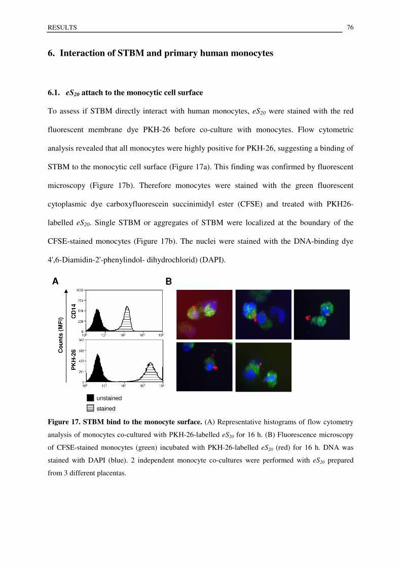
RESULTS 76
6. Interaction of STBM and primary human monocytes
6.1. eS20 attach to the monocytic cell surface
To assess if STBM directly interact with human monocytes, eS20 were stained with the red
fluorescent membrane dye PKH-26 before co-culture with monocytes. Flow cytometric
analysis revealed that all monocytes were highly positive for PKH-26, suggesting a binding of
STBM to the monocytic cell surface (Figure 17a). This finding was confirmed by fluorescent
microscopy (Figure 17b). Therefore monocytes were stained with the green fluorescent
cytoplasmic dye carboxyfluorescein succinimidyl ester (CFSE) and treated with PKH26-
labelled eS20. Single STBM or aggregates of STBM were localized at the boundary of the
CFSE-stained monocytes (Figure 17b). The nuclei were stained with the DNA-binding dye
4',6-Diamidin-2'-phenylindol- dihydrochlorid) (DAPI).
Figure 17. STBM bind to the monocyte surface. (A) Representative histograms of flow cytometry
analysis of monocytes co-cultured with PKH-26-labelled eS20 for 16 h. (B) Fluorescence microscopy
of CFSE-stained monocytes (green) incubated with PKH-26-labelled eS20 (red) for 16 h. DNA was
stained with DAPI (blue). 2 independent monocyte co-cultures were performed with eS20 prepared
from 3 different placentas.

RESULTS 77
6.2. eS20 mediated secretion of proinflammatory mediators is independent of
phagocytosis by human monocytes
Although Figure 17b does not suggest an engulfment of STBM, next, it was tested if
phagocytosis was important for the eS20-mediated cytokine secretion. To block phagocytosis,
monocytes were pre-treated with cytochalasin B, a cell-permeable mycotoxin that shortens
actin filaments by inhibiting monomer addition, before stimulation with eS20 (Figure 18).
Cytochalasin B did not reverse the eS20-induced secretion of IL-8 and IL-6 (Figure 18a and b),
showing that phagocytosis is not required for the induction of the inflammatory response.
However, the localization of eS20 at the monocytic cell surface suggested that monocytes
express cell surface molecules, which interact with eS20-associated markers and convey the
activation signal into the cell.
Figure 18. Inhibition of phagocytosis does not reverse STBM-mediated secretion of
proinflammatory mediators by monocytes. Monocytes were pre-treated with 1 or 10 µM of the
phagocytosis inhibitor cytochalasin B or with DMSO, the solvent of cytochalasin B, before addition of
300 µg/ml eS20. Release of IL-8 (A) and IL-6 (B) was quantified by ELISA. All data are presented as
mean ± SEM of 3 different monocyte co-culture experiments with eS20 prepared from 3 placentas.

RESULTS 78
6.3. STBM contain CD54
It has been previously proposed that monocytes strongly adhere via LFA-1 to CD54 displayed
by cultured syncytiotrophoblasts [240]. As it is likely that STBM express the same membrane
molecules as their parental cells, it was speculated that STBM-associated CD54 could bind
LFA-1 expressed by monocytes. Thus, in a next step, the potential existence of STBM-
associated CD54 was analyzed.
All STBM populations that activated monocytes, namely eS20 and pS prepared from healthy
term placentas and eS20 generated from preeclamptic placentas, contained CD54 (Table 4).
However, there was less microparticle-associated CD54 in pS, which always triggered the
highest STBM-mediated inflammatory response in monocytes, than in eS20, suggesting that
CD54 may not be the major molecule mediating the interaction.
Table 4. Presence of STBM-associated CD54 was measured by ELISA. All results are shown as mean
± SEM of STBM prepared from 3 different placentas for each condition. NP = normal pregnancy; PE
= preeclampsia.

RESULTS 79
6.4. STBM-associated CD54 is not responsible for eS20 mediated cytokine secretion
Although the concentration of STBM-associated CD54 was lower in pS than eS20, its possible
involvement in monocyte activation was evaluated. Therefore, the potential CD54-mediated
adhesion of eS20 to primary monocytes was blocked with an anti-human CD54 antagonistic
antibody. However, the secretion of IL-6 and IL-8 was not affected by the presence of the
blocking antibody (Figure 19a and b), showing that CD54 is not required for the eS20-
mediated inflammatory response.
Figure 19. STBM-associated CD54 is not responsible for the proinflammatory response of
monocytes. 100 µg/ml eS20 were pre-incubated with 10 µg/ml of anti-human CD54 adhesion antibody
before adding to monocytes. After 16 h of co-culture, IL-8 (A) and IL-6 (B) secretion by monocytes
was measured by ELISA. Bars represent mean ± SEM of 2 monocyte co-culture experiments
with eS20 prepared from 3 different placentas.
6.5. Basal expression of cell surface marker is different in Mono Mac 6 cells and
peripheral blood monocytes
Previous results from this work showed that eS20 triggered an inflammatory response in
primary human blood monocytes, but not in the monocytic cell line Mono Mac 6. The lack of
the cell surface expression of a specific molecule could be responsible for the
unresponsiveness of Mono Mac 6 cells towards STBM treatment. Thus, basal expression of

RESULTS 80
monocytic markers, which might be involved in the binding of eS20 to monocytes and the
subsequent intracellular signalling, was assessed on Mono Mac 6 cells and peripheral blood
monocytes (Figure 20a and b). Comparison of CD14 expression confirmed previous results.
Monocytes were highly positive for CD14, whereas Mono Mac 6 cells expressed low levels of
CD14 (17.1 ± 5.1%). Both cell types were positive for CD54 and CD11a (Figure 20a and b).
Peripheral blood monocytes and Mono Mac 6 highly expressed toll like receptor (TLR) 2
(98.9 ± 0.2% and 86.6 ± 2.1%, respectively), whereas they were negative for TLR4 (Figure
20a and b). However, there was a huge difference in the expression profile of TLR1: almost
all primary cells (86.2 ± 7.8%) were positive for TLR1, in contrast to the Mono Mac 6, which
did not express TLR1 (Figure 20a and b). In addition, primary monocytes (14.9 ± 3.9%)
displayed triggering receptor expressed on myeloid cells (TREM)-1, in contrast to Mono Mac
6 (Figure 20a and b).

RESULTS 81
Figure 20. Different basal expression of cell surface molecules on Mono Mac 6 and primary
monocytes. (A) Percentage of Mono Mac 6 cells and peripheral blood monocytes expressing CD14,
CD54, CD11a, TLR1, TLR2, TLR4 and TREM-1. Bars represent mean ± SEM of 2 experiments. (B)
Representative histograms of all cell surface molecules analysed.

RESULTS 82
6.6. MyD88 inhibitor peptide reduces eS20 mediated secretion of proinflammatory
mediators
TLR are potential receptors for STBM, as STBM are assembled of characteristic lipids [241].
In addition, the finding that TLR1 was differently expressed on primary monocytes and Mono
Mac 6, suggested that the eS20-induced monocyte activation may be mediated through TLR
signalling.
Myeloid differentiation primary response gene (MyD) 88 is an intracellular adaptor protein,
which is used by TLRs to induce the transcription factor NF-κB [242]. To investigate the
involvement of TLR activation in eS20-mediated secretion of proinflammatory mediators by
monocytes, TLR signalling was blocked by a MyD88 homodimerization inhibitory peptide
(Figure 21). Pre-treatment of monocytes with the inhibitory peptide reduced LPS-triggered
IL-6 and IL-8 secretion (Figure 21a and b). The highest inhibition was achieved when the
cells were treated with the lowest dose of LPS.
Treatment of monocytes with eS20 in the presence of the MyD88 inhibitory peptide impaired
monocyte activation, as shown by the reduced secretion of IL-6 (78.6 ± 16.5% and 87.1 ±
4.9%, respectively) and IL-8 (80.8 ± 3.2% and 72.7 ± 5.4%, respectively) compared to cells
cultured with eS20 alone (Figure 21a and b).
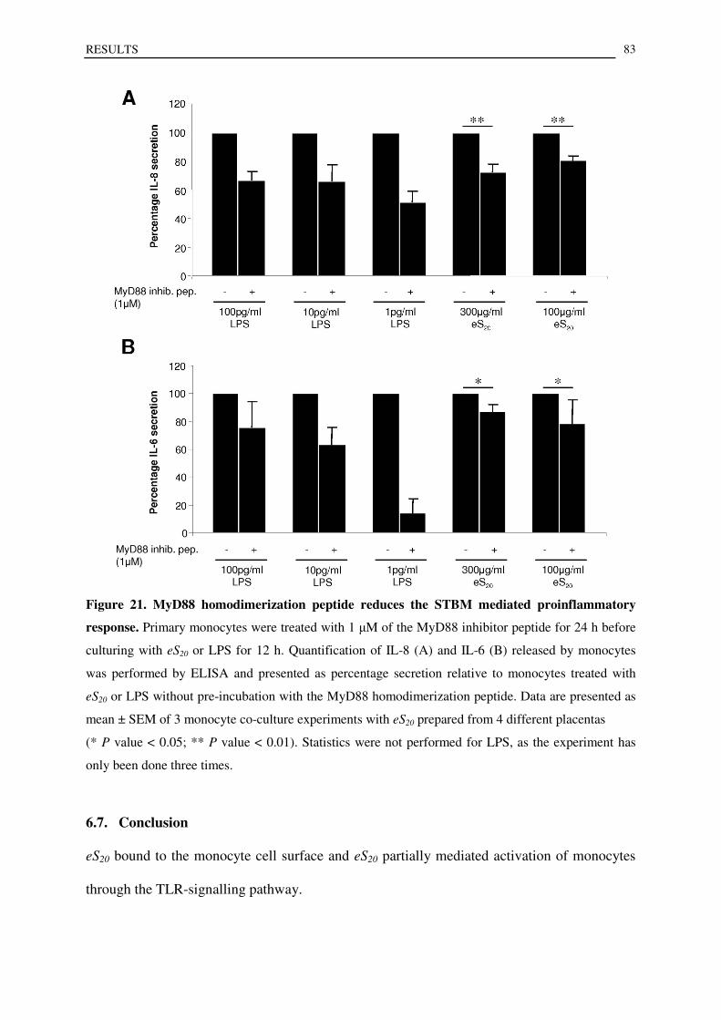
RESULTS 83
Figure 21. MyD88 homodimerization peptide reduces the STBM mediated proinflammatory
response. Primary monocytes were treated with 1 µM of the MyD88 inhibitor peptide for 24 h before
culturing with eS20 or LPS for 12 h. Quantification of IL-8 (A) and IL-6 (B) released by monocytes
was performed by ELISA and presented as percentage secretion relative to monocytes treated with
eS20 or LPS without pre-incubation with the MyD88 homodimerization peptide. Data are presented as
mean ± SEM of 3 monocyte co-culture experiments with eS20 prepared from 4 different placentas
(* P value < 0.05; ** P value < 0.01). Statistics were not performed for LPS, as the experiment has
only been done three times.
6.7. Conclusion
eS20 bound to the monocyte cell surface and eS20 partially mediated activation of monocytes
through the TLR-signalling pathway.

DISCUSSION 84
DISCUSSION
Syncytiotrophoblast microparticles (STBM) have been attributed potential functions in the
systemic maternal inflammatory response during normal human pregnancy and in the
exaggerated generalized inflammation found in preeclampsia [115]. My colleagues and others
showed, that STBM prepared from term placentas directly activate human cells of the
inflammatory network, comprising endothelial cells and neutrophils, in vitro [220, 226-228,
243].
In the current work, the monocytic cell line Mono Mac 6 and primary human monocytes were
used to investigate the potential contribution of STBM on the cellular viability, phenotype and
function of monocytes. Thereby, STBM were prepared from healthy term placentas by four
different in vitro approaches at conditions, which are believed to reflect either the physiologic
situation in normal pregnancy (STBM washed from the maternal side of a dually perfused
placental cotyledon and STBM shed by explants cultured in air) or the patho-physiologic one
found in preeclampsia (STBM isolated by mechanical dissection and STBM generated from
villous explant cultures incubated in hypoxic conditions). The dual perfusions of placental
cotyledons and the cultures of villous tissue explants may represent best the in vivo situation,
as the tissue structure and integrity are at least partially retained. In addition, Di Santo et al.
has previously shown that the trophoblast viability und functionality was even better
maintained during dual perfusions than in villous tissue explant cultures incubated in air, by
directly comparing tissues from the same placenta for 7 hours [244]. Although the conditions
of the present study are not exactly as those in Di Santo et al., one may assume that the same
would be true when comparing a dual perfusion of 30 minutes and a villous explant culture
incubated in air of 72 hours. In order to mimic the placental hypoxic conditions observed in
preeclampsia, villous tissue explants were cultured in 3% O2. Indeed, the current results
suggest that there is a shift from apoptotic to necrotic cell death in villous tissue cultured in

DISCUSSION 85
3% O2 in contrast to the villous tissue incubated in 20% O2. Furthermore, physical disruption
of tissue integrity by mechanical dissection of villous tissue is, in essence, a necrotic process.
None of the STBM populations triggered an inflammatory response in Mono Mac 6, although
an activated phenotype was inducible by LPS. However, decreased CD54 expression on
Mono Mac 6 cells incubated with mS could indicate changes in adhesion properties. Faas and
co-workers recently observed a similar decreased CD54 expression on Mono Mac 6 cells
following treatment with plasma derived from normal pregnant and preeclamptic women for
24 h, suggesting that STBM in the maternal plasma could mediate this reduced expression
[236].
The absence of responsiveness of Mono Mac 6 cells upon co-incubation with STBM, may be
attributed to its immature phenotype [245]. This is confirmed by the present results, which
revealed low frequency of Mono Mac 6 cells expressing CD14, in contrast to the mature
peripheral blood monocytes, which are highly positive for CD14. Thus, the inflammatory
potential of in vitro prepared STBM was assessed on primary human peripheral blood
monocytes. The data show, that different STBM populations triggered various responses in
primary monocytes without affecting cell viability, which confirms that Mono Mac 6 cells
and primary monocytes are not identical, concerning function and phenotype.
While the most patho-physiologic STBM populations (eS3 and mS) had no or only minor
influences on the monocytic phenotype and function, STBM generated in conditions
mimicking more closely the physiologic situation (eS20 and pS) induced a proinflammatory
response.
That the monocytic response varied according to the mode of preparation of the STBM is not
surprising in the light of previous findings with other cell types. For instance, it was shown
that mS triggered endothelial cell detachment from the collagen matrix and apoptosis, whereas
eS20 and pS induced a partial inhibition of endothelial cell proliferation, but no apoptosis [220,

DISCUSSION 86
226]. Furthermore, eS20 and mS significantly inhibited, whereas pS enhanced, the proliferation
of T lymphocytes induced with phorbol ester and Ca2+
ionophore [223].
The current data show that primary monocytes treated with pS and eS20 acquired an
activation-like phenotype and secreted proinflammatory immune mediators. Up-regulation of
the adhesion molecule CD54 is of interest in regard of a previous report documenting a
significant increase in CD54 expression on peripheral blood monocytes from third trimester
pregnancies [92]. The results suggest that placental microparticles could be the mediators of
this observation.
pS and eS20 also stimulated the production of the proinflammatory molecules IL-8, IL-6 and
IL-1β. These factors have been analysed because of their enhanced intracellular expression in
peripheral blood monocytes of preeclamptic women [187]. Furthermore, they are also present
in elevated concentrations in the serum of preeclamptic patients [183-185]. The current results
are also in line with a recent report, which showed that STBM prepared by dual perfusion of a
placental cotyledon, but not STBM generated by mechanical dissection of villous tissue,
induced a functional response in primary monocytes in vitro, as measured by the increased
intra-cellular production of TNF-α and IL-12p70 [173]. However, in the study of Germain
and co-workers, STBM were incubated with the whole peripheral blood mononuclear cell
(PBMC) fraction, leaving open the possibility that the up-regulation of cytokine expression in
monocytes could be indirect, for instance by cell-cell contact between monocytes and STBM-
activated third-party cells, or alternatively, through stimulation by a soluble mediator
produced by the latter. Our findings allow us to conclude that STBM are capable of directly
inducing monocytic cytokine production.
The monocytic response was time-dependent. As expected, IL-6 secretion increased steadily
with incubation time, whereas the secretion of IL-1β was rapid and sustained. This
observation is in agreement with the knowledge that IL-1β is a mediator of acute
inflammation, whereas IL-6 is a secondary pleiotropic regulator of inflammatory responses.

DISCUSSION 87
IL-1β is stored as an inactive precursor molecule in the cytoplasm, which is cleaved and
released as a proactive form in the extra-cellular compartment immediately after stimulation
[246].
The results also show that monocyte activation in response to STBM was dose-dependent.
Moreover, at identical protein concentration, the response stimulated by pS was much higher
than that induced by eS20. This could indicate that these two preparations of microparticles are
functionally different. On the other hand, this might also reflect a technical limitation of this
work. To conform to previous studies, the STBM-associated protein concentration was used
to quantify the STBM populations [173, 220]. However, this is a surrogate measurement for
microparticle numbers as the protein content of differentially prepared STBM may vary.
Another aspect that needs to be considered when interpreting the current results is that,
although the presence, in our STBM preparation, of syncytial-membrane microparticles could
be confirmed by the detection of syncytiotrophoblast-specific PLAP, a possible occurrence of
microparticles derived from non-trophoblastic elements of the villous tissue or from residual
maternal blood cell debris cannot be excluded.
eS20 prepared from preeclamptic placentas induced a proinflammatory reaction in peripheral
blood monocytes, as well. eS20 from normal as well as from preeclamptic placentas triggered
an equally strong and dose-dependent inflammatory response in primary monocytes,
suggesting that there are no or minor qualitative differences between the microparticles. From
these observations one may speculate that the overt maternal inflammation associated with
preeclampsia might be due to the higher concentrations of circulating STBM, rather than to
qualitative differences between the microparticles shed from the healthy and patho-
physiologic placentas.
eS20 partially induced the inflammatory response through NF-κB. NF-κB is a master
transcription factor involved in inflammatory pathways, which regulates the expression of
several genes, including CD54, IL-8, IL-6 and IL-1β [237]. eS20 triggered the transcription of

DISCUSSION 88
a number of NF-κB-regulated genes, which contribute to the amplification of an inflammatory
reaction, by either attracting or inducing production and differentiation of other immune cells.
Beside a tremendous increase in the transcription rate of IL-6, there was a high induction of
the small proinflammatory chemokine CCL2, which chemotactically attracts monocytes to
sites of inflammation [247]. Furthermore, eS20 induced the transcription of the cytokines CSF-
2 and -3, which control production, differentiation and function of macrophages and
granulocytes, TNF, which is a major mediator of acute inflammation, and the TNF family
member LT-α, which mediate various inflammatory and immunologic responses [248-250].
Although, eS20 triggered a huge release of IL-8 by monocytes, its transcription was only
marginally enhanced. This observation could point to the presence of an intracellular reservoir
of pre-stored IL-8, as it is the case in Weibel-Palade bodies in microvascular endothelial cells
[251]. However, it has to be considered that gene expression and secreted protein levels of IL-
8 were measured at different time points following eS20 treatment (after 4 and 16 hours,
respectively).
eS20 not only stimulated the secretion of IL-1β, but also its gene transcription. However, the
transcription of the other isoform of IL-1, namely IL-1α, was induced more by eS20. IL-1β and
IL-1α both stimulate chemokine production, when they bind IL-1 receptor 1 (IL-1R1).
eS20 also induced gene expression of the transcription factors EGR1 and JUN. EGR1 belongs
to a family of immediate early genes, which exhibit a critical role in cellular growth,
development and differentiation [252]. JUN either forms homodimers (JUN/JUN) or
heterodimers (JUN/FOS and JUN/ATF) to generate the functional AP-1 transcription factor,
which regulates gene expression of crucial molecules of the immune system, including
cytokines [253].
However, eS20 also triggered transcription of the anti-inflammatory cytokine IL-10,
representing a typical negative feedback regulator, which is usually produced by activated
cells to maintain homeostatic control and to prevent potential injury mediated by an overt

DISCUSSION 89
inflammatory reaction [254]. The eS20-mediated decrease in gene expression of TLR1, TLR6,
TLR7, TLR8, TLR9 and TICAM2, an intracellular adaptor molecule restricted to TLR4
signalling, may contribute to the resolution of the inflammatory response as well [255, 256].
In addition, the reduced transcription of the transcriptional co-activator of NF-κB BCL3 may
limit the transcriptional activity of NF-κB and, thus, support the termination of the
inflammatory reaction [257, 258].
On the one hand, down-regulated transcription of the cytoplasmic pattern recognition receptor
NOD1 (CARD4) could also account for the restriction of the inflammation, because NOD1
functions as activator of NF-κB [259]. On the other hand, reduced NOD1 transcription
prevents apoptosis, as NOD1-mediated activation of the pro-apoptotic molecule caspase 9 is
inhibited [259].
NF-κB is also known as a cell survival factor, and the eS20-mediated decrease in gene
expression of the apoptosis-inducing molecules FADD, caspase 8 and the TNF receptor
CD27, could explain the high viability of monocytes following STBM treatment, compared to
monocytes cultured in absence of any trigger [237, 260-262].
The requirement of NF-κB for the regulation of IL-6 and IL-8 production could be
demonstrated by the finding that the two NF-κB inhibitors 6AQ and PA partially prevented
the secretion of the proinflammatory mediators upon stimulation with eS20. eS20-triggered
release of proinflammatory factors was more reduced following treatment with 6AQ than with
PA. This is not surprising, as these two compounds interfere with different steps of NF-κB
activation. PA decreases intracellular calcium levels and thereby blocks the calcium-
dependent NF-κB pathway, only [239]. However, 6AQ is a low molecular weight inhibitor of
NF-κB transcriptional activation and might not be restricted to one of the pathways activating
NF-κB [238]. Activation of the NF-κB signal transduction pathway in PBMC of preeclamptic
women remains controversial [263, 264]. Luppi et al. showed an increased activation of NF-
κB compared to normal pregnant controls [263]. In contrast, McCracken and co-workers

DISCUSSION 90
published a suppression of the NF-κB activation pathway relative to normal pregnant and
non-pregnant subjects [264]. However, this suppression might rather be attributed to the T-
cell subset than the monocytes, as McCracken et al. recently documented the down-regulation
of NF-κB in peripheral blood T-cells of pregnant women [265].
The finding that there is a response in eS20-treated monocytes, raised the question of a direct
interaction between monocytes and STBM. Results obtained by flow cytometry and
fluorescence microscopy revealed, that eS20 attach to the monocytic surface. These results are
in line with the work of Germain and collaborators, who recently gave evidence for binding of
placental-derived microparticles on circulating monocytes in the peripheral blood of normal
pregnant and preeclamptic women [173].
The observed localization of eS20 at the boundary of monocytes, suggests that STBM interact
with monocytes via one or several receptor-ligand-pair(s).
pS and eS20 preparations led to decreased CD11a expression on monocytes. CD11a forms
together with CD18 the heterodimeric integrin lymphocyte function-associated antigen-1
(LFA-1). It has been suggested that monocytes strongly adhere via LFA-1 to CD54 displayed
by cultured syncytiotrophoblasts [240]. Since STBM are likely to encompass the same
membrane molecules as the parent cell from which they are derived, it has been speculated
that the observed changes in CD11a fluorescence intensity might be due to the interaction
between the STBM and the monocytes through these partner molecules, resulting in some
form of masking of the CD11a epitope to the antibody used for the present flow cytometry
analysis. The results obtained during this work confirmed the presence of CD54 on STBM.
Although binding was not analyzed, the adhesion blockade of CD54 did not affect the
production of IL-6 and IL-8 in primary monocytes induced by eS20.
The STBM used in this study also contained proinflammatory mediators. According to the
data, a role for IL-8 that is associated with eS20, or the proinflammatory cytokines IL-6, IL-1β
and TNF-α, which are present only in trace concentrations in all STBM populations, is

DISCUSSION 91
unlikely. However, it has been previously shown that STBM isolated by placental perfusion
of the maternal circuit contained the biologically active isoform of IL-1β [234]. In contrast to
the present work, where pS have been retrieved from placental washes lasting for 30 minutes,
Di Santo and co-workers prepared STBM after seven hours of perfusion of the maternal
compartment.
STBM are potential ligands for TLRs, as STBM are constituted of characteristic lipids and
contain DNA molecules [241, 266]. According to the data, TLR1 is a candidate receptor for
STBM, as Mono Mac 6 cells, which did not respond to STBM, were lacking TLR1
expression, whereas nearly all primary monocytes were positive for TLR1. TLR1 forms
heterodimers with TLR2 and binds preferentially triacylated lipopeptides [267]. Blocking the
TLR signalling by the cell permeable MyD88 homodimerization peptide reduced IL-6 and IL-
8 secretion, which gave further evidence for the involvement of TLRs. TLR signalling via the
adaptor molecule MyD88 results in NF-κB activation [268]. However, some TLRs, such as
TLR3 and TLR4, may trigger inflammation in a MyD88-independent way as well [268]. In
the case of TLR4 it is known, that MyD88-dependent signalling is already induced at the
plasma membrane, whereas activation of the MyD88-independent pathway needs endocytosis
of the receptor/ligand complex [269]. Provided that this observation of Kagan et al. is true for
other TLRs, which signal through MyD88, and knowing that engulfment of STBM is not
required for the proinflammatory response in monocytes, one could speculate, that STBM are
recognized on the monocyte membrane by one or several TLRs, which then trigger the
MyD88-dependent cascade and induce the inflammatory response in absence of TLR
internalisation.
However, the MyD88 inhibitory peptide did not completely block the inflammatory response
in eS20-treated monocytes. This could point out the involvement of further membrane
molecules, which signal through other pathways. A candidate molecule is TREM-1, which
was not expressed on Mono Mac 6, but present at low levels on primary monocytes. TREM-1

DISCUSSION 92
is known to transduce the extracellular signals through the immunoreceptor tyrosine-based
activation motif (ITAM) into the nucleus, inducing the production of proinflammatory
mediators, such as IL-8 and TNFα [270, 271]. However, the ligand of TREM-1 is still
illusive.
In conclusion, the present analysis suggests that STBM prepared by in vitro approaches,
which probably mimic best the physiologic conditions of human pregnancy, bear dose-
dependent proinflammatory properties for primary monocytes. This is interesting in regard to
the in vivo situation in normal pregnancy. The progressive monocytic activation in the
maternal peripheral blood may be caused by the steady increase in the load of placental
microparticles with gestational age. In addition, the present study suggests, that there are
minor qualitative differences between STBM prepared from healthy and preeclamptic
placentas. This could further indicate that the overt monocyte activation associated with
preeclampsia may be attributed to the enhanced circulatory concentrations of STBM, rather
than to a differential nature of the microparticles, compared to normal pregnancy.
The results also suggest that STBM induce the MyD88-dependent TLR signalling at the cell
surface, leading to NF-κB activation. In turn, NF-κB mediates the transcription of
proinflammatory mediators, including IL-1β, IL-6 and IL-8, and adhesion molecules, such as
CD54, resulting in a systemic inflammation.
In future, the following questions should be solved in order to understand the inflammation-
inducing components of STBM: which molecules displayed by STBM are recognized by
which TLR? Are there other receptors expressed by monocytes binding to respective ligands
on STBM? Although it is very demanding to get decent and pure amounts of naturally
occurring STBM from maternal blood, it would be interesting to compare their molecular
expression profile with the one of in vitro prepared placental microparticles.

DISCUSSION 93
To extend the knowledge of potential in vivo effects of the STBM populations, analyzed in
the present study, the next steps include their examination in pregnant mice. Possible xeno-
reactions should be ruled out by the administration of unspecific microparticles, for example
human red blood cell microparticles (RBC ghost).
However, preeclampsia is a multifactorial disorder and it is unlikely, that STBM alone are
responsible for the maternal syndrome. Thus, data from all lines of ongoing research in
preeclampsia should be considered, to get a more complete picture of the factors mediating
the disease, which hopefully can be translated into new diagnostic and screening tools.

ACKNOWLEDGEMENTS 94
ACKNOWLEDGEMENTS
I would like to thank my thesis advisor Prof. Dr. Sinuhe Hahn for giving me the opportunity
to do my PhD thesis in the Laboratory of Prenatal Medicine at the Department of Biomedicine
at the University Hospital Basel.
I am very thankful to Dr. Corinne Rusterholz for supervising my work, sharing her knowledge
and experience, and for helpful discussions.
I thank Prof. Dr. Antonius Rolink for giving me the chance to do my PhD at the Faculty of
Science in Basel, and Prof. Dr. Ian Sargent for being the co-referee of my thesis.
Many thanks go to the present and past members of the lab for the enjoyable time in- and
outside from the lab:
Vivian Kiefer, Inés Kaufmann, Simon Grill, Dorothy Huang, Iryna Perahud, Irina Banzola,
Kasemsri Srisupundit, Vara Prasad Kolla, Daniela Birri, Anurag Gupta, Aleksandra Kurak,
Flavio Cueni and Ying Li. I also thank Prof. Dr. Xiao Yan Zhong, Nicole Chiodetti, Corina
Kohler, Xiucheng Fan, Ramin Radpour, Zeinab Barekati, Reza Asadollah and Michal Sikora.
I am thankful to Dr. Olav Lapaire, Dr. Irene Hoesli and the clinical personnel of the
Department of Obstetrics of the University Hospital, Basel, for their help in collecting
placentas.
I would like to thank Dr. Karen May and Prof. Dr. Stefan Hansson of the Department of
Obstetrics and Gynecology, Lund, Sweden, for providing the placental washes of dually
perfused term placentas.
Special thanks go to my family and Simon for their love, moral support and constant belief in
me.

REFERENCES 95
REFERENCES
[1] Zinaman MJ, Clegg ED, Brown CC, O'Connor J, Selevan SG. Estimates of human
fertility and pregnancy loss. Fertil Steril 1996;65(3):503-9.
[2] Wilcox AJ, Weinberg CR, O'Connor JF, Baird DD, Schlatterer JP, Canfield RE,
Armstrong EG, Nisula BC. Incidence of early loss of pregnancy. N Engl J Med
1988;319(4):189-94.
[3] Norwitz ER, Schust DJ, Fisher SJ. Implantation and the survival of early pregnancy. N
Engl J Med 2001;345(19):1400-8.
[4] Moffett A, Loke C. Immunology of placentation in eutherian mammals. Nat Rev
Immunol 2006;6(8):584-94.
[5] Kliman HJ. Uteroplacental blood flow. The story of decidualization, menstruation, and
trophoblast invasion. Am J Pathol 2000;157(6):1759-68.
[6] Red-Horse K, Zhou Y, Genbacev O, Prakobphol A, Foulk R, McMaster M, Fisher SJ.
Trophoblast differentiation during embryo implantation and formation of the maternal-
fetal interface. J Clin Invest 2004;114(6):744-54.
[7] Mayhew TM, Leach L, McGee R, Ismail WW, Myklebust R, Lammiman MJ.
Proliferation, differentiation and apoptosis in villous trophoblast at 13-41 weeks of
gestation (including observations on annulate lamellae and nuclear pore complexes).
Placenta 1999;20(5-6):407-22.
[8] Huppertz B, Frank HG, Kingdom JC, Reister F, Kaufmann P. Villous cytotrophoblast
regulation of the syncytial apoptotic cascade in the human placenta. Histochem Cell
Biol 1998;110(5):495-508.
[9] Nelson DM. Apoptotic changes occur in syncytiotrophoblast of human placental villi
where fibrin type fibrinoid is deposited at discontinuities in the villous trophoblast.
Placenta 1996;17(7):387-91.
[10] Burton GJ, Jones CJ. Syncytial knots, sprouts, apoptosis, and trophoblast deportation
from the human placenta. Taiwan J Obstet Gynecol 2009;48(1):28-37.
[11] Huppertz B. The feto-maternal interface: setting the stage for potential immune
interactions. Semin Immunopathol 2007;29(2):83-94.
[12] Huppertz B, Kingdom JC. Apoptosis in the trophoblast--role of apoptosis in placental
morphogenesis. J Soc Gynecol Investig 2004;11(6):353-62.
[13] Jauniaux E, Poston L, Burton GJ. Placental-related diseases of pregnancy:
Involvement of oxidative stress and implications in human evolution. Hum Reprod
Update 2006;12(6):747-55.

REFERENCES 96
[14] Ezashi T, Das P, Roberts RM. Low O2 tensions and the prevention of differentiation
of hES cells. Proc Natl Acad Sci U S A 2005;102(13):4783-8.
[15] Jauniaux E, Watson A, Burton G. Evaluation of respiratory gases and acid-base
gradients in human fetal fluids and uteroplacental tissue between 7 and 16 weeks'
gestation. Am J Obstet Gynecol 2001;184(5):998-1003.
[16] Jauniaux E, Watson AL, Hempstock J, Bao YP, Skepper JN, Burton GJ. Onset of
maternal arterial blood flow and placental oxidative stress. A possible factor in human
early pregnancy failure. Am J Pathol 2000;157(6):2111-22.
[17] Jauniaux E, Hempstock J, Greenwold N, Burton GJ. Trophoblastic oxidative stress in
relation to temporal and regional differences in maternal placental blood flow in
normal and abnormal early pregnancies. Am J Pathol 2003;162(1):115-25.
[18] Sargent IL, Borzychowski AM, Redman CW. NK cells and human pregnancy--an
inflammatory view. Trends Immunol 2006;27(9):399-404.
[19] Rodesch F, Simon P, Donner C, Jauniaux E. Oxygen measurements in endometrial
and trophoblastic tissues during early pregnancy. Obstet Gynecol 1992;80(2):283-5.
[20] Wegmann TG, Lin H, Guilbert L, Mosmann TR. Bidirectional cytokine interactions in
the maternal-fetal relationship: is successful pregnancy a TH2 phenomenon? Immunol
Today 1993;14(7):353-6.
[21] Lin H, Mosmann TR, Guilbert L, Tuntipopipat S, Wegmann TG. Synthesis of T helper
2-type cytokines at the maternal-fetal interface. J Immunol 1993;151(9):4562-73.
[22] Saito S, Tsukaguchi N, Hasegawa T, Michimata T, Tsuda H, Narita N. Distribution of
Th1, Th2, and Th0 and the Th1/Th2 cell ratios in human peripheral and endometrial T
cells. Am J Reprod Immunol 1999;42(4):240-5.
[23] Krasnow JS, Tollerud DJ, Naus G, DeLoia JA. Endometrial Th2 cytokine expression
throughout the menstrual cycle and early pregnancy. Hum Reprod 1996;11(8):1747-
54.
[24] de Moraes-Pinto MI, Vince GS, Flanagan BF, Hart CA, Johnson PM. Localization of
IL-4 and IL-4 receptors in the human term placenta, decidua and amniochorionic
membranes. Immunology 1997;90(1):87-94.
[25] Sakai M, Tsuda H, Tanebe K, Sasaki Y, Saito S. Interleukin-12 secretion by peripheral
blood mononuclear cells is decreased in normal pregnant subjects and increased in
preeclamptic patients. Am J Reprod Immunol 2002;47(2):91-7.
[26] Chaouat G, Ledee-Bataille N, Dubanchet S, Zourbas S, Sandra O, Martal J. TH1/TH2
paradigm in pregnancy: paradigm lost? Cytokines in pregnancy/early abortion:
reexamining the TH1/TH2 paradigm. Int Arch Allergy Immunol 2004;134(2):93-119.

REFERENCES 97
[27] Kwak-Kim JY, Gilman-Sachs A, Kim CE. T helper 1 and 2 immune responses in
relationship to pregnancy, nonpregnancy, recurrent spontaneous abortions and
infertility of repeated implantation failures. Chem Immunol Allergy 2005;88:64-79.
[28] Chaouat G, Assal Meliani A, Martal J, Raghupathy R, Elliott JF, Mosmann T,
Wegmann TG. IL-10 prevents naturally occurring fetal loss in the CBA x DBA/2
mating combination, and local defect in IL-10 production in this abortion-prone
combination is corrected by in vivo injection of IFN-tau. J Immunol
1995;154(9):4261-8.
[29] Clark DA, Croitoru K. TH1/TH2,3 imbalance due to cytokine-producing NK,
gammadelta T and NK-gammadelta T cells in murine pregnancy decidua in success or
failure of pregnancy. Am J Reprod Immunol 2001;45(5):257-65.
[30] Rudolph MG, Stanfield RL, Wilson IA. How TCRs bind MHCs, peptides, and
coreceptors. Annu Rev Immunol 2006;24:419-66.
[31] Goker H, Haznedaroglu IC, Chao NJ. Acute graft-vs-host disease: pathobiology and
management. Exp Hematol 2001;29(3):259-77.
[32] Klein J, Sato A. The HLA system. Second of two parts. N Engl J Med
2000;343(11):782-6.
[33] King A, Burrows TD, Hiby SE, Bowen JM, Joseph S, Verma S, Lim PB, Gardner L,
Le Bouteiller P, Ziegler A, Uchanska-Ziegler B, Loke YW. Surface expression of
HLA-C antigen by human extravillous trophoblast. Placenta 2000;21(4):376-87.
[34] Ishitani A, Sageshima N, Hatake K. The involvement of HLA-E and -F in pregnancy.
J Reprod Immunol 2006;69(2):101-13.
[35] Le Bouteiller P, Rodriguez AM, Mallet V, Girr M, Guillaudeux T, Lenfant F.
Placental expression of HLA class I genes. Am J Reprod Immunol 1996;35(3):216-25.
[36] Schmidt CM, Orr HT. Maternal/fetal interactions: the role of the MHC class I
molecule HLA-G. Crit Rev Immunol 1993;13(3-4):207-24.
[37] Shobu T, Sageshima N, Tokui H, Omura M, Saito K, Nagatsuka Y, Nakanishi M,
Hayashi Y, Hatake K, Ishitani A. The surface expression of HLA-F on decidual
trophoblasts increases from mid to term gestation. J Reprod Immunol 2006;72(1-
2):18-32.
[38] Chu W, Fant ME, Geraghty DE, Hunt JS. Soluble HLA-G in human placentas:
synthesis in trophoblasts and interferon-gamma-activated macrophages but not
placental fibroblasts. Hum Immunol 1998;59(7):435-42.
[39] Ishitani A, Sageshima N, Lee N, Dorofeeva N, Hatake K, Marquardt H, Geraghty DE.
Protein expression and peptide binding suggest unique and interacting functional roles

REFERENCES 98
for HLA-E, F, and G in maternal-placental immune recognition. J Immunol
2003;171(3):1376-84.
[40] Solier C, Aguerre-Girr M, Lenfant F, Campan A, Berrebi A, Rebmann V, Grosse-
Wilde H, Le Bouteiller P. Secretion of pro-apoptotic intron 4-retaining soluble HLA-
G1 by human villous trophoblast. Eur J Immunol 2002;32(12):3576-86.
[41] Moffett-King A. Natural killer cells and pregnancy. Nat Rev Immunol 2002;2(9):656-
63.
[42] Bulmer JN, Lash GE. Human uterine natural killer cells: a reappraisal. Mol Immunol
2005;42(4):511-21.
[43] King A, Birkby C, Loke YW. Early human decidual cells exhibit NK activity against
the K562 cell line but not against first trimester trophoblast. Cell Immunol
1989;118(2):337-44.
[44] Kopcow HD, Allan DS, Chen X, Rybalov B, Andzelm MM, Ge B, Strominger JL.
Human decidual NK cells form immature activating synapses and are not cytotoxic.
Proc Natl Acad Sci U S A 2005;102(43):15563-8.
[45] Vacca P, Pietra G, Falco M, Romeo E, Bottino C, Bellora F, Prefumo F, Fulcheri E,
Venturini PL, Costa M, Moretta A, Moretta L, Mingari MC. Analysis of natural killer
cells isolated from human decidua: Evidence that 2B4 (CD244) functions as an
inhibitory receptor and blocks NK-cell function. Blood 2006;108(13):4078-85.
[46] Mandelboim O, Pazmany L, Davis DM, Vales-Gomez M, Reyburn HT, Rybalov B,
Strominger JL. Multiple receptors for HLA-G on human natural killer cells. Proc Natl
Acad Sci U S A 1997;94(26):14666-70.
[47] Gonen-Gross T, Achdout H, Gazit R, Hanna J, Mizrahi S, Markel G, Goldman-Wohl
D, Yagel S, Horejsi V, Levy O, Baniyash M, Mandelboim O. Complexes of HLA-G
protein on the cell surface are important for leukocyte Ig-like receptor-1 function. J
Immunol 2003;171(3):1343-51.
[48] Ashkar AA, Croy BA. Functions of uterine natural killer cells are mediated by
interferon gamma production during murine pregnancy. Semin Immunol
2001;13(4):235-41.
[49] Hanna J, Goldman-Wohl D, Hamani Y, Avraham I, Greenfield C, Natanson-Yaron S,
Prus D, Cohen-Daniel L, Arnon TI, Manaster I, Gazit R, Yutkin V, Benharroch D,
Porgador A, Keshet E, Yagel S, Mandelboim O. Decidual NK cells regulate key
developmental processes at the human fetal-maternal interface. Nat Med
2006;12(9):1065-74.
[50] Hiby SE, Walker JJ, O'Shaughnessy K M, Redman CW, Carrington M, Trowsdale J,
Moffett A. Combinations of maternal KIR and fetal HLA-C genes influence the risk of
preeclampsia and reproductive success. J Exp Med 2004;200(8):957-65.

REFERENCES 99
[51] Parham P. MHC class I molecules and KIRs in human history, health and survival.
Nat Rev Immunol 2005;5(3):201-14.
[52] Abrahams VM, Kim YM, Straszewski SL, Romero R, Mor G. Macrophages and
apoptotic cell clearance during pregnancy. Am J Reprod Immunol 2004;51(4):275-82.
[53] Bulmer JN, Johnson PM. Macrophage populations in the human placenta and
amniochorion. Clin Exp Immunol 1984;57(2):393-403.
[54] Heikkinen J, Mottonen M, Komi J, Alanen A, Lassila O. Phenotypic characterization
of human decidual macrophages. Clin Exp Immunol 2003;131(3):498-505.
[55] Renaud SJ, Graham CH. The role of macrophages in utero-placental interactions
during normal and pathological pregnancy. Immunol Invest 2008;37(5):535-64.
[56] Sakaguchi S, Sakaguchi N, Asano M, Itoh M, Toda M. Immunologic self-tolerance
maintained by activated T cells expressing IL-2 receptor alpha-chains (CD25).
Breakdown of a single mechanism of self-tolerance causes various autoimmune
diseases. J Immunol 1995;155(3):1151-64.
[57] Kingsley CI, Karim M, Bushell AR, Wood KJ. CD25+CD4+ regulatory T cells
prevent graft rejection: CTLA-4- and IL-10-dependent immunoregulation of
alloresponses. J Immunol 2002;168(3):1080-6.
[58] Somerset DA, Zheng Y, Kilby MD, Sansom DM, Drayson MT. Normal human
pregnancy is associated with an elevation in the immune suppressive CD25+ CD4+
regulatory T-cell subset. Immunology 2004;112(1):38-43.
[59] Sasaki Y, Sakai M, Miyazaki S, Higuma S, Shiozaki A, Saito S. Decidual and
peripheral blood CD4+CD25+ regulatory T cells in early pregnancy subjects and
spontaneous abortion cases. Mol Hum Reprod 2004;10(5):347-53.
[60] Petroff MG. Immune interactions at the maternal-fetal interface. J Reprod Immunol
2005;68(1-2):1-13.
[61] Riley JK. Trophoblast immune receptors in maternal-fetal tolerance. Immunol Invest
2008;37(5):395-426.
[62] Petroff MG, Chen L, Phillips TA, Azzola D, Sedlmayr P, Hunt JS. B7 family
molecules are favorably positioned at the human maternal-fetal interface. Biol Reprod
2003;68(5):1496-504.
[63] Petroff MG, Kharatyan E, Torry DS, Holets L. The immunomodulatory proteins B7-
DC, B7-H2, and B7-H3 are differentially expressed across gestation in the human
placenta. Am J Pathol 2005;167(2):465-73.

REFERENCES 100
[64] Guleria I, Khosroshahi A, Ansari MJ, Habicht A, Azuma M, Yagita H, Noelle RJ,
Coyle A, Mellor AL, Khoury SJ, Sayegh MH. A critical role for the programmed
death ligand 1 in fetomaternal tolerance. J Exp Med 2005;202(2):231-7.
[65] Greenwald RJ, Freeman GJ, Sharpe AH. The B7 family revisited. Annu Rev Immunol
2005;23:515-48.
[66] Freeman GJ, Long AJ, Iwai Y, Bourque K, Chernova T, Nishimura H, Fitz LJ,
Malenkovich N, Okazaki T, Byrne MC, Horton HF, Fouser L, Carter L, Ling V,
Bowman MR, Carreno BM, Collins M, Wood CR, Honjo T. Engagement of the PD-1
immunoinhibitory receptor by a novel B7 family member leads to negative regulation
of lymphocyte activation. J Exp Med 2000;192(7):1027-34.
[67] Taglauer ES, Trikhacheva AS, Slusser JG, Petroff MG. Expression and function of
PDCD1 at the human maternal-fetal interface. Biol Reprod 2008;79(3):562-9.
[68] Mellor AL, Munn DH. Tryptophan catabolism and T-cell tolerance:
immunosuppression by starvation? Immunol Today 1999;20(10):469-73.
[69] Mellor AL, Munn DH. IDO expression by dendritic cells: tolerance and tryptophan
catabolism. Nat Rev Immunol 2004;4(10):762-74.
[70] Hammer A, Dohr G. Expression of Fas-ligand in first trimester and term human
placental villi. J Reprod Immunol 2000;46(2):83-90.
[71] Selam B, Kayisli UA, Mulayim N, Arici A. Regulation of Fas ligand expression by
estradiol and progesterone in human endometrium. Biol Reprod 2001;65(4):979-85.
[72] Kauma SW, Huff TF, Hayes N, Nilkaeo A. Placental Fas ligand expression is a
mechanism for maternal immune tolerance to the fetus. J Clin Endocrinol Metab
1999;84(6):2188-94.
[73] Coumans B, Thellin O, Zorzi W, Melot F, Bougoussa M, Melen L, Zorzi D, Hennen
G, Igout A, Heinen E. Lymphoid cell apoptosis induced by trophoblastic cells: a
model of active foeto-placental tolerance. J Immunol Methods 1999;224(1-2):185-96.
[74] Mor G, Gutierrez LS, Eliza M, Kahyaoglu F, Arici A. Fas-fas ligand system-induced
apoptosis in human placenta and gestational trophoblastic disease. Am J Reprod
Immunol 1998;40(2):89-94.
[75] Abrahams VM, Straszewski-Chavez SL, Guller S, Mor G. First trimester trophoblast
cells secrete Fas ligand which induces immune cell apoptosis. Mol Hum Reprod
2004;10(1):55-63.
[76] Morgan BP. Physiology and pathophysiology of complement: progress and trends.
Crit Rev Clin Lab Sci 1995;32(3):265-98.

REFERENCES 101
[77] Fishelson Z, Attali G, Mevorach D. Complement and apoptosis. Mol Immunol
2001;38(2-3):207-19.
[78] Hsi BL, Hunt JS, Atkinson JP. Differential expression of complement regulatory
proteins on subpopulations of human trophoblast cells. J Reprod Immunol
1991;19(3):209-23.
[79] Song WC. Complement regulatory proteins and autoimmunity. Autoimmunity
2006;39(5):403-10.
[80] Holmes CH, Simpson KL, Okada H, Okada N, Wainwright SD, Purcell DF, Houlihan
JM. Complement regulatory proteins at the feto-maternal interface during human
placental development: distribution of CD59 by comparison with membrane cofactor
protein (CD46) and decay accelerating factor (CD55). Eur J Immunol
1992;22(6):1579-85.
[81] Holmes CH, Simpson KL, Wainwright SD, Tate CG, Houlihan JM, Sawyer IH,
Rogers IP, Spring FA, Anstee DJ, Tanner MJ. Preferential expression of the
complement regulatory protein decay accelerating factor at the fetomaternal interface
during human pregnancy. J Immunol 1990;144(8):3099-105.
[82] Nishikori K, Noma J, Hirakawa S, Amano T, Kudo T. The change of membrane
complement regulatory protein in chorion of early pregnancy. Clin Immunol
Immunopathol 1993;69(2):167-74.
[83] Johansen M, Redman CW, Wilkins T, Sargent IL. Trophoblast deportation in human
pregnancy--its relevance for pre-eclampsia. Placenta 1999;20(7):531-9.
[84] Knight M, Redman CW, Linton EA, Sargent IL. Shedding of syncytiotrophoblast
microvilli into the maternal circulation in pre-eclamptic pregnancies. Br J Obstet
Gynaecol 1998;105(6):632-40.
[85] Abumaree MH, Stone PR, Chamley LW. The effects of apoptotic, deported human
placental trophoblast on macrophages: possible consequences for pregnancy. J Reprod
Immunol 2006;72(1-2):33-45.
[86] Sacks GP, Studena K, Sargent K, Redman CW. Normal pregnancy and preeclampsia
both produce inflammatory changes in peripheral blood leukocytes akin to those of
sepsis. Am J Obstet Gynecol 1998;179(1):80-6.
[87] Smarason AK, Gunnarsson A, Alfredsson JH, Valdimarsson H. Monocytosis and
monocytic infiltration of decidua in early pregnancy. J Clin Lab Immunol
1986;21(1):1-5.
[88] Austgulen R, Lien E, Liabakk NB, Jacobsen G, Arntzen KJ. Increased levels of
cytokines and cytokine activity modifiers in normal pregnancy. Eur J Obstet Gynecol
Reprod Biol 1994;57(3):149-55.
REFERENCES 102
[89] Melczer Z, Banhidy F, Csomor S, Toth P, Kovacs M, Winkler G, Cseh K. Influence of
leptin and the TNF system on insulin resistance in pregnancy and their effect on
anthropometric parameters of newborns. Acta Obstet Gynecol Scand 2003;82(5):432-
8.
[90] Sacks GP, Seyani L, Lavery S, Trew G. Maternal C-reactive protein levels are raised
at 4 weeks gestation. Hum Reprod 2004;19(4):1025-30.
[91] Holthe MR, Staff AC, Berge LN, Lyberg T. Leukocyte adhesion molecules and
reactive oxygen species in preeclampsia. Obstet Gynecol 2004;103(5 Pt 1):913-22.
[92] Luppi P, Haluszczak C, Betters D, Richard CA, Trucco M, DeLoia JA. Monocytes are
progressively activated in the circulation of pregnant women. J Leukoc Biol
2002;72(5):874-84.
[93] Koumandakis E, Koumandaki I, Kaklamani E, Sparos L, Aravantinos D, Trichopoulos
D. Enhanced phagocytosis of mononuclear phagocytes in pregnancy. Br J Obstet
Gynaecol 1986;93(11):1150-4.
[94] Shibuya T, Izuchi K, Kuroiwa A, Okabe N, Shirakawa K. Study on nonspecific
immunity in pregnant women: increased chemiluminescence response of peripheral
blood phagocytes. Am J Reprod Immunol Microbiol 1987;15(1):19-23.
[95] Rusterholz C, Hahn S, Holzgreve W. Role of placentally produced inflammatory and
regulatory cytokines in pregnancy and the etiology of preeclampsia. Semin
Immunopathol 2007;29(2):151-62.
[96] Levine RJ, Maynard SE, Qian C, Lim KH, England LJ, Yu KF, Schisterman EF,
Thadhani R, Sachs BP, Epstein FH, Sibai BM, Sukhatme VP, Karumanchi SA.
Circulating angiogenic factors and the risk of preeclampsia. N Engl J Med
2004;350(7):672-83.
[97] Paradowska E, Blach-Olszewska Z, Gejdel E. Constitutive and induced cytokine
production by human placenta and amniotic membrane at term. Placenta 1997;18(5-
6):441-6.
[98] Kauma SW, Wang Y, Walsh SW. Preeclampsia is associated with decreased placental
interleukin-6 production. J Soc Gynecol Investig 1995;2(4):614-7.
[99] Keelan JA, Zhou RL, Mitchell MD. Activin A exerts both pro- and anti-inflammatory
effects on human term gestational tissues. Placenta 2000;21(1):38-43.
[100] Munno I, Chiechi LM, Lacedra G, Putignano G, Patimo C, Lobascio A, Loizzi P.
Spontaneous and induced release of prostaglandins, interleukin (IL)-1beta, IL-6, and
tumor necrosis factor-alpha by placental tissue from normal and preeclamptic
pregnancies. Am J Reprod Immunol 1999;42(6):369-74.
REFERENCES 103
[101] Sacks G, Sargent I, Redman C. An innate view of human pregnancy. Immunol Today
1999;20(3):114-8.
[102] Agarwal R, Loganath A, Roy AC, Wong YC, Lindoff C, Ng SC. Increased expression
of interleukin 6 in term compared to the first trimester human placental villi. Horm
Metab Res 2000;32(5):164-8.
[103] Hanna N, Hanna I, Hleb M, Wagner E, Dougherty J, Balkundi D, Padbury J, Sharma
S. Gestational age-dependent expression of IL-10 and its receptor in human placental
tissues and isolated cytotrophoblasts. J Immunol 2000;164(11):5721-8.
[104] Khaliq A, Li XF, Shams M, Sisi P, Acevedo CA, Whittle MJ, Weich H, Ahmed A.
Localisation of placenta growth factor (PIGF) in human term placenta. Growth Factors
1996;13(3-4):243-50,color plates I-II,pre bk cov.
[105] Tjwa M, Luttun A, Autiero M, Carmeliet P. VEGF and PlGF: two pleiotropic growth
factors with distinct roles in development and homeostasis. Cell Tissue Res
2003;314(1):5-14.
[106] Duff SE, Li C, Garland JM, Kumar S. CD105 is important for angiogenesis: evidence
and potential applications. FASEB J 2003;17(9):984-92.
[107] Cheifetz S, Bellon T, Cales C, Vera S, Bernabeu C, Massague J, Letarte M. Endoglin
is a component of the transforming growth factor-beta receptor system in human
endothelial cells. J Biol Chem 1992;267(27):19027-30.
[108] St-Jacques S, Forte M, Lye SJ, Letarte M. Localization of endoglin, a transforming
growth factor-beta binding protein, and of CD44 and integrins in placenta during the
first trimester of pregnancy. Biol Reprod 1994;51(3):405-13.
[109] Clark DE, Smith SK, Sharkey AM, Charnock-Jones DS. Localization of VEGF and
expression of its receptors flt and KDR in human placenta throughout pregnancy. Hum
Reprod 1996;11(5):1090-8.
[110] Clark DE, Smith SK, Licence D, Evans AL, Charnock-Jones DS. Comparison of
expression patterns for placenta growth factor, vascular endothelial growth factor
(VEGF), VEGF-B and VEGF-C in the human placenta throughout gestation. J
Endocrinol 1998;159(3):459-67.
[111] Venkatesha S, Toporsian M, Lam C, Hanai J, Mammoto T, Kim YM, Bdolah Y, Lim
KH, Yuan HT, Libermann TA, Stillman IE, Roberts D, D'Amore PA, Epstein FH,
Sellke FW, Romero R, Sukhatme VP, Letarte M, Karumanchi SA. Soluble endoglin
contributes to the pathogenesis of preeclampsia. Nat Med 2006;12(6):642-9.
[112] Myatt L, Webster RP. Vascular biology of preeclampsia. J Thromb Haemost
2009;7(3):375-84.

REFERENCES 104
[113] Strevens H, Wide-Swensson D, Hansen A, Horn T, Ingemarsson I, Larsen S, Willner
J, Olsen S. Glomerular endotheliosis in normal pregnancy and pre-eclampsia. BJOG
2003;110(9):831-6.
[114] Chaiworapongsa T, Romero R, Kim YM, Kim GJ, Kim MR, Espinoza J, Bujold E,
Goncalves L, Gomez R, Edwin S, Mazor M. Plasma soluble vascular endothelial
growth factor receptor-1 concentration is elevated prior to the clinical diagnosis of
pre-eclampsia. J Matern Fetal Neonatal Med 2005;17(1):3-18.
[115] Redman CW, Sargent IL. Microparticles and immunomodulation in pregnancy and
pre-eclampsia. J Reprod Immunol 2007;76(1-2):61-7.
[116] Magee LA, Ornstein MP, von Dadelszen P. Fortnightly review: management of
hypertension in pregnancy. BMJ 1999;318(7194):1332-6.
[117] Sibai B, Dekker G, Kupferminc M. Pre-eclampsia. Lancet 2005;365(9461):785-99.
[118] Lain KY, Roberts JM. Contemporary concepts of the pathogenesis and management of
preeclampsia. JAMA 2002;287(24):3183-6.
[119] Leitch CR, Cameron AD, Walker JJ. The changing pattern of eclampsia over a 60-
year period. Br J Obstet Gynaecol 1997;104(8):917-22.
[120] Khan KS, Wojdyla D, Say L, Gulmezoglu AM, Van Look PF. WHO analysis of
causes of maternal death: a systematic review. Lancet 2006;367(9516):1066-74.
[121] Lopez-Jaramillo P, Casas JP, Serrano N. Preeclampsia: from epidemiological
observations to molecular mechanisms. Braz J Med Biol Res 2001;34(10):1227-35.
[122] Abalos E, Duley L, Steyn DW, Henderson-Smart DJ. Antihypertensive drug therapy
for mild to moderate hypertension during pregnancy. Cochrane Database Syst Rev
2007(1):CD002252.
[123] Altman D, Carroli G, Duley L, Farrell B, Moodley J, Neilson J, Smith D. Do women
with pre-eclampsia, and their babies, benefit from magnesium sulphate? The Magpie
Trial: a randomised placebo-controlled trial. Lancet 2002;359(9321):1877-90.
[124] Sibai BM. Magnesium sulfate prophylaxis in preeclampsia: evidence from randomized
trials. Clin Obstet Gynecol 2005;48(2):478-88.
[125] Duley L, Henderson-Smart D. Magnesium sulphate versus phenytoin for eclampsia.
Cochrane Database Syst Rev 2003(4):CD000128.
[126] Duley L, Henderson-Smart D. Magnesium sulphate versus diazepam for eclampsia.
Cochrane Database Syst Rev 2003(4):CD000127.
REFERENCES 105
[127] Goldenberg RL, Culhane JF, Iams JD, Romero R. Epidemiology and causes of
preterm birth. Lancet 2008;371(9606):75-84.
[128] Redman CW, Sacks GP, Sargent IL. Preeclampsia: an excessive maternal
inflammatory response to pregnancy. Am J Obstet Gynecol 1999;180(2 Pt 1):499-506.
[129] Sibai BM. Biomarker for hypertension-preeclampsia: are we close yet? Am J Obstet
Gynecol 2007;197(1):1-2.
[130] Poston L, Briley AL, Seed PT, Kelly FJ, Shennan AH. Vitamin C and vitamin E in
pregnant women at risk for pre-eclampsia (VIP trial): randomised placebo-controlled
trial. Lancet 2006;367(9517):1145-54.
[131] Spinnato JA, 2nd, Freire S, Pinto ESJL, Cunha Rudge MV, Martins-Costa S, Koch
MA, Goco N, Santos Cde B, Cecatti JG, Costa R, Ramos JG, Moss N, Sibai BM.
Antioxidant therapy to prevent preeclampsia: a randomized controlled trial. Obstet
Gynecol 2007;110(6):1311-8.
[132] Wallenburg HC, Dekker GA, Makovitz JW, Rotmans P. Low-dose aspirin prevents
pregnancy-induced hypertension and pre-eclampsia in angiotensin-sensitive
primigravidae. Lancet 1986;1(8471):1-3.
[133] Knight M, Duley L, Henderson-Smart DJ, King JF. Antiplatelet agents for preventing
and treating pre-eclampsia. Cochrane Database Syst Rev 2000(2):CD000492.
[134] Wen SW, Chen XK, Rodger M, White RR, Yang Q, Smith GN, Sigal RJ, Perkins SL,
Walker MC. Folic acid supplementation in early second trimester and the risk of
preeclampsia. Am J Obstet Gynecol 2008;198(1):45 e1-7.
[135] Hofmeyr GJ, Duley L, Atallah A. Dietary calcium supplementation for prevention of
pre-eclampsia and related problems: a systematic review and commentary. BJOG
2007;114(8):933-43.
[136] Trumbo PR, Ellwood KC. Supplemental calcium and risk reduction of hypertension,
pregnancy-induced hypertension, and preeclampsia: an evidence-based review by the
US Food and Drug Administration. Nutr Rev 2007;65(2):78-87.
[137] Redman CW. Current topic: pre-eclampsia and the placenta. Placenta 1991;12(4):301-
8.
[138] Khong TY, De Wolf F, Robertson WB, Brosens I. Inadequate maternal vascular
response to placentation in pregnancies complicated by pre-eclampsia and by small-
for-gestational age infants. Br J Obstet Gynaecol 1986;93(10):1049-59.
[139] Huppertz B. Placental origins of preeclampsia: challenging the current hypothesis.
Hypertension 2008;51(4):970-5.

REFERENCES 106
[140] Ness RB, Roberts JM. Heterogeneous causes constituting the single syndrome of
preeclampsia: a hypothesis and its implications. Am J Obstet Gynecol
1996;175(5):1365-70.
[141] Roberts JM, Hubel CA. The two stage model of preeclampsia: variations on the
theme. Placenta 2009;30 Suppl A:S32-7.
[142] Hung TH, Skepper JN, Burton GJ. In vitro ischemia-reperfusion injury in term human
placenta as a model for oxidative stress in pathological pregnancies. Am J Pathol
2001;159(3):1031-43.
[143] Hung TH, Skepper JN, Charnock-Jones DS, Burton GJ. Hypoxia-reoxygenation: a
potent inducer of apoptotic changes in the human placenta and possible etiological
factor in preeclampsia. Circ Res 2002;90(12):1274-81.
[144] Hubel CA, Roberts JM, Taylor RN, Musci TJ, Rogers GM, McLaughlin MK. Lipid
peroxidation in pregnancy: new perspectives on preeclampsia. Am J Obstet Gynecol
1989;161(4):1025-34.
[145] De Wolf F, Robertson WB, Brosens I. The ultrastructure of acute atherosis in
hypertensive pregnancy. Am J Obstet Gynecol 1975;123(2):164-74.
[146] Gerretsen G, Huisjes HJ, Elema JD. Morphological changes of the spiral arteries in the
placental bed in relation to pre-eclampsia and fetal growth retardation. Br J Obstet
Gynaecol 1981;88(9):876-81.
[147] Salafia CM, Pezzullo JC, Ghidini A, Lopez-Zeno JA, Whittington SS. Clinical
correlations of patterns of placental pathology in preterm pre-eclampsia. Placenta
1998;19(1):67-72.
[148] Sheppard BL, Bonnar J. An ultrastructural study of utero-placental spiral arteries in
hypertensive and normotensive pregnancy and fetal growth retardation. Br J Obstet
Gynaecol 1981;88(7):695-705.
[149] Roberts JM, Hubel CA. Is oxidative stress the link in the two-stage model of pre-
eclampsia? Lancet 1999;354(9181):788-9.
[150] Roberts JM, Cooper DW. Pathogenesis and genetics of pre-eclampsia. Lancet
2001;357(9249):53-6.
[151] Wang X, Athayde N, Trudinger B. A proinflammatory cytokine response is present in
the fetal placental vasculature in placental insufficiency. Am J Obstet Gynecol
2003;189(5):1445-51.
[152] Roberts JM, Redman CW. Pre-eclampsia: more than pregnancy-induced hypertension.
Lancet 1993;341(8858):1447-51.

REFERENCES 107
[153] Strickland DM, Guzick DS, Cox K, Gant NF, Rosenfeld CR. The relationship between
abortion in the first pregnancy and development of pregnancy-induced hypertension in
the subsequent pregnancy. Am J Obstet Gynecol 1986;154(1):146-8.
[154] Robillard PY, Hulsey TC, Perianin J, Janky E, Miri EH, Papiernik E. Association of
pregnancy-induced hypertension with duration of sexual cohabitation before
conception. Lancet 1994;344(8928):973-5.
[155] Robertson SA, Bromfield JJ, Tremellen KP. Seminal 'priming' for protection from pre-
eclampsia-a unifying hypothesis. J Reprod Immunol 2003;59(2):253-65.
[156] Allaire AD, Ballenger KA, Wells SR, McMahon MJ, Lessey BA. Placental apoptosis
in preeclampsia. Obstet Gynecol 2000;96(2):271-6.
[157] Neale D, Demasio K, Illuzi J, Chaiworapongsa T, Romero R, Mor G. Maternal serum
of women with pre-eclampsia reduces trophoblast cell viability: evidence for an
increased sensitivity to Fas-mediated apoptosis. J Matern Fetal Neonatal Med
2003;13(1):39-44.
[158] DiFederico E, Genbacev O, Fisher SJ. Preeclampsia is associated with widespread
apoptosis of placental cytotrophoblasts within the uterine wall. Am J Pathol
1999;155(1):293-301.
[159] Genbacev O, DiFederico E, McMaster M, Fisher SJ. Invasive cytotrophoblast
apoptosis in pre-eclampsia. Hum Reprod 1999;14 Suppl 2:59-66.
[160] Ishihara N, Matsuo H, Murakoshi H, Laoag-Fernandez JB, Samoto T, Maruo T.
Increased apoptosis in the syncytiotrophoblast in human term placentas complicated
by either preeclampsia or intrauterine growth retardation. Am J Obstet Gynecol
2002;186(1):158-66.
[161] Colbern GT, Chiang MH, Main EK. Expression of the nonclassic histocompatibility
antigen HLA-G by preeclamptic placenta. Am J Obstet Gynecol 1994;170(5 Pt
1):1244-50.
[162] Hara N, Fujii T, Yamashita T, Kozuma S, Okai T, Taketani Y. Altered expression of
human leukocyte antigen G (HLA-G) on extravillous trophoblasts in preeclampsia:
immunohistological demonstration with anti-HLA-G specific antibody "87G" and
anti-cytokeratin antibody "CAM5.2". Am J Reprod Immunol 1996;36(6):349-58.
[163] Santoso DI, Rogers P, Wallace EM, Manuelpillai U, Walker D, Subakir SB.
Localization of indoleamine 2,3-dioxygenase and 4-hydroxynonenal in normal and
pre-eclamptic placentae. Placenta 2002;23(5):373-9.
[164] Hahn S, Gupta AK, Troeger C, Rusterholz C, Holzgreve W. Disturbances in placental
immunology: ready for therapeutic interventions? Springer Semin Immunopathol
2006;27(4):477-93.

REFERENCES 108
[165] Girardi G, Yarilin D, Thurman JM, Holers VM, Salmon JE. Complement activation
induces dysregulation of angiogenic factors and causes fetal rejection and growth
restriction. J Exp Med 2006;203(9):2165-75.
[166] Tedesco F, Radillo O, Candussi G, Nazzaro A, Mollnes TE, Pecorari D.
Immunohistochemical detection of terminal complement complex and S protein in
normal and pre-eclamptic placentae. Clin Exp Immunol 1990;80(2):236-40.
[167] Paeschke S, Chen F, Horn N, Fotopoulou C, Zambon-Bertoja A, Sollwedel A,
Zenclussen ML, Casalis PA, Dudenhausen JW, Volk HD, Zenclussen AC. Pre-
eclampsia is not associated with changes in the levels of regulatory T cells in
peripheral blood. Am J Reprod Immunol 2005;54(6):384-9.
[168] Toldi G, Svec P, Vasarhelyi B, Meszaros G, Rigo J, Tulassay T, Treszl A. Decreased
number of FoxP3+ regulatory T cells in preeclampsia. Acta Obstet Gynecol Scand
2008;87(11):1229-33.
[169] Prins JR, Boelens HM, Heimweg J, Van der Heide S, Dubois AE, Van Oosterhout AJ,
Erwich JJ. Preeclampsia is Associated with Lower Percentages of Regulatory T Cells
in Maternal Blood. Hypertens Pregnancy 2009:1-12.
[170] Saito S, Sakai M. Th1/Th2 balance in preeclampsia. J Reprod Immunol
2003;59(2):161-73.
[171] Saito S, Umekage H, Sakamoto Y, Sakai M, Tanebe K, Sasaki Y, Morikawa H.
Increased T-helper-1-type immunity and decreased T-helper-2-type immunity in
patients with preeclampsia. Am J Reprod Immunol 1999;41(5):297-306.
[172] Darmochwal-Kolarz D, Leszczynska-Gorzelak B, Rolinski J, Oleszczuk J. T helper 1-
and T helper 2-type cytokine imbalance in pregnant women with pre-eclampsia. Eur J
Obstet Gynecol Reprod Biol 1999;86(2):165-70.
[173] Germain SJ, Sacks GP, Sooranna SR, Sargent IL, Redman CW. Systemic
inflammatory priming in normal pregnancy and preeclampsia: the role of circulating
syncytiotrophoblast microparticles. J Immunol 2007;178(9):5949-56.
[174] Nakanishi K, Yoshimoto T, Tsutsui H, Okamura H. Interleukin-18 regulates both Th1
and Th2 responses. Annu Rev Immunol 2001;19:423-74.
[175] Roberts JM. Endothelial dysfunction in preeclampsia. Semin Reprod Endocrinol
1998;16(1):5-15.
[176] Redman CW, Sargent IL. Latest advances in understanding preeclampsia. Science
2005;308(5728):1592-4.
[177] Redman CW, Sargent IL. Placental debris, oxidative stress and pre-eclampsia.
Placenta 2000;21(7):597-602.
REFERENCES 109
[178] Terrone DA, Rinehart BK, May WL, Moore A, Magann EF, Martin JN, Jr.
Leukocytosis is proportional to HELLP syndrome severity: evidence for an
inflammatory form of preeclampsia. South Med J 2000;93(8):768-71.
[179] Canzoneri BJ, Lewis DF, Groome L, Wang Y. Increased Neutrophil Numbers
Account for Leukocytosis in Women with Preeclampsia. Am J Perinatol 2009.
[180] Vince GS, Starkey PM, Austgulen R, Kwiatkowski D, Redman CW. Interleukin-6,
tumour necrosis factor and soluble tumour necrosis factor receptors in women with
pre-eclampsia. Br J Obstet Gynaecol 1995;102(1):20-5.
[181] Greer IA, Lyall F, Perera T, Boswell F, Macara LM. Increased concentrations of
cytokines interleukin-6 and interleukin-1 receptor antagonist in plasma of women with
preeclampsia: a mechanism for endothelial dysfunction? Obstet Gynecol
1994;84(6):937-40.
[182] Stallmach T, Hebisch G, Joller H, Kolditz P, Engelmann M. Expression pattern of
cytokines in the different compartments of the feto-maternal unit under various
conditions. Reprod Fertil Dev 1995;7(6):1573-80.
[183] Jonsson Y, Ruber M, Matthiesen L, Berg G, Nieminen K, Sharma S, Ernerudh J,
Ekerfelt C. Cytokine mapping of sera from women with preeclampsia and normal
pregnancies. J Reprod Immunol 2006;70(1-2):83-91.
[184] Kauma S, Takacs P, Scordalakes C, Walsh S, Green K, Peng T. Increased endothelial
monocyte chemoattractant protein-1 and interleukin-8 in preeclampsia. Obstet
Gynecol 2002;100(4):706-14.
[185] Conrad KP, Miles TM, Benyo DF. Circulating levels of immunoreactive cytokines in
women with preeclampsia. Am J Reprod Immunol 1998;40(2):102-11.
[186] Gervasi MT, Chaiworapongsa T, Naccasha N, Blackwell S, Yoon BH, Maymon E,
Romero R. Phenotypic and metabolic characteristics of maternal monocytes and
granulocytes in preterm labor with intact membranes. Am J Obstet Gynecol
2001;185(5):1124-9.
[187] Luppi P, Deloia JA. Monocytes of preeclamptic women spontaneously synthesize pro-
inflammatory cytokines. Clin Immunol 2006;118(2-3):268-75.
[188] Lok CA, Jebbink J, Nieuwland R, Faas MM, Boer K, Sturk A, Van Der Post JA.
Leukocyte activation and circulating leukocyte-derived microparticles in
preeclampsia. Am J Reprod Immunol 2009;61(5):346-59.
[189] Savvidou MD, Hingorani AD, Tsikas D, Frolich JC, Vallance P, Nicolaides KH.
Endothelial dysfunction and raised plasma concentrations of asymmetric
dimethylarginine in pregnant women who subsequently develop pre-eclampsia. Lancet
2003;361(9368):1511-7.

REFERENCES 110
[190] Speer PD, Powers RW, Frank MP, Harger G, Markovic N, Roberts JM. Elevated
asymmetric dimethylarginine concentrations precede clinical preeclampsia, but not
pregnancies with small-for-gestational-age infants. Am J Obstet Gynecol
2008;198(1):112 e1-7.
[191] Nova A, Sibai BM, Barton JR, Mercer BM, Mitchell MD. Maternal plasma level of
endothelin is increased in preeclampsia. Am J Obstet Gynecol 1991;165(3):724-7.
[192] Lockwood CJ, Peters JH. Increased plasma levels of ED1+ cellular fibronectin
precede the clinical signs of preeclampsia. Am J Obstet Gynecol 1990;162(2):358-62.
[193] Perry KG, Jr., Martin JN, Jr. Abnormal hemostasis and coagulopathy in preeclampsia
and eclampsia. Clin Obstet Gynecol 1992;35(2):338-50.
[194] Konijnenberg A, Stokkers EW, van der Post JA, Schaap MC, Boer K, Bleker OP,
Sturk A. Extensive platelet activation in preeclampsia compared with normal
pregnancy: enhanced expression of cell adhesion molecules. Am J Obstet Gynecol
1997;176(2):461-9.
[195] Taylor RN, Crombleholme WR, Friedman SA, Jones LA, Casal DC, Roberts JM.
High plasma cellular fibronectin levels correlate with biochemical and clinical features
of preeclampsia but cannot be attributed to hypertension alone. Am J Obstet Gynecol
1991;165(4 Pt 1):895-901.
[196] Redman CW, Sargent IL. Preeclampsia and the systemic inflammatory response.
Semin Nephrol 2004;24(6):565-70.
[197] Maynard S, Epstein FH, Karumanchi SA. Preeclampsia and angiogenic imbalance.
Annu Rev Med 2008;59:61-78.
[198] Levine RJ, Lam C, Qian C, Yu KF, Maynard SE, Sachs BP, Sibai BM, Epstein FH,
Romero R, Thadhani R, Karumanchi SA. Soluble endoglin and other circulating
antiangiogenic factors in preeclampsia. N Engl J Med 2006;355(10):992-1005.
[199] Levine RJ, Qian C, Maynard SE, Yu KF, Epstein FH, Karumanchi SA. Serum sFlt1
concentration during preeclampsia and mid trimester blood pressure in healthy
nulliparous women. Am J Obstet Gynecol 2006;194(4):1034-41.
[200] Romero R, Nien JK, Espinoza J, Todem D, Fu W, Chung H, Kusanovic JP, Gotsch F,
Erez O, Mazaki-Tovi S, Gomez R, Edwin S, Chaiworapongsa T, Levine RJ,
Karumanchi SA. A longitudinal study of angiogenic (placental growth factor) and
anti-angiogenic (soluble endoglin and soluble vascular endothelial growth factor
receptor-1) factors in normal pregnancy and patients destined to develop preeclampsia
and deliver a small for gestational age neonate. J Matern Fetal Neonatal Med
2008;21(1):9-23.
[201] Aggarwal PK, Jain V, Sakhuja V, Karumanchi SA, Jha V. Low urinary placental
growth factor is a marker of pre-eclampsia. Kidney Int 2006;69(3):621-4.

REFERENCES 111
[202] Schrocksnadel H, Daxenbichler G, Artner E, Steckel-Berger G, Dapunt O. Tumor
markers in hypertensive disorders of pregnancy. Gynecol Obstet Invest
1993;35(4):204-8.
[203] Hahn S, Holzgreve W. Fetal cells and cell-free fetal DNA in maternal blood: new
insights into pre-eclampsia. Hum Reprod Update 2002;8(6):501-8.
[204] Huppertz B, Kingdom J, Caniggia I, Desoye G, Black S, Korr H, Kaufmann P.
Hypoxia favours necrotic versus apoptotic shedding of placental syncytiotrophoblast
into the maternal circulation. Placenta 2003;24(2-3):181-90.
[205] Huppertz B, Kadyrov M, Kingdom JC. Apoptosis and its role in the trophoblast. Am J
Obstet Gynecol 2006;195(1):29-39.
[206] Ratajczak J, Wysoczynski M, Hayek F, Janowska-Wieczorek A, Ratajczak MZ.
Membrane-derived microvesicles: important and underappreciated mediators of cell-
to-cell communication. Leukemia 2006;20(9):1487-95.
[207] Ginestra A, La Placa MD, Saladino F, Cassara D, Nagase H, Vittorelli ML. The
amount and proteolytic content of vesicles shed by human cancer cell lines correlates
with their in vitro invasiveness. Anticancer Res 1998;18(5A):3433-7.
[208] Shedden K, Xie XT, Chandaroy P, Chang YT, Rosania GR. Expulsion of small
molecules in vesicles shed by cancer cells: association with gene expression and
chemosensitivity profiles. Cancer Res 2003;63(15):4331-7.
[209] Kim HK, Song KS, Park YS, Kang YH, Lee YJ, Lee KR, Ryu KW, Bae JM, Kim S.
Elevated levels of circulating platelet microparticles, VEGF, IL-6 and RANTES in
patients with gastric cancer: possible role of a metastasis predictor. Eur J Cancer
2003;39(2):184-91.
[210] Baj-Krzyworzeka M, Szatanek R, Weglarczyk K, Baran J, Urbanowicz B, Branski P,
Ratajczak MZ, Zembala M. Tumour-derived microvesicles carry several surface
determinants and mRNA of tumour cells and transfer some of these determinants to
monocytes. Cancer Immunol Immunother 2006;55(7):808-18.
[211] Halicka HD, Bedner E, Darzynkiewicz Z. Segregation of RNA and separate packaging
of DNA and RNA in apoptotic bodies during apoptosis. Exp Cell Res
2000;260(2):248-56.
[212] Levine SJ. Mechanisms of soluble cytokine receptor generation. J Immunol
2004;173(9):5343-8.
[213] Taback B, Hoon DS. Circulating nucleic acids and proteomics of plasma/serum:
clinical utility. Ann N Y Acad Sci 2004;1022:1-8.
REFERENCES 112
[214] Sabatier F, Roux V, Anfosso F, Camoin L, Sampol J, Dignat-George F. Interaction of
endothelial microparticles with monocytic cells in vitro induces tissue factor-
dependent procoagulant activity. Blood 2002;99(11):3962-70.
[215] Nomura S, Tandon NN, Nakamura T, Cone J, Fukuhara S, Kambayashi J. High-shear-
stress-induced activation of platelets and microparticles enhances expression of cell
adhesion molecules in THP-1 and endothelial cells. Atherosclerosis 2001;158(2):277-
87.
[216] Satta N, Toti F, Feugeas O, Bohbot A, Dachary-Prigent J, Eschwege V, Hedman H,
Freyssinet JM. Monocyte vesiculation is a possible mechanism for dissemination of
membrane-associated procoagulant activities and adhesion molecules after stimulation
by lipopolysaccharide. J Immunol 1994;153(7):3245-55.
[217] MacKenzie A, Wilson HL, Kiss-Toth E, Dower SK, North RA, Surprenant A. Rapid
secretion of interleukin-1beta by microvesicle shedding. Immunity 2001;15(5):825-35.
[218] Gasser O, Schifferli JA. Activated polymorphonuclear neutrophils disseminate anti-
inflammatory microparticles by ectocytosis. Blood 2004;104(8):2543-8.
[219] Sadallah S, Eken C, Schifferli JA. Erythrocyte-derived ectosomes have
immunosuppressive properties. J Leukoc Biol 2008;84(5):1316-25.
[220] Gupta AK, Rusterholz C, Huppertz B, Malek A, Schneider H, Holzgreve W, Hahn S.
A comparative study of the effect of three different syncytiotrophoblast micro-
particles preparations on endothelial cells. Placenta 2005;26(1):59-66.
[221] Goswami D, Tannetta DS, Magee LA, Fuchisawa A, Redman CW, Sargent IL, von
Dadelszen P. Excess syncytiotrophoblast microparticle shedding is a feature of early-
onset pre-eclampsia, but not normotensive intrauterine growth restriction. Placenta
2006;27(1):56-61.
[222] Lok CA, Van Der Post JA, Sargent IL, Hau CM, Sturk A, Boer K, Nieuwland R.
Changes in microparticle numbers and cellular origin during pregnancy and
preeclampsia. Hypertens Pregnancy 2008;27(4):344-60.
[223] Gupta AK, Rusterholz C, Holzgreve W, Hahn S. Syncytiotrophoblast micro-particles
do not induce apoptosis in peripheral T lymphocytes, but differ in their activity
depending on the mode of preparation. J Reprod Immunol 2005;68(1-2):15-26.
[224] Gercel-Taylor C, O'Connor SM, Lam GK, Taylor DD. Shed membrane fragment
modulation of CD3-zeta during pregnancy: link with induction of apoptosis. J Reprod
Immunol 2002;56(1-2):29-44.
[225] Taylor DD, Akyol S, Gercel-Taylor C. Pregnancy-associated exosomes and their
modulation of T cell signaling. J Immunol 2006;176(3):1534-42.

REFERENCES 113
[226] Smarason AK, Sargent IL, Starkey PM, Redman CW. The effect of placental
syncytiotrophoblast microvillous membranes from normal and pre-eclamptic women
on the growth of endothelial cells in vitro. Br J Obstet Gynaecol 1993;100(10):943-9.
[227] Aly AS, Khandelwal M, Zhao J, Mehmet AH, Sammel MD, Parry S. Neutrophils are
stimulated by syncytiotrophoblast microvillous membranes to generate superoxide
radicals in women with preeclampsia. Am J Obstet Gynecol 2004;190(1):252-8.
[228] Gupta AK, Hasler P, Holzgreve W, Gebhardt S, Hahn S. Induction of neutrophil
extracellular DNA lattices by placental microparticles and IL-8 and their presence in
preeclampsia. Hum Immunol 2005;66(11):1146-54.
[229] Brinkmann V, Reichard U, Goosmann C, Fauler B, Uhlemann Y, Weiss DS,
Weinrauch Y, Zychlinsky A. Neutrophil extracellular traps kill bacteria. Science
2004;303(5663):1532-5.
[230] Martinelli S, Urosevic M, Daryadel A, Oberholzer PA, Baumann C, Fey MF, Dummer
R, Simon HU, Yousefi S. Induction of genes mediating interferon-dependent
extracellular trap formation during neutrophil differentiation. J Biol Chem
2004;279(42):44123-32.
[231] Geissmann F, Jung S, Littman DR. Blood monocytes consist of two principal subsets
with distinct migratory properties. Immunity 2003;19(1):71-82.
[232] Tacke F, Randolph GJ. Migratory fate and differentiation of blood monocyte subsets.
Immunobiology 2006;211(6-8):609-18.
[233] Cindrova-Davies T, Spasic-Boskovic O, Jauniaux E, Charnock-Jones DS, Burton GJ.
Nuclear factor-kappa B, p38, and stress-activated protein kinase mitogen-activated
protein kinase signaling pathways regulate proinflammatory cytokines and apoptosis
in human placental explants in response to oxidative stress: effects of antioxidant
vitamins. Am J Pathol 2007;170(5):1511-20.
[234] Di Santo S, Sager R, Andres AC, Guller S, Schneider H. Dual in vitro perfusion of an
isolated cotyledon as a model to study the implication of changes in the third trimester
placenta on preeclampsia. Placenta 2007;28 Suppl A:S23-32.
[235] Ziegler-Heitbrock HW, Thiel E, Futterer A, Herzog V, Wirtz A, Riethmuller G.
Establishment of a human cell line (Mono Mac 6) with characteristics of mature
monocytes. Int J Cancer 1988;41(3):456-61.
[236] Faas MM, Donker RB, van Pampus MG, Huls AM, Salomons J, de Vos P, Aarnoudse
JG. Plasma of pregnant and preeclamptic women activates monocytes in vitro. Am J
Obstet Gynecol 2008;199(1):84 e1-8.
[237] Karin M, Greten FR. NF-kappaB: linking inflammation and immunity to cancer
development and progression. Nat Rev Immunol 2005;5(10):749-59.

REFERENCES 114
[238] Tobe M, Isobe Y, Tomizawa H, Nagasaki T, Takahashi H, Fukazawa T, Hayashi H.
Discovery of quinazolines as a novel structural class of potent inhibitors of NF-kappa
B activation. Bioorg Med Chem 2003;11(3):383-91.
[239] Berchtold CM, Chen KS, Miyamoto S, Gould MN. Perillyl alcohol inhibits a calcium-
dependent constitutive nuclear factor-kappaB pathway. Cancer Res 2005;65(18):8558-
66.
[240] Garcia-Lloret MI, Winkler-Lowen B, Guilbert LJ. Monocytes adhering by LFA-1 to
placental syncytiotrophoblasts induce local apoptosis via release of TNF-alpha. A
model for hematogenous initiation of placental inflammations. J Leukoc Biol
2000;68(6):903-8.
[241] Gupta AK, Holzgreve W, Hahn S. Decrease in lipid levels of syncytiotrophoblast
micro-particles reduced their potential to inhibit endothelial cell proliferation. Arch
Gynecol Obstet 2008;277(2):115-9.
[242] Kenny EF, O'Neill LA. Signalling adaptors used by Toll-like receptors: an update.
Cytokine 2008;43(3):342-9.
[243] Cockell AP, Learmont JG, Smarason AK, Redman CW, Sargent IL, Poston L. Human
placental syncytiotrophoblast microvillous membranes impair maternal vascular
endothelial function. Br J Obstet Gynaecol 1997;104(2):235-40.
[244] Di Santo S, Malek A, Sager R, Andres AC, Schneider H. Trophoblast viability in
perfused term placental tissue and explant cultures limited to 7-24 hours. Placenta
2003;24(8-9):882-94.
[245] Abrink M, Gobl AE, Huang R, Nilsson K, Hellman L. Human cell lines U-937, THP-1
and Mono Mac 6 represent relatively immature cells of the monocyte-macrophage cell
lineage. Leukemia 1994;8(9):1579-84.
[246] Fantuzzi G, Dinarello CA. Interleukin-18 and interleukin-1 beta: two cytokine
substrates for ICE (caspase-1). J Clin Immunol 1999;19(1):1-11.
[247] Gu L, Tseng SC, Rollins BJ. Monocyte chemoattractant protein-1. Chem Immunol
1999;72:7-29.
[248] Metcalf D. The granulocyte-macrophage colony-stimulating factors. Science
1985;229(4708):16-22.
[249] Hehlgans T, Pfeffer K. The intriguing biology of the tumour necrosis factor/tumour
necrosis factor receptor superfamily: players, rules and the games. Immunology
2005;115(1):1-20.
[250] Goetz FW, Planas JV, MacKenzie S. Tumor necrosis factors. Dev Comp Immunol
2004;28(5):487-97.
REFERENCES 115
[251] Utgaard JO, Jahnsen FL, Bakka A, Brandtzaeg P, Haraldsen G. Rapid secretion of
prestored interleukin 8 from Weibel-Palade bodies of microvascular endothelial cells.
J Exp Med 1998;188(9):1751-6.
[252] Nagamura-Inoue T, Tamura T, Ozato K. Transcription factors that regulate growth
and differentiation of myeloid cells. Int Rev Immunol 2001;20(1):83-105.
[253] Rahmsdorf HJ. Jun: transcription factor and oncoprotein. J Mol Med 1996;74(12):725-
47.
[254] Moore KW, de Waal Malefyt R, Coffman RL, O'Garra A. Interleukin-10 and the
interleukin-10 receptor. Annu Rev Immunol 2001;19:683-765.
[255] Kawai T, Akira S. TLR signaling. Semin Immunol 2007;19(1):24-32.
[256] O'Neill LA, Bowie AG. The family of five: TIR-domain-containing adaptors in Toll-
like receptor signalling. Nat Rev Immunol 2007;7(5):353-64.
[257] Fujita T, Nolan GP, Liou HC, Scott ML, Baltimore D. The candidate proto-oncogene
bcl-3 encodes a transcriptional coactivator that activates through NF-kappa B p50
homodimers. Genes Dev 1993;7(7B):1354-63.
[258] Bours V, Franzoso G, Azarenko V, Park S, Kanno T, Brown K, Siebenlist U. The
oncoprotein Bcl-3 directly transactivates through kappa B motifs via association with
DNA-binding p50B homodimers. Cell 1993;72(5):729-39.
[259] Inohara N, Koseki T, del Peso L, Hu Y, Yee C, Chen S, Carrio R, Merino J, Liu D, Ni
J, Nunez G. Nod1, an Apaf-1-like activator of caspase-9 and nuclear factor-kappaB. J
Biol Chem 1999;274(21):14560-7.
[260] Chinnaiyan AM, O'Rourke K, Tewari M, Dixit VM. FADD, a novel death domain-
containing protein, interacts with the death domain of Fas and initiates apoptosis. Cell
1995;81(4):505-12.
[261] Cohen GM. Caspases: the executioners of apoptosis. Biochem J 1997;326 ( Pt 1):1-16.
[262] Prasad KV, Ao Z, Yoon Y, Wu MX, Rizk M, Jacquot S, Schlossman SF. CD27, a
member of the tumor necrosis factor receptor family, induces apoptosis and binds to
Siva, a proapoptotic protein. Proc Natl Acad Sci U S A 1997;94(12):6346-51.
[263] Luppi P, Tse H, Lain KY, Markovic N, Piganelli JD, DeLoia JA. Preeclampsia
activates circulating immune cells with engagement of the NF-kappaB pathway. Am J
Reprod Immunol 2006;56(2):135-44.
[264] McCracken SA, Drury CL, Lee HS, Morris JM. Pregnancy is associated with
suppression of the nuclear factor kappaB/IkappaB activation pathway in peripheral
blood mononuclear cells. J Reprod Immunol 2003;58(1):27-47.

REFERENCES 116
[265] McCracken SA, Hadfield K, Rahimi Z, Gallery ED, Morris JM. NF-kappaB-regulated
suppression of T-bet in T cells represses Th1 immune responses in pregnancy. Eur J
Immunol 2007;37(5):1386-96.
[266] Gupta AK, Holzgreve W, Huppertz B, Malek A, Schneider H, Hahn S. Detection of
fetal DNA and RNA in placenta-derived syncytiotrophoblast microparticles generated
in vitro. Clin Chem 2004;50(11):2187-90.
[267] Omueti KO, Beyer JM, Johnson CM, Lyle EA, Tapping RI. Domain exchange
between human toll-like receptors 1 and 6 reveals a region required for lipopeptide
discrimination. J Biol Chem 2005;280(44):36616-25.
[268] Arancibia SA, Beltran CJ, Aguirre IM, Silva P, Peralta AL, Malinarich F, Hermoso
MA. Toll-like receptors are key participants in innate immune responses. Biol Res
2007;40(2):97-112.
[269] Kagan JC, Su T, Horng T, Chow A, Akira S, Medzhitov R. TRAM couples
endocytosis of Toll-like receptor 4 to the induction of interferon-beta. Nat Immunol
2008;9(4):361-8.
[270] Bouchon A, Dietrich J, Colonna M. Cutting edge: inflammatory responses can be
triggered by TREM-1, a novel receptor expressed on neutrophils and monocytes. J
Immunol 2000;164(10):4991-5.
[271] Colonna M. TREMs in the immune system and beyond. Nat Rev Immunol
2003;3(6):445-53.

CURRICULUM VITAE 117
CURRICULUM VITAE
Name: Marianne Simone Messerli
Date of birth: July 10, 1982
Place of birth: Bern, Switzerland
Nationality: Swiss
Languages: Swiss German: mother tongue; German and English: fluent;
French: knowledge
EDUCATION:
2006 – 2009: PhD thesis in the Laboratory for Prenatal Medicine under the supervision of
Prof. Dr. Sinuhe Hahn
Examination of the inflammatory nature of different placental
syncytiotrophoblast microparticles (STBM) on maternal monocytes
Department of Biomedicine/University Hospital Basel, University of Basel,
Basel (CH)
2004-2005: Diploma thesis under the supervision of Prof. Dr. Christoph Mueller
Generation of a TREM-1-hFcγ1mut fusion protein as a novel therapeutic
strategy for the treatment of inflammatory disorders
Institute for Pathology, Division Immunopathology, University of Bern, Bern
(CH)
2001-2004 Studies in Biology
University of Bern, Bern (CH)
2001: Maturity (MAR)
Specialisation in biology and chemistry
Gymnasium Burgdorf, Burgdorf (CH)
TECHNICAL TOOLS:
Cell Biology: Cell and tissue culture, magnetic cell separation using MACS, flow
cytometry, ELISA, fluorescence microscopy
Molecular Biology: DNA and RNA isolation, cloning, transformation, transfections, PCR,
Real-Time PCR, Western Blot
SCIENTIFIC PUBLICATIONS:
M.Messerli, K.May, S.Hansson, H.Schneider, W.Holzgreve, S.Hahn and C.Rusterholz. Feto-
maternal interactions in pregnancies: Placental microparticles activate peripheral blood
monocytes. Placenta (2009), doi:10.1016/j.placenta.2009.11.011 (In press)

CURRICULUM VITAE 118
C.Rusterholz, M.Messerli, I.Hoesli and S.Hahn. Placental microparticles, DNA and mRNA in
preeclampsia. Hypertension in Pregnancy (2009, Submitted as invited Review)
M.Messerli, I.Hoesli, S.Hahn, C.Rusterholz. Activation of monocytes by placental
microparticles involves Toll-like receptors and NF-κB (2009, Manuscript in preparation)


![Review Article Immunomodulation and Anti-Inflammatory ...ammatory environment at high doses by shi ing- balance toward the type. [] Modulating cytokine secretion Preeclamptic placental](https://img.pdfslide.us/doc/110x75/613649f90ad5d2067647ed21/review-article-immunomodulation-and-anti-inflammatory-ammatory-environment-at.jpg)