Embed Size (px)
Citation preview
Examination Of The
Cardiovascular System
Charlie Goldberg, M.D.
Professor of Medicine, UCSD SOM
Review of Systems
• All organ systems have a review of
symptoms
• Questions designed to uncover problems
in that area
• Clinicians need to know the right questions
– as well as what the responses might
mean!
• Example:
http://meded.ucsd.edu/clinicalmed/ros.htm
Cardiovascular Exam • Includes Vital Signs (see earlier lecture), in
particular:
– Blood pressure
– Pulse: rate, rhythm, volume
• Includes Pulmonary Exam (coming soon)
• Includes (today) assessment distal
vasculature (legs, feet, carotids) - vascular
disease (atherosclerosis) systemic!
• 4 basic components:
– Observation, Palpation, Percussion
(omitted in cardiac exam) & Auscultation
Thoughts On Gown Management &
Appropriately/Respectfully Touching
Your Patients
• Several Sources of Tension: – Area examined reasonably exposed – yet patient
modesty preserved
– Palpate sensitive areas to perform accurate exam - requires touching people w/whom you’ve little acquaintance – awkward, particularly if opposite gender
– Exam not natural/normal part of interpersonal interactions - as newcomers to medicine, you’re particularly aware & hence sensitive a good thing!
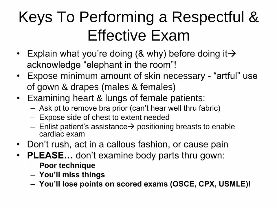
Keys To Performing a Respectful &
Effective Exam • Explain what you’re doing (& why) before doing it
acknowledge “elephant in the room”!
• Expose minimum amount of skin necessary - “artful” use
of gown & drapes (males & females)
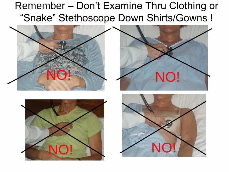
• Examining heart & lungs of female patients: – Ask pt to remove bra prior (can’t hear well thru fabric)
– Expose side of chest to extent needed
– Enlist patient’s assistance positioning breasts to enable cardiac exam
• Don’t rush, act in a callous fashion, or cause pain
• PLEASE… don’t examine body parts thru gown: – Poor technique
– You’ll miss things
– You’ll lose points on scored exams (OSCE, CPX, USMLE)!
Observation
• Pay attention to:
– Chest shape
– Shortness of breath (@ rest or walking)?
– Sitting upright? Able to speak?
– ? Visible impulse on chest wall from
vigorously contracting ventricle (rare)
Hammer & Nails icon indicates A Slide
Describing Skills You Should Perform In Lab
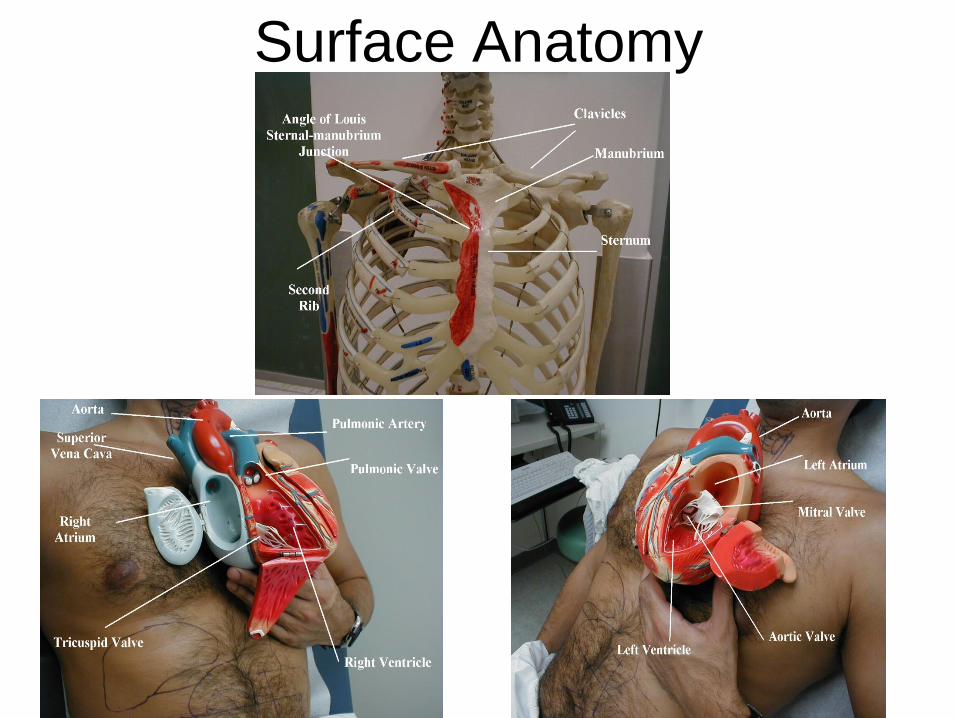
Surface Anatomy
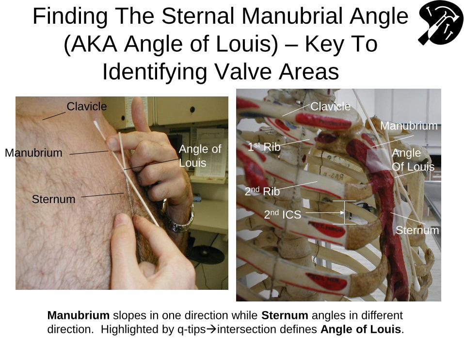
Finding The Sternal Manubrial Angle
(AKA Angle of Louis) – Key To
Identifying Valve Areas
Manubrium
Sternum
Angle of
Louis
Clavicle Clavicle
1st Rib
2nd Rib
Sternum
Manubrium
Angle
Of Louis
2nd ICS
Manubrium slopes in one direction while Sternum angles in different
direction. Highlighted by q-tipsintersection defines Angle of Louis.
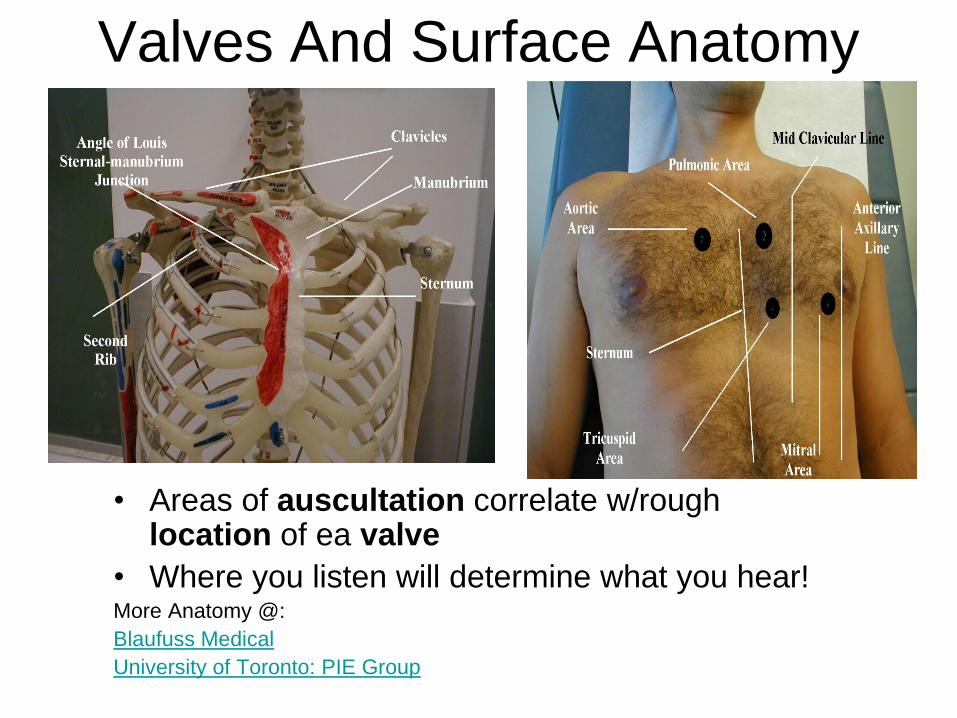
Valves And Surface Anatomy
• Areas of auscultation correlate w/rough location of ea valve
• Where you listen will determine what you hear! More Anatomy @:
Blaufuss Medical
University of Toronto: PIE Group
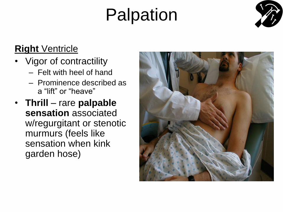
Palpation
Right Ventricle
• Vigor of contractility – Felt with heel of hand
– Prominence described as a “lift” or “heave”
• Thrill – rare palpable sensation associated w/regurgitant or stenotic murmurs (feels like sensation when kink garden hose)
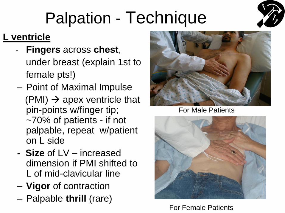
Palpation - Technique L ventricle
- Fingers across chest,
under breast (explain 1st to
female pts!)
– Point of Maximal Impulse
(PMI) apex ventricle that pin-points w/finger tip; ~70% of patients - if not palpable, repeat w/patient on L side
- Size of LV – increased dimension if PMI shifted to L of mid-clavicular line
– Vigor of contraction
– Palpable thrill (rare) For Female Patients
For Male Patients
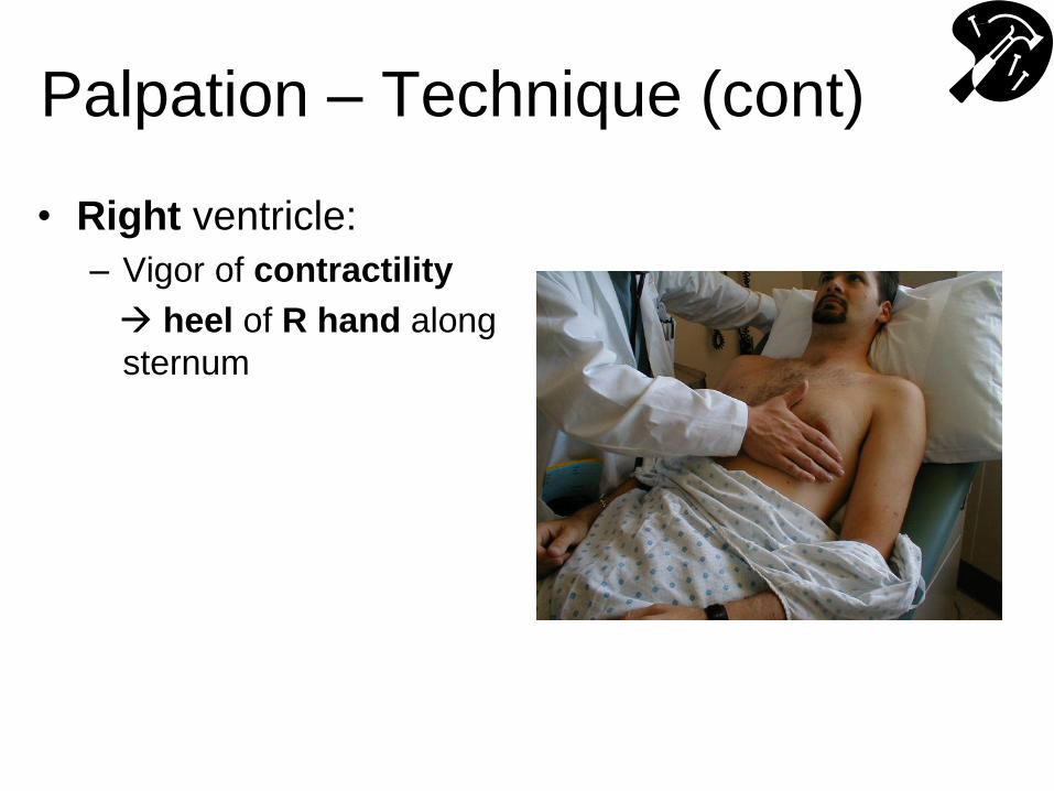
Palpation – Technique (cont)
• Right ventricle:
– Vigor of contractility
heel of R hand along
sternum
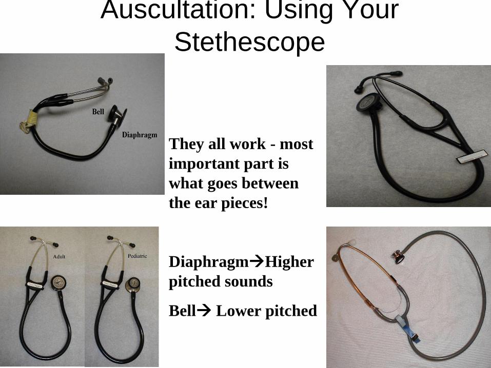
Auscultation: Using Your
Stethescope
They all work - most
important part is
what goes between
the ear pieces!
DiaphragmHigher
pitched sounds
Bell Lower pitched
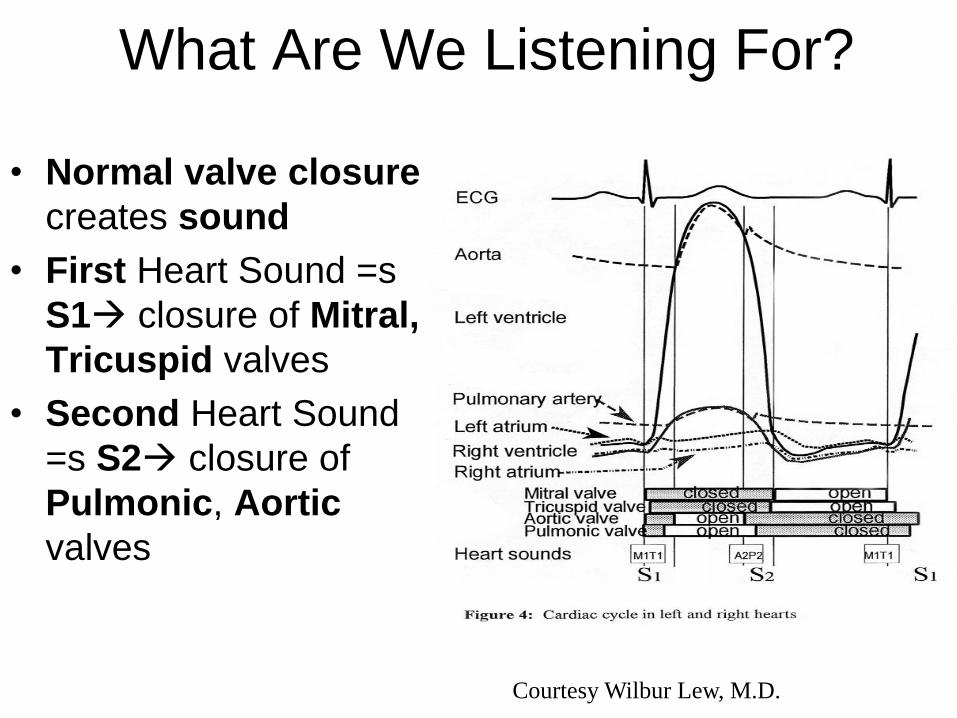
What Are We Listening For?
• Normal valve closure
creates sound
• First Heart Sound =s
S1 closure of Mitral,
Tricuspid valves
• Second Heart Sound
=s S2 closure of
Pulmonic, Aortic
valves
Courtesy Wilbur Lew, M.D.
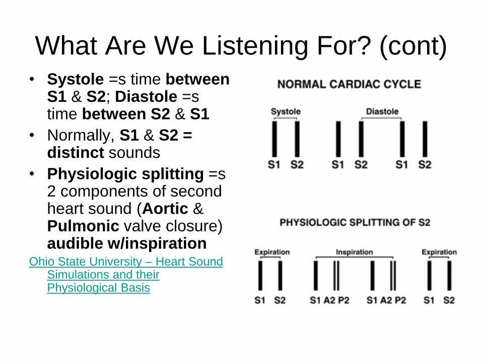
What Are We Listening For? (cont) • Systole =s time between
S1 & S2; Diastole =s time between S2 & S1
• Normally, S1 & S2 = distinct sounds
• Physiologic splitting =s 2 components of second heart sound (Aortic & Pulmonic valve closure) audible w/inspiration
Ohio State University – Heart Sound Simulations and their Physiological Basis
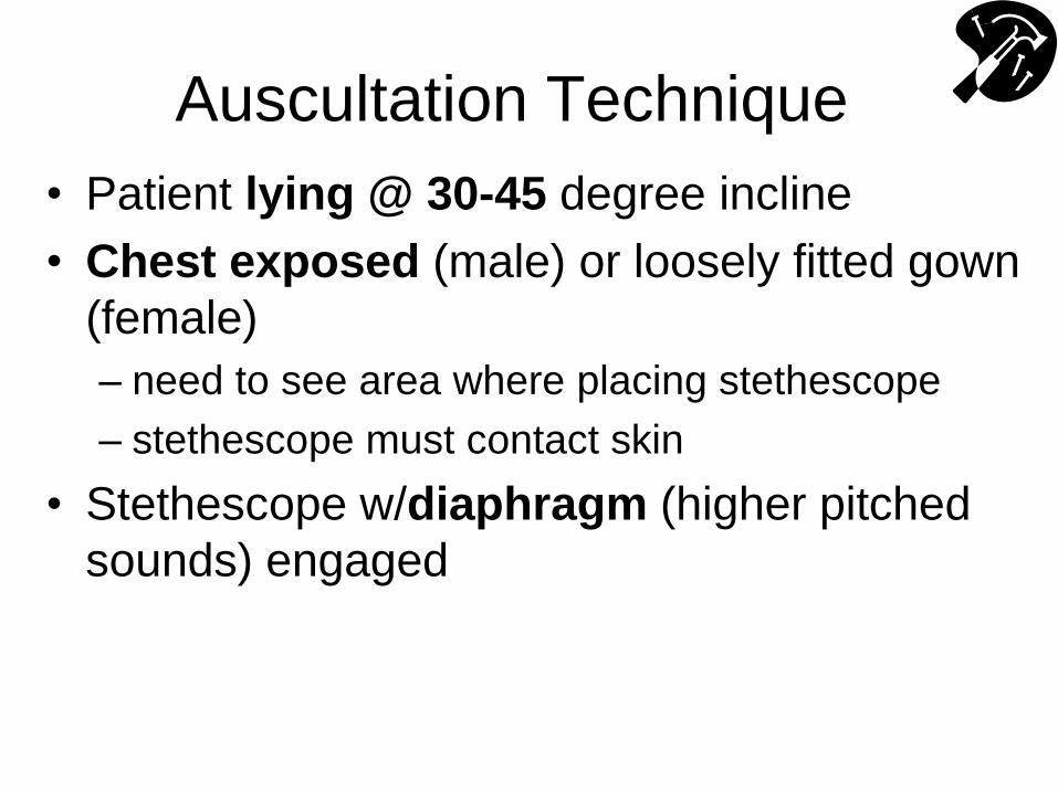
Auscultation Technique
• Patient lying @ 30-45 degree incline
• Chest exposed (male) or loosely fitted gown
(female)
– need to see area where placing stethescope
– stethescope must contact skin
• Stethescope w/diaphragm (higher pitched
sounds) engaged
NO! NO!
QuickTime™ and a decompressor
are needed to see this picture.
NO! NO!
Remember – Don’t Examine Thru Clothing or
“Snake” Stethoscope Down Shirts/Gowns !
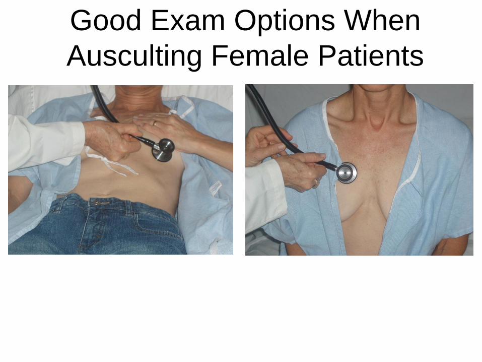
Good Exam Options When
Ausculting Female Patients
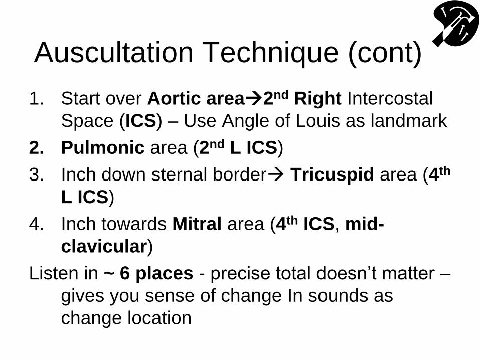
Auscultation Technique (cont)
1. Start over Aortic area2nd Right Intercostal
Space (ICS) – Use Angle of Louis as landmark
2. Pulmonic area (2nd L ICS)
3. Inch down sternal border Tricuspid area (4th
L ICS)
4. Inch towards Mitral area (4th ICS, mid-
clavicular)
Listen in ~ 6 places - precise total doesn’t matter –
gives you sense of change In sounds as
change location
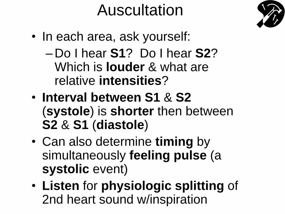
Auscultation
• In each area, ask yourself:
– Do I hear S1? Do I hear S2? Which is louder & what are relative intensities?
• Interval between S1 & S2 (systole) is shorter then between S2 & S1 (diastole)
• Can also determine timing by simultaneously feeling pulse (a systolic event)
• Listen for physiologic splitting of 2nd heart sound w/inspiration
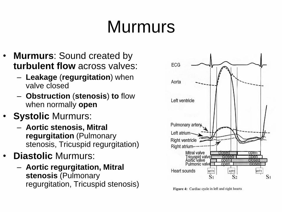
Murmurs
• Murmurs: Sound created by turbulent flow across valves: – Leakage (regurgitation) when
valve closed
– Obstruction (stenosis) to flow when normally open
• Systolic Murmurs: – Aortic stenosis, Mitral
regurgitation (Pulmonary stenosis, Tricuspid regurgitation)
• Diastolic Murmurs: – Aortic regurgitation, Mitral
stenosis (Pulmonary regurgitation, Tricuspid stenosis)
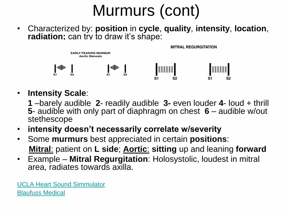
Murmurs (cont) • Characterized by: position in cycle, quality, intensity, location,
radiation; can try to draw it’s shape:
• Intensity Scale:
1 –barely audible 2- readily audible 3- even louder 4- loud + thrill 5- audible with only part of diaphragm on chest 6 – audible w/out stethescope
• intensity doesn’t necessarily correlate w/severity
• Some murmurs best appreciated in certain positions:
Mitral: patient on L side; Aortic: sitting up and leaning forward
• Example – Mitral Regurgitation: Holosystolic, loudest in mitral area, radiates towards axilla.
UCLA Heart Sound Simmulator
Blaufuss Medical
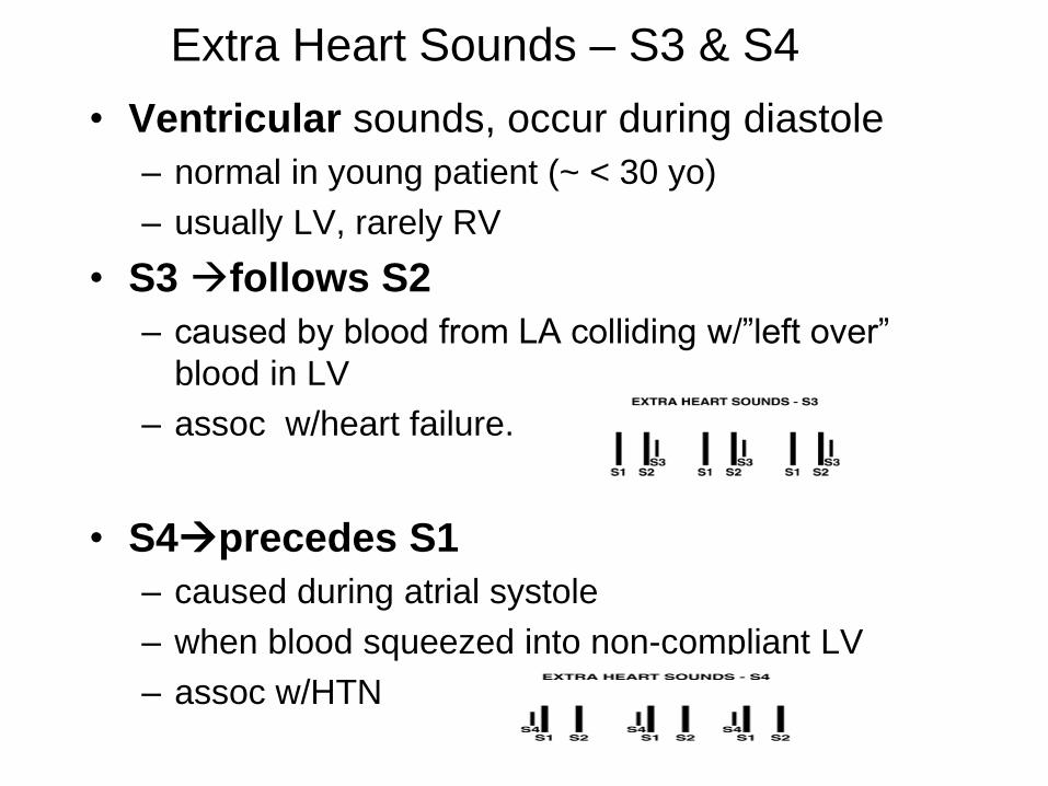
Extra Heart Sounds – S3 & S4
• Ventricular sounds, occur during diastole
– normal in young patient (~ < 30 yo)
– usually LV, rarely RV
• S3 follows S2
– caused by blood from LA colliding w/”left over”
blood in LV
– assoc w/heart failure.
• S4precedes S1
– caused during atrial systole
– when blood squeezed into non-compliant LV
– assoc w/HTN
Extra Heart Sound (cont) • S3 & S4 are soft, low pitched
• Best heart w/bell, laid over LV, w/patient lying on L side (brings apex of heart closer to chest wall) – can also check over RV (4th ICS, L parasternal)
• Abnormal beyond age ~30
• When present, S3 or S4 are referred to as “gallops”
S3 & S4 Simulator:
Ohio State University – Heart Sound Simulations and their Physiological Basis
Auscultation – An Ordered
Approach
• Do I hear S1? Do I hear S2? – Listen in ea major valvular area – think about which sound
should be loudest in ea location (S1 loudest region of TV & MV, S2 loudest AV & PV)
• Do I hear physiologic splitting of S2?
• Do I hear something before S1 (an S4) or after S2 (an S3)?
• Do I hear murmur in systole? In diastole?
• If a murmur present, note: – intensity, character, duration, radiation
• As listen, think about mechanical events that generate the sounds.
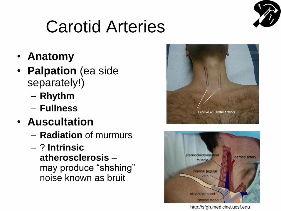
Carotid Arteries
• Anatomy
• Palpation (ea side separately!) – Rhythm
– Fullness
• Auscultation – Radiation of murmurs
– ? Intrinsic atherosclerosis – may produce “shshing” noise known as bruit
http://sfgh.medicine.ucsf.edu
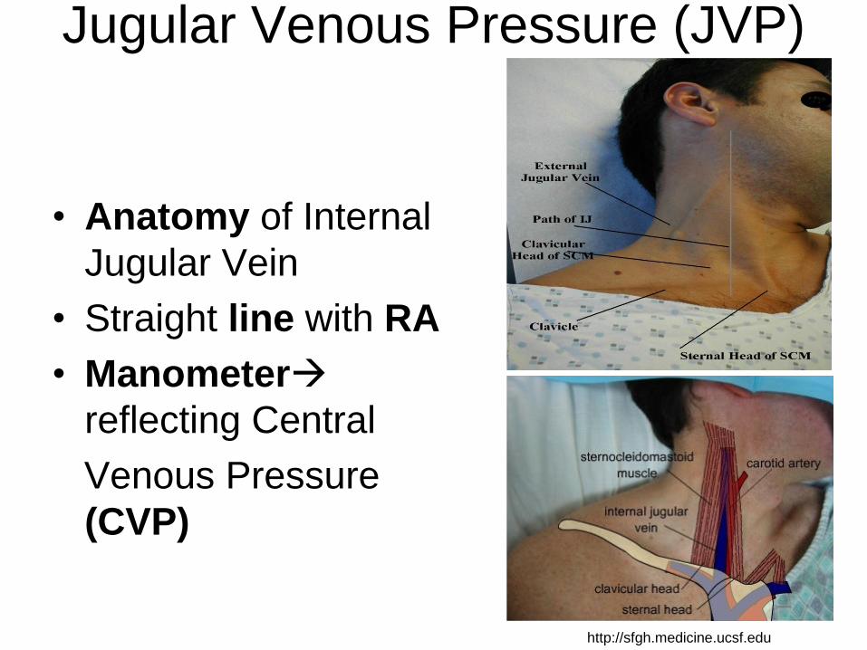
Jugular Venous Pressure (JVP)
• Anatomy of Internal
Jugular Vein
• Straight line with RA
• Manometer
reflecting Central
Venous Pressure
(CVP)
http://sfgh.medicine.ucsf.edu
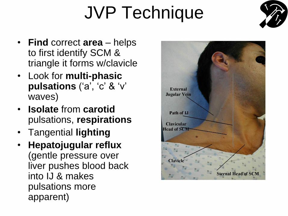
JVP Technique
• Find correct area – helps to first identify SCM & triangle it forms w/clavicle
• Look for multi-phasic pulsations (‘a’, ‘c’ & ‘v’ waves)
• Isolate from carotid pulsations, respirations
• Tangential lighting
• Hepatojugular reflux (gentle pressure over liver pushes blood back into IJ & makes pulsations more apparent)
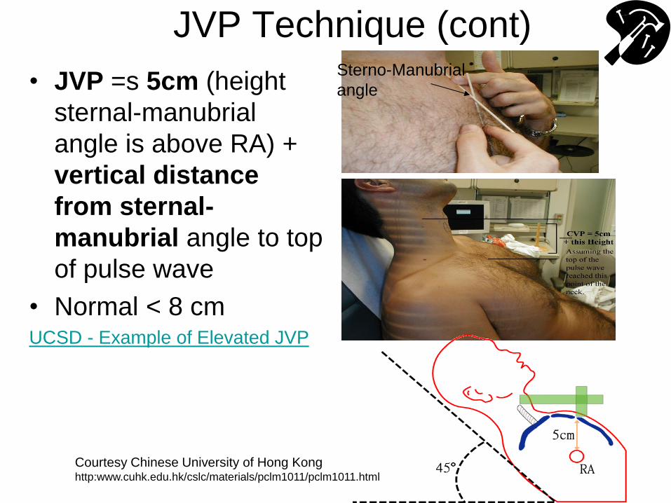
JVP Technique (cont)
• JVP =s 5cm (height
sternal-manubrial
angle is above RA) +
vertical distance
from sternal-
manubrial angle to top
of pulse wave
• Normal < 8 cm UCSD - Example of Elevated JVP
Sterno-Manubrial
angle
Courtesy Chinese University of Hong Kong http:www.cuhk.edu.hk/cslc/materials/pclm1011/pclm1011.html
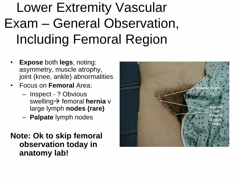
Lower Extremity Vascular
Exam – General Observation,
Including Femoral Region
• Expose both legs, noting: asymmetry, muscle atrophy, joint (knee, ankle) abnormalities
• Focus on Femoral Area:
– Inspect - ? Obvious swelling femoral hernia v large lymph nodes (rare)
– Palpate lymph nodes
Note: Ok to skip femoral observation today in anatomy lab!
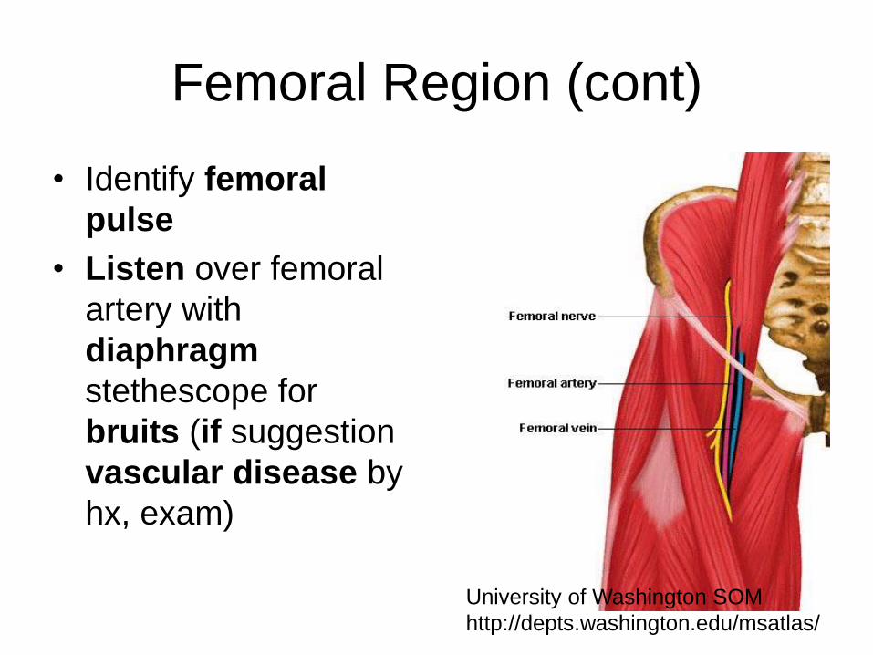
Femoral Region (cont)
• Identify femoral
pulse
• Listen over femoral
artery with
diaphragm
stethescope for
bruits (if suggestion
vascular disease by
hx, exam)
University of Washington SOM
http://depts.washington.edu/msatlas/
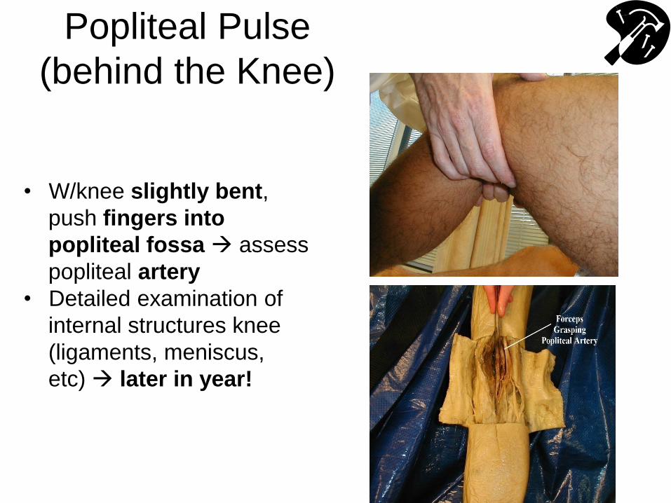
Popliteal Pulse
(behind the Knee)
• W/knee slightly bent,
push fingers into
popliteal fossa assess
popliteal artery
• Detailed examination of
internal structures knee
(ligaments, meniscus,
etc) later in year!
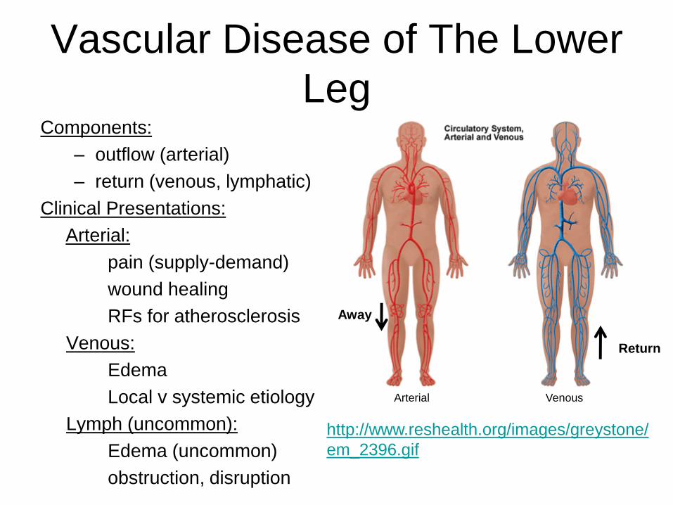
Vascular Disease of The Lower
Leg Components:
– outflow (arterial)
– return (venous, lymphatic)
Clinical Presentations:
Arterial:
pain (supply-demand)
wound healing
RFs for atherosclerosis
Venous:
Edema
Local v systemic etiology
Lymph (uncommon):
Edema (uncommon)
obstruction, disruption
http://www.reshealth.org/images/greystone/
em_2396.gif
Away
Return
Arterial Venous
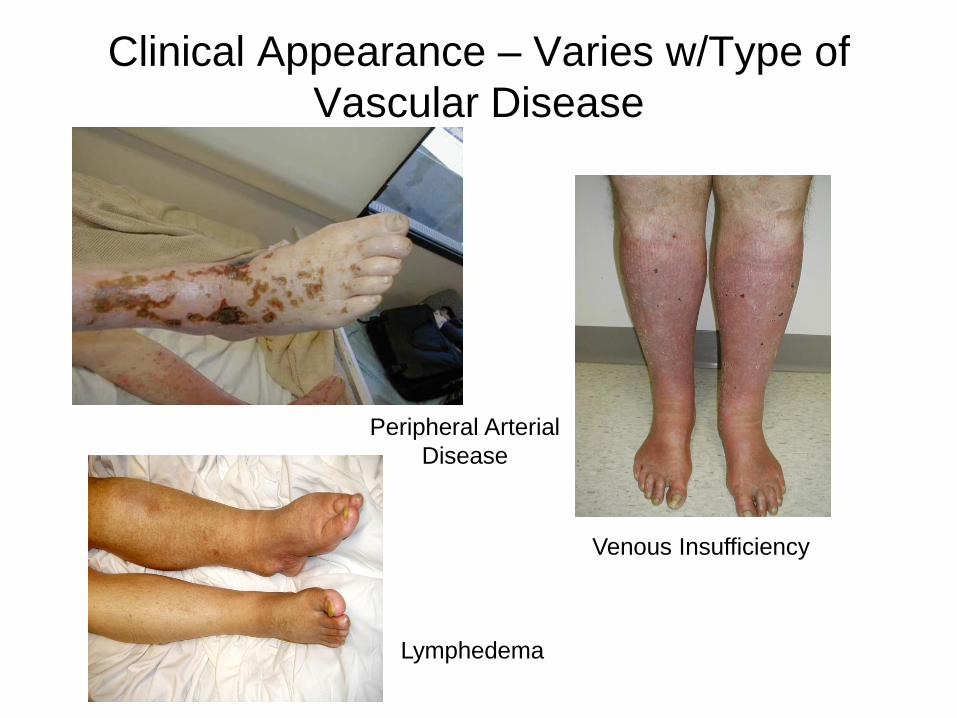
Clinical Appearance – Varies w/Type of
Vascular Disease
Venous Insufficiency
Lymphedema
Peripheral Arterial
Disease
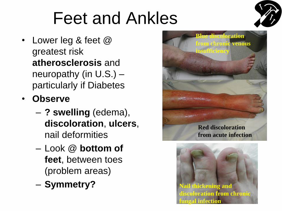
Feet and Ankles • Lower leg & feet @
greatest risk
atherosclerosis and
neuropathy (in U.S.) –
particularly if Diabetes
• Observe
– ? swelling (edema),
discoloration, ulcers,
nail deformities
– Look @ bottom of
feet, between toes
(problem areas)
– Symmetry?
Blue discoloration
from chronic venous
insufficiency
Red discoloration
from acute infection
Nail thickening and
discoloration from chronic
fungal infection
Feet and Ankles (cont)
• Palpation
– Temperature: Use back of examining hand -
warminflammation; coolatherosclerosis
&/or hypo-perfusion
– Capillary refill: push on end of toe or nail bed
& release color returns in < 2-3 seconds;
longer atheroscloerosis &/or hypo-perfusion
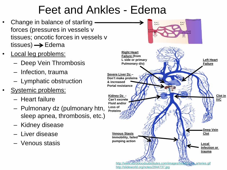
Feet and Ankles - Edema • Change in balance of starling
forces (pressures in vessels v
tissues; oncotic forces in vessels v
tissues) Edema
• Local leg problems:
– Deep Vein Thrombosis
– Infection, trauma
– Lymphatic obstruction
• Systemic problems:
– Heart failure
– Pulmonary dz (pulmonary htn,
sleep apnea, thrombosis, etc.)
– Kidney disease
– Liver disease
– Venous stasis
http://www.eurobloodsubstitutes.com/images/heartCircul_arteries.gif
http://slideworld.org/notes/2844737.jpg
Left Heart
Failure
Right Heart
Failure (from
L side or primary
Pulmonary d/o)
Severe Liver Dz –
Don’t make proteins
& increased
Portal resistance
Clot in
IVC
Deep Vein
Clot
Kidney Dz -
Can’t excrete
Fluid and/or
Loss of
Proteins
Venous Stasis
Immobility, failed
pumping action Local
Infection or
trauma
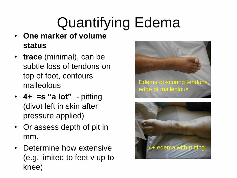
Quantifying Edema • One marker of volume
status
• trace (minimal), can be
subtle loss of tendons on
top of foot, contours
malleolous
• 4+ =s “a lot” - pitting
(divot left in skin after
pressure applied)
• Or assess depth of pit in
mm.
• Determine how extensive
(e.g. limited to feet v up to
knee)
Edema obscuring tendons,
edge of malleolous
4+ edema with pitting
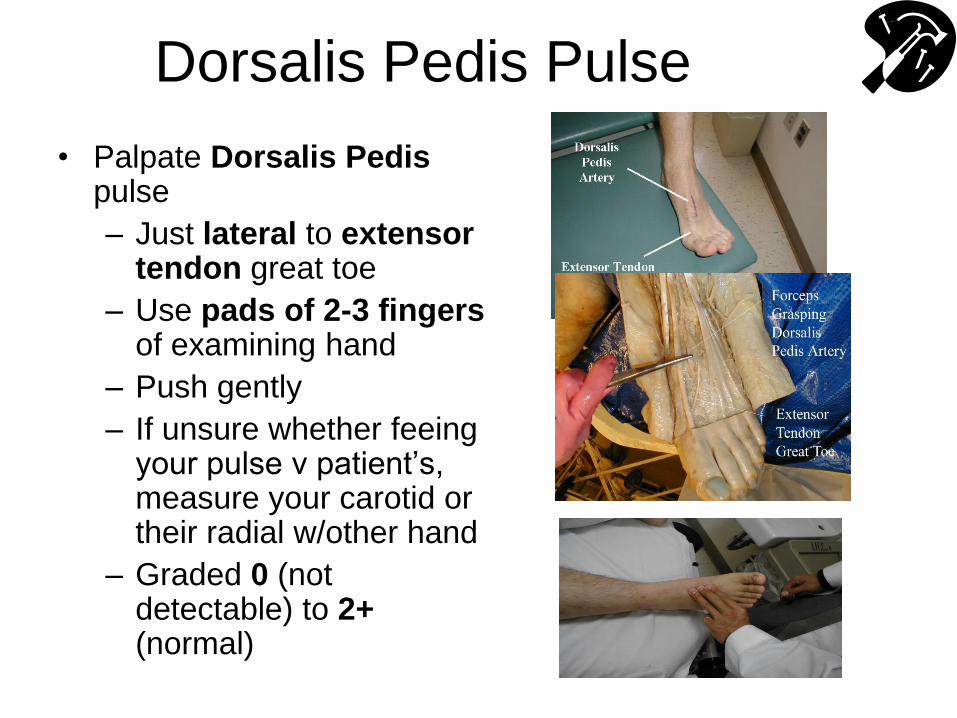
Dorsalis Pedis Pulse
• Palpate Dorsalis Pedis pulse
– Just lateral to extensor tendon great toe
– Use pads of 2-3 fingers of examining hand
– Push gently
– If unsure whether feeing your pulse v patient’s, measure your carotid or their radial w/other hand
– Graded 0 (not detectable) to 2+ (normal)

Posterior Tibial Pulse
• Palpate Posterior Tibial Pulse – Located posterior to
medial malleolous
– Start on top of mallelous & work towards achilles
– Use pads of 2-3 fingers, pushing gently
– Same rating scale as for dorsalis pedis
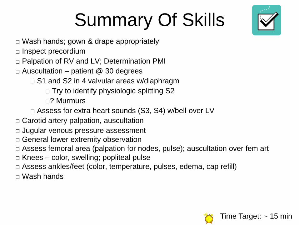
Summary Of Skills □ Wash hands; gown & drape appropriately
□ Inspect precordium
□ Palpation of RV and LV; Determination PMI
□ Auscultation – patient @ 30 degrees
□ S1 and S2 in 4 valvular areas w/diaphragm
□ Try to identify physiologic splitting S2
□? Murmurs
□ Assess for extra heart sounds (S3, S4) w/bell over LV
□ Carotid artery palpation, auscultation
□ Jugular venous pressure assessment
□ General lower extremity observation
□ Assess femoral area (palpation for nodes, pulse); auscultation over fem art
□ Knees – color, swelling; popliteal pulse
□ Assess ankles/feet (color, temperature, pulses, edema, cap refill)
□ Wash hands
Time Target: ~ 15 min