Embed Size (px)
Citation preview
RESEARCH ARTICLE
Evolutionary and biogeographical implications
of degraded LAGLIDADG endonuclease
functionality and group I intron occurrence in
stony corals (Scleractinia) and mushroom
corals (Corallimorpharia)
Juan Sebastian Celis1,2*, David R. Edgell3, Bjorn Stelbrink1, Daniel Wibberg4,
Torsten Hauffe1, Jochen Blom5, Jorn Kalinowski4, Thomas Wilke1,2
1 Animal Ecology and Systematics, Justus Liebig University, Giessen, Hessen, Germany, 2 Corporation
Center of Excellence in Marine Sciences CEMarin, Bogota, Colombia, 3 Department of Biochemistry, School
of Medicine and Dentistry, Western University, London, Ontario, Canada, 4 Department of Biochemistry,
Institute for Genome Research and Systems Biology CeBiTec, Bielefeld University, Bielefeld, Germany,
5 Bioinformatics and Systems Biology, Justus Liebig University, Giessen, Germany
Abstract
Group I introns and homing endonuclease genes (HEGs) are mobile genetic elements,
capable of invading target sequences in intron-less genomes. LAGLIDADG HEGs are the
largest family of endonucleases, playing a key role in the mobility of group I introns in a pro-
cess known as ‘homing’. Group I introns and HEGs are rare in metazoans, and can be
mainly found inserted in the COXI gene of some sponges and cnidarians, including stony
corals (Scleractinia) and mushroom corals (Corallimorpharia). Vertical and horizontal intron
transfer mechanisms have been proposed as explanations for intron occurrence in cnidari-
ans. However, the central role of LAGLIDADG motifs in intron mobility mechanisms remains
poorly understood. To resolve questions regarding the evolutionary origin and distribution of
group I introns and HEGs in Scleractinia and Corallimorpharia, we examined intron/HEGs
sequences within a comprehensive phylogenetic framework. Analyses of LAGLIDADG
motif conservation showed a high degree of degradation in complex Scleractinia and Coralli-
morpharia. Moreover, the two motifs lack the respective acidic residues necessary for
metal-ion binding and catalysis, potentially impairing horizontal intron mobility. In contrast,
both motifs are highly conserved within robust Scleractinia, indicating a fully functional endo-
nuclease capable of promoting horizontal intron transference. A higher rate of non-synony-
mous substitutions (Ka) detected in the HEGs of complex Scleractinia and Corallimorpharia
suggests degradation of the HEG, whereas lower Ka rates in robust Scleractinia are consis-
tent with a scenario of purifying selection. Molecular-clock analyses and ancestral inference
of intron type indicated an earlier intron insertion in complex Scleractinia and Corallimor-
pharia in comparison to robust Scleractinia. These findings suggest that the lack of horizon-
tal intron transfers in the former two groups is related to an age-dependent degradation of
PLOS ONE | DOI:10.1371/journal.pone.0173734 March 9, 2017 1 / 20
a1111111111
a1111111111
a1111111111
a1111111111
a1111111111
OPENACCESS
Citation: Celis JS, Edgell DR, Stelbrink B, Wibberg
D, Hauffe T, Blom J, et al. (2017) Evolutionary and
biogeographical implications of degraded
LAGLIDADG endonuclease functionality and group
I intron occurrence in stony corals (Scleractinia)
and mushroom corals (Corallimorpharia). PLoS
ONE 12(3): e0173734. doi:10.1371/journal.
pone.0173734
Editor: Alexander F. Palazzo, University of Toronto,
CANADA
Received: October 31, 2016
Accepted: February 24, 2017
Published: March 9, 2017
Copyright: © 2017 Celis et al. This is an open
access article distributed under the terms of the
Creative Commons Attribution License, which
permits unrestricted use, distribution, and
reproduction in any medium, provided the original
author and source are credited.
Data Availability Statement: All relevant data are
within the paper and its Supporting Information
files.
Funding: JSC was funded jointly by the German
Academic Exchange Service (Grant number
A1372429. https://www.daad.de/en/) and the
Corporation Center of Excellence in Marine
Sciences (Grant Number 80221287. http://cemarin.
org/). The funders had no role in study design, data
the endonuclease activity. Moreover, they also explain the peculiar geographical patterns of
introns in stony and mushroom corals.
Introduction
Group I introns are self-splicing genetic elements with conserved secondary and tertiary struc-
tures that are involved in ribozyme activity at the RNA level [1]. Many group I introns also
constitute mobile genetic elements at the DNA level due to mobility-promoting proteins,
termed homing endonucleases (HEs), which are encoded by a homing endonuclease gene
(HEG) inserted within the intron [2–5]. Currently, there are six known families of HEs [6, 7].
They are classified on the basis of conserved amino acid motifs that form the catalytic center
or structural core of the enzyme, with the LAGLIDADG family being the largest and most
diverse [4]. The intron/HEG association is a notable example of a composite mobile genetic
element that has persisted by exploiting cellular DNA repair and recombination pathways to
promote spread by a process known as ‘homing’ [8]. In the homing pathway, the intron-
encoded HE generates a double-strand break in cognate genes that lack the intron, stimulating
repair of the broken gene by using the intron-containing gene as template [2].
HEs tend to target conserved nucleotides that correspond to functionally critical amino
acids of cellular genes, ensuring that a target site will be present in related genomes [9–11].
Moreover, HEs can tolerate nucleotide substitutions within their target site [5], accommodat-
ing genetic drift and natural variation in an apparent adaptation to enable efficient intron
homing [2,12,13]. By inserting in phenotypically neutral locations within the intron (i.e., loop
regions), HEGs may greatly reduce deleterious effects to the host [14]. Furthermore, the close
association with introns allows the HEGs to spread horizontally through populations at high
rates [13,15]. Likewise, the conserved amino acid motifs within HEs play a key role in horizon-
tal transfer because they are directly involved in DNA endonuclease activity at target sequences
[3,4]. The class-defining LAGLIDADG motif comprises a subunit interface with acidic resi-
dues (Aspartic [D, underlined] or Glutamic acid [E]), co-ordinating and positioning divalent
metal ions necessary for hydrolysis of the DNA backbone [16,17]. Once all intron-less alleles
are occupied, the potential for further intron-spread diminishes, leading to decreased selective
pressure for maintaining endonuclease activity. Unless a population of intron-less alleles sup-
ports new insertions or the intron transposes to a new genomic location by reverse splicing or
endonuclease-mediated cleavage [18–20], two outcomes due to loss of selection are possible:
(i) degradation of the intron/HEG over evolutionary time, eventually leading to its deletion
and/or (ii) a shift towards a RNA maturase that promotes intron splicing by a variety of mech-
anisms [21]. Based on these observations, and phylogenetic analyses of intron distribution in
mitochondria, a cyclical life history of gain and loss was proposed for mobile group I introns
[14,15]. Briefly, the cycle comprises horizontal intron invasion targeting intron-less alleles, fix-
ation in the population, decreased selective pressure for maintaining endonuclease activity,
degradation of the HEG, and complete intron/HEG deletion leading to target sequence re-
establishment [13,15,22,23].
In metazoans, group I introns and HEGs are mainly known from sponges [24–26], placozo-
ans [27], sea anemones [22,23,28] and scleractinian corals [29]. The order Scleractinia (stony
corals) consists of two major phylogenetic groups, namely the ‘robust’ and the ‘complex’ clades
[30,31] (hereafter referred to as robust Scleractinia and complex Scleractinia, respectively).
However, there is still controversy regarding the monophyly of the Scleractinia, with the
Degraded endonucleases and introns in stony and mushroom corals
PLOS ONE | DOI:10.1371/journal.pone.0173734 March 9, 2017 2 / 20
collection and analysis, decision to publish, or
preparation of the manuscript.
Competing interests: The authors have declared
that no competing interests exist.
possibility of a sister group relationship between complex Scleractinia and Corallimorpharia
[32–35]. The presence of a group I intron, interrupting the COXI (cytochrome c oxidase sub-
unit I) gene, was previously reported by Fukami et al. in 20 species of robust Scleractinia [29].
Based on broadly similar topologies between host and intron-based phylogenies, they sug-
gested that the observed pattern can be explained by vertical inheritance of the intron,
although a sponge-coral horizontal transfer remained equally plausible as the origin of the
intron in corals [29]. Furthermore, the authors proposed that the intron was originally trans-
ferred to robust Scleractinia from a fungal donor, even though the intron insertion site in the
COXI gene differs between these two groups [29]. This contrasts with the general assumption
that introns inserted in different positions are phylogenetically not closely related [36]. The
exclusive vertical intron transfer proposed for robust Scleractinia is in disagreement with the
widely accepted combined horizontal/vertical transfer mechanisms shown in sponges and sea
anemones [22,23,25,26]. Moreover, the study of Fukami et al. did not include corals of com-
plex Scleractinia, which contains some of the most important and widespread reef-building
coral families [33].
Surprisingly, the functionality of the homing endonuclease assessed by the conservation
degree of the LAGLIDADG motifs has not been considered previously in explaining occurrence
of the intron in corals. Intron horizontal transfer requires an active endonuclease, since one
function of intron-encoded LAGLIDADG enzymes is to serve as site-specific endonucleases
within the mobility pathway of group I introns [37]. In comparison, inactive/degraded endonu-
cleases constrain intron transfer to vertical inheritance [15]. Moreover, in contrast to sea anem-
ones, the different stages of the intron cycle in corals [23], as well as the number of gains and/or
losses within an absolute temporal framework, remain largely unknown. Finally, the effect of a
restricted geographic occurrence of intron-containing taxa to the Indo-Pacific Ocean [29] has
not been considered in previous studies of group I intron evolution in Scleractinia.
Given this lack of knowledge of intron/HEGs evolution in scleractinian corals and coralli-
morpharians, the general aim of this study is to unravel the role of LAGLIDADG endonucle-
ases in the evolutionary history of group I intron occurrence in these groups. We used
scleractinian corals and corallimorpharians as a model system because they commonly feature
introns of potentially different origins [22,25], though being phylogenetically closely related
[32,33]. Our specific aims are: 1) to identify the putative functionality of LAGLIDADG motifs
and their role in the occurrence of group I introns; 2) to infer number and absolute times of
gains or losses of introns within a molecular-clock framework, and 3) to assess the geographic
distribution of intron-possessing species within an evolutionary context. We hypothesize that:
(i) differences in LAGLIDADG motif conservation and hence homing endonuclease function-
ality among major groups of corals informs about horizontal/vertical intron transmission by
comparing the putative LAGLIDADG motif composition between Scleractinia and Coralli-
morpharia; (ii) introns gained early during the evolutionary history of corals are more
degraded than introns acquired more recently due to a longer time for degradation as part of
the intron-cycle. Molecular-clock analyses in combination with information on LAGLIDADG
motif composition and the inference of intron acquisition allow linking the time-dependent
degradation of the LAGLIDADG motif, and (iii) introns obtained more recently show a more
restricted geographical distribution than older introns. Biogeographic patterns of intron-con-
taining taxa together with time-calibrated phylogenies and the LAGLIDADG motif assessment
help explaining the intron occurrence over space and time.
By using a comprehensive phylogenetic framework, our results will likely provide insights
into the occurrence of mitochondrial group I introns based on homing endonuclease func-
tionality. Moreover, our approach has the potential of addressing evolutionary questions
related to the evolutionary history of introns in other metazoans.
Degraded endonucleases and introns in stony and mushroom corals
PLOS ONE | DOI:10.1371/journal.pone.0173734 March 9, 2017 3 / 20
Materials and methods
COXI intron-exon sequence alignment and LAGLIDADG homing
endonuclease homology validation
In order to retrieve the available homologous COXI intron sequences and their 5´ and 3´ flank-
ing exons from Scleractinia and Corallimorpharia, a BLASTN [38,39] search was performed
taking the Porites rus (complex Scleractinia) intron that we annotated previously [40], and the
reported intron of Physogyra lichtensteini (robust Scleractinia) [29] as query sequences. This
step was performed in an iterative procedure in which sequences identified during each step
were used to refine the search [41]. We stopped the search when no new homologs were
detected. By the time of searching (March 1st, 2016), 14 intron sequences for complex Sclerac-
tinia, 23 for robust Scleractinia and 11 for Corallimorpharia were retrieved (S1 Table). Using
this methodology we retrieved only group I introns present in the COXI gene. Intron
sequences belonging to complex Scleractinia lacked annotation of the HEG [hereafter referred
to as LAGLIDADG Open Reading Frames (ORFs)]. Therefore the LAGLIDADG ORFs were
annotated using the GenDB platform [42] and GLIMMER 3.0.2 [43]. We obtained informa-
tion concerning intron presence/absence by performing a comprehensive literature and data-
base search. All retrieved intron sequences were aligned in BioEdit version 7.2.5 [44] with the
ClustalW algorithm [45] and default settings. Additionally, we aligned intron sequences found
in complex Scleractinia using information on the intron secondary structure (S1 Fig). Intron
insertion sites were defined following the guidelines proposed by the Human Genome Varia-
tion Society [46], using the reference human COXI gene (Cambridge human mtDNA
sequence; GenBank accession number NC_012920.1). We evaluated the conservation of
potential target sequences for intron-encoded HEs by aligning selected COXI exon sequences
from Scleractinia and Corallimorpharia. Taxa were chosen to represent the COXI intron inser-
tion sites known among Scleractinia (S2 Fig).
Protein structure modeling of the intron-encoded LAGLIDADG homing endonuclease
(LHE) found in P. rus (complex Scleractinia), P. lichtensteini (robust Scleractinia) and Ricordeaflorida (Corallimorpharia) was performed in Phyre2 [47] in order to validate their similarity to
known LHEs. The generated LHE structure models were constructed based on the experimen-
tally validated crystal structure of the I-SmaMI LHE found in the fungus Sordaria macrospora(Molecular Modeling Data Base ID: 123349). Both the predicted secondary structures and the
known and predicted secondary structure of S.macrospora are used in conjunction with the
sequence information in generating the alignment. By this means, is possible to identify the
two LAGLIDADG motifs as part of the two core α helices, consistent with the accepted canon-
ical LHE structure [7].
Synonymous vs. non-synonymous ratios (Ka/Ks) and sequence logo
representation of amino acid conservation
To determine whether the LAGLIDADG ORFs of Scleractinia and Corallimorpharia are under
different types of selection, ratios of synonymous versus non-synonymous substitutions (Ka/
Ks) were calculated following Emblem et al. [23]. Thereby, a Ka/Ks ratio>1 indicates substitu-
tions that more frequently result in a change of amino acid identity, consistent with positive
selection. Ka/Ks ratios <1 indicate substitutions that retain amino acid identity, as expected for
purifying (negative) selection, while a Ka/Ks ratio of ~1 reflects neutral evolution [23].
The predicted amino acid sequences encoded by each of the LAGLIDADG ORFs of 23
robust Scleractinia, 14 complex Scleractinia, and 11 Corallimorpharia species were reverse
translated to generate nucleotide sequence alignments in MEGA version 7 [48]. We excluded
Degraded endonucleases and introns in stony and mushroom corals
PLOS ONE | DOI:10.1371/journal.pone.0173734 March 9, 2017 4 / 20
codons containing alignment gaps in any sequence from subsequent analyses. The final HEG
alignment lengths were 925, 753, and 453 bp for robust Scleractinia, complex Scleractinia, and
Corallimorpharia, respectively. Ka/Ks calculations of the HEG alignments were performed for
each alignment in MEGA using the mold-yeast mitochondrial genetic code [23,32]. Substitu-
tions rates were calculated using the Nei-Gojobori nucleotide diversity estimates [49] and
applying the Jukes-Cantor correction for multiple substitutions. Pairwise comparisons of the
HEG were plotted using a custom script for the R statistical environment v.3.2.1 [50]. A one-
sided codon-based Z-test of selection [48], as implemented in MEGA, was used to test for puri-
fying selection acting on the translated LAGLIDADG ORFs (H0: Ka = Ks vs. HA: Ka<Ks) with
variance estimated with 1000 bootstrap replicates. This analysis was performed by averaging
over all sequence pairs of the aligned HEs datasets of robust Scleractinia, complex Scleractinia,
and Corallimorpharia (Table 1). The Ka/Ks ratios of the COXI Folmer region [51], a 658 bp
fragment widely used for phylogenetic inference, of the intron-containing taxa were compared
to the ratios calculated for the LAGLIDADG ORFs in order to test for relaxed purifying selec-
tion (S3 Fig). Sequence logo representation of amino acid conservation in the LAGLIDADG
motifs was generated for complex Scleractinia, robust Scleractinia, and Corallimorpharia
endonucleases using WebLogo [52].
Estimation of divergence times and ancestral intron types
For the molecular-clock analysis, we used a 171-taxon dataset of two mitochondrial genes;
COXI and cyt b, available from GenBank (see S2 Table for accession numbers). Mitochondrial
genes have been widely used for phylogenetic studies in corals [29,32,33,53] given their low
substitution rates in this group [33]. The dataset included 149 of the c. 780 zooxanthellate
scleractinian species (19%), 12 corallimorpharian and 10 outgroup taxa belonging to the Acti-
niaria, Antipatharia, Zoantharia, Octocorallia, and Porifera.
We removed the intron-containing species Caulastraea echinulata (robust Scleractinia)
from the molecular-clock analysis due to the lack of respective cyt b sequences in GenBank.
The concatenated dataset had a total length of 1349 bp. Divergence times were inferred in
BEAST v. 1.8.2 [54]. An initial uncorrelated relaxed-clock analysis using substitution models
for COXI and cyt b (jModelTest, 24 candidate substitutions models, best AIC: GTR+I+G) [55]
and relative rates was performed in order to retrieve the topology. In the final analysis, a set of
fossil calibration points was used as suggested by Simpson et al. [53]. Given the uncertainty of
these calibration points, Simpson et al. used uniform priors for the node calibration. However,
since such priors represent hard constraints and often result in initial posterior probabilities to
be zero, we here used normal distribution priors. Moreover, three out of the eleven suggested
fossil calibration points had to be excluded due to a slightly different sampling design. Thus,
the final analyses included the following eight fossil calibration points used by Simpson et al.
[53]: (1) MRCA Scleractinia, 245–271 My (BEAST settings: mean = 258, standard
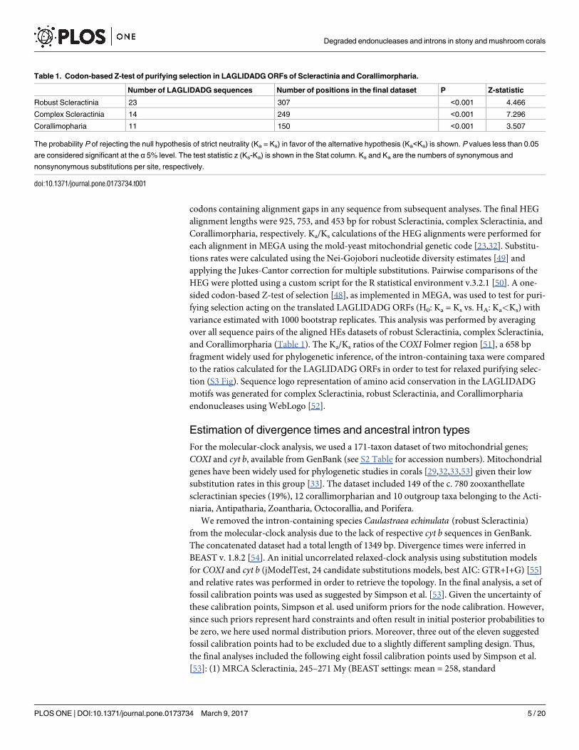
Table 1. Codon-based Z-test of purifying selection in LAGLIDADG ORFs of Scleractinia and Corallimorpharia.
Number of LAGLIDADG sequences Number of positions in the final dataset P Z-statistic
Robust Scleractinia 23 307 <0.001 4.466
Complex Scleractinia 14 249 <0.001 7.296
Corallimopharia 11 150 <0.001 3.507
The probability P of rejecting the null hypothesis of strict neutrality (Ka = Ks) in favor of the alternative hypothesis (Ka<Ks) is shown. P values less than 0.05
are considered significant at the α 5% level. The test statistic z (Ks-Ka) is shown in the Stat column. Ks and Ka are the numbers of synonymous and
nonsynonymous substitutions per site, respectively.
doi:10.1371/journal.pone.0173734.t001
Degraded endonucleases and introns in stony and mushroom corals
PLOS ONE | DOI:10.1371/journal.pone.0173734 March 9, 2017 5 / 20
deviation = 6.6); (2) Isopora–Acropora, 5.3–15 My (mean = 10.15, SD = 2.5); (3) Acropora–
Montipora, 37.2–67.7 My (mean = 52.45, SD = 7.8); (4) Acropora–Stephanocoenia, 63.4–90.1
My (mean = 76.75, SD = 6.8); (5); Lobophyllia–Symphyllia, 19–31.3 My (mean = 25.15,
SD = 3.1); (6) Pocillopora–Seriatopora, 28.4–42.7 My (mean = 35.55, SD = 3.7); (7) Echino-pora–Oulophyllia, 21.9–49.3 My (mean = 35.6, SD = 7.0); and (8) Cladocora–Hydnophora 100–
183.3 My (mean = 141.65, SD = 21.2). Two replicates were run on the CIPRES Science Gate-
way web portal [56] using the following settings: ngen = 100,000,000; samplefreq = 5,000;
birth-death (BD) model. Log files were visualized in Tracer v. 1.5 [57] for congruency and
combined in LogCombiner v. 1.8.2 (BEAST package; 50% burnin). TreeAnnotator v. 1.8.2
(BEAST package; no additional burn-in) was used for combining the tree files and for identify-
ing the maximum clade credibility (MCC) tree.
In order to infer intron ancestral states and frequency of gains and/or losses, ancestral infer-
ence of intron-type was performed employing stochastic character mapping [58] available in
the package phytools version 0.5.10 [59] for R. This method can handle uncertainty of intron
presence by permitting the input of a matrix of prior probabilities of intron state at the tips of
the phylogeny. The analysis used the COXI/cyt b topology obtained from the divergence time
inference. We defined five character states based on intron presence/absence and intron type:
1) intron absence; 2) intron missing information; 3) intron 720; 4) intron 884; 5) intron 867.
In the case where intron presence/absence was unknown (i.e., missing) the character state was
defined by a uniform prior. This approach not only infers ancestral states at internal nodes but
also provides posterior probabilities of intron state for tips with unknown intron presence/
absence [59].
Results
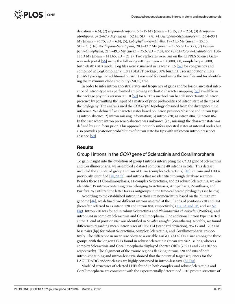
Group I introns in the COXI gene of Scleractinia and Corallimorpharia
To gain insight into the evolution of group I introns interrupting the COXI gene of Scleractinia
and Corallimorpharia, we assembled a dataset comprising 48 introns in total. This dataset
included the annotated group I intron of P. rus (complex Scleractinia) [40], introns and HEGs
previously identified [28,29,32], and introns that we identified through database searches.
Besides these 11 Corallimorpharia, 14 complex Scleractinia, and 23 robust Scleractinia, we also
identified 19 intron-containing taxa belonging to Actiniaria, Antipatharia, Zoantharia, and
Porifera. We utilized the latter taxa as outgroups in the time-calibrated phylogeny (see below).
According to the established intron insertion site nomenclature based on the human mt
genome [46], we defined two different introns inserted at the 3´ ends of positions 720 and 884
(hereafter referred to as intron 720 and intron 884, respectively) (Fig 1A and 1B, and see S2
Fig). Intron 720 was found in robust Scleractinia and Plakinastrella cf. onkodes (Porifera), and
intron 884 in complex Scleractinia and Corallimorpharia. One additional intron type inserted
at the 3´ end of position 867 was identified in Savalia savaglia (Zoantharia). Notably, we found
differences regarding mean intron sizes of 1086±24 (standard deviation), 967±7 and 1203±28
base pairs (bp) for robust Scleractinia, complex Scleractinia, and Corallimorpharia, respec-
tively. The difference in mean size obeys to a variable LAGLIDADG ORF size among the three
groups, with the longest ORFs found in robust Scleractinia (mean size 962±31 bp), whereas
complex Scleractinia and Corallimorpharia displayed shorter ORFs (755±1 and 778±207 bp,
respectively). The alignment of the exonic regions flanking introns 720 and 884 of both
intron-containing and intron-less taxa showed that the potential target sequences for the
LAGLIDADG endonucleases are highly conserved in intron-less taxa (S2 Fig).
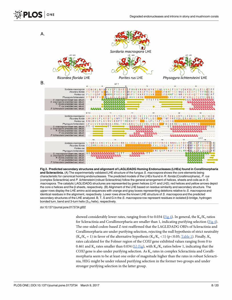
Modeled structures of selected LHEs found in both complex and robust Scleractinia and
Corallimorpharia are consistent with the experimentally determined LHE protein structure of
Degraded endonucleases and introns in stony and mushroom corals
PLOS ONE | DOI:10.1371/journal.pone.0173734 March 9, 2017 6 / 20
the fungus S.macrospora. Thus, the two LAGLIDADG motifs were placed in the two central
helices at the protein’s domain interface (Fig 2A). Likewise, the alignment of the LHEs of P.
rus, P. lichtensteini and R. florida, based on residue similarity and predicted secondary struc-
ture is consistent with the known crystal structure of the LHE of S.macrospora (Fig 2B).
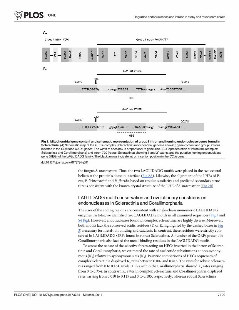
LAGLIDADG motif conservation and evolutionary constrains on
endonucleases in Scleractinia and Corallimorpharia
The sizes of the coding regions are consistent with single-chain monomeric LAGLIDADG
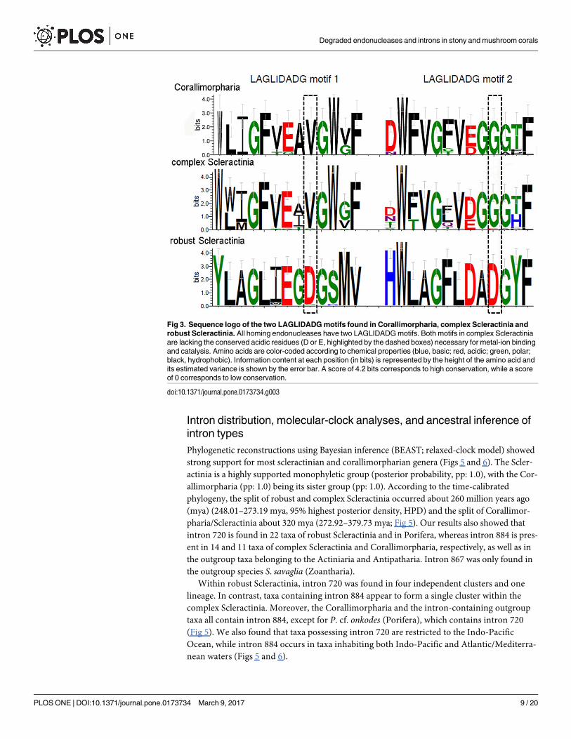
enzymes. In total, we identified two LAGLIDADG motifs in all examined sequences (Fig 3 and
S4 Fig). However, endonucleases found in complex Scleractinia are highly diverse. Moreover,
both motifs lack the conserved acidic residues (D or E, highlighted by the dashed boxes in Fig
3) necessary for metal-ion binding and catalysis. In contrast, these residues were strictly con-
served in LAGLIDADG ORFs found in robust Scleractinia. A number of the ORFs present in
Corallimorpharia also lacked the metal-binding residues in the LAGLIDADG motifs.
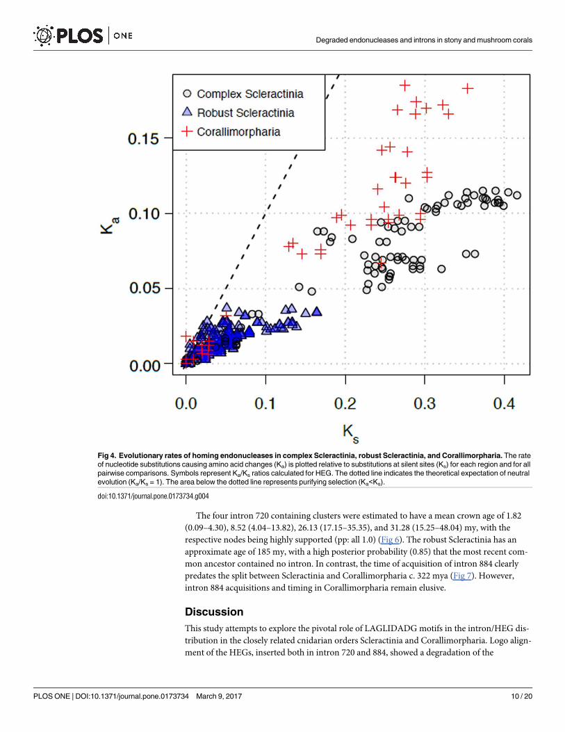
To assess the nature of the selective forces acting on HEGs inserted in the intron of Sclerac-
tinia and Corallimorpharia, we estimated the rate of nucleotide substitutions at non-synony-
mous (Ka) relative to synonymous sites (Ks). Pairwise comparisons of HEGs sequences of
complex Scleractinia displayed Ks rates between 0.007 and 0.416. The rates for robust Scleracti-
nia ranged from 0 to 0.164, while HEGs within the Corallimorpharia showed Ks rates ranging
from 0 to 0.354. In contrast, Ka rates in complex Scleractinia and Corallimorpharia displayed
rates varying from 0.010 to 0.115 and 0 to 0.185, respectively; whereas robust Scleractinia
Fig 1. Mitochondrial gene content and schematic representation of group I intron and homing endonuclease genes found in
Scleractinia. (A) Schematic map of the P. rus (complex Scleractinia) mitochondrial genome showing gene content and group I introns
inserted in the COXI and NAD5 genes. The width of each box is proportional to gene size. (B) Representation of intron 884 (complex
Scleractinia and Corallimorpharia) and intron 720 (robust Scleractinia) showing 5´and 3´ exons, and the putative homing endonuclease
gene (HEG) of the LAGLIDADG family. The black arrows indicate intron insertion position in the COXI gene.
doi:10.1371/journal.pone.0173734.g001
Degraded endonucleases and introns in stony and mushroom corals
PLOS ONE | DOI:10.1371/journal.pone.0173734 March 9, 2017 7 / 20
showed considerably lower rates, ranging from 0 to 0.034 (Fig 4). In general, the Ka/Ks ratios
for Scleractinia and Corallimorpharia are smaller than 1, indicating purifying selection (Fig 4).
The one-sided codon-based Z-test reaffirmed that the LAGLIDADG ORFs of Scleractinia and
Corallimorpharia are under purifying selection, rejecting the null hypothesis of strict neutrality
(Ka/Ks = 1) in favor of the alternative hypothesis (Ka/Ks <1) (p<0.05; Table 1). Finally, Ks
rates calculated for the Folmer region of the COXI gene exhibited values ranging from 0 to
0.461 and Ka rates smaller than 0.034 (S3 Fig), with Ka/Ks ratios below 1, indicating that the
COXI gene is also under purifying selection. As Ka rates in complex Scleractinia and Coralli-
morpharia seem to be at least one order of magnitude higher than the rates in robust Scleracti-
nia, HEG might be under relaxed purifying selection in the former two groups and under
stronger purifying selection in the latter group.
Fig 2. Predicted secondary structures and alignment of LAGLIDADG Homing Endonucleases (LHEs) found in Corallimorpharia
and Scleractinia. (A) The experimentally validated LHE structure of the fungus S. macrospora shows the core elements being
characteristic for canonical homing endonucleases. The predicted models of the LHEs found in R. florida (Corallimorpharia), P. rus
(complex Scleractinia) and P. lichtensteini (robust Scleractinia) follow the general arrangement of helices, sheets and coils as in S.
macrospora. The catalytic LAGLIDADG structures are represented by green helices (LH1 and LH2); red helices and yellow arrows depict
the core α helices and the β sheets, respectively. (B) Alignment of the LHE based on residue similarity and secondary structure. The
upper rows display the LHE amino acid sequences with orange and gray boxes representing deletions relative to S. macrospora and
identical residues in the alignment, respectively. Lower rows show the known LHE structure of S. macrospora and the predicted
secondary structures of the LHE analyzed. B, T, S and G in the S. macrospora row represent residues in isolated β-bridge, hydrogen
bonded turn, bend and 3-turn helix (310 helix), respectively.
doi:10.1371/journal.pone.0173734.g002
Degraded endonucleases and introns in stony and mushroom corals
PLOS ONE | DOI:10.1371/journal.pone.0173734 March 9, 2017 8 / 20
Intron distribution, molecular-clock analyses, and ancestral inference of
intron types
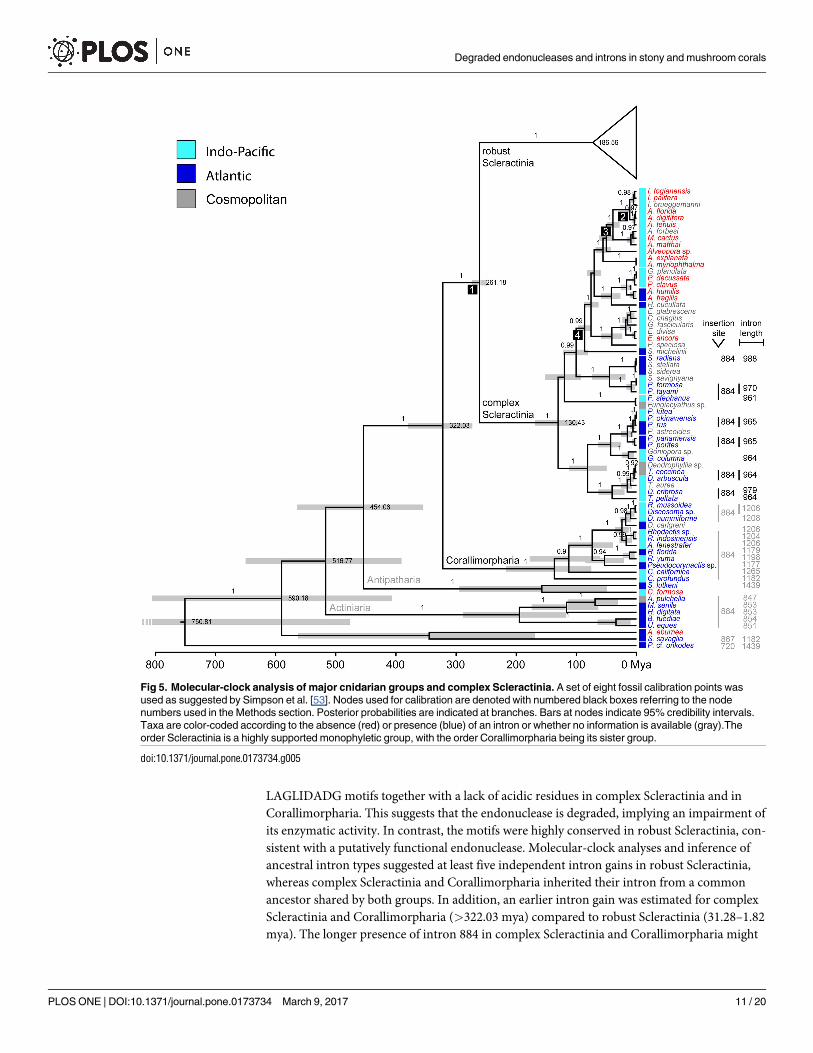
Phylogenetic reconstructions using Bayesian inference (BEAST; relaxed-clock model) showed
strong support for most scleractinian and corallimorpharian genera (Figs 5 and 6). The Scler-
actinia is a highly supported monophyletic group (posterior probability, pp: 1.0), with the Cor-
allimorpharia (pp: 1.0) being its sister group (pp: 1.0). According to the time-calibrated
phylogeny, the split of robust and complex Scleractinia occurred about 260 million years ago
(mya) (248.01–273.19 mya, 95% highest posterior density, HPD) and the split of Corallimor-
pharia/Scleractinia about 320 mya (272.92–379.73 mya; Fig 5). Our results also showed that
intron 720 is found in 22 taxa of robust Scleractinia and in Porifera, whereas intron 884 is pres-
ent in 14 and 11 taxa of complex Scleractinia and Corallimorpharia, respectively, as well as in
the outgroup taxa belonging to the Actiniaria and Antipatharia. Intron 867 was only found in
the outgroup species S. savaglia (Zoantharia).
Within robust Scleractinia, intron 720 was found in four independent clusters and one
lineage. In contrast, taxa containing intron 884 appear to form a single cluster within the
complex Scleractinia. Moreover, the Corallimorpharia and the intron-containing outgroup
taxa all contain intron 884, except for P. cf. onkodes (Porifera), which contains intron 720
(Fig 5). We also found that taxa possessing intron 720 are restricted to the Indo-Pacific
Ocean, while intron 884 occurs in taxa inhabiting both Indo-Pacific and Atlantic/Mediterra-
nean waters (Figs 5 and 6).
Fig 3. Sequence logo of the two LAGLIDADG motifs found in Corallimorpharia, complex Scleractinia and
robust Scleractinia. All homing endonucleases have two LAGLIDADG motifs. Both motifs in complex Scleractinia
are lacking the conserved acidic residues (D or E, highlighted by the dashed boxes) necessary for metal-ion binding
and catalysis. Amino acids are color-coded according to chemical properties (blue, basic; red, acidic; green, polar;
black, hydrophobic). Information content at each position (in bits) is represented by the height of the amino acid and
its estimated variance is shown by the error bar. A score of 4.2 bits corresponds to high conservation, while a score
of 0 corresponds to low conservation.
doi:10.1371/journal.pone.0173734.g003
Degraded endonucleases and introns in stony and mushroom corals
PLOS ONE | DOI:10.1371/journal.pone.0173734 March 9, 2017 9 / 20
The four intron 720 containing clusters were estimated to have a mean crown age of 1.82
(0.09–4.30), 8.52 (4.04–13.82), 26.13 (17.15–35.35), and 31.28 (15.25–48.04) my, with the
respective nodes being highly supported (pp: all 1.0) (Fig 6). The robust Scleractinia has an
approximate age of 185 my, with a high posterior probability (0.85) that the most recent com-
mon ancestor contained no intron. In contrast, the time of acquisition of intron 884 clearly
predates the split between Scleractinia and Corallimorpharia c. 322 mya (Fig 7). However,
intron 884 acquisitions and timing in Corallimorpharia remain elusive.
Discussion
This study attempts to explore the pivotal role of LAGLIDADG motifs in the intron/HEG dis-
tribution in the closely related cnidarian orders Scleractinia and Corallimorpharia. Logo align-
ment of the HEGs, inserted both in intron 720 and 884, showed a degradation of the
Fig 4. Evolutionary rates of homing endonucleases in complex Scleractinia, robust Scleractinia, and Corallimorpharia. The rate
of nucleotide substitutions causing amino acid changes (Ka) is plotted relative to substitutions at silent sites (Ks) for each region and for all
pairwise comparisons. Symbols represent Ka/Ks ratios calculated for HEG. The dotted line indicates the theoretical expectation of neutral
evolution (Ka/Ks = 1). The area below the dotted line represents purifying selection (Ka<Ks).
doi:10.1371/journal.pone.0173734.g004
Degraded endonucleases and introns in stony and mushroom corals
PLOS ONE | DOI:10.1371/journal.pone.0173734 March 9, 2017 10 / 20
LAGLIDADG motifs together with a lack of acidic residues in complex Scleractinia and in
Corallimorpharia. This suggests that the endonuclease is degraded, implying an impairment of
its enzymatic activity. In contrast, the motifs were highly conserved in robust Scleractinia, con-
sistent with a putatively functional endonuclease. Molecular-clock analyses and inference of
ancestral intron types suggested at least five independent intron gains in robust Scleractinia,
whereas complex Scleractinia and Corallimorpharia inherited their intron from a common
ancestor shared by both groups. In addition, an earlier intron gain was estimated for complex
Scleractinia and Corallimorpharia (>322.03 mya) compared to robust Scleractinia (31.28–1.82
mya). The longer presence of intron 884 in complex Scleractinia and Corallimorpharia might
Fig 5. Molecular-clock analysis of major cnidarian groups and complex Scleractinia. A set of eight fossil calibration points was
used as suggested by Simpson et al. [53]. Nodes used for calibration are denoted with numbered black boxes referring to the node
numbers used in the Methods section. Posterior probabilities are indicated at branches. Bars at nodes indicate 95% credibility intervals.
Taxa are color-coded according to the absence (red) or presence (blue) of an intron or whether no information is available (gray).The
order Scleractinia is a highly supported monophyletic group, with the order Corallimorpharia being its sister group.
doi:10.1371/journal.pone.0173734.g005
Degraded endonucleases and introns in stony and mushroom corals
PLOS ONE | DOI:10.1371/journal.pone.0173734 March 9, 2017 11 / 20
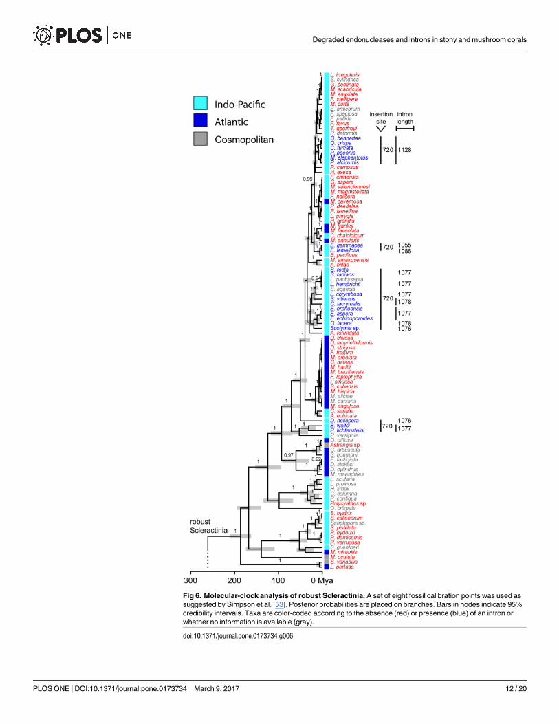
Fig 6. Molecular-clock analysis of robust Scleractinia. A set of eight fossil calibration points was used as
suggested by Simpson et al. [53]. Posterior probabilities are placed on branches. Bars in nodes indicate 95%
credibility intervals. Taxa are color-coded according to the absence (red) or presence (blue) of an intron or
whether no information is available (gray).
doi:10.1371/journal.pone.0173734.g006
Degraded endonucleases and introns in stony and mushroom corals
PLOS ONE | DOI:10.1371/journal.pone.0173734 March 9, 2017 12 / 20
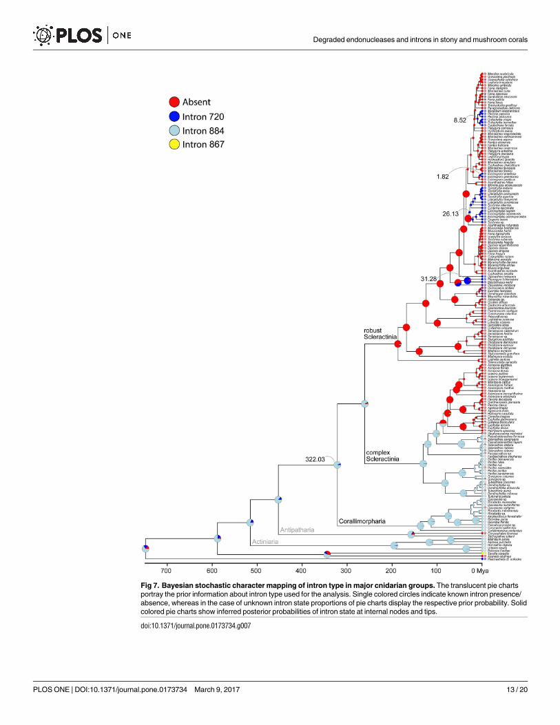
Fig 7. Bayesian stochastic character mapping of intron type in major cnidarian groups. The translucent pie charts
portray the prior information about intron type used for the analysis. Single colored circles indicate known intron presence/
absence, whereas in the case of unknown intron state proportions of pie charts display the respective prior probability. Solid
colored pie charts show inferred posterior probabilities of intron state at internal nodes and tips.
doi:10.1371/journal.pone.0173734.g007
Degraded endonucleases and introns in stony and mushroom corals
PLOS ONE | DOI:10.1371/journal.pone.0173734 March 9, 2017 13 / 20
be related to the degradation of the homing endonuclease gene, consistent with the intron
cycle hypothesis. This is further supported by the higher Ka rates detected for the endonuclease
in complex Scleractinia and Corallimorpharia than in robust Scleractinia, suggesting that the
LAGLIDADG endonuclease is under a relaxed purifying selection in the former two groups,
allowing higher rates of amino acid replacements. Finally, we noticed a clear geographic
restriction of intron 720 to taxa inhabiting the Indo-Pacific Ocean, while taxa containing
intron 884 are cosmopolitan species.
The LAGLIDADG motif as a major driver of group I intron transfer in
Scleractinia and Corallimorpharia
Intron-encoded LAGLIDADG enzymes function primarily as site-specific endonucleases
within the mobility pathway of group I introns. LAGLIDADG proteins can also act as RNA
maturase by binding to and stabilizing the RNA secondary structure of the group I intron and
thus promoting efficient RNA splicing. These two activities are not mutually exclusive, as
some LAGLIDADG enzymes promote both mobility and splicing [4]. It has been proposed
that the maturase activity of LAGLIDADG enzymes could evolve when opportunities to pro-
mote intron mobility are limited or non-existent because all potential target sites within a pop-
ulation are saturated with introns [3,4]. In this case, purifying selection on LAGLIDADG
coding regions would be relaxed, allowing higher rates of non-synonymous substitutions lead-
ing to amino acid replacements. Variants of the motif with RNA maturase activity are not
being degraded and eventually deleted [3,4,60]. Interestingly, we found that all LAGLIDADG
enzymes in complex Scleractinia and Corallimorpharia are highly degraded. In addition, com-
plex Scleractinia lacks the catalytic metal-binding acidic residues (D or E), while they are
poorly conserved in Corallimorpharia. Thus, they might lack endonuclease activity or use a
different mechanism for DNA hydrolysis than canonical enzymes. A further possibility is that
HEs have switched to another function, for instance, acting as RNA maturase [3]. This might
be consistent with the fact that all members of Corallimorpharia and one of the clusters of
complex Scleractinia (i.e., the cluster containing Porites rus) possess intron 884. This indicates
that the HEGs are probably within the extinction phase of the intron/endonuclease life cycle
[15,22]. Thus, we suggest that group I introns and their associated HEGs experience a cycle of
invasion, mutational degradation of its form and function, and deletion from the host genome,
re-establishing the target sequence [61]. This hypothesis is supported by the shorter size of the
homing endonuclease observed in complex Scleractinia and Corallimorpharia in comparison
to robust Scleractinia (Figs 5 and 6). The size of the coding region serves as evidence for HEG
degradation, indicating that shorter HEGs are placed in advanced stages of the intron degrada-
tion cycle as previously shown for sea anemones [23].
The putative degradation of LAGLIDADG motifs has profound implications for intron
mobility. On the one hand, endonucleases with conserved LAGLIDADG motifs are potentially
capable of invading intron-less alleles via horizontal transfer. On the other hand, degraded
endonucleases most likely do no longer function as site-specific endonucleases. In that case, the
intron/HEG transmission depends largely on vertical transmission via mitochondrial maternal
inheritance. We propose that a different functionality of LAGLIDADG motifs accounts for the
two patterns of intron distribution observed in our phylogeny: a probably single origin of
intron 884 in complex Scleractinia and Corallimorpharia, together with a lack of endonuclease
activity, and at least five independent origins of intron 720 in robust Scleractinia due to a func-
tional endonuclease. Given the highly conserved endonuclease, intron 720 in robust Scleracti-
nia has the potential of being transmitted horizontally among populations and across species.
This is consistent with the idea that frequent horizontal transmission is necessary for the long-
Degraded endonucleases and introns in stony and mushroom corals
PLOS ONE | DOI:10.1371/journal.pone.0173734 March 9, 2017 14 / 20
term persistence of homing endonuclease genes [15]. In contrast, the evolutionary history of
intron 884 in complex Scleractinia and Corallimorpharia is largely driven by vertical inheri-
tance, due to the degraded homing endonuclease that limits intron mobility.
Finally, the homology of the HEGs was validated through the comparative analyses of sec-
ondary and tertiary protein structures, which allow the identification of homologous regions
in spite of differences found at the primary sequence level [37,47]. The key principles for such
analyses are: (i) the protein structure is assumed to be more conserved than the protein
sequence, and (ii) only a finite and relatively small (1,000–10,000) number of unique protein
folds occur in nature [62].
More recent intron gains in robust Scleractinia than in complex
Scleractinia and Corallimorpharia
The completion of the intron degradation cycle requires a certain amount of time as shown for
some cnidarians in which a rate of one cycle per 100 my may be sufficient to allow invasion,
cycling, and persistence of HEGs [22]. The evolutionary history of Scleractinia and Coralli-
morpharia covers a time frame sufficiently long to permit intron invasion, fixation, and poste-
rior degradation. However, previous divergence time estimates of Scleractinia differ
considerably depending on the genes used. Time-calibrated phylogenies based on mitochon-
drial 12S and 16S rRNAs, and on the nuclear 28S rRNA suggest an age for the MRCA of
robust/complex Scleractinia of about 415 my [63], whereas another study based on COXI/cyt bgenes and a more sophisticated calibration approach indicates a considerably younger age
(250 mya) [53]. As we used the same calibration strategy as in the latter study, our time esti-
mates are very similar, suggesting that the split of robust and complex Scleractinia occurred c.
260 mya, i.e., shortly before the onset of a massive coral diversification during the mid-Triassic
(250 mya) [64–66]. The long time frame together with conserved target sequences may have
provided repeated opportunities for intron homing in Scleractinia and Corallimorpharia.
Thus, the presence of introns/HEGs in different stages of the intron cycle can be expected. The
high conservation of the potential target sequences for the endonuclease may also be explained
by a low mitochondrial substitution rate of only 0.03% per my [67] for scleractinian mitochon-
drial genomes [32,33,35].
The observed patterns of intron distribution and the ancestral intron type inference indi-
cate at least five independent intron gains in robust Scleractinia during the last c. 31 my. In
contrast, the number and timing of intron 884 acquisitions in complex Scleractinia and Coral-
limorpharia remains elusive. However, considering the intron life cycle framework, we pro-
pose that intron 884 was inserted early in the evolutionary history of cnidarians, and then
rapidly invaded the available intron-less alleles via horizontal transfer. After fixation, the selec-
tive pressure for maintaining endonuclease activity decreased, finally leading to the degrada-
tion of the HEG as previously shown for sea anemones [23]. Given these findings, we
hypothesize that intron 720, which so far has not been reported in complex Scleractinia, may
potentially appear in species of this group. In contrast, intron 884 will probably remain absent
in robust Scleractinia due to its non-functional endonucleases.
Intron occurrence in Scleractinia and Corallimorpharia: Biogeographic
implications
Our phylogeny (Figs 5 and 6) revealed a distinct geographic pattern of intron type occurrence.
Taxa containing intron 720 are only found in the Indo-Pacific Ocean (see also [29]), whereas
intron 884 is found in species inhabiting both the Indo-Pacific and the Atlantic Ocean/Medi-
terranean. Evidence from recent models of coral reef biodiversity dynamics [68] and from the
Degraded endonucleases and introns in stony and mushroom corals
PLOS ONE | DOI:10.1371/journal.pone.0173734 March 9, 2017 15 / 20
coral fossil record [69] suggest that the location of the major marine biodiversity hotspot has
moved across the globe during the last 50 my. During the mid/late Eocene—Oligocene (35–40
mya), the Tethys closure was associated with a narrowing coastal corridor that allowed a faunal
migration of Tethys elements into the Indo-Australian Archipelago [68]. Thus, we propose that
intron 720 was probably transferred from a still unknown donor to robust Scleractinia during
the Tethys closure, isolating the intron-containing taxa from their Atlantic counterparts, and
preventing opportunities for intron 720 transfer. Likewise, the cosmopolitan geographic occur-
rence of taxa containing intron 884 might be explained by the earlier insertion event.
Conclusions
This study represents the first assessment of the role of the LAGLIDADG motifs during the
evolutionary history of group I introns and homing endonuclease genes in the orders Scleracti-
nia and Corallimorpharia. We showed that the LAGLIDADG motifs of complex Scleractinia
and Corallimorpharia are degraded, with a lack of the conserved acidic residues necessary for
hydrolysis of the DNA backbone of the target sequence. In contrast, both motifs are well
defined in robust Scleractinia. Therefore, we suggest that vertical inheritance of group I introns
due to non-functional homing explains intron occurrence in complex Scleractinia and Coralli-
morpharia, whereas both horizontal and vertical transfer gave rise to the present intron distri-
bution in robust Scleractinia. Molecular-clock analyses and ancestral intron type inference
indicated at least five independent intron gains in robust Scleractinia during the last c. 31 my.
Timing and number of intron gains for complex Scleractinia and Corallimorpharia, however,
remain elusive. We further hypothesize that the timing of intron 720 transfer to robust Sclerac-
tinia corresponds to the evolution of the Tethys Sea, isolating the intron-containing taxa from
their Atlantic counterparts. In summary, our data suggest a complex evolutionary history of
introns in Scleractinia and Corallimorpharia mainly driven by endonuclease functionality.
Supporting information
S1 Fig. Intron alignment found in complex Scleractinia using secondary structure ele-
ments. Secondary structures of intron 884 of complex Scleractinia are similar based on the
number of loop and stem regions (P1-P9) of canonical group I intron structures. The only
exception was found in Siderastrea radians intron, which differs in the P5 region. Boxes indi-
cate LAGLIDADG ORF start and stop codons, which are found in frame with the intron
sequence.
(TIF)
S2 Fig. Schematic representation of selected COXI exons of complex Scleractinia (blue
bar), robust Scleractinia (green bar), and Corallimorpharia (red bar). Boxes show insertion
position of intron 720 (orange) and 884 (purple). Symbols (+) and (-) in the insertion sites
boxes indicate intron presence or absence, respectively. Shaded areas represent conserved
sequences. N indicates no available sequences for those sites. Due to space constraints, only a
small number of COXI sequences is portrayed here.
(TIF)
S3 Fig. Evolutionary rates of the COXI gene and of the LAGLIDADG homing endonuclease
gene in robust and complex Scleractinia. The rate of nucleotide substitutions causing amino
acid changes (Ka) is plotted relative to substitutions at silent sites (Ks). Circles represent Ka/Ks
ratios. The dotted line indicates the theoretical expectation of neutral evolution (Ka/Ks = 1).
The area below the dotted line represents purifying selection (Ka<Ks).
(TIF)
Degraded endonucleases and introns in stony and mushroom corals
PLOS ONE | DOI:10.1371/journal.pone.0173734 March 9, 2017 16 / 20
S4 Fig. Alignment of the putative LAGLIDADG motifs found in Scleractinia and Coralli-
morpharia. Both motifs are well conserved in robust Scleractinia, whereas complex Scleracti-
nia and Corallimorpharia displayed degraded motifs.
(TIF)
S1 Table. Taxonomic information, accession numbers, and intron insertion sites of the
intron-containing taxa studied. �Insertion site based on the human COXI gene as a counting
reference. ��The COXI gene of the sponge P. cf. onkodes has two introns in positions 720 and
711. Intron 711 does not contain any ORF. ���Excluded from the molecular-clock analysis due
to missing data for cyt b.
(DOC)
S2 Table. GenBank accession numbers for the COXI and cyt b genes used for the divergence
time estimations. Taxa are sorted alphabetically.
(DOC)
Acknowledgments
The present work is part of the Global Change simulation project ‘Ocean 2100’ at Justus Liebig
University Giessen, initiated by the Colombian-German Center of Excellence in Marine Sci-
ences (CEMarin). Steinar Daae Johansen and an anonymous referee provided constructive
criticism to an earlier version of this study. We thank Patrick Schubert and Jessica Reichert for
providing the Porites rusmaterials from the aquacultures at Justus Liebig University Giessen.
We also acknowledge the stimulating discussions with Catalina Ramırez, Diana Delicado,
Elena Quintanilla, and Pauline Gauffre (Justus Liebig University Giessen).
Author Contributions
Conceptualization: JSC DRE TW JK.
Data curation: JSC DW JB.
Formal analysis: JSC DRE BS TH.
Funding acquisition: TW.
Investigation: JSC.
Methodology: JSC DRE BS TH TW.
Supervision: TW.
Writing – original draft: JSC.
Writing – review & editing: JSC DRE BS TH TW.
References1. Cech TR, Damberger SH, Gutell RR. Representation of the secondary and tertiary structure of group I
introns. Nat Struct Mol Biol. 1994; 1: 273–280.
2. Belfort M, Derbyshire V, Parker MM, Cousineau B, Lambowitz AM. Mobile introns: pathways and pro-
teins. In: Craig NL, Craigie R, Gellert M, Lambowitz AM, editors. Mobile DNA II. Washington D.C.:
ASM Press; 2002. pp. 761–783.
3. Edgell DR, Chalamcharla VR, Belfort M. Learning to live together: mutualism between self-splicing
introns and their hosts. BMC Biol. 2011; 9: 22. doi: 10.1186/1741-7007-9-22 PMID: 21481283
4. Hausner G, Hafez M, Edgell DR. Bacterial group I introns: mobile RNA catalysts. Mob DNA. 2014; 5: 8.
doi: 10.1186/1759-8753-5-8 PMID: 24612670
Degraded endonucleases and introns in stony and mushroom corals
PLOS ONE | DOI:10.1371/journal.pone.0173734 March 9, 2017 17 / 20
5. Stoddard BL. Homing endonucleases from mobile group I introns: discovery to genome engineering.
Mob DNA 2014; 5: 7. doi: 10.1186/1759-8753-5-7 PMID: 24589358
6. Sandegren L, Nord D, Sjoberg BM. SegH and Hef: two novel homing endonucleases whose genes
replace the mobC and mobE genes in several T4-related phages. Nucleic Acids Res. 2005; 33: 6203–
6213. doi: 10.1093/nar/gki932 PMID: 16257983
7. Stoddard BL. Homing endonucleases: from microbial genetic invaders to reagents for targeted DNA
modification. Structure 2011; 19: 7–15. doi: 10.1016/j.str.2010.12.003 PMID: 21220111
8. Cho Y, Palmer JD. Multiple acquisitions via horizontal transfer of a group I intron in the mitochondrial
cox1 gene during evolution of the Araceae family. Mol Biol Evol. 1999; 16: 1155–1165. PMID:
10486971
9. Edgell DR, Stanger MJ, Belfort M. Importance of a single base pair for discrimination between intron-
containing and intronless alleles by endonuclease I-BmoI. Curr Biol. 2003; 13: 973–978. PMID:
12781137
10. Scalley-Kim M, McConnell-Smith A, Stoddard BL. Coevolution of a homing endonuclease and its host
target sequence. J Mol Biol. 2007; 372: 1305–1319. doi: 10.1016/j.jmb.2007.07.052 PMID: 17720189
11. Swithers KS, Senejani AG, Fournier GP, Gogarten JP. Conservation of intron and intein insertion sites:
implications for life histories of parasitic genetic elements. BMC Evol Biol. 2009; 9: 303. doi: 10.1186/
1471-2148-9-303 PMID: 20043855
12. Jacquier A, Dujon B. An intron-encoded protein is active in a gene conversion process that spreads an
intron into a mitochondrial gene. Cell 1985; 41: 383–394. PMID: 3886163
13. Koufopanou V, Goddard MR, Burt A. Adaptation for horizontal transfer in a homing endonuclease. Mol
Biol Evol. 2002; 19: 239–246. PMID: 11861883
14. Burt A, Trivers R. Genes in conflict. The biology of selfish genetic elements. Cambridge: Harvard Uni-
versity Press; 2006.
15. Goddard MR, Burt A. Recurrent invasion and extinction of a selfish gene. Proc Natl Acad Sci. 1999; 96:
13880–13885. PMID: 10570167
16. Lucas P, Otis C, Mercier JP, Turmel M, Lemieux C. Rapid evolution of the DNA-binding site in LAGLI-
DADG homing endonucleases. Nucleic Acids Res. 2001; 29: 960–969. PMID: 11160929
17. Haugen P, Bhattacharya D. The spread of LAGLIDADG homing endonuclease genes in rDNA. Nucleic
Acids Res. 2004; 32: 2049–2057. doi: 10.1093/nar/gkh520 PMID: 15069127
18. Loizos N, Tillier ER, Belfort M. Evolution of mobile group I introns: recognition of intron sequences by an
intron-encoded endonuclease. Proc Natl Acad Sci. 1994; 91: 11983–11987. PMID: 7991569
19. Roman J, Woodson SA. Integration of the Tetrahymena group I intron into bacterial rRNA by reverse
splicing in vivo. Proc Natl Acad Sci. 1998; 95: 2134–2139. PMID: 9482851
20. Roy AC, Wilson GG, Edgell DR. Perpetuating the homing endonuclease life cycle: identification of
mutations that modulate and change I-TevI cleavage preference. Nucleic Acids Res. 2016; 44: 7350–
7359. doi: 10.1093/nar/gkw614 PMID: 27387281
21. Lambowitz AM, Perlman PS. Involvement of aminoacyl-tRNA synthetases and other proteins in group I
and group II intron splicing. Trends Biochem Sci. 1990; 15: 440–444. PMID: 2278103
22. Goddard MR, Leigh J, Roger AJ, Pemberton AJ. Invasion and persistence of a selfish gene in the Cni-
daria. PLoS ONE 2006; 1: e3. doi: 10.1371/journal.pone.0000003 PMID: 17183657
23. Emblem Å, Okkenhaug S, Weiss ES, Denver DR, Karlsen BO, Moum T, et al. Sea anemones possess
dynamic mitogenome structures. Mol Phylogenet Evol. 2014; 75: 184–193. doi: 10.1016/j.ympev.2014.
02.016 PMID: 24613805
24. Wang X, Lavrov DV. Seventeen new complete mtDNA sequences reveal extensive mitochondrial
genome evolution within the Demospongiae. PLoS ONE 2008; 3: e2723. doi: 10.1371/journal.pone.
0002723 PMID: 18628961
25. Szitenberg A, Rot C, Ilan M, Huchon D. Diversity of sponge mitochondrial introns revealed by cox 1
sequences of Tetillidae. BMC Evol Biol. 2010; 10: 288. doi: 10.1186/1471-2148-10-288 PMID:
20849667
26. Huchon D, Szitenberg A, Shefer S, Ilan M, Feldstein T. Mitochondrial group I and group II introns in the
sponge orders Agelasida and Axinellida. BMC Evol Biol. 2015; 15: 278. doi: 10.1186/s12862-015-
0556-1 PMID: 26653218
27. Dellaporta SL, Xu A, Sagasser S, Jakob W, Moreno MA, Buss LW, et al. Mitochondrial genome of Tri-
choplax adhaerens supports Placozoa as the basal lower metazoan phylum. Proc Natl Acad Sci. 2006;
103: 8751–8756. doi: 10.1073/pnas.0602076103 PMID: 16731622
Degraded endonucleases and introns in stony and mushroom corals
PLOS ONE | DOI:10.1371/journal.pone.0173734 March 9, 2017 18 / 20
28. Beagley CT, Okada NA, Wolstenholme DR. Two mitochondrial group I introns in a metazoan, the sea
anemone Metridium senile: one intron contains genes for subunits 1 and 3 of NADH dehydrogenase.
Proc Natl Acad Sci. 1996; 93: 5619–5623. PMID: 8643626
29. Fukami H, Chen CA, Chiou CY, Knowlton N. Novel group I introns encoding a putative homing endonu-
clease in the mitochondrial cox1 gene of scleractinian corals. J Mol Evol. 2007; 64: 591–600. doi: 10.
1007/s00239-006-0279-4 PMID: 17437148
30. Romano SL, Palumbi SR. Evolution of scleractinian corals inferred from molecular systematics. Science
1996; 271: 640–642.
31. Romano S, Cairns SD. Molecular phylogenetic hypotheses for the evolution of scleractinian corals. Bull
Mar Sci. 2000; 67: 1043–1068.
32. Medina M, Collins AG, Takaoka TL, Kuehl JV, Boore JL. Naked corals: skeleton loss in Scleractinia.
Proc Natl Acad Sci. 2006; 103: 9096–9100. doi: 10.1073/pnas.0602444103 PMID: 16754865
33. Kitahara MV, Cairns SD, Stolarski J, Blair D, Miller DJ. A comprehensive phylogenetic analysis of the
Scleractinia (Cnidaria, Anthozoa) based on mitochondrial CO1 sequence data. PLoS ONE 2010; 5:
e11490. doi: 10.1371/journal.pone.0011490 PMID: 20628613
34. Kitahara MV, Lin MF, Forêt S, Huttley G, Miller DJ, Chen CA. ‘The Naked Coral’ hypothesis revisited—
evidence for and against scleractinian monophyly. PLoS ONE 2014; 9: e94774. doi: 10.1371/journal.
pone.0094774 PMID: 24740380
35. Lin MF, Kitahara MV, Luo H, Tracey D, Geller J, Fukami H, et al. Mitochondrial genome rearrangements
in the Scleractinia/Corallimorpharia complex: implications for coral phylogeny. Genome Biol Evol. 2014;
6: 1086–1095. doi: 10.1093/gbe/evu084 PMID: 24769753
36. Haugen P, Simon DM, Bhattacharya D. The natural history of group I introns. Trends Genet. 2005; 21:
111–119. doi: 10.1016/j.tig.2004.12.007 PMID: 15661357
37. Chevalier B, Monat RJ, Stoddard BL. The LAGLIDADG homing endonuclease family. In: Belfort M, edi-
tor. Homing Endonucleases and Inteins. Berlin: Springer-Verlag; 2005. pp. 33–47.
38. Altschul SF, Gish W, Miller W, Myers EW, Lipman DJ. Altschul et al. Basic Local Alignment Search
Tool. J Mol Biol. 1990; 215: 403–410. doi: 10.1016/S0022-2836(05)80360-2 PMID: 2231712
39. Altschul SF, Madden TL, Schaffer AA, Zhang J, Zhang Z, Miller W, et al. Gapped BLAST and PSI-
BLAST: a new generation of protein database search programs. Nucleid Acid Res. 1997; 25: 3389–
3402.
40. Celis JS, Wibberg D, Winkler A, Wilke T, Kalinowski J. Complete mitochondrial genome of the scleracti-
nian coral Porites rus. Mitochondrial DNA Part A 2016; 27: 3695–3696.
41. Anisimova M, Liberles D, Philippe H, Provan J, Pupko T, von Haeseler A. State-of-the-art methodolo-
gies dictate new standards for phylogenetic analysis. BMC Evol Biol. 2013; 13: 161. doi: 10.1186/1471-
2148-13-161 PMID: 23914788
42. Meyer F, Goesmann A, McHardy AC, Bartels D, Bekel T, Clausen J, et al. GenDB—an open source
genome annotation system for prokaryote genomes. Nucleic Acids Res. 2003; 31: 2187–2195. PMID:
12682369
43. Delcher AL, Bratke KA, Powers EC, Salzberg SL. Identifying bacterial genes and endosymbiont DNA
with Glimmer. Bioinformatics 2007; 23: 673–679. doi: 10.1093/bioinformatics/btm009 PMID: 17237039
44. Hall TA. BioEdit: a user-friendly biological sequence alignment editor and analysis program for Windows
95/98/NT. Nucleic Acids Symp Ser. 1999; 41: 95–98.
45. Larkin MA, Blackshields G, Brown NP, Chenna R, Mcgettigan PA, McWilliam H, et al. Clustal W and
Clustal X version 2.0. Bioinformatics 2007; 23: 2947–2948. doi: 10.1093/bioinformatics/btm404 PMID:
17846036
46. den Dunnen JT, Dalgleish R, Maglott DR, Hart RK, Greenblatt MS, Mcgowan-Jordan J, et al. HGVS
Recommendations for the description of sequence variants: 2016 update. Hum Mutat. 2016; 37: 564–
569. doi: 10.1002/humu.22981 PMID: 26931183
47. Kelley LA, Mezulis S, Yates CM, Wass MN, Sternberg MJE. The Phyre2 web portal for protein model-
ing, prediction and analysis. Nat Protoc. 2015; 10: 845–858. doi: 10.1038/nprot.2015.053 PMID:
25950237
48. Kumar S, Stecher G, Tamura K. MEGA7: Molecular Evolutionary Genetics Analysis version 7.0 for big-
ger datasets. Mol Evol Gen. 2016; 33: 1870–1874.
49. Nei M, Gojobori T. Simple methods for estimating the numbers of synonymous and nonsynonymous
nucleotide substitutions. Mol Biol Evol. 1986; 3: 418–426. PMID: 3444411
50. R Core Team. R: a language and environment for statistical computing. R Foundation for Statistical
Computing, Vienna, Austria. URL http://www.R-project.org. 2015.
Degraded endonucleases and introns in stony and mushroom corals
PLOS ONE | DOI:10.1371/journal.pone.0173734 March 9, 2017 19 / 20
51. Folmer O, Black M, Hoeh W, Lutz R, Vrijenhoek R. DNA primers for amplification of mitochondrial cyto-
chrome c oxidase subunit I from diverse metazoan invertebrates. Mol Mar Biol Biotechnol. 1994; 3:
294–299. PMID: 7881515
52. Crooks GE, Hon G, Chandonia JM, Brenner SE. WebLogo: a sequence logo generator. Genome Res.
2004; 14: 1188–1190. doi: 10.1101/gr.849004 PMID: 15173120
53. Simpson C, Kiessling W, Mewis H, Baron-Szabo RC, Muller J. Evolutionary diversification of reef corals:
a comparison of the molecular and fossil records. Evolution 2011; 65: 3274–3284. doi: 10.1111/j.1558-
5646.2011.01365.x PMID: 22023591
54. Drummond AJ, Suchard MA, Xie D, Rambaut A. Bayesian phylogenetics with BEAUti and the BEAST
1.7. Mol Biol Evol. 2012; 29: 1969–1973. doi: 10.1093/molbev/mss075 PMID: 22367748
55. Posada D. jModelTest: phylogenetic model averaging. Mol Biol Evol. 2008; 25: 1253–1256. doi: 10.
1093/molbev/msn083 PMID: 18397919
56. Miller MA, Pfeiffer W, Schwartz T. Creating the CIPRES Science Gateway for inference of large phylo-
genetic trees. New Orleans: Proceedings of the Gateway Computing Environments Workshop (GCE);
2010. pp. 1–8.
57. Rambaut A, Drummond AJ. Tracer v.1.5 http://tree.bio.ed.ac.uk/software/tracer (assessed September,
2016).
58. Huelsenbeck JP, Nielsen R, Bollback JP. Stochastic mapping of morphological characters. Syst Biol.
2003; 52: 131–158. PMID: 12746144
59. Revell LJ. phytools: an R package for phylogenetic comparative biology (and other things). Methods
Ecol Evol. 2012; 3: 217–223.
60. Edgell DR. Selfish DNA: homing endonucleases find a home. Curr Biol. 2009; 19: 115–117.
61. Burt A, Koufopanou V. Homing endonuclease genes: the rise and fall and rise again of a selfish ele-
ment. Curr Opin Genet Dev. 2004; 14: 609–615. doi: 10.1016/j.gde.2004.09.010 PMID: 15531154
62. Koonin EV, Wolf YI, Karev GP. The structure of the protein universe and genome evolution. Nature.
2002; 420: 218–223. doi: 10.1038/nature01256 PMID: 12432406
63. Stolarski J, Kitahara MV, Miller DJ, Cairns SD, Mazur M, Meibom A. The ancient evolutionary origins of
Scleractinia revealed by azooxanthellate corals. BMC Evol Biol. 2011; 11: 316. doi: 10.1186/1471-
2148-11-316 PMID: 22034946
64. Flugel E. Triassic reef patterns. In: Kiessling W, Flugel E, Golonka J, editors. Phanerozoic Reef Pat-
terns. Tulsa: Society for Sedimentary Geology; 2002. pp 391–463.
65. Kiessling W, Roniewicz E, Villier L, Leonide P, Struck U. An early Hettangian coral reef in southern
France: implications for the end-Triassic reef crisis. Palaios 2009; 24: 657–671.
66. Kiessling W, Simpson C. On the potential for ocean acidification to be a general cause of ancient reef
crises. Glob Chang Biol. 2011; 17: 56–67.
67. Fukami H, Knowlton N. Analysis of complete mitochondrial DNA sequences of three members of the
Montastraea annularis coral species complex. Cnidaria, Anthozoa, Scleractinia. Coral Reefs 2005; 24:
410–417.
68. Leprieur F, Descombes P, Gaboriau T, Cowman PF, Parravicini V. Plate tectonics drive tropical reef
biodiversity dynamics. Nat Commun. 2016; 7: 11461. doi: 10.1038/ncomms11461 PMID: 27151103
69. Renema W, Wallace C, Kiessling W, Pandolfi J, Webster J, Bosellini F, et al. Are coral reefs victims of
their own past success? Sci Adv. 2016; 2: e1500850. doi: 10.1126/sciadv.1500850 PMID: 27152330
Degraded endonucleases and introns in stony and mushroom corals
PLOS ONE | DOI:10.1371/journal.pone.0173734 March 9, 2017 20 / 20