Embed Size (px)
Citation preview
Published Ahead of Print 15 November 2013. 2014, 13(1):143. DOI: 10.1128/EC.00158-13. Eukaryotic Cell
Gaston and Andrew J. RogerAnastasios D. Tsaousis, Eleni Gentekaki, Laura Eme, Daniel EukaryotesBlastocystis Species and Other MicrobialCluster Assembly Machinery in Evolution of the Cytosolic Iron-Sulfur
http://ec.asm.org/content/13/1/143Updated information and services can be found at:
These include:
SUPPLEMENTAL MATERIAL Supplemental material
REFERENCEShttp://ec.asm.org/content/13/1/143#ref-list-1at:
This article cites 50 articles, 28 of which can be accessed free
CONTENT ALERTS more»articles cite this article),
Receive: RSS Feeds, eTOCs, free email alerts (when new
http://journals.asm.org/site/misc/reprints.xhtmlInformation about commercial reprint orders: http://journals.asm.org/site/subscriptions/To subscribe to to another ASM Journal go to:
on February 24, 2014 by D
alhousie University
http://ec.asm.org/
Dow
nloaded from
on February 24, 2014 by D
alhousie University
http://ec.asm.org/
Dow
nloaded from
Evolution of the Cytosolic Iron-Sulfur Cluster Assembly Machinery inBlastocystis Species and Other Microbial Eukaryotes
Anastasios D. Tsaousis,a,b Eleni Gentekaki,a Laura Eme,a Daniel Gaston,a Andrew J. Rogera
‹Centre for Comparative Genomics and Evolutionary Bioinformatics, Dalhousie University, Department of Biochemistry and Molecular Biology, Halifax, Nova Scotia,Canadaa; Laboratory of Molecular and Evolutionary Parasitology, School of Biosciences, University of Kent, Canterbury, United Kingdomb
The cytosolic iron/sulfur cluster assembly (CIA) machinery is responsible for the assembly of cytosolic and nuclear iron/sulfurclusters, cofactors that are vital for all living cells. This machinery is uniquely found in eukaryotes and consists of at least eightproteins in opisthokont lineages, such as animals and fungi. We sought to identify and characterize homologues of the CIA sys-tem proteins in the anaerobic stramenopile parasite Blastocystis sp. strain NandII. We identified transcripts encoding six of thecomponents—Cia1, Cia2, MMS19, Nbp35, Nar1, and a putative Tah18 —and showed using immunofluorescence microscopy,immunoelectron microscopy, and subcellular fractionation that the last three of them localized to the cytoplasm of the cell. Wethen used comparative genomic and phylogenetic approaches to investigate the evolutionary history of these proteins. Whilemost Blastocystis homologues branch with their eukaryotic counterparts, the putative Blastocystis Tah18 seems to have a sepa-rate evolutionary origin and therefore possibly a different function. Furthermore, our phylogenomic analyses revealed that alleight CIA components described in opisthokonts originated before the diversification of extant eukaryotic lineages and werelikely already present in the last eukaryotic common ancestor (LECA). The Nbp35, Nar1 Cia1, and Cia2 proteins have been con-served during the subsequent evolutionary diversification of eukaryotes and are present in virtually all extant lineages, whereasthe other CIA proteins have patchy phylogenetic distributions. Cia2 appears to be homologous to SufT, a component of the pro-karyotic sulfur utilization factors (SUF) system, making this the first reported evolutionary link between the CIA and any otherFe/S biogenesis pathway. All of our results suggest that the CIA machinery is an ubiquitous biosynthetic pathway in eukaryotes,but its apparent plasticity in composition raises questions regarding how it functions in nonmodel organisms and how it inter-faces with various iron/sulfur cluster systems (i.e., the iron/sulfur cluster, nitrogen fixation, and/or SUF system) found in eu-karyotic cells.
The assembly of iron/sulfur (Fe/S) clusters is considered one ofthe basic biosynthetic functions in all living cells. Both pro-
karyotic and eukaryotic organisms have at least one pathway ded-icated to Fe/S cluster biosynthesis. In eukaryotes, these clusters areassembled by distinct biosynthetic pathways, which are localizedin different compartments of the cell. The sulfur utilization factors(SUF) system is typically found in plastid-bearing organisms andensures maturation of apoproteins within plastids, whereas thisfunction is performed by the iron/sulfur cluster (ISC) machineryin mitochondria and mitochondrion-related organelles (MROs).The ISC system is indirectly functionally linked with the cytosoliciron/sulfur cluster assembly (CIA) machinery, which is involvedin the maturation of cytosolic and nuclear apoproteins (1). Manyof these Fe/S cluster-bearing proteins are involved in key enzy-matic activities, such as DNA replication and repair, rRNA pro-cessing, and telomere stability (2, 3). Consequently, depletions ofcomponents of the CIA system are lethal in Saccharomyces cerevi-siae, corroborating the significant role of this biosynthetic path-way in the cell (4–8).
Fe/S cluster biosynthetic machineries typically consist of fivemain parts: a desulfurase, an iron donor, an electron transfermechanism, a scaffold, and Fe/S cluster transfer proteins. It isnoteworthy that while the SUF, nitrogen fixation (NIF), and ISCsystems share several homologous components, no evolutionarylink has yet been reported between CIA system proteins and theircounterparts in other Fe/S cluster machineries. Currently, it isknown that the CIA pathway involves at least eight proteins inyeast and humans—Dre2, Tah18, Nbp35, Cfd1, Cia1, Cia2, Nar1,and MMS19 (see Fig. S1 in the supplemental material)—and that
their function is dependent on the mitochondrial ISC machinery(9). The pathway starts when one of the two Fe/S clusters in Dre2(human Ciapin1) is reduced by the diflavin reductase Tah18 (hu-man Ndor1) (10). Nbp35 and Cfd1 then form a scaffold complexfor assembling transiently bound Fe/S clusters (11). In a laterstage, mature Fe/S clusters are transferred to apoproteins via theCia1 (human CIA01) and Nar1 (human IOP1) proteins, a processfacilitated through the formation of a CIA-targeting complex withthe recently discovered chaperone protein MMS19 (also known asMET18 in yeast) (9, 12, 13) and Cia2 (9, 12).
In most microbial eukaryotes, the role of the CIA machinery inthe maturation of the cytosolic and nuclear apoproteins is unclear,as are its functional interactions with other Fe/S cluster matura-tion pathways, in particular, in organisms bearing more than onecytosolic Fe/S system. For example, the microaerophiles Entam-oeba and Mastigamoeba both possess functionally reduced MROsthat do not contain the typical ISC pathway. Instead, their ge-
Received 29 July 2013 Accepted 12 November 2013
Published ahead of print 15 November 2013
Address correspondence to Anastasios D. Tsaousis,[email protected], or Andrew J. Roger, [email protected].
A.D.T., E.G., and L.E. contributed equally to this article.
Supplemental material for this article may be found at http://dx.doi.org/10.1128/EC.00158-13.
Copyright © 2014, American Society for Microbiology. All Rights Reserved.
doi:10.1128/EC.00158-13
January 2014 Volume 13 Number 1 Eukaryotic Cell p. 143–153 ec.asm.org 143
on February 24, 2014 by D
alhousie University
http://ec.asm.org/
Dow
nloaded from

nomes encode NIF system components that were acquired by lat-eral gene transfer (LGT) from epsilonproteobacteria and that lo-calize both in the cytosol and in their organelles (14–16). Anotherexample is found in Blastocystis, an obligate anaerobic parasitethat encodes a functional fused version of the SufC and SufB pro-teins, SufCB, that was also acquired by LGT, in this case, from amethanoarchaeal lineage, and that functions in the cytosol of theorganism (17). The presence of both this SUF protein and com-ponents of the CIA Fe/S cluster biosynthetic pathway in Blastocys-tis (18) raises questions regarding their respective cytosolic roles.
In this study, we focused on the CIA system in Blastocystis sp.We show that this organism expresses homologues of six ofthe CIA system proteins—Nbp35, Nar1, Cia1, Cia2, Tah18, andMMS19 —and illustrate by immunomicroscopy and subcellularfractionation that three of them are localized in the cytosol. Usingcomparative genomic and phylogenetic approaches, we then in-vestigated the distribution and evolutionary histories of the CIAcomponents among eukaryotes.
MATERIALS AND METHODSBlastocystis culture and maintenance. Blastocystis sp. strain NandII cul-tures were obtained from the American Type Culture Collection (ATCC)and maintained in Locke’s medium egg slants at 35.6°C in an anaerobicchamber.
Protein extraction and Western blotting. Blastocystis cells were pel-leted at 800 � g for 10 min and then washed twice in 1� Locke’s solutionbefore being suspended in 0.15 M NaCl. Cells were disrupted using ultra-sonication (for 30 s at 30-s intervals, 6 cycles) (17, 19) on ice. Followingdisruption, protease inhibitor cocktail (10 �l; Sigma) was added to thesample, and the mixture was then centrifuged at 10,000 � g for 10 min at4°C. Afterwards, the supernatant was collected and 10 �l of ice-cold nu-clease buffer (20 mM Tris-HCl, pH 8.8, 2 mM CaCl2) along with 10 �l ofprotease inhibitor cocktail was added. Thirty microliters of a DNase-RNase mix (50 mM MgCl2, 0.5 M Tris-HCl, pH 7.0) was added and themixture was incubated on ice for 3 min. Subsequently, 10 �l of 3% SDS–10% mercaptoethanol was added and the mix was passed through a finesyringe. Samples were then stored at �20°C in NuPAGE lithium dodecylsulfate (LDS) sample buffer along with 10� sample reducing agent (In-vitrogen). Depending on the amount of protein, 5 to 20 �l of the super-natant (�10%) was analyzed using a polyacrylamide minigel.
Different cell fractions were isolated following procedures previouslydescribed (19, 20). Blastocystis cells (well-grown in 20 egg-slant tubes for 5days) were harvested by centrifugation at 1,200 � g for 10 min at 4°C. Cellswere resuspended in Locke’s solution (pH 7.4) and pelleted again at thesame speed for the same duration. Cells were then broken with 40 strokesin a 10-ml Potter-Elvehjem tissue homogenizer at 4°C in isotonic buffer(200 mM sucrose, pH 7.2, 30 mM phosphate, 15 mM �-mercaptoethanol,30 mM NaCl, 0.6 mM CaCl2, 0.6 mM KCl). Broken cells were then dilutedwith isotonic buffer and then centrifuged at 700 � g for 10 min using aSorvall RC-2B centrifuge to remove unbroken cells. The supernatant wascollected and centrifuged at 5,000 � g for 20 min to pellet the large gran-ular fraction (LGF), where MROs are found (see references 21 and 22).The LGF was resuspended (washed) in isotonic buffer and pelleted asdescribed above. Finally, all fractions were stored at �20°C in NuPAGELDS sample buffer along with 10� sample reducing agent (Invitrogen).Depending on the amount of protein, 5 to 20 �l of the supernatant wasanalyzed using a polyacrylamide minigel.
For Western blot analysis, proteins were transferred to immunoblotpolyvinylidene difluoride (PVDF) membranes (Bio-Rad), visualized byPonceau staining, and then blocked for an hour with 5% skimmed milk inTBS (10 mM Tris, 0.2 M NaCl, 0.2% bovine serum albumin [BSA], pH8.1)– 0.1% Tween. Membranes were washed with 0.5% skimmed milk inTBS– 0.1% Tween three times for 10 min each time. The primary antibod-ies (see below) were diluted in 1% skimmed milk in TBS– 0.1% Tween
(anti-Blastocystis SufCB, 1:500; anti-Saccharomyces Nbp35 [6], 1:500; an-ti-Saccharomyces Nar1 [8], 1:300; anti-Saccharomyces Tah18 [10], 1:200)and applied to the membrane overnight at 4°C. Membranes were washedas described above and incubated with the secondary antibody diluted in1% skimmed milk in TBS– 0.1% Tween. Membranes were washed in TBSthree times for 10 min each time and incubated with enhanced chemilu-minescence (ECL) reagent (GE Healthcare), and fluorescence was moni-tored by autoradiography.
Immunolocalization of CIA components in Blastocystis. Blastocystiscells were fixed with 4% paraformaldehyde at 37°C for 15 min, followed bythree 10-min washes with 1� TBS. Fixed cells were permeabilized withice-cold acetone and washed three times for 10 min each time with 1�TBS– 0.1% Triton. Fixed cells were incubated for 30 min with a blockingsolution of 5% skimmed milk powder and 1� TBS– 0.1% Triton solution(wt/vol). They were then rinsed with 0.5% milk–1� TBS– 0.1% Tritonsolution for 30 min. The cells were then incubated with a dilution of theantiserum (anti-SufCB, 1:200; anti-Nbp35, 1:100; anti-Nar1, 1:100; anti-Tah18, 1:50) in 1% milk–TBS– 0.1% Triton solution overnight at 4°C.Three different dilutions of each antiserum were tested to determine op-timal conditions. After three rinses in 1% milk–TBS– 0.1% Triton, theslides were incubated with fluorescent dye (Alexa 488 green and Alexa 594red)-labeled secondary antibodies at a dilution of 1:200. Finally, the slideswere incubated with DRAQ5 (Cell Signaling Technology) stain for 5 min(dilution, 1:1,000) and washed three times for 1 min each time with 1�TBS. Coverslips were mounted with antifade mounting medium(Vectashield) and observed under a laser scanning confocal microscope(Zeiss LSM 510 Meta) using a �100 oil immersion lens.
For the immunolocalization experiments using transmission electronmicroscopy, Blastocystis NandII cells were fixed and manipulated usingthe protocols described previously (17). The prepared grids were incu-bated in sodium borohydride (1 mg/ml) for 10 min, followed by a 10-minincubation in glycine buffer (30 mM glycine in 0.1 M borate buffer, pH9.6). The grids were then incubated in blocking solution (TBS buffer with1% skim milk, 1% BSA) for 45 min, followed by a quick rinse with TBSbuffer. The grids were then incubated overnight with a dilution of theantiserum (anti-Nbp35, 1:20; anti-Nar1, 1:15; anti-Tah18, 1:5) in TBSbuffer at 4°C. Three different dilutions of each antiserum were tested todetermine the optimal conditions. The grids were then rinsed three times(15 min each) in washing buffer (10 mM Tris, 0.3 M NaCl, 0.1% BSA, pH8.1), followed by an hour incubation with secondary antirabbit antibodyconjugated with 10-nm gold particles (Sigma) diluted in TBS buffer. Thegrids were then rinsed three times (15 min each) in washing buffer, incu-bated for 15 min in 2.5% glutaraldehyde, rinsed three times (3 min each)in distilled water, and stained with 2% uranyl acetate and lead citrate.Samples were viewed with a JEOL JEM 1230 transmission electron micro-scope to determine quality before proceeding with immunolabeling.
Transcriptomic data obtained by 454 pyrosequencing of cDNAfrom Blastocystis sp. NandII. Total RNA from Blastocystis cells was iso-lated using the TRIzol reagent according to the manufacturer’s specifica-tions with the following modification: following separation of the organicphase, the supernatant was collected and underwent a second round ofTRIzol extraction. cDNA was constructed by Vertis Biotechnologies AG(Germany), and 454 pyrosequencing was performed by Genome Quebecin a 4SLX titanium platform. The Mira assembly program (version 3.0)(18) was used to assemble the reads into contigs. A BLAST database wascreated using the resulting Mira contigs. The CIA proteins of the Blasto-cystis hominis S7 strain (23) and Saccharomyces cerevisiae were used asseeds to perform a local tblastn search against the Blastocystis NandIIcontig database and extract the corresponding homologues.
Database searches and data set assembly. Prokaryotic and eukaryotichomologues of the CIA system protein sequences were retrieved fromGenBank using blastp and tblastn searches with S. cerevisiae and Homosapiens as the initial seed query sequences (24). Identical and highly sim-ilar sequences were removed. Additional databases that were searched foreukaryotic homologues included the Joint Genome Institute (JGI) data-
Tsaousis et al.
144 ec.asm.org Eukaryotic Cell
on February 24, 2014 by D
alhousie University
http://ec.asm.org/
Dow
nloaded from
base (http://www.jgi.doe.gov), the Broad Institute database (http://www.broadinstitute.org), the Cyanidioschyzon merolae Genome Project data-base (http://merolae.biol.s.u-tokyo.ac.jp/), GiardiaDB (http://giardiadb.org), AmoebaDB (http://amoebadb.org), NemaGENETAG (http://elegans.imbb.forth.gr/nemagenetag), and the Wellcome Trust Sanger Institutedatabase (http://www.sanger.ac.uk). For a given protein, the absence of ahomologue in a taxon was further verified by employing tblastn on thecorresponding genomes using multiple queries as seeds. For organismswhere the tblastn search did not result in any hits to a particular proteinfamily, a Hidden Markov Model (HMM) profile of the alignment wasbuilt and used to search the predicted proteomes using the hmmbuild andhmmsearch programs of the HMMER package, version 3.0 (http://hmmer.janelia.org).
Protein domain identification. Protein domains were identified us-ing the SMART program (http://smart.embl-heidelberg.de/) (25) and byperforming HMMER searches against the PFAM (version 26.0) database(http://pfam.sanger.ac.uk) (21).
Multiple-sequence alignment and phylogenetic analysis. Protein se-quences were aligned by use of the MAFFT program (version 6.903b)(22). Furthermore, the alignments were inspected by eye to detect cases ofobviously misaligned regions. Subsequently, the alignments were maskedto remove regions of ambiguous alignment using the Block Mapping andGathering with Entropy (BMGE) program (version 1.1) with default pa-rameters (26).
After trimming, the final alignments contained 59 taxa and 225 sitesfor the Cia1 protein, 78 taxa and 96 sites for the Dre2 protein, 87 taxa and55 sites for the MMS19 protein, 81 taxa and 233 sites for the Nar1 protein,95 taxa and 191 sites for the Nbp35/Cfd1 proteins, 110 taxa and 157 sitesfor the Tah18 protein, and 60 taxa and 98 sites for the Cia2 protein.Phylogenetic trees were constructed for each individual protein of the CIAmachinery. Maximum likelihood (ML) trees were computed using theRAxML program (version 7.2.8) (27) and the Le and Gascuel (LG) aminoacid substitution model (28). To account for rate heterogeneity acrossamino acid sites, the gamma option was also implemented. For each pro-tein data set, bootstrap support was assessed from 100 bootstrap replicatesthat were subsequently mapped onto the best-scoring ML tree. Phylog-enies for the CIA components that displayed interesting patterns (e.g.,Nbp35/Cfd1 and Tah18) are shown in Fig. 2 and 3, whereas those with lessnotable evolutionary histories are included as figures in the supplementalmaterial.
Topology testing. In the case of Tah18, we tested whether variousphylogenetic hypotheses could be significantly rejected by the data. Wetested two alternative phylogenetic placements of putative Blastocystis sp.Tah18 sequences: (i) grouping with the four Blastocystis sequences of thepyruvate-NADPH oxidoreductase (PNO) clade (sequences indicated by awhite square in Fig. 2) (case 1) or (ii) grouping as a monophyletic groupwith other stramenopile Tah18 orthologues (sequences indicated by ablack square in Fig. 2) (case 2). We used the approximately unbiased (AU)test implemented in the CONSEL program (29). Since this test requires alarge sample of good trees, in addition to the test topologies, to accuratelyestimate P values (30), 500 bootstrap trees were included in the analyses.The maximum likelihood tree given a specific constraint (i.e., corre-sponding to a phylogenetic hypothesis) was obtained using the �gRAxML option with all other parameters set, as previously described. Case1 could not be rejected (P � 0.228, likelihood � �51,718.8), whereas case2 was rejected (P � 0.036, likelihood � �51,781.3).
Nucleotide sequence accession numbers. The sequences of the Blas-tocystis sp. NandII transcripts are deposited in GenBank with the acces-sion numbers KF438229 to KF438233 and KF841440, and the accessionnumbers of homologous sequences are provided in the supplemental dataset (Table S1) in the supplemental material.
RESULTS AND DISCUSSIONCharacterization/localization of components of BlastocystisCIA machinery. In a previous study, we showed that three differ-
ent Fe/S cluster biosynthetic machineries were likely present in theanaerobic parasite Blastocystis (17). We localized and functionallycharacterized the mitochondrial Fe/S cluster (ISC) machinery andthe cytosolic SUF machinery, but only limited data regarding thepresence of the CIA machinery were available. Here, we reporthomologues of six CIA proteins (Nbp35, Nar1, Cia1, Cia2, Tah18,and MMS19) in our transcriptomic data. The homology of threeof these proteins was further confirmed by the identification ofconserved features/motifs shared with better-characterized eu-karyotic homologues (e.g., human, yeast) (see Fig. S2 to S4 in thesupplemental material).
We employed various methods to assess the localization of CIAcomponents in Blastocystis. Using heterologous antibodies raisedagainst the yeast proteins Nbp35 (6), Nar1 (8), and Tah18 (10), wedemonstrated their specificity against the Blastocystis homologuesin Western blots of total protein extracts (see Fig. S5 in the sup-plemental material). To further confirm the localization of theseproteins, we performed subcellular fractionations employing atwo-step centrifugation method followed by a Western blot assay.We used Nbp35, Nar1, and Tah18 antibodies against the corre-sponding proteins obtained from the fractions. In all cases, theantibodies were localized exclusively in the cytosolic fraction ofthe cell extracts (see Fig. S5 in the supplemental material).
Immunofluorescence microscopy demonstrated that the Blas-tocystis Nbp35, Nar1, and Tah18 proteins localized in the cytosolof the cell; these proteins colocalized with the cytosolic proteinSufCB as well (17) (Fig. 1), and they did not colocalize with Mito-Tracker, a dye that labels the MROs of the parasite (see Fig. S6 inthe supplemental material). Moreover, immunogold transmis-sion electron microscopy using the same antibodies against theCIA proteins revealed an abundance of gold particles in the cyto-sol and their virtual absence from the MROs, vacuole, and nucleus(Fig. 1d and e; see Fig. S7 to S9 in the supplemental material). Asimilar immunogold labeling pattern was demonstrated for thefused cytosolic SufCB protein of the Blastocystis sp. (Fig. 1e) (17).
This work, along with a previous study (17), demonstrated thatkey components of the ISC and CIA systems are encoded by genesin the Blastocystis sp. NandII genome and that they localize in thisorganism’s MROs and cytosol, respectively. However, the Blasto-cystis sp. also has a cytosolic SUF system (17), a machinery neverbefore described in the eukaryotic cytoplasmic compartment (1).With these two cytosolic Fe/S cluster biogenesis systems, the Blas-tocystis sp. resembles Entamoeba and Mastigamoeba, both ofwhich possess novel cytosolic NIF systems (16, 31), in addition toCIA components (presumed to be cytosolic in these organisms)(Table 1). For these organisms, it is unclear how Fe/S cluster bio-genesis is partitioned between the two cytosolic systems andwhether or not they function independently or in a coordinatedfashion. Currently, it is not possible to genetically manipulatesome of these organisms, making it difficult to study these systemsin more detail.
Distribution and evolutionary history of the CIA machineryhomologues in other microbial eukaryotes. Although most ofthe eight CIA system components present in yeast are essential forits viability (9), our survey of Blastocystis sp. data (23, 32) allowedthe identification of only five (potentially six; see the discussion ofTah18 below) of these proteins. Consequently, it is unclear howthis pathway functions in Blastocystis and whether other eu-karyotes also lack some of the components. To address this ques-
CIA Machinery in Microbial Eukaryotes
January 2014 Volume 13 Number 1 ec.asm.org 145
on February 24, 2014 by D
alhousie University
http://ec.asm.org/
Dow
nloaded from

tion, we carried out a phylogenomic analysis of each individualcomponent.
(i) Dre2. Dre2 is essential for the assembly of virtually all cyto-solic and nuclear target Fe/S proteins in yeast and localizes mostlyin the cytosol and partially in the mitochondrial intermembranespace (IMS) (33). The latter finding seems to suggest that Dre2acts before the rest of the CIA components, an observation furthersupported by additional experimental evidence (10). Interest-ingly, Dre2 itself is a Fe/S cluster-containing protein whose mat-uration seems to occur independently of the CIA system (10). Thissuggests a putative functional dependence of Dre2 (and, thus, theCIA system) on another Fe/S cluster assembly pathway.
Dre2 had a highly conserved C terminus corresponding to aeukaryote-specific Ciapin1 domain that contained two Fe/S clus-ter-binding motifs, CX2CXC and CX2CX7CX2C, while the N ter-minus is more divergent (see Fig. S10 in the supplemental mate-rial). In some chlorophytes/embryophytes and metazoans, the Nterminus contains a methyltransferase domain (see Fig. S10 in thesupplemental material), supporting the recently proposed hy-pothesis that this part of Dre2 is homologous to the S-adenosyl-methionine (SAM) methyltransferase protein family (34). Al-though the interaction of Dre2 and Tah18 has been shown to beindispensable for yeast survival, a few parasitic taxa (includingBlastocystis, Entamoeba, Giardia, and Trichomonas) appear to belacking a Dre2 homologue (Table 1). Apart from these absences(presumably because of secondary loss), the phylogenetic analysiswas consistent with the vertical inheritance of Dre2 from the lasteukaryotic common ancestor (LECA) within eukaryotes (see Fig.S11 in the supplemental material). The taxonomic distribution ofthe SAM domain seems to suggest that Dre2 in LECA had a SAM-Ciapin1 domain composition. It is possible that eukaryotic se-quences in which a SAM domain was not detected have evolvedbeyond recognition, since most of them have an N-terminal se-quence that aligns well with (and thus seems to be homologous to)the SAM domain of green plant/metazoan sequences.
(ii) Tah18. Tah18 is a reductase with binding motifs for flavinmononucleotide (FMN), flavin adenine dinucleotide (FAD), andNAD cofactors (Fig. 2). Tah18 forms a complex with Dre2 thatfunctions as an electron transfer chain by transferring two reduc-ing equivalents from NADPH to Dre2 (10, 34, 35). Experimentalevidence suggests that the Tah18/Dre2 complex disassociates un-der oxidative stress conditions, and as a result, Tah18 retargets tomitochondria in yeast (35), leading to the speculation that Tah18promotes apoptosis under oxygen stress (34). Phylogenetic anal-yses show that Tah18 belongs to a large multiprotein family, mostmembers of which share the canonical FMN-FAD-NAD domainorganization and are involved in redox reactions. The pyruvate-NADPH oxidoreductase (PNO) and NADPH cytochrome P450reductase (CPR) subfamilies are the closest homologues of Tah18(Fig. 2). Our analyses show that the putative Blastocystis Tah18sequences (Fig. 2) do not group with the rest of the stramenopileTah18 homologues (Fig. 2, black square) but instead branch withthe PNO sequences. While most eukaryotes possess an orthologueof Tah18, the protein seems to be absent in Entamoeba (Table 1)and potentially in Giardia, whose homologues are unclearly re-lated to Tah18 or CPR (Fig. 2). There seems to be a similar phylo-genetic profile of the presence/absence of canonical Tah18 andDre2 (Table 1) that is consistent with them forming a functionalmodule in the CIA system.
(iii) Nbp35/Cfd1. Nbp35 and Cfd1 are homologous Mrp-likeproteins and members of the P-loop NTPase family, which alsoincludes the more distantly related mitochondrial Fe/S apoproteinIND1 (36). Both Nbp35 and Cfd1 proteins possess a Walker A boxfor ATP hydrolysis and two conserved C-terminal cysteines thatserve as the binding sites of Fe/S clusters (11) (see Fig. S4 in thesupplemental material). Nbp35 has an N-terminal ferredoxin-likedomain that Cfd1 lacks (7) (see Fig. S4 in the supplemental mate-rial). While Nbp35 is universally distributed among eukaryotes,Cfd1 has a by far more patchy distribution: it is found only inopisthokonts, amoebozoa, discicristate excavates, and the crypto-phyte Guillardia theta (Table 1). These results are in contrast tothose presented in a recent publication (37) claiming the universal
FIG 1 Immunolocalization of Nbp35, Nar1, and Tah18 in the Blastocystis sp.(a) Cellular localization of Nbp35 and SufCB in Blastocystis cells. (i) Rabbitanti-yeast Nbp35 antibody (1:200) detects the Blastocystis Nbp35 protein; (ii)localization of the Blastocystis SufCB antibody; (iii) DRAQ5 staining of thenuclei of Blastocystis; (iv) differential interference contrast (DIC) image of thecells used for immunofluorescence; (v) overlapping of the previous imagesshowing the localization pattern of these proteins. (b) Cellular localization ofcomponents of Nar1 and SufCB in Blastocystis cells. (i) Rabbit anti-yeast Nar1antibody (1:100) detects the Blastocystis Nbp35 protein; (ii) localization of theBlastocystis SufCB antibody; (iii) DRAQ5 staining of the nuclei of Blastocystis;(iv) differential interference contrast image of the cells used for immunofluo-rescence; (v) overlapping of the previous images showing the localization pat-tern of these proteins. (c) Cellular localization of components of Tah18 andSufCB in Blastocystis cells. (i) Rabbit anti-yeast Tah18 antibody (1:100) detectsthe Blastocystis Nbp35 protein; (ii) localization of the Blastocystis SufCB anti-body; (iii) DRAQ5 staining of the nuclei of Blastocystis; (iv) differential inter-ference contrast image of the cells used for immunofluorescence; (v) overlap-ping of the previous images showing the localization pattern of these proteins.(d) Immunogold microscopy image demonstrating the localization pattern ofthe anti-Nbp35 antibody. A higher-magnification image is shown in Fig. S7 inthe supplemental material. (e) The densities of immunogold particle labelingin different compartments of Blastocystis cells suggest that Nbp35, Nar1, andTah18, along with the previously published SufCB (18) proteins, are mainlylocalized in the cytosol of the parasite.
Tsaousis et al.
146 ec.asm.org Eukaryotic Cell
on February 24, 2014 by D
alhousie University
http://ec.asm.org/
Dow
nloaded from

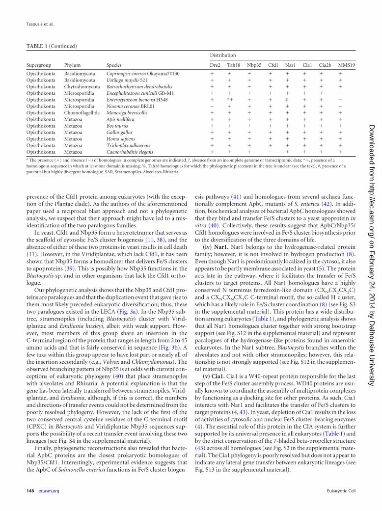
TABLE 1 Distribution of homologues of the CIA machinery among publicly available genomes or expressed sequence tag data from eukaryote taxaa
Supergroup Phylum Species
Distribution
Dre2 Tah18 Nbp35 Cfd1 Nar1 Cia1 Cia2b MMS19
SAR Apicomplexa Babesia bovis � *� � � � � � �SAR Apicomplexa Cryptosporidium hominis � � � � � � � �SAR Apicomplexa Cryptosporidium muris RN66 � � � � � � � �SAR Apicomplexa Cryptosporidium parvum Iowa II � � � � � � � �SAR Apicomplexa Plasmodium berghei � *� � � � � � #SAR Apicomplexa Plasmodium chabaudi � *� � � � � � #SAR Apicomplexa Plasmodium falciparum � *� � � � � � #SAR Apicomplexa Plasmodium knowlesi strain H � *� � � � � � #SAR Apicomplexa Plasmodium vivax � *� � � # � � #SAR Apicomplexa Theileria annulata strain Ankara � *� � � � � � �SAR Apicomplexa Theileria parva � *� � � � � � �SAR Apicomplexa Toxoplasma gondii � � � � � � � #SAR Ciliophora Paramecium tetraurelia � � � � � � � #SAR Ciliophora Tetrahymena thermophila SB210 � � � � � � � #SAR Perkinsida Perkinsus marinus ATCC 50983 � *� � � � � � #SAR Stramenopila Aureococcus anophagefferens � � � � � � � �SAR Stramenopila Hyaloperonospora � � � � � � � �SAR Stramenopila Phaeodactylum tricornutum CCAP 1055/1 � � � � � � � �SAR Stramenopila Phytophthora infestans T30-4 � � � � � � � �SAR Stramenopila Phytophthora ramorum � � � � � � � �SAR Stramenopila Phytophthora sojae � � � � � � � �SAR Stramenopila Saprolegnia parasitica CBS 223.65 � � � � � � � �SAR Stramenopila Nannochloropsis gaditana CCMP526 � � � � � � � �SAR Stramenopila Thalassiosira pseudonana CCMP1335 � � � � � � � �SAR Stramenopila Ectocarpus siliculosus � � � � � � � �SAR Stramenopila Blastocystis sp. strain NandII � % � � � � � #SAR Stramenopila Blastocystis sp. strain Sub 7 � % � � � � � #
SAR Rhizaria Bigelowiella natans � � � � � � � �Hyptophyceae Emiliania huxleyi � � � � # � � �Cryptophyta Guillardia theta � � � � � � � �
Amoebozoa Dictyostelium purpureum � � � � � � � �Amoebozoa Entamoeba dispar SAW760 � � � � � � � �Amoebozoa Entamoeba histolytica HM-1:IMSS � � � � � � � �Amoebozoa Mastigamoeba balamuthi ? ? � � � � ? ?Amoebozoa Polysphondylium pallidum PN500 � � � � � � � �Archaeplastida Viridiplantae Chlamydomonas reinhardtii � � � � � � � �Archaeplastida Viridiplantae Chlorella variabilis � � � � � � � �Archaeplastida Viridiplantae Coccomyxa subellipsoidea C-169 � � � � � � � �Archaeplastida Viridiplantae Micromonas pusilla � � � � � � � �Archaeplastida Viridiplantae Micromonas sp. � � � � � � � �Archaeplastida Viridiplantae Ostreococcus lucimarinus � � � � � � � �Archaeplastida Viridiplantae Ostreococcus tauri � � � � � � � �Archaeplastida Viridiplantae Volvox carteri � � � � � � � �Archaeplastida Rhodophyta Cyanidioschyzon merolae � � � � � � � �Archaeplastida Viridiplantae Arabidopsis thaliana � � � � � � � �Archaeplastida Viridiplantae Brachypodium distachyon � � � � � � � �Archaeplastida Viridiplantae Glycine max � � � � � � � �Excavata Euglenozoa Leishmania braziliensis MHOM/BR/75/M2904 � � � � � � � #Excavata Euglenozoa Leishmania infantum JPCM5 � � � � � � � #Excavata Euglenozoa Leishmania major strain Friedlin � � � � � � � #Excavata Euglenozoa Trypanosoma brucei � � � � � � � #Excavata Euglenozoa Trypanosoma cruzi � � � � � � � #Excavata Fornicata Giardia intestinalis � % � � � � � �Excavata Parabasalia Trichomonas vaginalis G3 � � � � � � � �Excavata Heterolobosea Naegleria gruberi � � � � � � � #Opisthokonta Ascomycota Saccharomyces cerevisiae � � � � � � � �Opisthokonta Ascomycota Schizosaccharomyces pombe � � � � � � � �Opisthokonta Ascomycota Gibberella zeae PH-1 � � � � � � � �
(Continued on following page)
CIA Machinery in Microbial Eukaryotes
January 2014 Volume 13 Number 1 ec.asm.org 147
on February 24, 2014 by D
alhousie University
http://ec.asm.org/
Dow
nloaded from
presence of the Cfd1 protein among eukaryotes (with the excep-tion of the Plantae clade). As the authors of the aforementionedpaper used a reciprocal blast approach and not a phylogeneticanalysis, we suspect that their approach might have led to a mis-identification of the two paralogous families.
In yeast, Cfd1 and Nbp35 form a heterotetramer that serves asthe scaffold of cytosolic Fe/S cluster biogenesis (11, 38), and theabsence of either of these two proteins in yeast results in cell death(11). However, in the Viridiplantae, which lack Cfd1, it has beenshown that Nbp35 forms a homodimer that delivers Fe/S clustersto apoproteins (39). This is possibly how Nbp35 functions in theBlastocystis sp. and in other organisms that lack the Cfd1 ortho-logue.
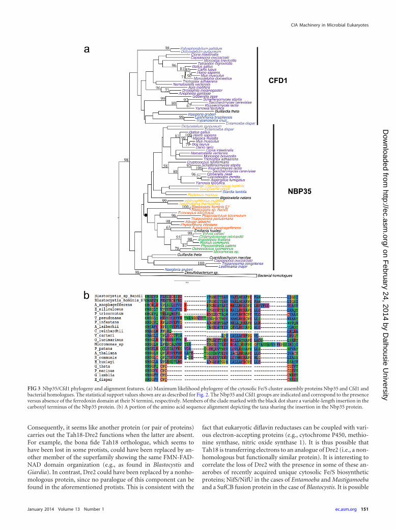
Our phylogenetic analysis shows that the Nbp35 and Cfd1 pro-teins are paralogues and that the duplication event that gave rise tothem most likely preceded eukaryotic diversification; thus, thesetwo paralogues existed in the LECA (Fig. 3a). In the Nbp35 sub-tree, stramenopiles (including Blastocystis) cluster with Virid-iplantae and Emiliania huxleyi, albeit with weak support. How-ever, most members of this group share an insertion in theC-terminal region of the protein that ranges in length from 2 to 45amino acids and that is fairly conserved in sequence (Fig. 3b). Afew taxa within this group appear to have lost part or nearly all ofthe insertion secondarily (e.g., Volvox and Chlamydomonas). Theobserved branching pattern of Nbp35 is at odds with current con-ceptions of eukaryotic phylogeny (40) that place stramenopileswith alveolates and Rhizaria. A potential explanation is that thegene has been laterally transferred between stramenopiles, Virid-iplantae, and Emiliania, although, if this is correct, the numbersand directions of transfer events could not be determined from thepoorly resolved phylogeny. However, the lack of the first of thetwo conserved central cysteine residues of the C-terminal motif(CPXC) in Blastocystis and Viridiplantae Nbp35 sequences sup-ports the possibility of a recent transfer event involving these twolineages (see Fig. S4 in the supplemental material).
Finally, phylogenetic reconstructions also revealed that bacte-rial ApbC proteins are the closest prokaryotic homologues ofNbp35/Cfd1. Interestingly, experimental evidence suggests thatthe ApbC of Salmonella enterica functions in Fe/S cluster biogen-
esis pathways (41) and homologues from several archaea func-tionally complement ApbC mutants of S. enterica (42). In addi-tion, biochemical analyses of bacterial ApbC homologues showedthat they bind and transfer Fe/S clusters to a yeast apoprotein invitro (40). Collectively, these results suggest that ApbC/Nbp35/Cfd1 homologues were involved in Fe/S cluster biosynthesis priorto the diversification of the three domains of life.
(iv) Nar1. Nar1 belongs to the hydrogenase-related proteinfamily; however, it is not involved in hydrogen production (8).Even though Nar1 is predominantly localized in the cytosol, it alsoappears to be partly membrane associated in yeast (5). The proteinacts late in the pathway, where it facilitates the transfer of Fe/Sclusters to target proteins. All Nar1 homologues have a highlyconserved N terminus ferredoxin-like domain (CXNCX2CX2C)and a CXNCXNCX3C C-terminal motif, the so-called H cluster,which has a likely role in Fe/S cluster coordination (8) (see Fig. S3in the supplemental material). This protein has a wide distribu-tion among eukaryotes (Table 1), and phylogenetic analysis showsthat all Nar1 homologues cluster together with strong bootstrapsupport (see Fig. S12 in the supplemental material) and representparalogues of the hydrogenase-like proteins found in anaerobiceukaryotes. In the Nar1 subtree, Blastocystis branches within thealveolates and not with other stramenopiles; however, this rela-tionship is not strongly supported (see Fig. S12 in the supplemen-tal material).
(v) Cia1. Cia1 is a W40-repeat protein responsible for the laststep of the Fe/S cluster assembly process. WD40 proteins are usu-ally known to coordinate the assembly of multiprotein complexesby functioning as a docking site for other proteins. As such, Cia1interacts with Nar1 and facilitates the transfer of Fe/S clusters totarget proteins (4, 43). In yeast, depletion of Cia1 results in the lossof activities of cytosolic and nuclear Fe/S cluster-bearing enzymes(4). The essential role of this protein in the CIA system is furthersupported by its universal presence in all eukaryotes (Table 1) andby the strict conservation of the 7-bladed beta-propeller structure(43) across all homologues (see Fig. S2 in the supplemental mate-rial). The Cia1 phylogeny is poorly resolved but does not appear toindicate any lateral gene transfer between eukaryotic lineages (seeFig. S13 in the supplemental material).
TABLE 1 (Continued)
Supergroup Phylum Species
Distribution
Dre2 Tah18 Nbp35 Cfd1 Nar1 Cia1 Cia2b MMS19
Opisthokonta Basidiomycota Coprinopsis cinerea Okayama7#130 � � � � � � � �Opisthokonta Basidiomycota Ustilago maydis 521 � � � � � � � �Opisthokonta Chytridiomycota Batrachochytrium dendrobatidis � � � � � � � �Opisthokonta Microsporidia Encephalitozoon cuniculi GB-M1 � � � � � � � �Opisthokonta Microsporidia Enterocytozoon bieneusi H348 � *� � � # � � �Opisthokonta Microsporidia Nosema ceranae BRL01 � � � � � � � �Opisthokonta Choanoflagellida Monosiga brevicollis � � � � � � � �Opisthokonta Metazoa Apis mellifera � � � � � � � �Opisthokonta Metazoa Bos taurus � � � � � � � �Opisthokonta Metazoa Gallus gallus � � � � � � � �Opisthokonta Metazoa Homo sapiens � � � � � � � �Opisthokonta Metazoa Trichoplax adhaerens � � � � � � � �Opisthokonta Metazoa Caenorhabditis elegans � � � � � � � �a The presence (�) and absence (�) of homologues in complete genomes are indicated. ?, absence from an incomplete genome or transcriptomic data; *�, presence of ahomologous sequence in which at least one domain is missing; %, Tah18 homologues for which the phylogenetic placement in the tree is unclear (see the text); #, presence of apotential but highly divergent homologue. SAR, Stramenopiles-Alveolates-Rhizaria.
Tsaousis et al.
148 ec.asm.org Eukaryotic Cell
on February 24, 2014 by D
alhousie University
http://ec.asm.org/
Dow
nloaded from

POR_N Transketolase POR EKR 4Fe-4S TPP
enzyme Flavodoxin FAD NAD
4Fe-4S TPPenzyme Flavodoxin FAD NADEKRPOR_N
Flavodoxin FAD NAD
FAD NAD
Flavodoxin FAD
b
c
c, dcc
b
a
b
b
d
c
NADPH cytochrome P450 reductase
Flavodoxin FAD NAD
Flavodoxin FAD NAD
FAD NAD
FAD NADFAD
POR FAD NAD
POR_N FAD NAD
POR_N FAD NAD
POR_N FAD NAD
POR_N FAD NAD
FADFAD
Flavodoxin FAD NAD NAD
Flavodoxin
FAD NADFADFlavodoxin
Flavodoxin
FADFlavodoxin
FADFADFlavodoxin
FADFADFlavodoxin
FADFADFlavodoxinFlavodoxin FAD NAD
FADFlavodoxinFlavodoxin
Flavodoxin FAD NAD Flavodoxin
FADFlavodoxin
FAD NADFADFlavodoxin
Opisthokonta Tah18
POR
a
d
e
PFO
PFO
98
5378
100
100
9199
67
72
5598 84
81 100
98
8974
84100
73 100
61
100100
9964
60
100
83 62
9254
10075
8462
80
9799
6167
99
100
9659
10059
52
10093
putative tah18
PNO clade
tah18 clade
CIA Machinery in Microbial Eukaryotes
January 2014 Volume 13 Number 1 ec.asm.org 149
on February 24, 2014 by D
alhousie University
http://ec.asm.org/
Dow
nloaded from

(vi) MMS19. MMS19 is the most recently discovered compo-nent of the CIA machinery (9, 12). This chaperone forms a com-plex with Nar1 and Cia1 components. This protein is patchilydistributed among eukaryotes, suggesting that it has been lost nu-merous times along various lineages (Table 1). However, a highdegree of divergence between homologous sequences was ob-served. We thus cannot exclude the possibility that other eukary-otic homologues have diverged beyond recognition and thatMMS19 is universally present but unrecognizable at the sequencelevel in many eukaryotes (see Materials and Methods). In partic-ular, the large degree of sequence divergence observed specificallyin homologues from Blastocystis spp., alveolates, and excavatessuggests that the protein may have a different function in theselineages. Although the phylogeny is poorly resolved, the presenceof MMS19 homologues in most organisms of all major groups, aswell as the absence of obvious LGT between eukaryotic lineages,suggests that this component already existed in LECA (see Fig. S14in the supplemental material). As no homologues of this proteinhave been identified in prokaryotes, this component likely evolvedafter the divergence of eukaryotes from their prokaryotic ancestor,although here again, the high rate of evolution of this gene mightpreclude the identification of prokaryotic homologues.
(vii) Cia2. The Cia2 protein family has recently been identifiedas acting at the end of the CIA pathway (9, 12). In animals, twoparalogous members of this family, Cia2A and Cia2B, are present.Cia2B associates with Cia1 and MMS19 to form the so-called CIA-targeting complex, which binds directly to the coordinating cys-teines of Fe/S clusters of most nuclear/cytosolic apoproteins thatare matured by the complex (44). This suggests that Cia2B has arole in connecting donor and acceptor Fe/S proteins (9). In con-trast, Cia2A seems to be specifically involved in cellular iron ho-meostasis through the maturation of iron regulatory protein 1 andthe stabilization of iron regulatory protein 2 (45).
Phylogenetic analyses showed that virtually all eukaryotes, in-cluding Blastocystis, possess at least one homologue of the Cia2family (see Fig. S15 in the supplemental material). The phyloge-netic tree is poorly supported overall but seems to show a basalsplit between two monophyletic groups. The first one includes thehuman Cia2B homologue and contains sequences from all eu-karyotic lineages (with very few exceptions), suggesting that thisprotein was already present in LECA. The second group, whichincludes the human Cia2A, displays a much narrower taxonomicdistribution, with orthologues present only in some lineages ofanimals, Amoebozoa, ciliates, Trypanosoma, and red algae. Giventhe very weak support for this clade and its sparse taxonomicdistribution, the existence of a second paralogue in these lineagescan be explained by three different hypotheses: (i) the two paral-ogues were already present in LECA and have been independentlylost many times along the eukaryotic tree, (ii) the duplicationevent occurred in one of these eukaryotic lineages and the gene hassubsequently been laterally transferred to the other lineages rep-resented in the Cia2A clade, or (iii) several duplication events
occurred independently and the second copy of each lineage arti-factually groups with the others in the tree.
Proteins of the Cia2 families are composed of a unique DUF59domain and represent the only proteins carrying this domain ineukaryotic organisms (with the exception of the HCF101 protein,discussed below). Cia2 possess homologues in both archaea andbacteria. Surprisingly, the closest prokaryotic homologues can befound among bacteria and correspond to a DUF59-containingprotein called SufT. The function of SufT is unknown, but mem-bers of this family are commonly found in operons for the iron/sulfur cluster biosynthesis SUF system. This is particularly note-worthy since, to our knowledge, it is the first reported evidence foran evolutionary link between the SUF and the CIA pathways.
Other bacterial and archaeal homologues are found in operonsassociated with phenylacetic acid degradation pathways and arerepresented by the PaaD protein (46, 47). PaaD is essential in vivoand is thought to be involved in the maturation of the other Fe/Scluster-baring members of the operon (48). Finally, the only othereukaryotic protein carrying a DUF59 domain is the plastidHCF101 (high chlorophyll fluorescence 101) protein. HCF101 isessential for the accumulation of two [4Fe-4S]-containing chlo-roplast proteins (49). Interestingly, this protein is a fusion of aDUF59 domain, a member of the P-loop NTPase superfamily (asuperfamily that also contains the Nbp35 and Cfd1 proteins of theCIA pathway), and a DUF971 domain.
On the origin and evolution of the CIA machinery. Our phy-logenetic analyses suggest that all of the eight known componentsof the CIA system originated before the diversification of extanteukaryotic lineages and that LECA had a complete and functionalCIA pathway, likely resembling the one found in most animalsand fungi. While some components have been universally con-served during eukaryotic evolution (i.e., Nbp35, Nar1, Cia1, andCia2), a few of the CIA proteins, such as Cfd1 and MMS19, displaya very patchy distribution (Table 1). According to the correspond-ing phylogenetic trees, this distribution is more likely to be ex-plained by multiple loss events than by lateral gene transfers (seeabove). This presence/absence pattern also has no obvious corre-lation with organismal lifestyle. However, we can speculate that itmay correlate with the complement of Fe/S cluster-containingproteins and the type of Fe/S clusters needed and assembledthrough the CIA pathway in these organisms. In contrast, thepresence/absence pattern of Tah18 and Dre2 seems to have aclearer correlation with organismal lifestyle. With the exception ofCryptosporidium species, all of the protists that live in hypoxicenvironments and contain mitochondrion-related organelles(i.e., Entamoeba, Mastigamoeba, Trichomonas, Giardia, and Blas-tocystis) lack Dre2 and a canonical Tah18 orthologue (Table 1).This is unexpected because the NADPH-Tah18-Dre2 electrontransfer chain was shown (in yeast) to be required early in thebiosynthetic pathway for the incorporation of the Fe/S cluster intoall proteins carrying stable Fe/S clusters (i.e., not scaffold pro-teins), including the CIA Fe/S proteins Nbp35 and Nar1 (10).
FIG 2 Tah18 phylogeny. A maximum likelihood phylogeny of the cytosolic Fe/S cluster assembly protein Tah18 and its two closest homologues, NADPHcytochrome P450 reductase and pyruvate ferredoxin oxidoreductase, is shown. Numerical values on the branches represent statistical support in the form ofbootstrap values, and the scale bar indicates the branch length in terms of the number of substitutions per site. Only bootstrap support values greater than 50 areshown. Thick branches highlight clades whose members have common protein domain structure. Black and white squares indicate the two alternative phylo-genetic branching positions of the putative Blastocystis Tah18 orthologues considered in AU tests (see Materials and Methods). Letters underneath the branchesindicate the number and order of the domains identified by letter at the bottom. Protein domains are individually depicted for taxa that possess numbers ofdomains different from those of most members of their clade. TPP, thiamine pyrophosphate; PFO, pyruvate:ferredoxin oxidoreductase.
Tsaousis et al.
150 ec.asm.org Eukaryotic Cell
on February 24, 2014 by D
alhousie University
http://ec.asm.org/
Dow
nloaded from
Consequently, it seems like another protein (or pair of proteins)carries out the Tah18-Dre2 functions when the latter are absent.For example, the bona fide Tah18 orthologue, which seems tohave been lost in some protists, could have been replaced by an-other member of the superfamily showing the same FMN-FAD-NAD domain organization (e.g., as found in Blastocystis andGiardia). In contrast, Dre2 could have been replaced by a nonho-mologous protein, since no paralogue of this component can befound in the aforementioned protists. This is consistent with the
fact that eukaryotic diflavin reductases can be coupled with vari-ous electron-accepting proteins (e.g., cytochrome P450, methio-nine synthase, nitric oxide synthase 1). It is thus possible thatTah18 is transferring electrons to an analogue of Dre2 (i.e., a non-homologous but functionally similar protein). It is interesting tocorrelate the loss of Dre2 with the presence in some of these an-aerobes of recently acquired unique cytosolic Fe/S biosyntheticproteins; NifS/NifU in the cases of Entamoeba and Mastigamoebaand a SufCB fusion protein in the case of Blastocystis. It is possible
FIG 3 Nbp35/Cfd1 phylogeny and alignment features. (a) Maximum likelihood phylogeny of the cytosolic Fe/S cluster assembly proteins Nbp35 and Cfd1 andbacterial homologues. The statistical support values shown are as described for Fig. 2. The Nbp35 and Cfd1 groups are indicated and correspond to the presenceversus absence of the ferredoxin domain at their N termini, respectively. Members of the clade marked with the black dot share a variable-length insertion in thecarboxyl terminus of the Nbp35 protein. (b) A portion of the amino acid sequence alignment depicting the taxa sharing the insertion in the Nbp35 protein.
CIA Machinery in Microbial Eukaryotes
January 2014 Volume 13 Number 1 ec.asm.org 151
on February 24, 2014 by D
alhousie University
http://ec.asm.org/
Dow
nloaded from
that the NIF and SUF components have a Dre2-like role in theelectron transfer chain and therefore interface with the remainderof the CIA system in these organisms.
In addition, the Tah18-Dre2 complex putatively interfaceswith the mitochondrial ISC system in yeast and is implicated inprocessing its product, compound X (1). Compound X is thoughtto be a sulfur-containing compound of an as yet unknown naturethat serves as the sulfur donor for the CIA pathway. It is producedby the mitochondrion-localized ISC assembly machinery and ex-ported from the mitochondrial matrix to the cytosol. In Giardiamitosomes, the only proteins carrying Fe/S clusters are the com-ponents of the ISC Fe/S cluster assembly machinery itself. Conse-quently, one of the main roles of mitosomes could be to exportcompound X to other cellular compartments (50). The potentiallyessential role of compound X for the proper functioning of theCIA pathway is another line of evidence that a Tah18-Dre2 ana-logue must exist in this organism.
The various patterns of the presence/absence of componentsacross eukaryotes suggest that while there are a number of coreessential components of the CIA system (e.g., Nbp35, Cia1, Cia2,and Nar1), the remaining set of proteins appears to be more evo-lutionarily plastic. Since this pathway has been extensively studiedin only a few opisthokont lineages, further investigations focusingon microbial eukaryotes will likely provide us with insights intoother novel components involved in the CIA system that mayfunctionally replace some of the less conserved known CIA pro-teins. In any case, a broader study of the CIA system and its targetapoproteins across eukaryotic diversity should illuminate bothuniversal ancestral and lineage-specific functions of this essentialpathway.
ACKNOWLEDGMENTS
This work, E.G., and D.G. were supported by operating grant MOP-62809from the Canadian Institutes of Health Research awarded to A.J.R. A.D.T.was supported by a Marie Curie international outgoing fellowship, andL.E. was supported by a postdoctoral fellowship from the Tula Founda-tion. A.J.R. acknowledges support from the Canadian Institute for Ad-vanced Research Program in Integrated Microbial Biodiversity.
We thank Roland Lill for providing us with the antibodies against theNbp35, Nar1, and Tah18 proteins. We also thank Mary Ann Trevors forher assistance on the electron microscopy and Stephen Whitefield for hisguidance on using the Cellular & Molecular Digital Imaging facility. Fi-nally, we are thankful to the three anonymous reviewers for valuable com-ments.
REFERENCES1. Lill R. 2009. Function and biogenesis of iron-sulphur proteins. Nature
460:831– 838. http://dx.doi.org/10.1038/nature08301.2. Balk J, Lobreaux S. 2005. Biogenesis of iron-sulfur proteins in plants.
Trends Plant Sci. 10:324 –331. http://dx.doi.org/10.1016/j.tplants.2005.05.002.
3. Lill R, Muhlenhoff U. 2008. Maturation of iron-sulfur proteins in eu-karyotes: mechanisms, connected processes, and diseases. Annu. Rev.Biochem. 77:669 –700. http://dx.doi.org/10.1146/annurev.biochem.76.052705.162653.
4. Balk J, Aguilar Netz DJ, Tepper K, Pierik AJ, Lill R. 2005. The essentialWD40 protein Cia1 is involved in a late step of cytosolic and nucleariron-sulfur protein assembly. Mol. Cell. Biol. 25:10833–10841. http://dx.doi.org/10.1128/MCB.25.24.10833-10841.2005.
5. Balk J, Pierik AJ, Netz DJ, Muhlenhoff U, Lill R. 2004. The hydrogenase-like Nar1p is essential for maturation of cytosolic and nuclear iron-sulphur proteins. EMBO J. 23:2105–2115. http://dx.doi.org/10.1038/sj.emboj.7600216.
6. Hausmann A, Aguilar Netz DJ, Balk J, Pierik AJ, Muhlenhoff U, Lill R.
2005. The eukaryotic P loop NTPase Nbp35: an essential component ofthe cytosolic and nuclear iron-sulfur protein assembly machinery. Proc.Natl. Acad. Sci. U. S. A. 102:3266 –3271. http://dx.doi.org/10.1073/pnas.0406447102.
7. Roy A, Solodovnikova N, Nicholson T, Antholine W, Walden WE.2003. A novel eukaryotic factor for cytosolic Fe-S cluster assembly. EMBOJ. 22:4826 – 4835. http://dx.doi.org/10.1093/emboj/cdg455.
8. Urzica E, Pierik AJ, Muhlenhoff U, Lill R. 2009. Crucial role of con-served cysteine residues in the assembly of two iron-sulfur clusters on theCIA protein Nar1. Biochemistry 48:4946 – 4958. http://dx.doi.org/10.1021/bi900312x.
9. Stehling O, Vashisht AA, Mascarenhas J, Jonsson ZO, Sharma T, NetzDJ, Pierik AJ, Wohlschlegel JA, Lill R. 2012. MMS19 assembles iron-sulfur proteins required for DNA metabolism and genomic integrity. Sci-ence 337:195–199. http://dx.doi.org/10.1126/science.1219723.
10. Netz DJ, Stumpfig M, Dore C, Muhlenhoff U, Pierik AJ, Lill R. 2010.Tah18 transfers electrons to Dre2 in cytosolic iron-sulfur protein biogen-esis. Nat. Chem. Biol. 6:758 –765. http://dx.doi.org/10.1038/nchembio.432.
11. Netz DJ, Pierik AJ, Stumpfig M, Bill E, Sharma AK, Pallesen LJ, WaldenWE, Lill R. 2012. A bridging [4Fe-4S] cluster and nucleotide binding areessential for function of the Cfd1-Nbp35 complex as a scaffold in iron-sulfur protein maturation. J. Biol. Chem. 287:12365–12378. http://dx.doi.org/10.1074/jbc.M111.328914.
12. Gari K, Leon Ortiz AM, Borel V, Flynn H, Skehel JM, Boulton SJ. 2012.MMS19 links cytoplasmic iron-sulfur cluster assembly to DNA metabo-lism. Science 337:243–245. http://dx.doi.org/10.1126/science.1219664.
13. Papatriantafyllou M. 2012. DNA metabolism: MMS19: CIA agent forDNA-linked affairs. Nat. Rev. Mol. Cell Biol. 13:538 –539. http://dx.doi.org/10.1038/nrm3411.
14. Maralikova B, Ali V, Nakada-Tsukui K, Nozaki T, van der Giezen M,Henze K, Tovar J. 2010. Bacterial-type oxygen detoxification and iron-sulfur cluster assembly in amoebal relict mitochondria. Cell. Microbiol.12:331–342. http://dx.doi.org/10.1111/j.1462-5822.2009.01397.x.
15. Mi-ichi F, Abu Yousuf M, Nakada-Tsukui K, Nozaki T. 2009. Mito-somes in Entamoeba histolytica contain a sulfate activation pathway. Proc.Natl. Acad. Sci. U. S. A. 106:21731–21736. http://dx.doi.org/10.1073/pnas.0907106106.
16. Nyvltova E, Sutak R, Harant K, Sedinova M, Hrdy I, Paces J, Vlcek C,Tachezy J. 2013. NIF-type iron-sulfur cluster assembly system is dupli-cated and distributed in the mitochondria and cytosol of Mastigamoebabalamuthi. Proc. Natl. Acad. Sci. U. S. A. 110:7371–7376. http://dx.doi.org/10.1073/pnas.1219590110.
17. Tsaousis AD, Ollagnier de Choudens S, Gentekaki E, Long S, Gaston D,Stechmann A, Vinella D, Py B, Fontecave M, Barras F, Lukes J, RogerAJ. 2012. Evolution of Fe/S cluster biogenesis in the anaerobic parasiteBlastocystis. Proc. Natl. Acad. Sci. U. S. A. 109:10426 –10431. http://dx.doi.org/10.1073/pnas.1116067109.
18. Chevreux B, Pfisterer T, Drescher B, Driesel AJ, Muller WE, Wetter T,Suhai S. 2004. Using the miraEST assembler for reliable and automatedmRNA transcript assembly and SNP detection in sequenced ESTs. Ge-nome Res. 14:1147–1159. http://dx.doi.org/10.1101/gr.1917404.
19. Nasirudeen AM, Tan KS. 2004. Isolation and characterization of themitochondrion-like organelle from Blastocystis hominis. J. Microbiol.Methods 58:101–109. http://dx.doi.org/10.1016/j.mimet.2004.03.008.
20. Lantsman Y, Tan KS, Morada M, Yarlett N. 2008. Biochemical charac-terization of a mitochondrial-like organelle from Blastocystis sp. subtype7. Microbiology 154:2757–2766. http://dx.doi.org/10.1099/mic.0.2008/017897-0.
21. Finn RD, Mistry J, Tate J, Coggill P, Heger A, Pollington JE, Gavin OL,Gunasekaran P, Ceric G, Forslund K, Holm L, Sonnhammer EL, EddySR, Bateman A. 2010. The Pfam protein families database. Nucleic AcidsRes. 38:D211–D222. http://dx.doi.org/10.1093/nar/gkp985.
22. Katoh K, Misawa K, Kuma K, Miyata T. 2002. MAFFT: a novel methodfor rapid multiple sequence alignment based on fast Fourier transform.Nucleic Acids Res. 30:3059 –3066. http://dx.doi.org/10.1093/nar/gkf436.
23. Denoeud F, Roussel M, Noel B, Wawrzyniak I, Da Silva C, Diogon M,Viscogliosi E, Brochier-Armanet C, Couloux A, Poulain J, Segurens B,Anthouard V, Texier C, Blot N, Poirier P, Ng GC, Tan KS, ArtiguenaveF, Jaillon O, Aury JM, Delbac F, Wincker P, Vivares CP, El Alaoui H.2011. Genome sequence of the stramenopile Blastocystis, a human anaer-obic parasite. Genome Biol. 12:R29. http://dx.doi.org/10.1186/gb-2011-12-3-r29.
Tsaousis et al.
152 ec.asm.org Eukaryotic Cell
on February 24, 2014 by D
alhousie University
http://ec.asm.org/
Dow
nloaded from
24. Altschul SF, Madden TL, Schäffer AA, Zhang J, Zhang Z, Miller W,Lipman DJ. 1997. Gapped BLAST and PSI-BLAST: a new generation ofprotein database search programs. Nucleic Acids Res. 25:3389 –3402. http://dx.doi.org/10.1093/nar/25.17.3389.
25. Schultz J, Milpetz F, Bork P, Ponting CP. 1998. SMART, a simplemodular architecture research tool: identification of signaling domains.Proc. Natl. Acad. Sci. U. S. A. 95:5857–5864. http://dx.doi.org/10.1073/pnas.95.11.5857.
26. Criscuolo A, Gribaldo S. 2010. BMGE (block mapping and gatheringwith entropy): a new software for selection of phylogenetic informativeregions from multiple sequence alignments. BMC Evol. Biol. 10:210. http://dx.doi.org/10.1186/1471-2148-10-210.
27. Stamatakis A. 2006. RAxML-VI-HPC: maximum likelihood-based phy-logenetic analyses with thousands of taxa and mixed models. Bioinformat-ics 22:2688 –2690. http://dx.doi.org/10.1093/bioinformatics/btl446.
28. Le SQ, Gascuel O. 2008. An improved general amino acid replacementmatrix. Mol. Biol. Evol. 25:1307–1320. http://dx.doi.org/10.1093/molbev/msn067.
29. Shimodaira H, Hasegawa M. 2001. CONSEL: for assessing the confidenceof phylogenetic tree selection. Bioinformatics 17:1246 –1247. http://dx.doi.org/10.1093/bioinformatics/17.12.1246.
30. Shi X, Gu H, Susko E, Field C. 2005. The comparison of the confidenceregions in phylogeny. Mol. Biol. Evol. 22:2285–2296. http://dx.doi.org/10.1093/molbev/msi226.
31. Ali V, Nozaki T. 2013. Iron-sulphur clusters, their biosynthesis, andbiological functions in protozoan parasites. Adv. Parasitol. 83:1–92. http://dx.doi.org/10.1016/B978-0-12-407705-8.00001-X.
32. Stechmann A, Hamblin K, Perez-Brocal V, Gaston D, Richmond GS,van der Giezen M, Clark CG, Roger AJ. 2008. Organelles in Blastocystisthat blur the distinction between mitochondria and hydrogenosomes.Curr. Biol. 18:580 –585. http://dx.doi.org/10.1016/j.cub.2008.03.037.
33. Zhang Y, Lyver ER, Nakamaru-Ogiso E, Yoon H, Amutha B, Lee DW, BiE, Ohnishi T, Daldal F, Pain D, Dancis A. 2008. Dre2, a conserved eukary-otic Fe/S cluster protein, functions in cytosolic Fe/S protein biogenesis. Mol.Cell. Biol. 28:5569–5582. http://dx.doi.org/10.1128/MCB.00642-08.
34. Soler N, Craescu CT, Gallay J, Frapart YM, Mansuy D, Raynal B,Baldacci G, Pastore A, Huang ME, Vernis L. 2012. A S-adenosyl-methionine methyltransferase-like domain within the essential, Fe-S-containing yeast protein Dre2. FEBS J. 279:2108 –2119. http://dx.doi.org/10.1111/j.1742-4658.2012.08597.x.
35. Vernis L, Facca C, Delagoutte E, Soler N, Chanet R, Guiard B, Faye G,Baldacci G. 2009. A newly identified essential complex, Dre2-Tah18, con-trols mitochondria integrity and cell death after oxidative stress in yeast.PLoS One 4:e4376. http://dx.doi.org/10.1371/journal.pone.0004376.
36. Leipe DD, Wolf YI, Koonin EV, Aravind L. 2002. Classification andevolution of P-loop GTPases and related ATPases. J. Mol. Biol. 317:41–72.http://dx.doi.org/10.1006/jmbi.2001.5378.
37. Basu S, Leonard JC, Desai N, Mavridou DA, Tang KH, Goddard AD,Ginger ML, Lukes J, Allen JW. 2013. Divergence of Erv1-associatedmitochondrial import and export pathways in trypanosomes and anaero-bic protists. Eukaryot. Cell 12:343–355. http://dx.doi.org/10.1128/EC.00304-12.
38. Netz DJ, Pierik AJ, Stumpfig M, Muhlenhoff U, Lill R. 2007. TheCfd1-Nbp35 complex acts as a scaffold for iron-sulfur protein assembly in
the yeast cytosol. Nat. Chem. Biol. 3:278 –286. http://dx.doi.org/10.1038/nchembio872.
39. Bych K, Netz DJ, Vigani G, Bill E, Lill R, Pierik AJ, Balk J. 2008. Theessential cytosolic iron-sulfur protein Nbp35 acts without Cfd1 partner inthe green lineage. J. Biol. Chem. 283:35797–35804. http://dx.doi.org/10.1074/jbc.M807303200.
40. Adl SM, Simpson AG, Lane CE, Lukes J, Bass D, Bowser SS, Brown MW,Burki F, Dunthorn M, Hampl V, Heiss A, Hoppenrath M, Lara E, Le Gall L,Lynn DH, McManus H, Mitchell EA, Mozley-Stanridge SE, Parfrey LW,Pawlowski J, Rueckert S, Shadwick L, Schoch CL, Smirnov A, Spiegel FW.2012.Therevisedclassificationofeukaryotes.J.Eukaryot.Microbiol.59:429–493.http://dx.doi.org/10.1111/j.1550-7408.2012.00644.x.
41. Boyd JM, Pierik AJ, Netz DJ, Lill R, Downs DM. 2008. Bacterial ApbCcan bind and effectively transfer iron-sulfur clusters. Biochemistry 47:8195– 8202. http://dx.doi.org/10.1021/bi800551y.
42. Boyd JM, Drevland RM, Downs DM, Graham DE. 2009. ArchaealApbC/Nbp35 homologs function as iron-sulfur cluster carrier proteins. J.Bacteriol. 191:1490 –1497. http://dx.doi.org/10.1128/JB.01469-08.
43. Srinivasan V, Netz DJ, Webert H, Mascarenhas J, Pierik AJ, Michel H,Lill R. 2007. Structure of the yeast WD40 domain protein Cia1, a compo-nent acting late in iron-sulfur protein biogenesis. Structure 15:1246 –1257. http://dx.doi.org/10.1016/j.str.2007.08.009.
44. van Wietmarschen N, Moradian A, Morin GB, Lansdorp PM, UringaEJ. 2012. The mammalian proteins MMS19, MIP18, and ANT2 are in-volved in cytoplasmic iron-sulfur cluster protein assembly. J. Biol. Chem.287:43351– 43358. http://dx.doi.org/10.1074/jbc.M112.431270.
45. Stehling O, Mascarenhas J, Vashisht AA, Sheftel AD, Niggemeyer B,Rosser R, Pierik AJ, Wohlschlegel JA, Lill R. 2013. Human CIA2A-FAM96A and CIA2B-FAM96B integrate iron homeostasis and matura-tion of different subsets of cytosolic-nuclear iron-sulfur proteins. CellMetab. 18:187–198. http://dx.doi.org/10.1016/j.cmet.2013.06.015.
46. Ferrandez A, Minambres B, Garcia B, Olivera ER, Luengo JM, GarciaJL, Diaz E. 1998. Catabolism of phenylacetic acid in Escherichia coli.Characterization of a new aerobic hybrid pathway. J. Biol. Chem. 273:25974 –25986.
47. Olivera ER, Minambres B, Garcia B, Muniz C, Moreno MA, FerrandezA, Diaz E, Garcia JL, Luengo JM. 1998. Molecular characterization of thephenylacetic acid catabolic pathway in Pseudomonas putida U: the pheny-lacetyl-CoA catabolon. Proc. Natl. Acad. Sci. U. S. A. 95:6419 – 6424. http://dx.doi.org/10.1073/pnas.95.11.6419.
48. Grishin AM, Ajamian E, Tao L, Zhang L, Menard R, Cygler M. 2011.Structural and functional studies of the Escherichia coli phenylacetyl-CoAmonooxygenase complex. J. Biol. Chem. 286:10735–10743. http://dx.doi.org/10.1074/jbc.M110.194423.
49. Schwenkert S, Netz DJ, Frazzon J, Pierik AJ, Bill E, Gross J, Lill R,Meurer J. 2010. Chloroplast HCF101 is a scaffold protein for [4Fe-4S]cluster assembly. Biochem. J. 425:207–214. http://dx.doi.org/10.1042/BJ20091290.
50. Jedelsky PL, Dolezal P, Rada P, Pyrih J, Smid O, Hrdy I, Sedinova M,Marcincikova M, Voleman L, Perry AJ, Cambo Beltran N, Lithgow T,Tachezy J. 2011. The minimal proteome in the reduced mitochondrion ofthe parasitic protist Giardia intestinalis. PLoS One 6:e17285. http://dx.doi.org/10.1371/journal.pone.0017285.
CIA Machinery in Microbial Eukaryotes
January 2014 Volume 13 Number 1 ec.asm.org 153
on February 24, 2014 by D
alhousie University
http://ec.asm.org/
Dow
nloaded from


![Glutaredoxin GRXS17 Associates with the Cytosolic … · Glutaredoxin GRXS17 Associates with the Cytosolic Iron-Sulfur Cluster Assembly Pathway1[OPEN] Sabrina Iñigo2, Astrid Nagels](https://img.pdfslide.us/doc/110x75/5b9f750709d3f267388b4bda/glutaredoxin-grxs17-associates-with-the-cytosolic-glutaredoxin-grxs17-associates.jpg)


![Cytosolic [Ca]](https://img.pdfslide.us/doc/110x75/56814e3f550346895dbbac79/cytosolic-ca.jpg)