Embed Size (px)
Citation preview

Emerging Infectious Diseases • www.cdc.gov/eid • Vol. 19, No. 10, October 2013 1635
Evolution of Influenza A Virus
H7 and N9 Subtypes,
Eastern AsiaCamille Lebarbenchon, Justin D. Brown,
and David E. Stallknecht
Influenza A viruses are a threat to poultry and human health. We investigated evolution of influenza A virus H7 and N9 subtypes in wild and domestic birds. Influenza A(H7N9) virus probably emerged after a long silent circulation in live poultry markets in eastern Asia.
Emergence of influenza A(H7N9) virus in China raised concerns about potential virus adaptation to mam-
mals and human-to-human transmission (1,2). Investiga-tions of virus sources and vectors are needed because they will provide useful information about influenza A(H7N9) virus subtype evolution and adaptation processes. Wild waterbirds are natural hosts for influenza A viruses and are sources for introduction of virus into poultry, in which the viruses adapt and sometimes evolve toward increased viru-lence (H5 and H7 virus subtypes). Although H7 subtype in-fluenza A viruses have been isolated from wild birds world-wide, the role of these hosts in emergence, maintenance, and potential intercontinental spread of influenza A(H7N9) virus has not been determined.
We analyzed molecular evolution of H7 (hemaggluti-nin) and N9 (neuraminidase) subtypes of avian influenza vi-rus. The purpose of this study was to investigate the recent evolutionary history of H7 and N9 virus subtypes in eastern Asia and identify the most recent wild bird ancestor of in-fluenza A(H7N9) virus hemagglutinin and neuraminidase.
The StudyTo assess global phylogeny of influenza A virus H7
and N9 subtypes, we analyzed 715 hemagglutinin and 309 neuraminidase nucleotide sequences of viruses isolated during 1927–2012 worldwide (online Technical Appendix, wwwnc.cdc.gov/EID/article/19/10/13-0609-Techapp
1.pdf). Bayesian Markov Chain Monte Carlo coalescent analyses were conducted to investigate recent evolutionary history of influenza A virus H7 and N9 subtypes in eastern Asia by using BEAST version 1.7.4 (3,4).
Phylogenetic analyses showed maintenance of influ-enza A virus H7 subtypes in wild birds in eastern Asia since 1999 (clade A) (Figure 1, panel A). More specifi-cally, circulation has been restricted mainly to the eastern Asia flyway; most viruses isolated from wild birds were from Japan and South Korea. This local perpetuation in wild birds has favored several independent introductions of viruses into poultry in Japan, South Korea, eastern China (Jiangxi and Zhejiang Provinces), and Thailand. A geneti-cally different virus H7 subtype lineage was detected in Eu-rope and Asia during 2006–2012 (clade B) (Figure 1, panel B). These results suggest that ≥2 influenza A virus H7 sub-types co-circulated in eastern Asia during that period. In Japan, replacement of influenza A virus H7 subtypes that were circulating in wild ducks during 2008 (clade A) may have occurred because recent viruses isolated from ducks (2011–2012) all belong to clade B. The same pattern was observed in Thailand: influenza A virus H7 subtypes iso-lated in 2011 were genetically different from most viruses isolated in 2010.
This pattern suggests that the old genetic lineage of influenza A virus H7 subtypes that circulated in eastern Asia since 1999 (clade A) may have been progressively replaced by a more recent lineage (clade B). Clustering of clade B viruses with those isolated in Europe suggest that gene flow has recently occurred in Eurasia, probably as the result of waterfowl migrations and poultry trade. However, the European origin of viruses from Asia was not support-ed on the basis of phylogeographic analysis (online Tech-nical Appendix).
Consistent with results of previous studies (1,2,5,6), our results indicate that hemagglutinin of human influenza A(H7N9) viruses belongs to clade A and is most genetical-ly related to influenza viruses isolated from domestic ducks at live-poultry markets in Zhejiang Province, China (7). Our findings further indicate that hemagglutinin of human influenza virus did not evolve from the H7 HA circulating in these domestic birds but was derived from a common an-cestral influenza A virus circulating in an unidentified host during 2010. The most recent common ancestral influenza A virus among A/Hangzhou/1/2013(H7N9) virus, Zheji-ang domestic duck(H7N3) virus, and influenza A virus H7 subtype circulating in wild birds could be dated to 2004 (Figure 1, panel A), indicating that silent introduction and circulation of influenza A virus H7 subtypes in domestic animals might have occurred in this virus before influenza A(H7N9) virus was identified in humans (8).
Limited epidemiologic and genetic information about influenza A virus H7 subtype circulating in eastern China
Author affiliations: Centre de Recherche et de Veille sur les Mala-dies Émergentes dans l’Océan Indien, Sainte Clotilde, Reunion (C. Lebarbenchon); Université de la Réunion, Saint-Denis, Reunion (C. Lebarbenchon); and College of Veterinary Medicine, University of Georgia, Athens, Georgia, USA (J.D. Brown, D.E. Stallknecht)
DOI: http://dx.doi.org/10.3201/eid1910.130609
DISPATCHES
1636 Emerging Infectious Diseases • www.cdc.gov/eid • Vol. 19, No. 10, October 2013
during 2004–2011 precludes more precise conclusions on origins of human influenza A(H7N9) virus and relatedness to influenza A virus circulating in wild birds. However, on the basis of genetic analyses of recently isolated viruses from chickens, pigeons, and the environment, maintenance and genetic reassortment of emerging influenza A(H7N9) virus might have occurred in live poultry markets in Shang-hai, China (5,7).
The phylogenetic structure we observed for influenza A virus subtype N9 suggests that gene flow has occurred since 1996 among Europe, Africa, Asia, and Oceania (Figure 2; online Technical Appendix). Analyses showed circulation of influenza A virus subtype N9 (mainly H11N9 subtype) (Figure 2) in eastern Asia since 2003, and evidence of virus dispersal to Europe and Australia and reassortments with hemagglutinin of avian influenza
Figure 1. Maximum clade credibility trees for co-circulating influenza A virus H7 subtype genetic lineages, eastern Asia. A) Clade A. B) Clade B. Values along the branches are posterior probability values >0.8. Gray bars indicate 95% highest posterior density for times of the most recent common ancestors; blue indicates viruses isolated in Asia; green indicates viruses isolated in Europe (details on locations and associated posterior probabilities are shown in the online Technical Appendix, wwwnc.cdc.gov/EID/article/19/10/13-0609-Techapp1.pdf); red indicates A/Hangzhou/1/2013(H7N9) virus; and yellow star indicates most recent common ancestral influenza virus among A/Hangzhou/1/2013(H7N9) virus, Zhejiang domestic ducks viruses (H7N3), and influenza A virus H7 subtype circulating in wild birds.
Emerging Infectious Diseases • www.cdc.gov/eid • Vol. 19, No. 10, October 2013 1637
virus subtypes H5, H6, and H7. Consistent with results of other studies (1,2,5,6), we found that neuraminidase of A/Hangzhou/1/2013(H7N9) virus was closely related to that of A/wild bird/Korea/A3/2011(H7N9) virus (9). Howev-er, our estimate of the time of the most recent common ancestral influenza A virus between these 2 viruses was earlier (2008) than suggested (6).
ConclusionsOur findings suggest that neuraminidase of human
influenza A(H7N9) virus might have originated from influenza A(H11N9) viruses that circulated in eastern China, although limited information about influenza A virus N9 subtypes circulating in wild birds in this region represents a major challenge to identifying the donor of
influenza A(H7N9) virus neuraminidase. Reassortments between influenza A(H11N9) viruses from Asia and in-fluenza A(H1N3 and H7N3) viruses circulating in live-poultry markets in Zhejiang Province were documented in domestic duck in 2011 in nearby Jiangsu Province (A/duck/Jiangsu/10-d4/2011(H11N3) (10) In a similar fash-ion, influenza A(H7N9) virus could have resulted from silent circulation and reassortment between influenza (H7N3 and H11N9) viruses in live-poultry markets in the Shanghai region.
As in a study on influenza A virus H7 subtype evolu-tion in wild birds and poultry (11), we found no evidence of spillover of influenza A virus H7 subtype from domestic to wild birds and subsequent long-term maintenance in east-ern Asia. Although we cannot formally exclude that local
Figure 2. Maximum clade credibility tree for influenza A virus N9 subtype genetic lineages in Eurasia. Values along branches are posterior probability values >0.8. Gray bars indicate the 95% highest posterior density for times of the most recent common ancestors. Blue indicates viruses isolated in Asia; green indicates viruses isolated in Europe; purple indicates viruses isolated in Oceania; orange indicates viruses isolated in Africa (details on locations and associated posterior probabilities are shown in the online Technical Appendix, wwwnc.cdc.gov/EID/article/19/10/13-0609-Techapp1.pdf); red indicates A/Hangzhou/1/2013(H7N9) virus; and yellow star indicates the basis of influenza A(H11N9) virus genetic lineage from Asia.
Evolution of Influenza A Virus H7 and N9 Subtypes
DISPATCHES
1638 Emerging Infectious Diseases • www.cdc.gov/eid • Vol. 19, No. 10, October 2013
transmission of influenza A virus from domestic to wild birds has occurred, lack of evidence for reintroduction of poultry-adapted viruses into wild birds suggests there has been little to no dissemination of influenza A(H7N9) vi-rus by waterfowl along their migratory flyways. Increas-ing adaptation of this virus to mammals (2) is unlikely to favor spillover and spread by migratory birds. However, development and maintenance of influenza A virus surveil-lance programs for wild waterfowls worldwide are needed to confirm this possibility (12,13).
In eastern Asia, 2 major influenza A virus H7 subtype genetic lineages have recently circulated in wild and do-mestic birds, and there has been potential replacement of the older lineage (clade A). Hemagglutinin of influenza A(H7N9) virus belongs to clade A. Genetic data indicate that the most recent ancestral wild bird–origin virus for A/Hangzhou/1/2013(H7N9) virus and Zhejiang domestic duck viruses can be dated to 2004. The influenza A(H11N9) virus that circulated in eastern Asia for ≈10 years, with as-sociated intercontinental gene flows and reassortments, may be the donor of influenza A(H7N9) virus neuramini-dase. Hosts and areas in which ancestral viruses have been maintained are unknown. However, influenza A(H7N9) vi-rus probably emerged after a long silent circulation in live poultry markets in eastern Asia.
This study was supported by the National Institute of Allergy and Infectious Diseases, National Institutes of Health (contract no. HHSN266200700007C), and the European Union Seventh Framework Programme (FP7/2007–2013) under grant no. 263958 (RUN-Emerge Project).
Dr Lebarbenchon is a postdoctoral research associate at the Centre de Recherche et de Veille sur les Maladies Émergen-tes dans l’Océan Indien, Sainte Clotilde, Reunion. His research focuses on the ecology and evolution of emerging viruses.
References
1. Gao R, Cao B, Hu Y, Feng Z, Wang D, Hu W, et al. Human infection with a novel avian-origin influenza A (H7N9) virus. N Engl J Med. 2013;368:1888–97. http://dx.doi.org/10.1056/NEJMoa1304459
2. Kageyama T, Fujisaki S, Takashita E, Xu H, Yamada S, Uchida Y, et al. Genetic analysis of novel avian A(H7N9) influenza viruses isolated from patients in China, February to April 2013. Euro Surveill. 2013;18:20453.
3. Drummond AJ, Rambaut A. BEAST: Bayesian evolutionary analysis by sampling trees. BMC Evol Biol. 2007;7:214. http://dx.doi.org/10.1186/1471-2148-7-214
4. Lemey P, Rambaut A, Drummond AJ, Suchard MA. Bayesian phylogeography finds its roots. PLOS Comput Biol. 2009;5:e1000520. http://dx.doi.org/10.1371/journal.pcbi.1000520
5. Shi JZ, Deng GH, Liu PH, Zhou JP, Guan LZ, Li WH, et al. Isolation and characterization of H7N9 viruses from live poultry markets: implication of the source of current H7N9 infection in humans. Chinese Science Bulletin. 2013; 58:1857–63. http://dx.doi.org/10.1007/s11434-013-5873-4
6. Liu D, Shi W, Shi Y, Wang D, Xiao H, Li W, et al. Origin and diversity of novel avian influenza A H7N9 viruses causing human infection: phylogenetic, structural and coalescent analyses. Lancet. 2013;381:1926–32. http://dx.doi.org/10.1016/S0140-6736 (13)60938-1
7. Hai-bo W, Ru-feng L, En-kang W, Jin-biao Y, Yi-ting W, Qiao-gang W, et al. Sequence and phylogenetic analysis of H7N3 avian influenza viruses isolated from poultry in China in 2011. Arch Virol. 2012;157:2017–21. http://dx.doi.org/10.1007/s00705-012-1370-3
8. Jonges M, Meijer A, Fouchier RA, Koch G, Li J, Pan JC, et al. Guiding outbreak management by the use of influenza A(H7Nx) virus sequence analysis. Euro Surveill. 2013;18:20460.
9. Kim HR, Park CK, Lee YJ, Oem JK, Kang HM, Choi JG, et al. Low pathogenic H7 subtype avian influenza viruses isolated from domestic ducks in South Korea and the close association with isolates of wild birds. J Gen Virol. 2012;93:1278–87. http://dx.doi.org/10.1099/vir.0.041269-0
10. Chen C, Zhao G, Gu X, Gu M, Hu J, Li Q, et al. Complete genomic sequence of a novel reassortant H11N3 influenza virus isolated from domestic ducks in Jiangsu, China. J Virol. 2012;86:11950–1. http://dx.doi.org/10.1128/JVI.02167-12
11. Lebarbenchon C, Stallknecht DE. Host shifts and molecular evolu-tion of H7 avian influenza virus hemagglutinin. Virol J. 2011;8:328. http://dx.doi.org/10.1186/1743-422X-8-328
12. Munster VJ, Wallensten A, Baas C, Rimmelzwaan GF, Schutten M, Olsen B, et al. Mallards and highly pathogenic avian influenza ancestral viruses, northern Europe. Emerg Infect Dis. 2005;11:1545–51. http://dx.doi.org/10.3201/eid1110.050546
13. Lebarbenchon C, Feare CJ, Renaud F, Thomas F, Gauthier-Clerc M. Persistence of highly pathogenic avian influenza viruses in natu-ral ecosystems. Emerg Infect Dis. 2010;16:1057–62. http://dx.doi.org/10.3201/eid1607.090389
Address for correspondence: Camille Lebarbenchon, Centre de Recherche et de Veille sur les Maladies Émergentes dans l’Océan Indien, 2 Rue Maxime Riviere, BP 80005, 97491 Sainte Clotilde Cedex, Reunion; email: [email protected].
®
Have you renewed your print subscription to
Subscribe or renew now at http://wwwnc.cdc.gov/eid/subscribe.htmand have the print issue delivered
?
Page 1 of 6
Article DOI: http://dx.doi.org/10.3201/eid1910.130609
Evolution of Influenza A Virus H7 and N9 Subtypes, Eastern Asia
Technical Appendix
Global Phylogeny of Influenza A Virus H7 and N9 Subtypes
A total of 714 hemagglutinin and 308 neuraminidase complete nucleotide sequences of
influenza A viruses isolated in wild and domestic birds were downloaded from the Influenza
Sequence Database (1) on April 15, 2013. The hemagglutinin and neuraminidase sequences of
the A/Hangzhou/1/2013 (H7N9) virus human isolate were also included. Sequences with
unidentified hosts, duplicate sequences from the same strain, and those identified as reflecting
potential laboratory errors were excluded from the dataset (2,3). The coding region of nucleotide
sequences was aligned by using the CLC Sequence Viewer version 6.6.2 (CLC Bio, Aarhus,
Denmark) (sequence alignments are available from the authors). A maximum-likelihood analysis
was performed by using R 2.14.1 software (www.R-project.org) and Phangorn version 1.6.4 (4)
with the general time reversible evolutionary model, an estimation of the proportion of invariable
sites, and the nucleotide heterogeneity of substitution rates. Nodal supports were assessed with
1,000 bootstrap replicates. Phylogenetic trees are shown in Technical Appendix Figures 1, 2.
Phylogeography of Recent Influenza A Virus H7 and N9 Subtype Genetic Lineages in Eastern Asia
Bayesian Markov Chain Monte Carlo coalescent analyses were performed to investigate
the recent evolutionary history of influenza A virus H7 and N9 subtypes in eastern Asia.
Location states and associated posterior probabilities for internal nodes were obtained by using
the program BEAST 1.7.4 (5) according to a described method (6). The uncorrelated exponential
molecular clock was selected according to Bayes factors comparison with estimates obtained for
strict and uncorrelated lognormal local clocks. The SRD06 nucleotide substitution model (7) and
a Bayesian skyline coalescent tree prior were used in all simulations (8). Analyses were
Page 2 of 6
performed with a chain length of 30–60 million generations sampled every 1,000 iterations; the
first 10% of trees were discarded as burn-in. Complementary information for results derived
from these analyses is shown in Technical Appendix Figures 3, 4.
References
1. Bao Y, Bolotov P, Dernovoy D, Kiryutin B, Zaslavsky L, Tatusova T, et al. The influenza virus
resource at the National Center for Biotechnology Information. J Virol. 2008;82:596–601.
PubMed http://dx.doi.org/10.1128/JVI.02005-07
2. Krasnitz M, Levine AJ, Rabadan R. Anomalies in the influenza virus genome database: new biology or
laboratory errors? J Virol. 2008;82:8947–50. PubMed http://dx.doi.org/10.1128/JVI.00101-08
3. Lebarbenchon C, Stallknecht DE. Host shifts and molecular evolution of H7 avian influenza virus
hemagglutinin. Virol J. 2011;8:328. PubMed http://dx.doi.org/10.1186/1743-422X-8-328
4. Schliep KP. Phangorn: phylogenetic analysis in R. Bioinformatics. 2011;27:592–3. PubMed
http://dx.doi.org/10.1093/bioinformatics/btq706
5. Drummond AJ, Rambaut A. BEAST: Bayesian evolutionary analysis by sampling trees. BMC Evol
Biol. 2007;7:214. PubMed http://dx.doi.org/10.1186/1471-2148-7-214
6. Lemey P, Rambaut A, Drummond AJ, Suchard MA. Bayesian phylogeography finds its roots. PLOS
Comput Biol. 2009;5:e1000520. PubMed http://dx.doi.org/10.1371/journal.pcbi.1000520
7. Shapiro B, Rambaut A, Drummond AJ. Choosing appropriate substitution models for the phylogenetic
analysis of protein-coding sequences. Mol Biol Evol. 2006;23:7–9. PubMed
http://dx.doi.org/10.1093/molbev/msj021
8. Drummond AJ, Rambaut A, Shapiro B, Pybus OG. Bayesian coalescent inference of past population
dynamics from molecular sequences. Mol Biol Evol. 2005;22:1185–92. PubMed
http://dx.doi.org/10.1093/molbev/msi103

Page 3 of 6
Technical Appendix Figure 1 (click figure to enlarge). Maximum
likelihood consensus tree derived from 715 influenza A virus H7
subtype hemagglutinin nucleotide sequences. Red branches indicate
recent genetic lineages of H7 subtype viruses that circulated in eastern
Asia, and for which detailed evolutionary history was investigated by
using coalescent analyses. Scale bar indicates nucleotide substitutions
per site.
Page 4 of 6
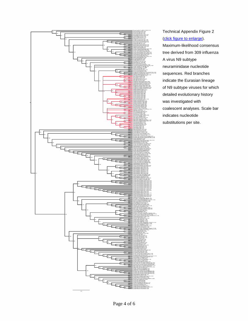
Technical Appendix Figure 2
(click figure to enlarge).
Maximum-likelihood consensus
tree derived from 309 influenza
A virus N9 subtype
neuraminidase nucleotide
sequences. Red branches
indicate the Eurasian lineage
of N9 subtype viruses for which
detailed evolutionary history
was investigated with
coalescent analyses. Scale bar
indicates nucleotide
substitutions per site.
Page 5 of 6
Technical Appendix Figure 3. Maximum clade credibility tree for clade B of influenza A virus H7
hemagglutinin subtypes. Branches are colored according to most probable location, as obtained by
coalescent analysis. Green indicates Europe and blue indicates Asia. For internal nodes, locations and
associated posterior probabilities are reported when >0.8. Supporting value for the European ancestor of
the Asian lineage (red dot) is indicated in italics.
Page 6 of 6
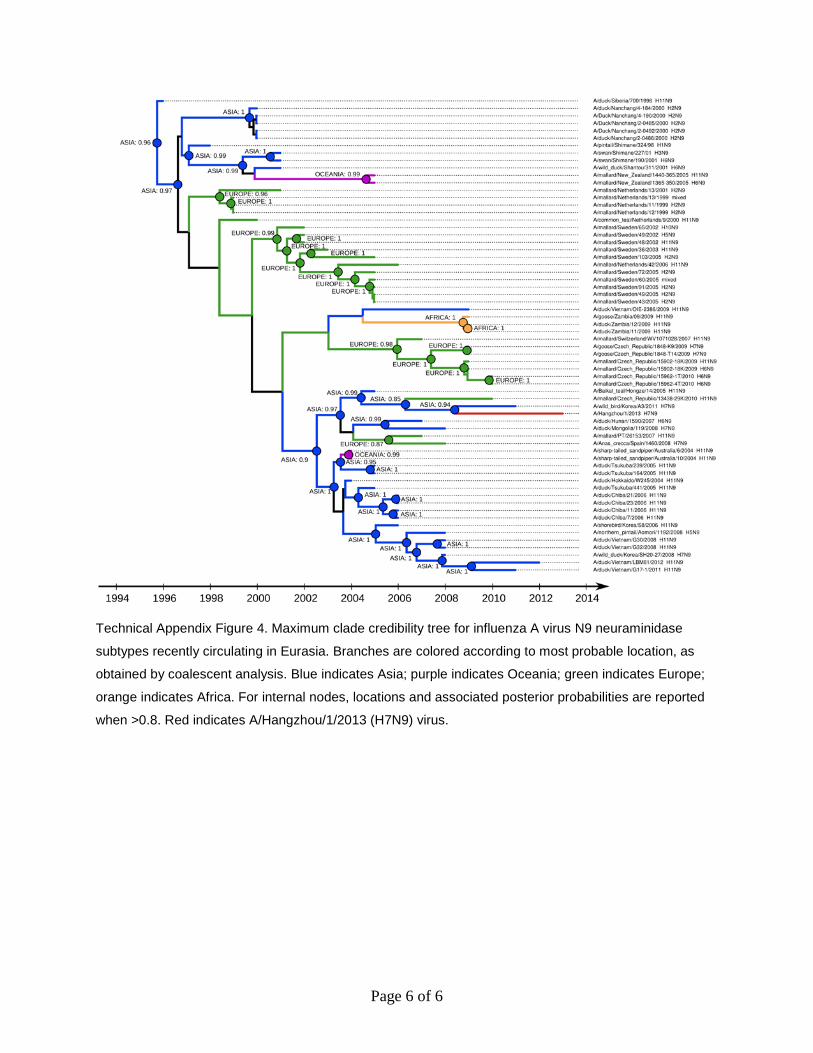
Technical Appendix Figure 4. Maximum clade credibility tree for influenza A virus N9 neuraminidase
subtypes recently circulating in Eurasia. Branches are colored according to most probable location, as
obtained by coalescent analysis. Blue indicates Asia; purple indicates Oceania; green indicates Europe;
orange indicates Africa. For internal nodes, locations and associated posterior probabilities are reported
when >0.8. Red indicates A/Hangzhou/1/2013 (H7N9) virus.