Embed Size (px)
Citation preview
Evolution and Development: Rise ofthe Little Squirts
Dispatch
Anthony Graham
It used to be thought that only vertebrates possessneural crest cells, but a recent study hasdemonstrated the existence of neural crest-likecells in an ascidian urochordate. This alters ourviews on the evolution of the neural crest and ofthe vertebrates.
It has long been held that the neural crest is a definingfeature of vertebrates. The neural crest arises at thedorsal aspect of the neural tube and then migrateswidely in the embryo, giving rise to a range ofderivatives which are distinctly vertebrate, such as theneurons and glia of the peripheral nervous system,melanocytes, and, additionally in the head, cartilage,bone and teeth. The other members of the phylumChordata, the urochordates and the cephalochor-dates, the nearest living relatives of the vertebrates,have been thought to lack neural crest cells. Indeed,the evolution of the neural crest was believed to havebeen concomitant with, and pivotal to, the evolution ofthe vertebrates [1].
Excitingly, however, a recent paper [2] has directlychallenged current received wisdom with the demon-stration that urochordates possess neural crest-likecells. Thus it would seem that neural crest cells didnot evolve with the vertebrates but that they have amore ancient history. The results presented here alsohave serious implications for how we view therelationships between the vertebrates, the cephalo-chordates and the urochordates, as they may suggestthat it is the urochordates that are the true sistergroup of the vertebrates and not, as is generallyaccepted, the cephalochordates.
Given the importance of the neural crest tovertebrates, there have been numerous previousstudies looking at how neural crest cells evolved. Byand large, these studies focused on a cephalochor-date, amphioxus, and on a few urochordate species,primarily Ciona intestinalis. They found that cells atthe neural plate border in these species expressorthologues of some of the genes known to beinvolved in specifying dorsal neural tube fates,including neural crest cells, in vertebrates [3–8]. Theydid not, however, find direct evidence for theexistence of migratory neural crest cells. Rather,these studies suggested that the neural crestevolved, with the vertebrates, from dorsal neural tubecells, and that both are linked by a shared develop-mental programme.
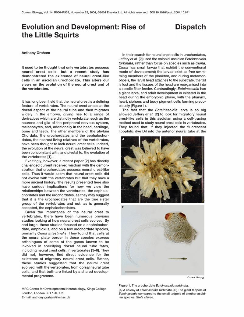
In their search for neural crest cells in urochordates,Jeffery et al. [2] used the colonial ascidian Ecteinascidiaturbinata, rather than focus on species such as Ciona.Ciona has small larvae that exhibit the conventionalmode of development; the larvae exist as free swim-ming members of the plankton, and during metamor-phosis, the larval head attaches to the substrate, the tailis lost and the tissues of the head are reorganised intoa sessile filter feeder. Contrastingly, Ecteinascidia hasa giant larva, and adult development is initiated in thehead during the embryonic phase, with the pharynx,heart, siphons and body pigment cells forming preco-ciously (Figure 1).
The fact that the Ecteinascidia larva is so bigallowed Jeffery et al. [2] to look for migratory neuralcrest-like cells in this ascidian using a cell-tracingmethod used to study neural crest cells in vertebrates.They found that, if they injected the fluorescentlipophilic dye DiI into the anterior neural tube at the
Current Biology, Vol. 14, R956–R958, November 23, 2004, ©2004 Elsevier Ltd. All rights reserved. DOI 10.1016/j.cub.2004.10.041
MRC Centre for Developmental Neurobiology, Kings CollegeLondon, London SE1 1UL, UK. E-mail: [email protected]
Figure 1. The urochordate Ecteinascidia turbinata.
(A) A colony of Ecteinascidia turbinata. (B) The giant tadpole ofEcteinascidia compared to the small tadpole of another ascid-ian species, Stela clavae.
early tailbud stage, they could, with time, observecells migrating away from the neural primordiumtowards the developing siphons and body wall.Although the posterior neural tube was not found torelease migratory cells at these early stages, injectionsat later stages did highlight the production of suchcells by this region, and again these cells migratedinto the body wall.
Just as in vertebrates, therefore, migratory cellsemerge from the neural tube during Ecteinascidiadevelopment and, again like vertebrates, they do so inan anterior to posterior sequence. This cell tracinganalysis also revealed a further startling feature ofthese migratory cells. Jeffery et al. [2] followed the fateof the cells that migrated to the siphons and bodywall, and found that they differentiated as pigmentcells. Again, this directly mirrors what is seen invertebrates; the neural crest cells that populate theskin generate melanocytes. The authors furtherprobed whether these migratory cells in Ecteinascidiaexpressed any of the markers associated withvertebrate neural crest cells. They found that, indeed,the pigment cells of the siphons and body wall couldbe highlighted using two general vertebrate neuralcrest markers, staining with the HNK-1 antibody [9]and expression of a Zic gene [10].
Collectively, these results demonstrate that ascidianurochordates possess neural crest-like cells, and theyprovide us with the basis of a scenario for neural crestevolution in chordates. As Jeffery et al. [2] suggest,the first step in the evolution of the neural crest mayhave been, as is seen in Ecteinascidia, the emergenceof the capacity of the neural tube to release migratorypigment cell precursors in an ascidian-like chordateancestor, possibly as a means of protection from theharmful effects of sunlight in a shallow marine envi-ronment. The fact that these cells are derived from theneural tube makes it fairly easy to imagine how neuralcrest cells could go on to form the neurons and glia ofthe vertebrate peripheral nervous system. Theevolution of the vertebrates, however, would furtherrequire that these migratory neural-tube-derived cellsfurther attained the ability to form skeletal tissues, andthis would most likely have been driven by alterationsto the embryonic environment and the correspondingresponse of these migratory neural derived cells.
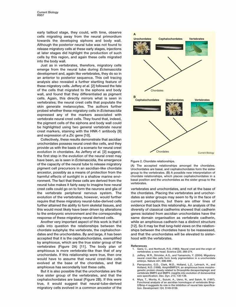
Another very important aspect of this work is that itcalls into question the relationships between thechordate subphyla: the vertebrate, the cephalochor-dates and the urochordates. By and large, it has beenaccepted that it is the cephalochordates, exemplifiedby amphioxus, which are the true sister group of thevertebrates (Figure 2A) [11]. The body plan ofamphioxus is more vertebrate-like than that of anyurochordate. If this relationship were true, then onewould have to assume that neural crest-like cellsevolved at the base of the chordates, and thatamphioxus has secondarily lost these cells.
But it is also possible that the urochordates are thetrue sister group of the vertebrates, and that thecephalochordates are basal (Figure 2B). If this weretrue, it would suggest that neural-tube-derivedmigratory cells evolved in a common ancestor of the
vertebrates and urochordates, and not at the base ofthe chordates. Placing the vertebrates and urochor-dates as sister groups may seem to fly in the face ofcurrent perceptions, but there are other lines ofevidence that back this relationship. An analysis of thediversity of classical cadherins showed that cadheringenes isolated from ascidian urochordates have thesame domain organisation as vertebrate cadherin,while an amphioxus cadherin has a distinct structure[12]. So it may be that long-held views on the relation-ships between the chordates have to be reassessed,and that the urochordates will be elevated to sister-hood with the vertebrates.
References1. Gans, C., and Northcutt, R.G. (1983). Neural crest and the origin of
vertebrates: a new head. Science 220, 268-274.2. Jeffery, W.R., Strickler, A.G., and Yamamoto, Y. (2004). Migratory
neural crest-like cells form body pigmentation in a urochordateembryo. Nature 431, 696-699 .
3. Panopoulou, G.D., Clark, M.D., Holland, L.Z., Lehrach, H., andHolland, N.D. (1998). AmphiBMP2/4, an amphioxus bone morpho-genetic protein closely related to Drosophila decapentaplegic andvertebrate BMP2 and BMP4: insights into evolution of dorsoventralaxis specification. Dev. Dyn. 213, 130-139.
4. Miya, T., Morita, K., Suzuki, A., Ueno, N., and Satoh, N. (1997).Functional analysis of an ascidian homologue of vertebrate Bmp-2/Bmp-4 suggests its role in the inhibition of neural fate specifica-tion. Development 124, 5149-5159.
Current BiologyR957
Figure 2. Chordate relationships.
(A) The accepted relationships amongst the chordates.Urochordates are basal, and cephalochordates form the sistergroup to the vertebrates. (B) A possible new interpretation ofchordate relationships, which places cephalochordates in abasal position and the urochordates as the sister group to thevertebrates.
Urochordates Cephalochordates Vertebrates
Chordates
Cephalochordates Urochordates Vertebrates
Chordates
A
B
Current Biology
DispatchR958
5. Langeland, J.A., Tomsa, J.M., Jackman, W.R., Jr., and Kimmel, C.B.(1998). An amphioxus snail gene: expression in paraxial mesodermand neural plate suggests a conserved role in patterning thechordate embryo. Dev. Genes Evol. 208, 569-577.
6. Erives, A., Corbo, J.C., and Levine, M. (1998). Lineage-specificregulation of the Ciona snail gene in the embryonic mesoderm andneuroectoderm. Dev. Biol. 194, 213-225.
7. Ma, L., Swalla, B.J., Zhou, J., Dobias, S.L., Bell, J.R., Chen, J.,Maxson, R.E., and Jeffery, W.R. (1996). Expression of an Msxhomeobox gene in ascidians: insights into the archetypal chordateexpression pattern. Dev. Dyn. 205, 308-318.
8. Sharman, A.C., Shimeld, S.M., and Holland, P.W. (1999). Anamphioxus Msx gene expressed predominantly in the dorsal neuraltube. Dev. Genes Evol. 209, 260-263.
9. Tucker, G.C., Aoyama, H., Lipinski, M., Tursz, T., and Thiery, J.P.(1984). Identical reactivity of monoclonal antibodies HNK-1 and NC-1: conservation in vertebrates on cells derived from the neuralprimordium and on some leukocytes. Cell Differ. 14, 223-230.
10. Nakata, K., Nagai, T., Aruga, J., and Mikoshiba, K. (1998). XenopusZic family and its role in neural and neural crest development.Mech. Dev. 75, 43-51.
11. Schaeffer, B. (1987). Deuterostome monophyly and phylogeny.Evol. Biol. 21, 179-234.
12. Oda, H., Wada, H., Tagawa, K., Akiyama-Oda, Y., Satoh, N.,Humphreys, T., Zhang, S., and Tsukita, S. (2002). A novelamphioxus cadherin that localizes to epithelial adherens junctionshas an unusual domain organization with implications for chordatephylogeny. Evol. Dev. 4, 426-434.