Embed Size (px)
Citation preview

Evil Humors: Thoracic Manifestations of
Immunoglobulin-Related Disease
Matthew Lee MD, Cristopher Meyer MD, Jeffrey Kanne MD
IntroductionDisorders of humoral immunity result in a variety of clinically
significant immunoglobulin(Ig)-related diseases.
Ig-related diseases are systemic disorders that have
characteristic serologic profiles and thoracic imaging findings.
These are broadly categorized into disorders of overproduction,
underproduction, and specific antibody-related conditions.
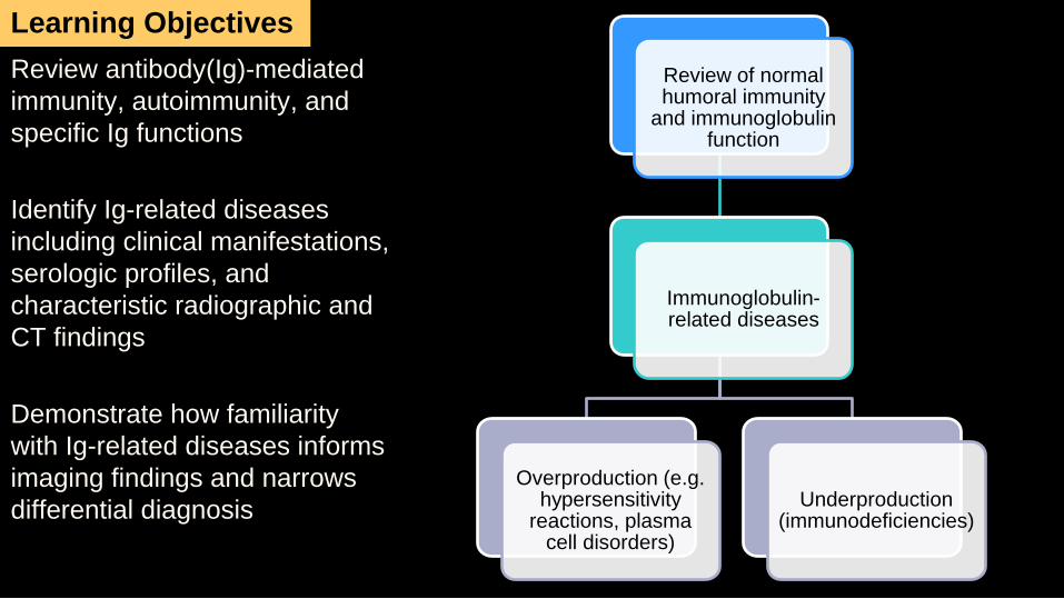
Review antibody(Ig)-mediated
immunity, autoimmunity, and
specific Ig functions
Identify Ig-related diseases
including clinical manifestations,
serologic profiles, and
characteristic radiographic and
CT findings
Demonstrate how familiarity
with Ig-related diseases informs
imaging findings and narrows
differential diagnosis
Learning Objectives
Review of normal humoral immunity
and immunoglobulin function
Immunoglobulin-related diseases
Overproduction (e.g. hypersensitivity
reactions, plasma cell disorders)
Underproduction (immunodeficiencies)
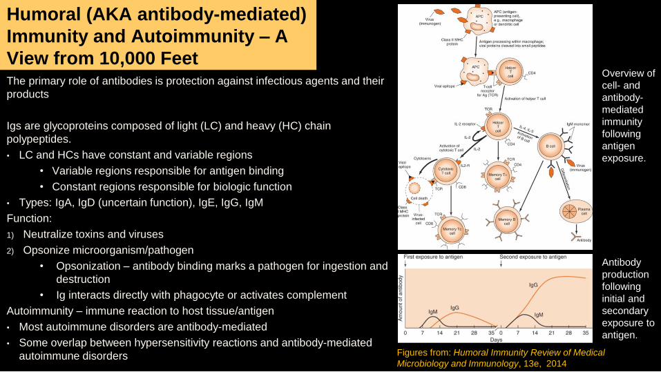
The primary role of antibodies is protection against infectious agents and their
products
Igs are glycoproteins composed of light (LC) and heavy (HC) chain
polypeptides.
• LC and HCs have constant and variable regions
• Variable regions responsible for antigen binding
• Constant regions responsible for biologic function
• Types: IgA, IgD (uncertain function), IgE, IgG, IgM
Function:
1) Neutralize toxins and viruses
2) Opsonize microorganism/pathogen
• Opsonization – antibody binding marks a pathogen for ingestion and
destruction
• Ig interacts directly with phagocyte or activates complement
Autoimmunity – immune reaction to host tissue/antigen
• Most autoimmune disorders are antibody-mediated
• Some overlap between hypersensitivity reactions and antibody-mediated
autoimmune disorders
Humoral (AKA antibody-mediated)
Immunity and Autoimmunity – A
View from 10,000 FeetOverview of
cell- and
antibody-
mediated
immunity
following
antigen
exposure.
Figures from: Humoral Immunity Review of Medical
Microbiology and Immunology, 13e, 2014
Antibody
production
following
initial and
secondary
exposure to
antigen.
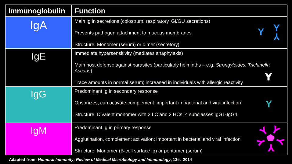
Immunoglobulin Function
IgAMain Ig in secretions (colostrum, respiratory, GI/GU secretions)
Prevents pathogen attachment to mucous membranes
Structure: Monomer (serum) or dimer (secretory)
IgEImmediate hypersensitivity (mediates anaphylaxis)
Main host defense against parasites (particularly helminths – e.g. Strongyloides, Trichinella,
Ascaris)
Trace amounts in normal serum; increased in individuals with allergic reactivity
IgGPredominant Ig in secondary response
Opsonizes, can activate complement; important in bacterial and viral infection
Structure: Divalent monomer with 2 LC and 2 HCs; 4 subclasses IgG1-IgG4
IgMPredominant Ig in primary response
Agglutination, complement activation; important in bacterial and viral infection
Structure: Monomer (B-cell surface Ig) or pentamer (serum)
Adapted from: Humoral Immunity; Review of Medical Microbiology and Immunology, 13e, 2014
Y
Y YY

Overproduction
Overproduction
IgE-related
Hypersensitivity/Eosinophilic Lung Disease
IgG-related
Specific Ig-Mediated ANCA-associated vasculitides IgG4
Mixed Ig- Mediated
Amyloid Plasma Cell Disorder
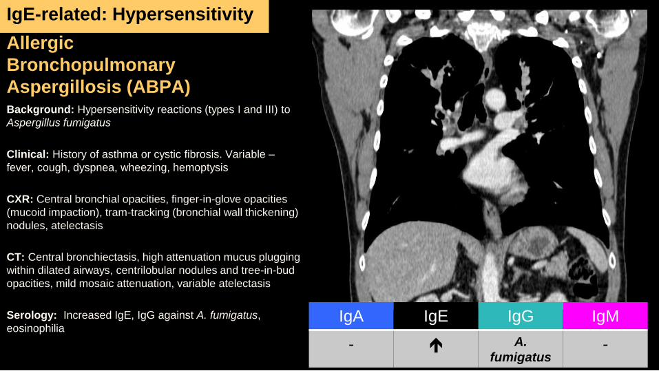
Allergic
Bronchopulmonary
Aspergillosis (ABPA)Background: Hypersensitivity reactions (types I and III) to
Aspergillus fumigatus
Clinical: History of asthma or cystic fibrosis. Variable –
fever, cough, dyspnea, wheezing, hemoptysis
CXR: Central bronchial opacities, finger-in-glove opacities
(mucoid impaction), tram-tracking (bronchial wall thickening)
nodules, atelectasis
CT: Central bronchiectasis, high attenuation mucus plugging
within dilated airways, centrilobular nodules and tree-in-bud
opacities, mild mosaic attenuation, variable atelectasis
Serology: Increased IgE, IgG against A. fumigatus,
eosinophilia
IgE-related: Hypersensitivity
IgA IgE IgG IgM
- A.
fumigatus-
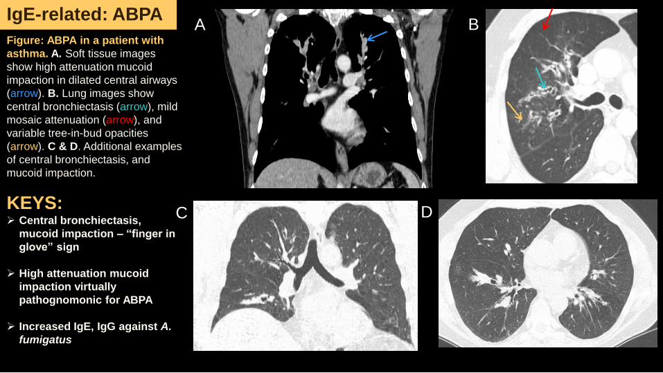
Figure: ABPA in a patient with
asthma. A. Soft tissue images
show high attenuation mucoid
impaction in dilated central airways
(arrow). B. Lung images show
central bronchiectasis (arrow), mild
mosaic attenuation (arrow), and
variable tree-in-bud opacities
(arrow). C & D. Additional examples
of central bronchiectasis, and
mucoid impaction.
KEYS: Central bronchiectasis,
mucoid impaction – “finger in
glove” sign
High attenuation mucoid
impaction virtually
pathognomonic for ABPA
Increased IgE, IgG against A.
fumigatus
A B
C
IgE-related: ABPA
D
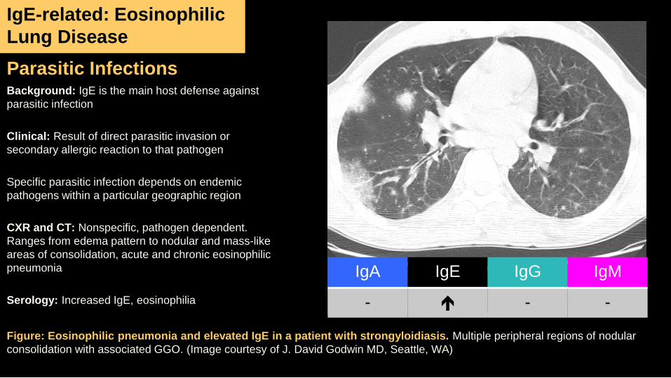
Parasitic InfectionsBackground: IgE is the main host defense against
parasitic infection
Clinical: Result of direct parasitic invasion or
secondary allergic reaction to that pathogen
Specific parasitic infection depends on endemic
pathogens within a particular geographic region
CXR and CT: Nonspecific, pathogen dependent.
Ranges from edema pattern to nodular and mass-like
areas of consolidation, acute and chronic eosinophilic
pneumonia
Serology: Increased IgE, eosinophilia
IgE-related: Eosinophilic
Lung Disease
IgA IgE IgG IgM
- - -
Figure: Eosinophilic pneumonia and elevated IgE in a patient with strongyloidiasis. Multiple peripheral regions of nodular
consolidation with associated GGO. (Image courtesy of J. David Godwin MD, Seattle, WA)
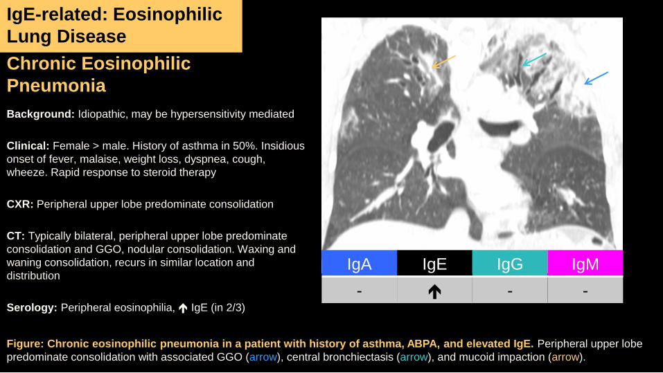
Chronic Eosinophilic
Pneumonia
Background: Idiopathic, may be hypersensitivity mediated
Clinical: Female > male. History of asthma in 50%. Insidious
onset of fever, malaise, weight loss, dyspnea, cough,
wheeze. Rapid response to steroid therapy
CXR: Peripheral upper lobe predominate consolidation
CT: Typically bilateral, peripheral upper lobe predominate
consolidation and GGO, nodular consolidation. Waxing and
waning consolidation, recurs in similar location and
distribution
Serology: Peripheral eosinophilia, IgE (in 2/3)
IgA IgE IgG IgM
- - -
Figure: Chronic eosinophilic pneumonia in a patient with history of asthma, ABPA, and elevated IgE. Peripheral upper lobe
predominate consolidation with associated GGO (arrow), central bronchiectasis (arrow), and mucoid impaction (arrow).
IgE-related: Eosinophilic
Lung Disease
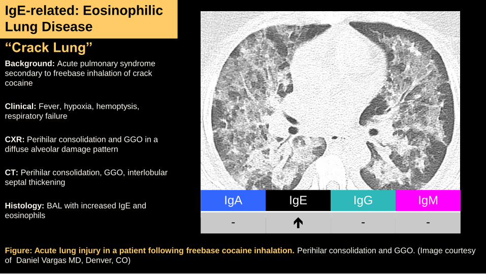
“Crack Lung”Background: Acute pulmonary syndrome
secondary to freebase inhalation of crack
cocaine
Clinical: Fever, hypoxia, hemoptysis,
respiratory failure
CXR: Perihilar consolidation and GGO in a
diffuse alveolar damage pattern
CT: Perihilar consolidation, GGO, interlobular
septal thickening
Histology: BAL with increased IgE and
eosinophils
IgA IgE IgG IgM
- - -
Figure: Acute lung injury in a patient following freebase cocaine inhalation. Perihilar consolidation and GGO. (Image courtesy
of Daniel Vargas MD, Denver, CO)
IgE-related: Eosinophilic
Lung Disease
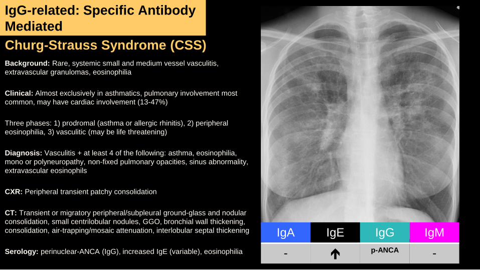
Churg-Strauss Syndrome (CSS)
Background: Rare, systemic small and medium vessel vasculitis,
extravascular granulomas, eosinophilia
Clinical: Almost exclusively in asthmatics, pulmonary involvement most
common, may have cardiac involvement (13-47%)
Three phases: 1) prodromal (asthma or allergic rhinitis), 2) peripheral
eosinophilia, 3) vasculitic (may be life threatening)
Diagnosis: Vasculitis + at least 4 of the following: asthma, eosinophilia,
mono or polyneuropathy, non-fixed pulmonary opacities, sinus abnormality,
extravascular eosinophils
CXR: Peripheral transient patchy consolidation
CT: Transient or migratory peripheral/subpleural ground-glass and nodular
consolidation, small centrilobular nodules, GGO, bronchial wall thickening,
consolidation, air-trapping/mosaic attenuation, interlobular septal thickening
Serology: perinuclear-ANCA (IgG), increased IgE (variable), eosinophilia
IgG-related: Specific Antibody
Mediated
IgA IgE IgG IgM
- p-ANCA -
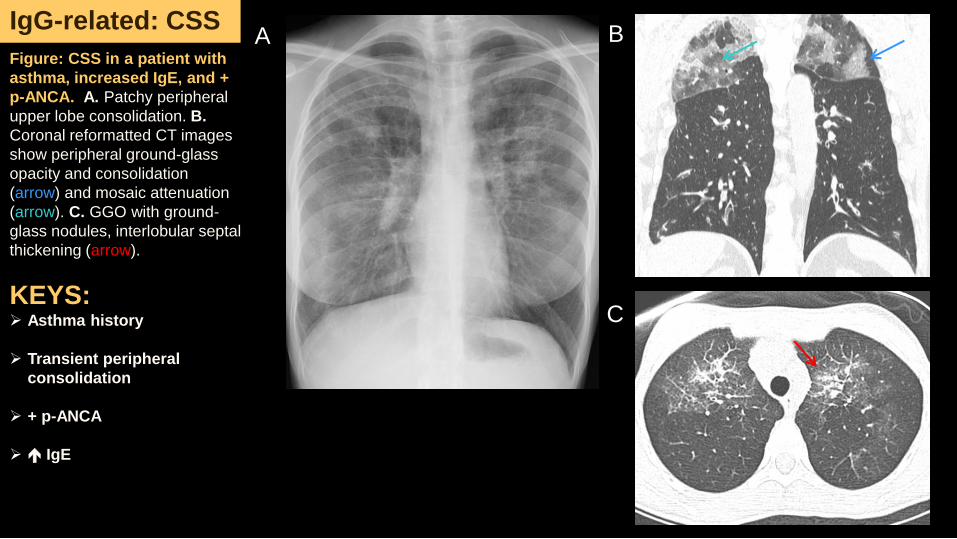
Figure: CSS in a patient with
asthma, increased IgE, and +
p-ANCA. A. Patchy peripheral
upper lobe consolidation. B.
Coronal reformatted CT images
show peripheral ground-glass
opacity and consolidation
(arrow) and mosaic attenuation
(arrow). C. GGO with ground-
glass nodules, interlobular septal
thickening (arrow).
KEYS: Asthma history
Transient peripheral
consolidation
+ p-ANCA
IgE
BIgG-related: CSS
A
C
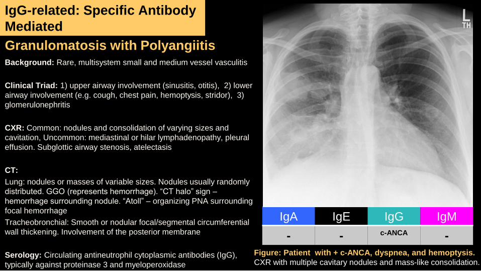
Granulomatosis with PolyangiitisBackground: Rare, multisystem small and medium vessel vasculitis
Clinical Triad: 1) upper airway involvement (sinusitis, otitis), 2) lower
airway involvement (e.g. cough, chest pain, hemoptysis, stridor), 3)
glomerulonephritis
CXR: Common: nodules and consolidation of varying sizes and
cavitation, Uncommon: mediastinal or hilar lymphadenopathy, pleural
effusion. Subglottic airway stenosis, atelectasis
CT:
Lung: nodules or masses of variable sizes. Nodules usually randomly
distributed. GGO (represents hemorrhage). “CT halo” sign –
hemorrhage surrounding nodule. “Atoll” – organizing PNA surrounding
focal hemorrhage
Tracheobronchial: Smooth or nodular focal/segmental circumferential
wall thickening. Involvement of the posterior membrane
Serology: Circulating antineutrophil cytoplasmic antibodies (IgG),
typically against proteinase 3 and myeloperoxidase
IgG-related: Specific Antibody
Mediated
IgA IgE IgG IgM
- - c-ANCA -
Figure: Patient with + c-ANCA, dyspnea, and hemoptysis.
CXR with multiple cavitary nodules and mass-like consolidation.
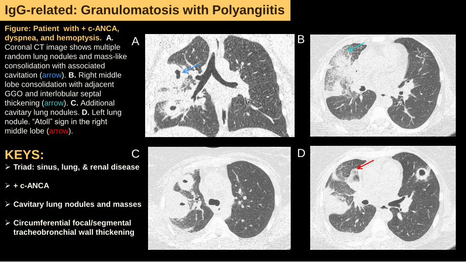
Figure: Patient with + c-ANCA,
dyspnea, and hemoptysis. A.
Coronal CT image shows multiple
random lung nodules and mass-like
consolidation with associated
cavitation (arrow). B. Right middle
lobe consolidation with adjacent
GGO and interlobular septal
thickening (arrow). C. Additional
cavitary lung nodules. D. Left lung
nodule. “Atoll” sign in the right
middle lobe (arrow).
A B
C
IgG-related: Granulomatosis with Polyangiitis
DKEYS: Triad: sinus, lung, & renal disease
+ c-ANCA
Cavitary lung nodules and masses
Circumferential focal/segmental
tracheobronchial wall thickening
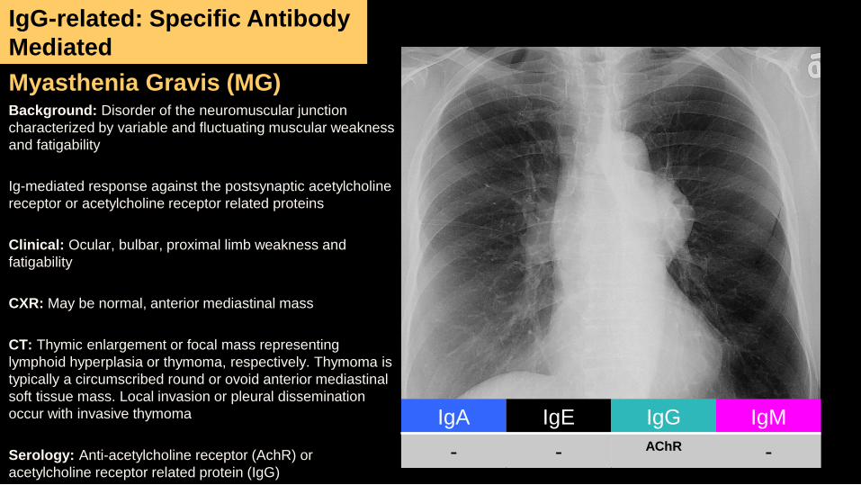
Myasthenia Gravis (MG)Background: Disorder of the neuromuscular junction
characterized by variable and fluctuating muscular weakness
and fatigability
Ig-mediated response against the postsynaptic acetylcholine
receptor or acetylcholine receptor related proteins
Clinical: Ocular, bulbar, proximal limb weakness and
fatigability
CXR: May be normal, anterior mediastinal mass
CT: Thymic enlargement or focal mass representing
lymphoid hyperplasia or thymoma, respectively. Thymoma is
typically a circumscribed round or ovoid anterior mediastinal
soft tissue mass. Local invasion or pleural dissemination
occur with invasive thymoma
Serology: Anti-acetylcholine receptor (AchR) or
acetylcholine receptor related protein (IgG)
IgA IgE IgG IgM
- - AChR -
IgG-related: Specific Antibody
Mediated
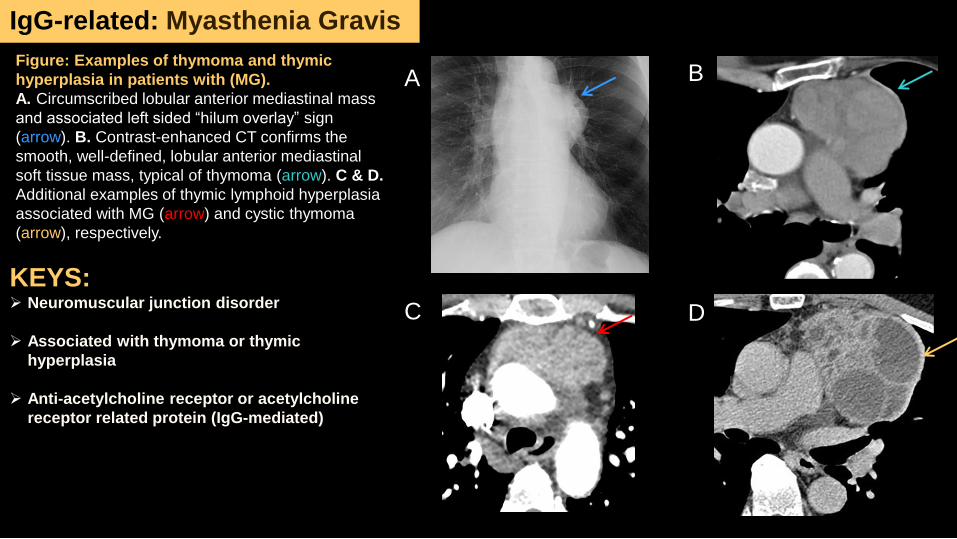
Figure: Examples of thymoma and thymic
hyperplasia in patients with (MG).
A. Circumscribed lobular anterior mediastinal mass
and associated left sided “hilum overlay” sign
(arrow). B. Contrast-enhanced CT confirms the
smooth, well-defined, lobular anterior mediastinal
soft tissue mass, typical of thymoma (arrow). C & D.
Additional examples of thymic lymphoid hyperplasia
associated with MG (arrow) and cystic thymoma
(arrow), respectively.
IgG-related: Myasthenia Gravis
A
C
B
D
KEYS: Neuromuscular junction disorder
Associated with thymoma or thymic
hyperplasia
Anti-acetylcholine receptor or acetylcholine
receptor related protein (IgG-mediated)
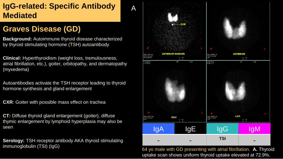
Graves Disease (GD)Background: Autoimmune thyroid disease characterized
by thyroid stimulating hormone (TSH) autoantibody
Clinical: Hyperthyroidism (weight loss, tremulousness,
atrial fibrillation, etc.), goiter, orbitopathy, and dermatopathy
(myxedema)
Autoantibodies activate the TSH receptor leading to thyroid
hormone synthesis and gland enlargement
CXR: Goiter with possible mass effect on trachea
CT: Diffuse thyroid gland enlargement (goiter), diffuse
thymic enlargement by lymphoid hyperplasia may also be
seen
Serology: TSH receptor antibody AKA thyroid stimulating
immunoglobulin (TSI) (IgG)
IgG-related: Specific Antibody
Mediated
IgA IgE IgG IgM
- - TSI -64 yo male with GD presenting with atrial fibrillation. A. Thyroid
uptake scan shows uniform thyroid uptake elevated at 72.9%.
A
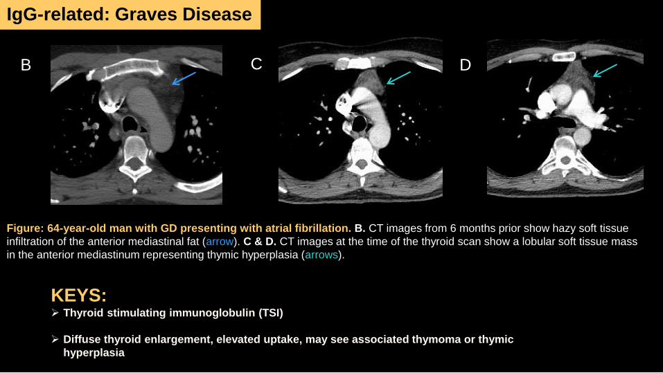
Figure: 64-year-old man with GD presenting with atrial fibrillation. B. CT images from 6 months prior show hazy soft tissue
infiltration of the anterior mediastinal fat (arrow). C & D. CT images at the time of the thyroid scan show a lobular soft tissue mass
in the anterior mediastinum representing thymic hyperplasia (arrows).
B C D
IgG-related: Graves Disease
KEYS: Thyroid stimulating immunoglobulin (TSI)
Diffuse thyroid enlargement, elevated uptake, may see associated thymoma or thymic
hyperplasia
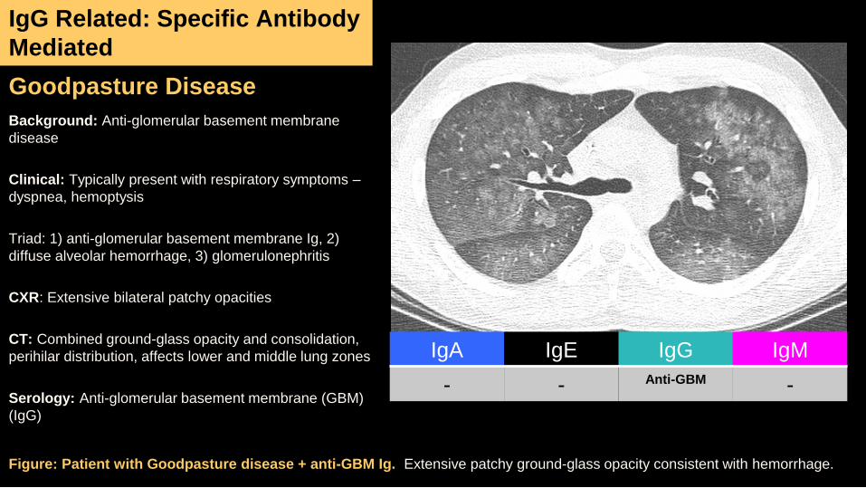
Goodpasture Disease
Background: Anti-glomerular basement membrane
disease
Clinical: Typically present with respiratory symptoms –
dyspnea, hemoptysis
Triad: 1) anti-glomerular basement membrane Ig, 2)
diffuse alveolar hemorrhage, 3) glomerulonephritis
CXR: Extensive bilateral patchy opacities
CT: Combined ground-glass opacity and consolidation,
perihilar distribution, affects lower and middle lung zones
Serology: Anti-glomerular basement membrane (GBM)
(IgG)
IgG Related: Specific Antibody
Mediated
IgA IgE IgG IgM
- - Anti-GBM -
Figure: Patient with Goodpasture disease + anti-GBM Ig. Extensive patchy ground-glass opacity consistent with hemorrhage.
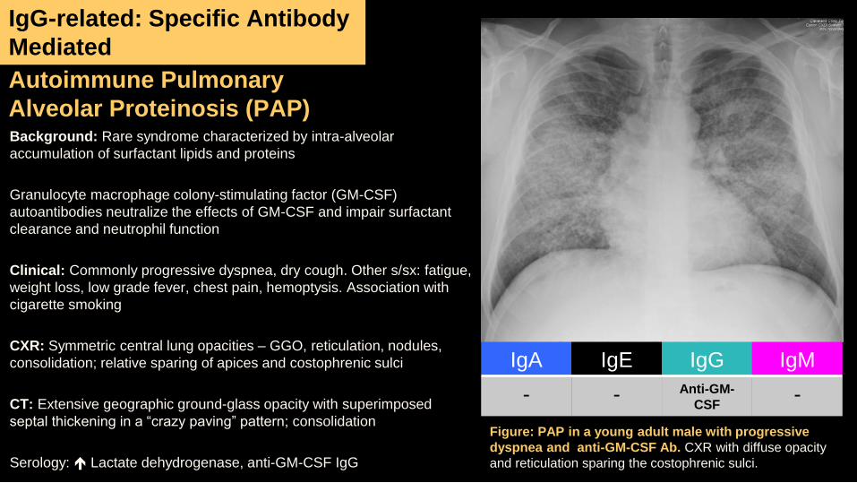
Autoimmune Pulmonary
Alveolar Proteinosis (PAP)Background: Rare syndrome characterized by intra-alveolar
accumulation of surfactant lipids and proteins
Granulocyte macrophage colony-stimulating factor (GM-CSF)
autoantibodies neutralize the effects of GM-CSF and impair surfactant
clearance and neutrophil function
Clinical: Commonly progressive dyspnea, dry cough. Other s/sx: fatigue,
weight loss, low grade fever, chest pain, hemoptysis. Association with
cigarette smoking
CXR: Symmetric central lung opacities – GGO, reticulation, nodules,
consolidation; relative sparing of apices and costophrenic sulci
CT: Extensive geographic ground-glass opacity with superimposed
septal thickening in a “crazy paving” pattern; consolidation
Serology: Lactate dehydrogenase, anti-GM-CSF IgG
IgG-related: Specific Antibody
Mediated
IgA IgE IgG IgM
- - Anti-GM-
CSF-
Figure: PAP in a young adult male with progressive
dyspnea and anti-GM-CSF Ab. CXR with diffuse opacity
and reticulation sparing the costophrenic sulci.
IgG-related: Autoimmune PAP
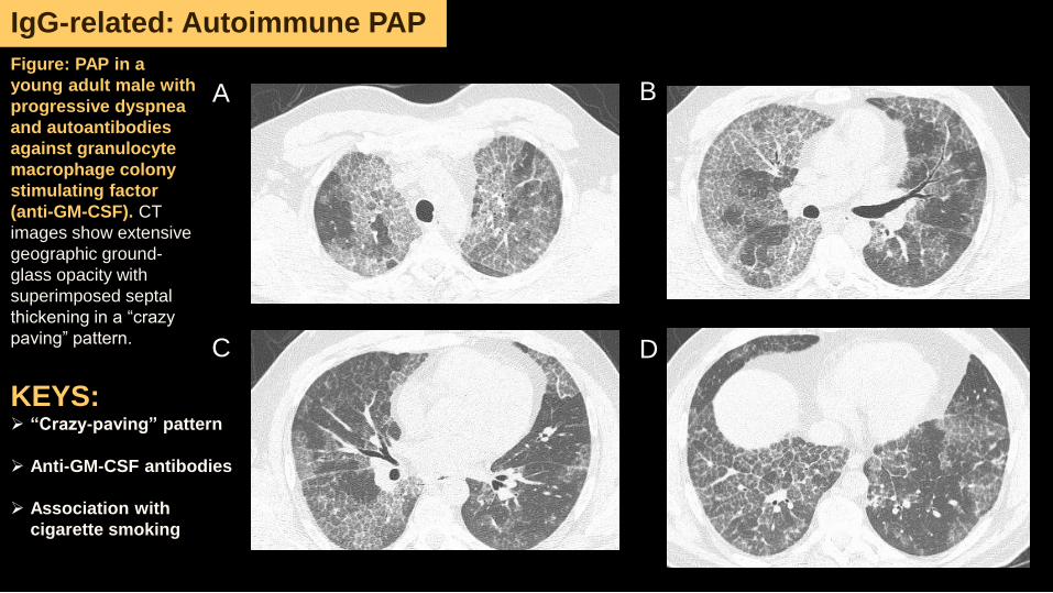
Figure: PAP in a
young adult male with
progressive dyspnea
and autoantibodies
against granulocyte
macrophage colony
stimulating factor
(anti-GM-CSF). CT
images show extensive
geographic ground-
glass opacity with
superimposed septal
thickening in a “crazy
paving” pattern.
A B
C D
KEYS: “Crazy-paving” pattern
Anti-GM-CSF antibodies
Association with
cigarette smoking
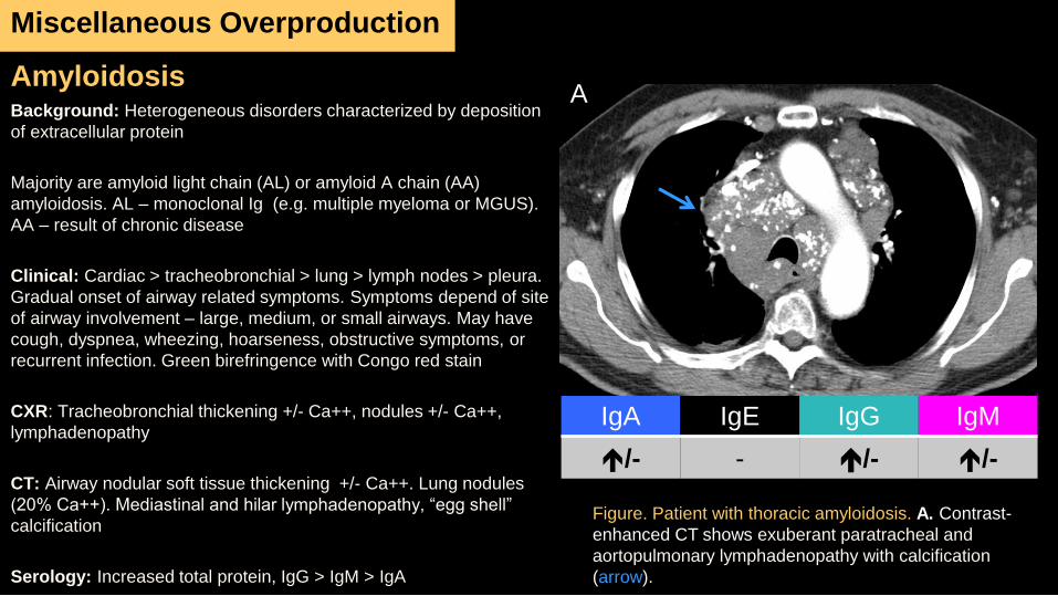
AmyloidosisBackground: Heterogeneous disorders characterized by deposition
of extracellular protein
Majority are amyloid light chain (AL) or amyloid A chain (AA)
amyloidosis. AL – monoclonal Ig (e.g. multiple myeloma or MGUS).
AA – result of chronic disease
Clinical: Cardiac > tracheobronchial > lung > lymph nodes > pleura.
Gradual onset of airway related symptoms. Symptoms depend of site
of airway involvement – large, medium, or small airways. May have
cough, dyspnea, wheezing, hoarseness, obstructive symptoms, or
recurrent infection. Green birefringence with Congo red stain
CXR: Tracheobronchial thickening +/- Ca++, nodules +/- Ca++,
lymphadenopathy
CT: Airway nodular soft tissue thickening +/- Ca++. Lung nodules
(20% Ca++). Mediastinal and hilar lymphadenopathy, “egg shell”
calcification
Serology: Increased total protein, IgG > IgM > IgA
Miscellaneous Overproduction
IgA IgE IgG IgM
/- - /- /-
Figure. Patient with thoracic amyloidosis. A. Contrast-
enhanced CT shows exuberant paratracheal and
aortopulmonary lymphadenopathy with calcification
(arrow).
A
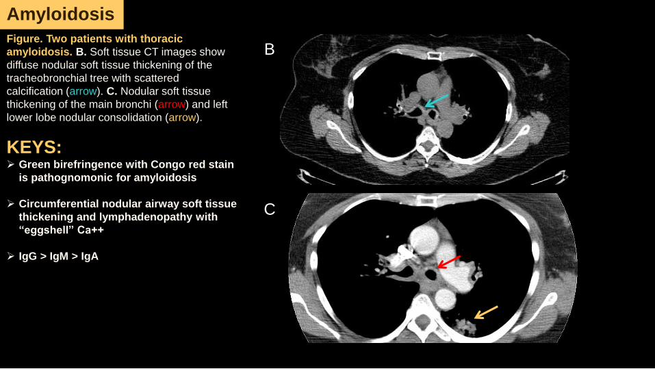
Figure. Two patients with thoracic
amyloidosis. B. Soft tissue CT images show
diffuse nodular soft tissue thickening of the
tracheobronchial tree with scattered
calcification (arrow). C. Nodular soft tissue
thickening of the main bronchi (arrow) and left
lower lobe nodular consolidation (arrow).
KEYS: Green birefringence with Congo red stain
is pathognomonic for amyloidosis
Circumferential nodular airway soft tissue
thickening and lymphadenopathy with
“eggshell” Ca++
IgG > IgM > IgA
B B
C
Amyloidosis
D
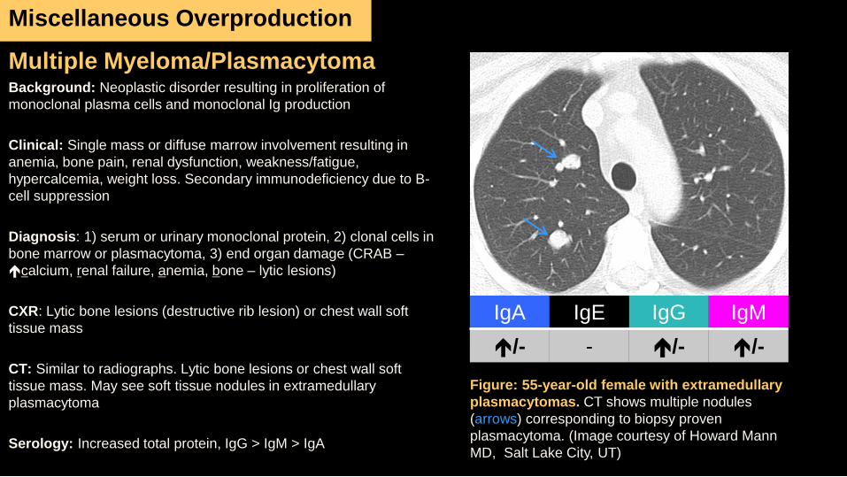
Multiple Myeloma/PlasmacytomaBackground: Neoplastic disorder resulting in proliferation of
monoclonal plasma cells and monoclonal Ig production
Clinical: Single mass or diffuse marrow involvement resulting in
anemia, bone pain, renal dysfunction, weakness/fatigue,
hypercalcemia, weight loss. Secondary immunodeficiency due to B-
cell suppression
Diagnosis: 1) serum or urinary monoclonal protein, 2) clonal cells in
bone marrow or plasmacytoma, 3) end organ damage (CRAB –
calcium, renal failure, anemia, bone – lytic lesions)
CXR: Lytic bone lesions (destructive rib lesion) or chest wall soft
tissue mass
CT: Similar to radiographs. Lytic bone lesions or chest wall soft
tissue mass. May see soft tissue nodules in extramedullary
plasmacytoma
Serology: Increased total protein, IgG > IgM > IgA
Miscellaneous Overproduction
Figure: 55-year-old female with extramedullary
plasmacytomas. CT shows multiple nodules
(arrows) corresponding to biopsy proven
plasmacytoma. (Image courtesy of Howard Mann
MD, Salt Lake City, UT)
IgA IgE IgG IgM
/- - /- /-
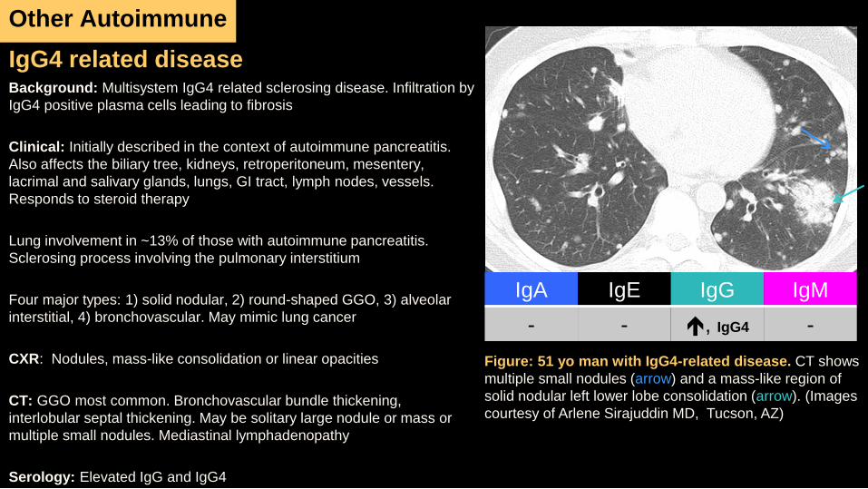
IgG4 related diseaseBackground: Multisystem IgG4 related sclerosing disease. Infiltration by
IgG4 positive plasma cells leading to fibrosis
Clinical: Initially described in the context of autoimmune pancreatitis.
Also affects the biliary tree, kidneys, retroperitoneum, mesentery,
lacrimal and salivary glands, lungs, GI tract, lymph nodes, vessels.
Responds to steroid therapy
Lung involvement in ~13% of those with autoimmune pancreatitis.
Sclerosing process involving the pulmonary interstitium
Four major types: 1) solid nodular, 2) round-shaped GGO, 3) alveolar
interstitial, 4) bronchovascular. May mimic lung cancer
CXR: Nodules, mass-like consolidation or linear opacities
CT: GGO most common. Bronchovascular bundle thickening,
interlobular septal thickening. May be solitary large nodule or mass or
multiple small nodules. Mediastinal lymphadenopathy
Serology: Elevated IgG and IgG4
Other Autoimmune
IgA IgE IgG IgM
- - , IgG4 -
Figure: 51 yo man with IgG4-related disease. CT shows
multiple small nodules (arrow) and a mass-like region of
solid nodular left lower lobe consolidation (arrow). (Images
courtesy of Arlene Sirajuddin MD, Tucson, AZ)
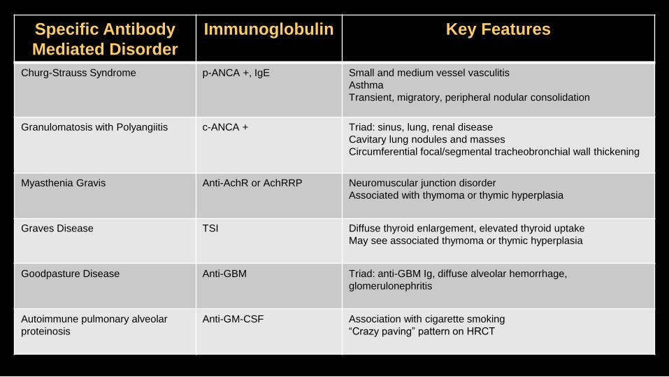
Specific Antibody
Mediated Disorder
Immunoglobulin Key Features
Churg-Strauss Syndrome p-ANCA +, IgE Small and medium vessel vasculitis
Asthma
Transient, migratory, peripheral nodular consolidation
Granulomatosis with Polyangiitis c-ANCA + Triad: sinus, lung, renal disease
Cavitary lung nodules and masses
Circumferential focal/segmental tracheobronchial wall thickening
Myasthenia Gravis Anti-AchR or AchRRP Neuromuscular junction disorder
Associated with thymoma or thymic hyperplasia
Graves Disease TSI Diffuse thyroid enlargement, elevated thyroid uptake
May see associated thymoma or thymic hyperplasia
Goodpasture Disease Anti-GBM Triad: anti-GBM Ig, diffuse alveolar hemorrhage,
glomerulonephritis
Autoimmune pulmonary alveolar
proteinosis
Anti-GM-CSF Association with cigarette smoking
“Crazy paving” pattern on HRCT

Underproduction
Underproduction
Common variable immunodeficiency
disease
Ig subtype deficiency
IgA-related IgG-related
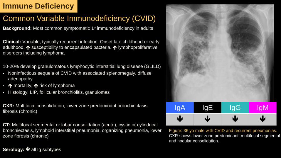
Background: Most common symptomatic 1o immunodeficiency in adults
Clinical: Variable, typically recurrent infection. Onset late childhood or early
adulthood. susceptibility to encapsulated bacteria. lymphoproliferative
disorders including lymphoma
10-20% develop granulomatous lymphocytic interstitial lung disease (GLILD)
• Noninfectious sequela of CVID with associated splenomegaly, diffuse
adenopathy
• mortality, risk of lymphoma
• Histology: LIP, follicular bronchiolitis, granulomas
CXR: Multifocal consolidation, lower zone predominant bronchiectasis,
fibrosis (chronic)
CT: Multifocal segmental or lobar consolidation (acute), cystic or cylindrical
bronchiectasis, lymphoid interstitial pneumonia, organizing pneumonia, lower
zone fibrosis (chronic)
Serology: all Ig subtypes
Common Variable Immunodeficiency (CVID)
IgA IgE IgG IgM
Figure: 36 yo male with CVID and recurrent pneumonias.
CXR shows lower zone predominant, multifocal segmental
and nodular consolidation.
Immune Deficiency
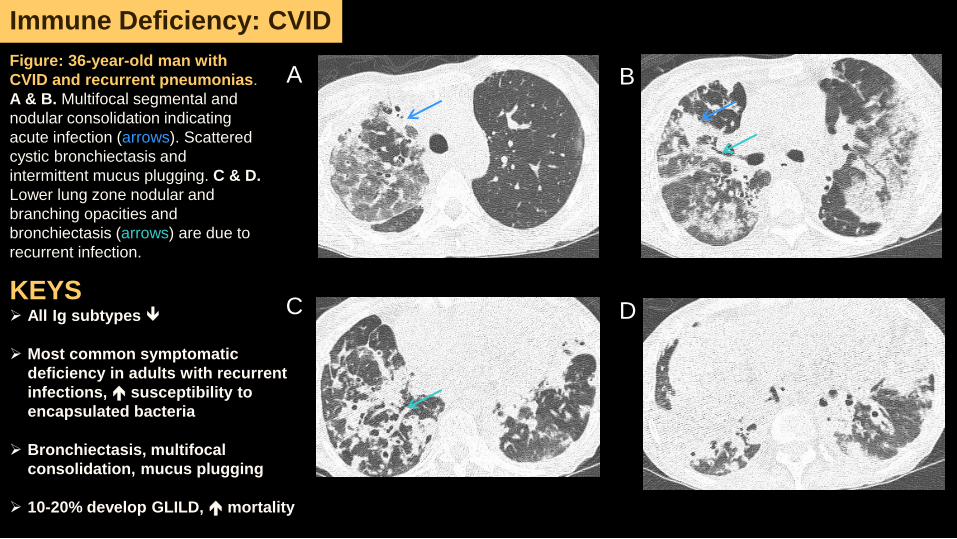
Immune Deficiency: CVID
Figure: 36-year-old man with
CVID and recurrent pneumonias.
A & B. Multifocal segmental and
nodular consolidation indicating
acute infection (arrows). Scattered
cystic bronchiectasis and
intermittent mucus plugging. C & D.
Lower lung zone nodular and
branching opacities and
bronchiectasis (arrows) are due to
recurrent infection.
A
C
B
DKEYS All Ig subtypes
Most common symptomatic
deficiency in adults with recurrent
infections, susceptibility to
encapsulated bacteria
Bronchiectasis, multifocal
consolidation, mucus plugging
10-20% develop GLILD, mortality
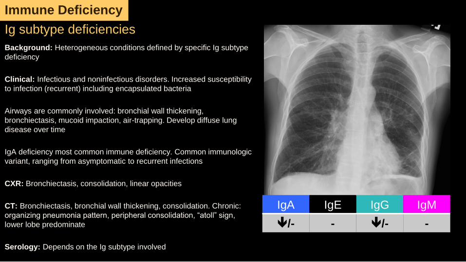
Background: Heterogeneous conditions defined by specific Ig subtype
deficiency
Clinical: Infectious and noninfectious disorders. Increased susceptibility
to infection (recurrent) including encapsulated bacteria
Airways are commonly involved: bronchial wall thickening,
bronchiectasis, mucoid impaction, air-trapping. Develop diffuse lung
disease over time
IgA deficiency most common immune deficiency. Common immunologic
variant, ranging from asymptomatic to recurrent infections
CXR: Bronchiectasis, consolidation, linear opacities
CT: Bronchiectasis, bronchial wall thickening, consolidation. Chronic:
organizing pneumonia pattern, peripheral consolidation, “atoll” sign,
lower lobe predominate
Serology: Depends on the Ig subtype involved
Ig subtype deficiencies
IgA IgE IgG IgM
/- - /- -
Immune Deficiency
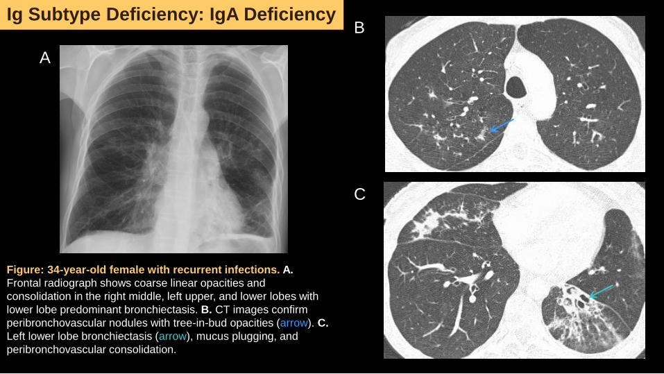
Figure: 34-year-old female with recurrent infections. A.
Frontal radiograph shows coarse linear opacities and
consolidation in the right middle, left upper, and lower lobes with
lower lobe predominant bronchiectasis. B. CT images confirm
peribronchovascular nodules with tree-in-bud opacities (arrow). C.
Left lower lobe bronchiectasis (arrow), mucus plugging, and
peribronchovascular consolidation.
A
B
C
Ig Subtype Deficiency: IgA Deficiency
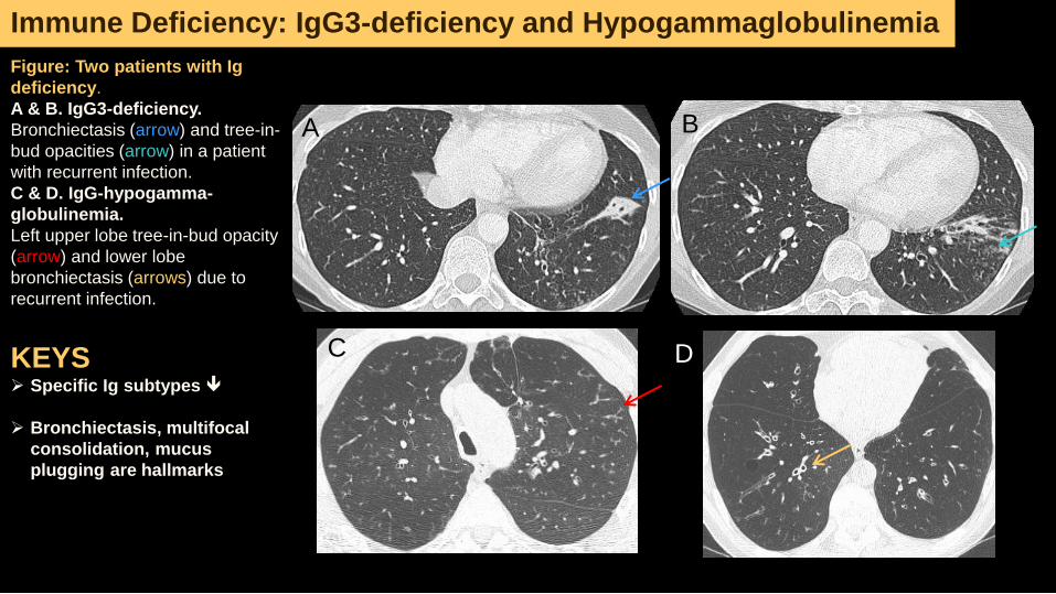
Immune Deficiency: IgG3-deficiency and Hypogammaglobulinemia
Figure: Two patients with Ig
deficiency.
A & B. IgG3-deficiency.
Bronchiectasis (arrow) and tree-in-
bud opacities (arrow) in a patient
with recurrent infection.
C & D. IgG-hypogamma-
globulinemia.
Left upper lobe tree-in-bud opacity
(arrow) and lower lobe
bronchiectasis (arrows) due to
recurrent infection.
BA
KEYS Specific Ig subtypes
Bronchiectasis, multifocal
consolidation, mucus
plugging are hallmarks
DC
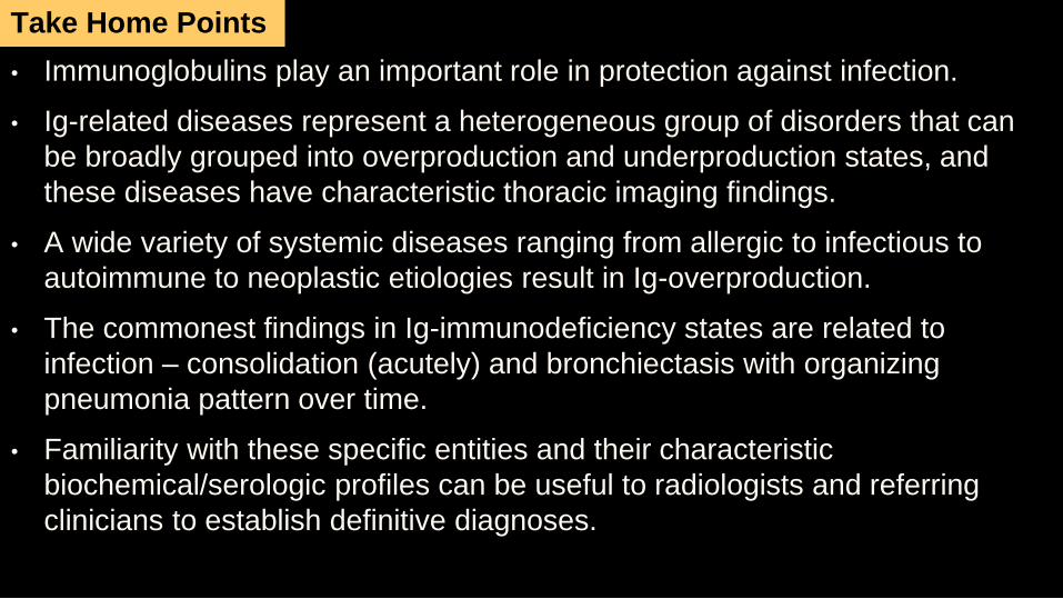
• Immunoglobulins play an important role in protection against infection.
• Ig-related diseases represent a heterogeneous group of disorders that can
be broadly grouped into overproduction and underproduction states, and
these diseases have characteristic thoracic imaging findings.
• A wide variety of systemic diseases ranging from allergic to infectious to
autoimmune to neoplastic etiologies result in Ig-overproduction.
• The commonest findings in Ig-immunodeficiency states are related to
infection – consolidation (acutely) and bronchiectasis with organizing
pneumonia pattern over time.
• Familiarity with these specific entities and their characteristic
biochemical/serologic profiles can be useful to radiologists and referring
clinicians to establish definitive diagnoses.
Take Home Points
References
1. Aylwin ACB, Gishen P, Copley SJ. Imaging appearance of thoracic amyloidosis. Journal of Thoracic Imaging. 2005;20(1):41-6.
2. Bierry G, Boileau J, Barnig C, et al. Thoracic Manifestations of Primary Humoral Immunodeficiency: A Comprehensive Review. Radiographics.
2009;29(7):1909-U77.
3. Borhani AA, Hosseinzadeh K, Almusa O, Furlan A, Nalesnik M. Imaging of Posttransplantation Lymphoproliferative Disorder after Solid Organ
Transplantation. Radiographics. 2009;29(4):981-U9.
4. Bonella F, Bauer PC, Griese M, Ohshimo S, Guzman J, Costabel U. Pulmonary alveolar proteinosis: New insights from a single-center cohort of 70
patients. Respiratory Medicine. 2011;105(12):1908-16.
5. Chase NM, Verbsky JW, Hintermeyer MK, et al. Use of Combination Chemotherapy for Treatment of Granulomatous and Lymphocytic Interstitial
Lung Disease (GLILD) in Patients with Common Variable Immunodeficiency (CVID). Journal of Clinical Immunology. 2013;33(1):30-9.
6. Chung MP, Yi CA, Lee HY, Han J, Lee KS. Imaging of Pulmonary Vasculitis. Radiology. 2010;255(2):322-41.
7. Frazier AA, Franks TJ, Cooke EO, Mohammed TLH, Pugatch RD, Galvin JR. From the archives of the AFIP - Pulmonary alveolar proteinosis.
Radiographics. 2008;28(3):883-99.
8. Georgiades CS, Neyman EG, Barish MA, Fishman EK. Amyloidosis: Review and CT manifestations. Radiographics. 2004;24(2):405-16.
9. Inoue D, Zen Y, Abo H, et al. Immunoglobulin G4-related Lung Disease: CT Findings with Pathologic Correlations. Radiology. 2009;251(1):260-70.
10. Jeong YJ, Kim KI, Seo IJ, et al. Eosinophilic lung diseases: A clinical, radiologic, and pathologic overview. Radiographics. 2007;27(3):617-U45.
11. Levinson W. Humoral Immunity. In: Levinson W. eds. Review of Medical Microbiology and Immunology, 13e. New York, NY: McGraw-Hill;
2014.http://accessmedicine.mhmedical.com.ezproxy.library.wisc.edu/content.aspx?bookid=1023&Sectionid=57053507. Accessed October 29, 2014
12. Martinez F, Chung JH, Digumarthy SR, et al. Common and Uncommon Manifestations of Wegener Granulomatosis at Chest CT: Radiologic-
Pathologic Correlation. Radiographics. 2012;32(1):51-69.
13. Naughton M, Fahy J, Fitzgerald MX. CHRONIC EOSINOPHILIC PNEUMONIA - A LONG-TERM FOLLOW-UP OF 12 PATIENTS. Chest.
1993;103(1):162-5.
14. Oh YW, Effmann EL, Godwin JD. Pulmonary infections in immunocompromised hosts: The importance of correlating the conventional radiologic
appearance with the clinical setting. Radiology. 2000;217(3):647-56.
15. Rose ME, Lang DM. Evaluating and managing hypogammaglobulinemia. Cleveland Clinic Journal of Medicine. 2006;73(2):133-+.
16. Vlachou PA, Khalili K, Jang H-J, Fischer S, Hirschfield GM, Kim TK. IgG4-related Sclerosing Disease: Autoimmune Pancreatitis and
Extrapancreatic Manifestations. Radiographics. 2011;31(5):1379-402.