Embed Size (px)
Citation preview
524 © 2017 Deutsche Dermatologische Gesellschaft (DDG). Published by John Wiley & Sons Ltd. | JDDG | 1610-0379/2017/1505
Original Article
Evidence for silver in wound care – meta-analysis of clinical studies from 2000–2015
Summary Background: Given that the scientific evidence for silver in wound care is generally considered insufficient, there is uncertainty among users regarding its clinical use. Material and methods: A group of experts evaluated the clinical studies on silver in wound management published from 2000–2015. Results: Overall, 851 articles were identified, 173 of which were included and cate-gorized. There were 31 randomized controlled trials (RCTs) and eight cohort studies. Twenty-eight of these studies showed statistically significant outcome parameters in support of silver. While nine of these studies investigated burn injuries, 20 addressed other indications (venous leg ulcers: 9; pressure ulcers: 3; chronic wounds: 2; diabetic foot ulcer: 1; other types of wounds: 5). In 16 studies, the primary parameter was wound healing, whereas quality of life including pain was assessed in twelve studies; cost-effectiveness, in eight studies; reduction of bacterial load, in three studies. Based on these results, a treatment algorithm for the clinical use of silver in wound care has been developed. Conclusions: The present meta-analysis shows that the evidence base for silver in wound management is significantly better than perceived in the current scientific debate. Thus, if used selectively and for a limited period of time, silver not only has antimicrobial effects but is also characterized by an improvement in quality of life and good cost-effectiveness.
Joachim Dissemond 1 , Johannes Georg Böttrich 2 , Horst Braunwarth 3 , Jörg Hilt 4 , Patricia Wilken 5 , Karl-Christian Münter 6
(1) Department of Dermatology , Vene-reology, and Allergology , University Hospital Essen , Essen , Germany (2) B. Braun Melsungen AG , Melsungen , Germany (3) Coloplast GmbH , Hamburg , Germany (4) Smith & Nephew GmbH , Hamburg , Germany (5) URGO GmbH , Sulzbach , Germany (6) Joint Dermatology Practice Bramfeld , Hamburg , Germany
DOI: 10.1111/ddg.13233 Submitted: 15.8.2016 Accepted: 27.10.2016
Introduction The use of silver in wound management has a very long tra-dition. In Egypt, the application of silver leaf for wounds was practiced as early as 1850 BC, and silver-coated ves-sels were used in ancient times for the storage of water and food to keep them from spoiling. While the antimicrobial effects of silver were known, they were not understood in greater detail. In his textbooks, Hippocrates, too, described the positive effects of silver on wound healing. At the end of the 19 th century, many medications for the treatment of ocular diseases, skin disorders, and infectious diseases con-tained silver. In particular, Credé’s prophylaxis, which was
Introduced in 1881 and continued for nearly a century, con-sisted of the instillation of silver nitrate 1 % solution into the eyes of newborns in an effort to prevent gonorrhea. Around 1920, colloidal silver was approved for wound treatment by the US FDA (Food and Drug Administration). In 1968, silver sulfadiazine (SSD) was introduced into clinical practice, and – for some years – continued to be the gold standard in topi-cal antimicrobial treatment, especially in the management of burn injuries [ 1 ] .
Despite this very long tradition of silver in medical thera-py, its scientifi c evidence has frequently been critically asses-sed, especially with respect to wound treatment. Searching for clinical studies published until 2009, the most recent
Conflict of interest JD: Convatec, Coloplast, Mölnlycke, Urgo, Smith & Nephew. JGB: emplo-yee of B. Braun Melsungen AG. HB: employee of Coloplast GmbH. JH: employee of Smith & Nephew GmbH. PW: employee of URGO GmbH. KCM: Coloplast, Urgo, Smith & Nephew.
Original Article Silver in wound care
525© 2017 Deutsche Dermatologische Gesellschaft (DDG). Published by John Wiley & Sons Ltd. | JDDG | 1610-0379/2017/1505
(2010) Cochrane meta-analysis of scientifi c data on silver in wound treatment found 26 randomized controlled trials (RCTs) with a total of 2,066 patients. The authors concluded that the evidence hitherto available was insuffi cient to deter-mine whether silver-containing dressings promoted wound healing or prevented wound infections. It was pointed out, however, that the heterogeneity of the products as well as the parameters and results of the RCTs greatly limited the validity of the analysis [ 2 ] . Consequently, the evidence for silver-containing wound care agents, as for most therapeutic agents employed in wound treatment, has to date been con-sidered insuffi cient [ 3 ] . While current guidelines do highlight the potential benefi ts of most wound care agents, it remains up to the individual experience of the treating physician as to when, how long, and in which patients these agents are to be used.
Recommendations in scientifi c medical guidelines are based on clinical evidence and/or expert consensus. The underlying scientifi c evidence is divided into categories that refl ect the validity of the study type and the data obtained. However, with respect to infected wounds and those marked by bacterial colonization, these recommendations are insuf-fi cient for topical antimicrobial treatment in routine clinical practice [ 4 ] . The aim of the authors was therefore to review the scientifi c basis of wound treatment using silver, taking into account the numerous studies of recent years as the basis for a practical recommendation for its clinical use.
Material and methods
A group of experts conducted a structured search in the Pubmed, Embase, and Cochrane databases for the period from January 1, 2000 to June 30, 2015. The following se-arch items were used: silver (all forms), wound, antimicrobial wound dressings, biofi lm, wound healing, preclinical studies (in vitro and animal studies), health technology assessment (HTA), meta-analyses, reviews, randomized controlled trials (RCTs), clinical effi cacy/tolerability studies including recom-mendation, guidelines, and best practice. This was supple-mented by a manual search.
Articles without (direct) signifi cance for the listed topics, such as chemotherapy, coated catheters and implants, as well as articles with antiseptic principles other than silver were excluded. Included were RCTs (evidence level 1b) and com-parative studies (evidence level 2b individual cohort study) on silver.
In the context of this meta-analysis, the studies were not scientifi cally reevaluated as all studies included had been published in peer-reviewed journals following independent scientifi c reviews. The studies were classifi ed by indications and study type, listed by topic, and categorized by endpoints. Separate statistical analysis was not performed.
Results
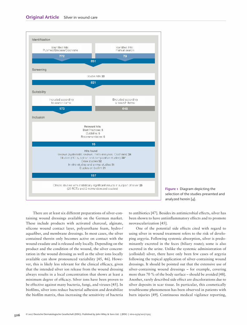
Overall, 851 articles were identifi ed using the aforementi-oned search parameters. A subsequent review showed that 30 articles had been collected twice. Following a closer look at the abstracts of the remaining 821 articles, 157 articles were determined to be studies that met the inclusion criteria and warranted further analysis. These studies were broken down into the following categories (Figure 1 ) [ 5 ] : 34 review articles (systematic reviews, meta-analyses) 39 clinical studies (RCTs and comparative cohort
studies) 32 case series or case studies 31 preclinical studies (in vitro studies, animal studies) 21 studies on biofi lm
With respect to the primary objective of the present ana-lysis, the 39 clinical studies were of particular interest. They included 31 RCTs and eight controlled cohort studies. Of these 39 clinical studies, 28 showed statistically signifi cant outcome parameters in favor of silver (Tables 1–5 ). One study revealed a comparable reduction in wound pain and bacte-rial load for silver and polyhexanide (PHMB). While nine of these studies investigated burn injuries (including eight RCTs), 20 addressed other indications (9: venous leg ulcers; 3: pressure ulcers; 2: chronic wounds; 1: diabetic foot ulcer; and 5: other types of wounds). In 16 studies, the primary parameter was wound resolution (healing, wound closure, wound size/area reduction, completed re-epithelialization). Twelve studies examined quality of life including pain; eight, cost-effectiveness; and three, the reduction of bacterial load (bioburden) (Table 6 ).
Ten clinical studies (only) partly showed statistically signifi cant outcome parameters in favor of silver (Table 7 ). In other clinical studies, other endpoints were partially supported the use of silver, such as surgical site infec-tions (SSIs), reduction in wound size or resistance to silver ( Table 8 ).
Discussion
In its elemental form, silver is nonreactive and has thus no an-timicrobial effects. Silver atoms (Ag or Ag 0 ) only acquire such properties when they lose an electron and become positively charged silver ions (Ag + ). The latter bind to peptidoglycans in the bacterial cell membrane, thus leading to the destruction of the bacterial cell wall. Silver ions transported into the cell disrupt numerous cell functions by binding to proteins and interfering with energy production, enzyme function, and cell replication. Given these highly diverse effects on various target structures, development of bacterial resistance to silver is rather unlikely and clinically irrelevant [ 44 ] .
Original Article Silver in wound care
526 © 2017 Deutsche Dermatologische Gesellschaft (DDG). Published by John Wiley & Sons Ltd. | JDDG | 1610-0379/2017/1505
There are at least six different preparations of silver-con-taining wound dressings available on the German market. These include products with activated charcoal, alginate, silicone wound contact layer, polyurethane foam, hydro-/aquafi ber, and membrane dressings. In most cases, the silver contained therein only becomes active on contact with the wound exudate and is released only locally. Depending on the product and the condition of the wound, the silver concent-ration in the wound dressing as well as the silver ions locally available can show pronounced variability [ 45, 46 ] . Howe-ver, this is likely less relevant for the clinical effi cacy, given that the intended silver ion release from the wound dressing always results in a local concentration that shows at least a minimum degree of effi cacy. Silver ions have been proven to be effective against many bacteria, fungi, and viruses [ 45 ] . In biofi lms, silver ions reduce bacterial adhesion and destabilize the biofi lm matrix, thus increasing the sensitivity of bacteria
to antibiotics [ 47 ] . Besides its antimicrobial effects, silver has been shown to have antiinfl ammatory effects and to promote neovascularization [ 45 ] .
One of the potential side effects cited with regard to using silver in wound treatment refers to the risk of develo-ping argyria. Following systemic absorption, silver is predo-minantly excreted in the feces (biliary route); some is also excreted in the urine. Unlike the systemic administration of (colloidal) silver, there have only been few cases of argyria following the topical application of silver-containing wound dressings. It should be pointed out that the extensive use of silver-containing wound dressings – for example, covering more than 70 % of the body surface – should be avoided [ 48 ] . Another, rarely described side effect are discolorations due to silver deposits in scar tissue. In particular, this cosmetically troublesome phenomenon has been observed in patients with burn injuries [ 49 ] . Continuous medical vigilance reporting,
Figure 1 Diagram depicting the selection of the studies presented and analyzed herein [ 4 ] .
Original Article Silver in wound care
527© 2017 Deutsche Dermatologische Gesellschaft (DDG). Published by John Wiley & Sons Ltd. | JDDG | 1610-0379/2017/1505
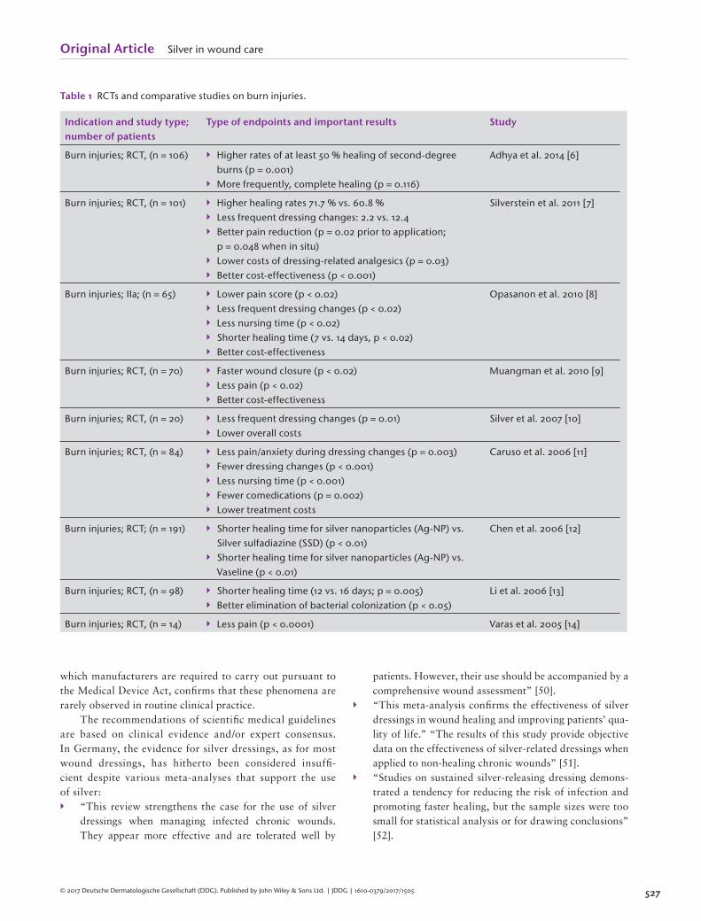
Table 1 RCTs and comparative studies on burn injuries.
Indication and study type; number of patients
Type of endpoints and important results Study
Burn injuries; RCT, (n = 106) Higher rates of at least 50 % healing of second-degree burns (p = 0.001)
More frequently, complete healing (p = 0.116)
Adhya et al. 2014 [ 6 ]
Burn injuries; RCT, (n = 101) Higher healing rates 71.7 % vs. 60.8 % Less frequent dressing changes: 2.2 vs. 12.4 Better pain reduction (p = 0.02 prior to application;
p = 0.048 when in situ) Lower costs of dressing-related analgesics (p = 0.03) Better cost-effectiveness (p < 0.001)
Silverstein et al. 2011 [ 7 ]
Burn injuries ; IIa; (n = 65) Lower pain score (p < 0.02) Less frequent dressing changes (p < 0.02) Less nursing time (p < 0.02) Shorter healing time (7 vs. 14 days, p < 0.02) Better cost-effectiveness
Opasanon et al. 2010 [ 8 ]
Burn injuries; RCT, (n = 70) Faster wound closure (p < 0.02) Less pain (p < 0.02) Better cost-effectiveness
Muangman et al. 2010 [ 9 ]
Burn injuries; RCT, (n = 20) Less frequent dressing changes (p = 0.01) Lower overall costs
Silver et al. 2007 [ 10 ]
Burn injuries; RCT, (n = 84) Less pain/anxiety during dressing changes (p = 0.003) Fewer dressing changes (p < 0.001) Less nursing time (p < 0.001) Fewer comedications (p = 0.002) Lower treatment costs
Caruso et al. 2006 [ 11 ]
Burn injuries; RCT; (n = 191) Shorter healing time for silver nanoparticles (Ag-NP) vs. Silver sulfadiazine (SSD) (p < 0.01)
Shorter healing time for silver nanoparticles (Ag-NP) vs. Vaseline (p < 0.01)
Chen et al. 2006 [ 12 ]
Burn injuries; RCT, (n = 98) Shorter healing time (12 vs. 16 days; p = 0.005) Better elimination of bacterial colonization (p < 0.05)
Li et al. 2006 [ 13 ]
Burn injuries; RCT, (n = 14) Less pain (p < 0.0001) Varas et al. 2005 [ 14 ]
which manufacturers are required to carry out pursuant to the Medical Device Act, confi rms that these phenomena are rarely observed in routine clinical practice.
The recommendations of scientifi c medical guidelines are based on clinical evidence and/or expert consensus. In Germany, the evidence for silver dressings, as for most wound dressings, has hitherto been considered insuffi -cient despite various meta-analyses that support the use of silver: “This review strengthens the case for the use of silver
dressings when managing infected chronic wounds. They appear more effective and are tolerated well by
patients. However, their use should be accompanied by a comprehensive wound assessment” [ 50 ] .
“This meta-analysis confi rms the effectiveness of silver dressings in wound healing and improving patients’ qua-lity of life.” “The results of this study provide objective data on the effectiveness of silver-related dressings when applied to non-healing chronic wounds” [ 51 ] .
“Studies on sustained silver-releasing dressing demons-trated a tendency for reducing the risk of infection and promoting faster healing, but the sample sizes were too small for statistical analysis or for drawing conclusions” [ 52 ] .
Original Article Silver in wound care
528 © 2017 Deutsche Dermatologische Gesellschaft (DDG). Published by John Wiley & Sons Ltd. | JDDG | 1610-0379/2017/1505
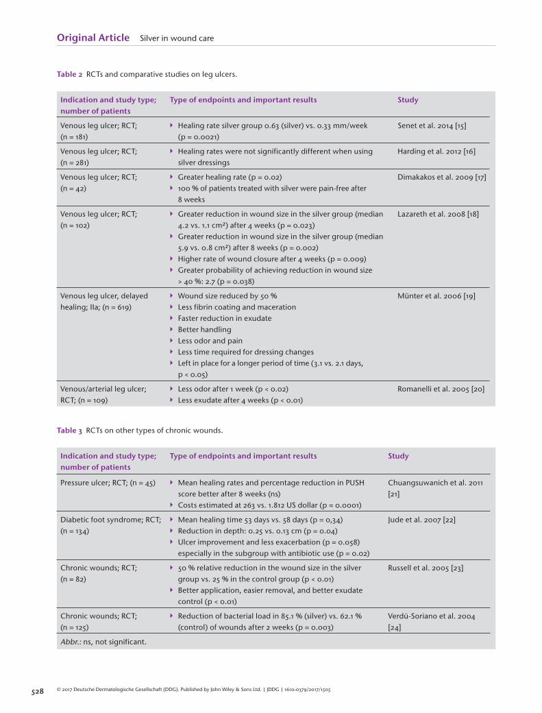
Table 2 RCTs and comparative studies on leg ulcers.
Indication and study type; number of patients
Type of endpoints and important results Study
Venous leg ulcer; RCT; (n = 181)
Healing rate silver group 0.63 (silver) vs. 0.33 mm/week (p = 0.0021)
Senet et al. 2014 [ 15 ]
Venous leg ulcer; RCT; (n = 281)
Healing rates were not significantly different when using silver dressings
Harding et al. 2012 [ 16 ]
Venous leg ulcer; RCT; (n = 42)
Greater healing rate (p = 0.02) 100 % of patients treated with silver were pain-free after
8 weeks
Dimakakos et al. 2009 [ 17 ]
Venous leg ulcer; RCT; (n = 102)
Greater reduction in wound size in the silver group (median 4.2 vs. 1.1 cm²) after 4 weeks (p = 0.023)
Greater reduction in wound size in the silver group (median 5.9 vs. 0.8 cm²) after 8 weeks (p = 0.002)
Higher rate of wound closure after 4 weeks (p = 0.009) Greater probability of achieving reduction in wound size
> 40 %: 2.7 (p = 0.038)
Lazareth et al. 2008 [ 18 ]
Venous leg ulcer, delayed healing ; IIa; (n = 619)
Wound size reduced by 50 % Less fibrin coating and maceration Faster reduction in exudate Better handling Less odor and pain Less time required for dressing changes Left in place for a longer period of time (3.1 vs. 2.1 days,
p < 0.05)
Münter et al. 2006 [ 19 ]
Venous/arterial leg ulcer; RCT; (n = 109)
Less odor after 1 week (p < 0.02) Less exudate after 4 weeks (p < 0.01)
Romanelli et al. 2005 [ 20 ]
Table 3 RCTs on other types of chronic wounds.
Indication and study type; number of patients
Type of endpoints and important results Study
Pressure ulcer; RCT; (n = 45) Mean healing rates and percentage reduction in PUSH score better after 8 weeks (ns)
Costs estimated at 263 vs. 1.812 US dollar (p = 0.0001)
Chuangsuwanich et al. 2011 [ 21 ]
Diabetic foot syndrome; RCT; (n = 134)
Mean healing time 53 days vs. 58 days (p = 0,34) Reduction in depth: 0.25 vs. 0.13 cm (p = 0.04) Ulcer improvement and less exacerbation (p = 0.058)
especially in the subgroup with antibiotic use (p = 0.02)
Jude et al. 2007 [ 22 ]
Chronic wounds; RCT; (n = 82)
50 % relative reduction in the wound size in the silver group vs. 25 % in the control group (p < 0.01)
Better application, easier removal, and better exudate control (p < 0.01)
Russell et al. 2005 [ 23 ]
Chronic wounds; RCT; (n = 125)
Reduction of bacterial load in 85.1 % (silver) vs. 62.1 % (control) of wounds after 2 weeks (p = 0.003)
Verdú-Soriano et al. 2004 [ 24 ]
Abbr.: ns, not significant.
Original Article Silver in wound care
529© 2017 Deutsche Dermatologische Gesellschaft (DDG). Published by John Wiley & Sons Ltd. | JDDG | 1610-0379/2017/1505
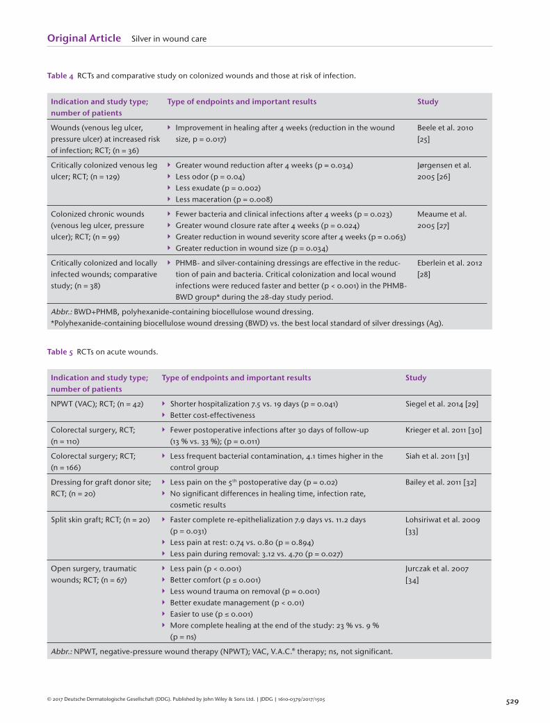
Table 4 RCTs and comparative study on colonized wounds and those at risk of infection.
Indication and study type; number of patients
Type of endpoints and important results Study
Wounds (venous leg ulcer, pressure ulcer) at increased risk of infection; RCT; (n = 36)
Improvement in healing after 4 weeks (reduction in the wound size, p = 0.017)
Beele et al. 2010 [ 25 ]
Critically colonized venous leg ulcer; RCT; (n = 129)
Greater wound reduction after 4 weeks (p = 0.034) Less odor (p = 0.04) Less exudate (p = 0.002) Less maceration (p = 0.008)
Jørgensen et al. 2005 [ 26 ]
Colonized chronic wounds (venous leg ulcer, pressure ulcer); RCT; (n = 99)
Fewer bacteria and clinical infections after 4 weeks (p = 0.023) Greater wound closure rate after 4 weeks (p = 0.024) Greater reduction in wound severity score after 4 weeks (p = 0.063) Greater reduction in wound size (p = 0.034)
Meaume et al. 2005 [ 27 ]
Critically colonized and locally infected wounds; comparative study; (n = 38)
PHMB- and silver-containing dressings are effective in the reduc-tion of pain and bacteria . Critical colonization and local wound infections were reduced faster and better (p < 0.001) in the PHMB-BWD group* during the 28-day study period.
Eberlein et al. 2012 [ 28 ]
Abbr.: BWD+PHMB, polyhexanide-containing biocellulose wound dressing. *Polyhexanide-containing biocellulose wound dressing (BWD) vs. the best local standard of silver dressings (Ag).
Table 5 RCTs on acute wounds.
Indication and study type; number of patients
Type of endpoints and important results Study
NPWT (VAC); RCT; (n = 42) Shorter hospitalization 7.5 vs. 19 days (p = 0.041) Better cost-effectiveness
Siegel et al. 2014 [ 29 ]
Colorectal surgery, RCT; (n = 110)
Fewer postoperative infections after 30 days of follow-up (13 % vs. 33 %); (p = 0.011)
Krieger et al. 2011 [ 30 ]
Colorectal surgery; RCT; (n = 166)
Less frequent bacterial contamination, 4.1 times higher in the control group
Siah et al. 2011 [ 31 ]
Dressing for graft donor site; RCT; (n = 20)
Less pain on the 5 th postoperative day (p = 0.02) No significant differences in healing time, infection rate,
cosmetic results
Bailey et al. 2011 [ 32 ]
Split skin graft; RCT; (n = 20) Faster complete re-epithelialization 7.9 days vs. 11.2 days (p = 0.031)
Less pain at rest: 0.74 vs. 0.80 (p = 0.894) Less pain during removal: 3.12 vs. 4.70 (p = 0.027)
Lohsiriwat et al. 2009 [ 33 ]
Open surgery, traumatic wounds; RCT; (n = 67)
Less pain (p < 0.001) Better comfort (p ≤ 0.001) Less wound trauma on removal (p = 0.001) Better exudate management (p < 0.01) Easier to use (p ≤ 0.001) More complete healing at the end of the study: 23 % vs. 9 %
(p = ns)
Jurczak et al. 2007 [ 34 ]
Abbr.: NPWT, negative-pressure wound therapy (NPWT); VAC, V.A.C. ® therapy; ns, not significant.
Original Article Silver in wound care
530 © 2017 Deutsche Dermatologische Gesellschaft (DDG). Published by John Wiley & Sons Ltd. | JDDG | 1610-0379/2017/1505
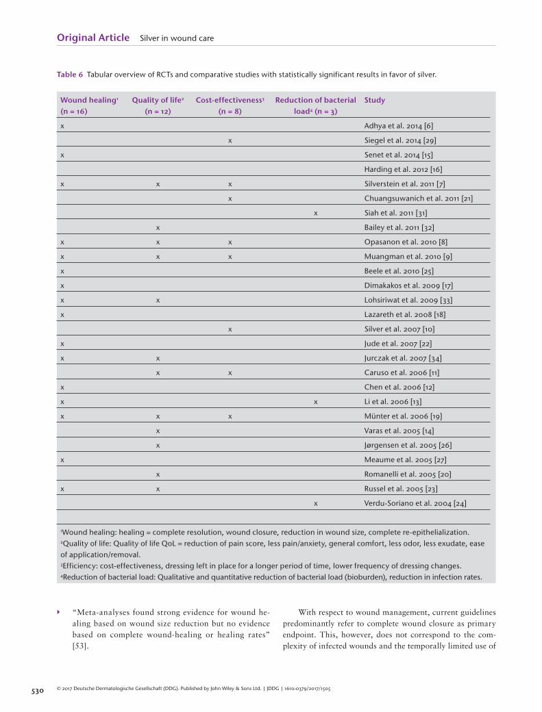
Table 6 Tabular overview of RCTs and comparative studies with statistically significant results in favor of silver.
Wound healing 1 (n = 16)
Quality of life 2 (n = 12)
Cost-effectiveness 3 (n = 8)
Reduction of bacterial load 4 (n = 3)
Study
x Adhya et al. 2014 [ 6 ]
x Siegel et al. 2014 [ 29 ]
x Senet et al. 2014 [ 15 ]
Harding et al. 2012 [ 16 ]
x x x Silverstein et al. 2011 [ 7 ]
x Chuangsuwanich et al. 2011 [ 21 ]
x Siah et al. 2011 [31]
x Bailey et al. 2011 [ 32 ]
x x x Opasanon et al. 2010 [ 8 ]
x x x Muangman et al. 2010 [ 9 ]
x Beele et al. 2010 [ 25 ]
x Dimakakos et al. 2009 [ 17 ]
x x Lohsiriwat et al. 2009 [ 33 ]
x Lazareth et al. 2008 [ 18 ]
x Silver et al. 2007 [ 10 ]
x Jude et al. 2007 [ 22 ]
x x Jurczak et al. 2007 [ 34 ]
x x Caruso et al. 2006 [ 11 ]
x Chen et al. 2006 [ 12 ]
x x Li et al. 2006 [ 13 ]
x x x Münter et al. 2006 [ 19 ]
x Varas et al. 2005 [ 14 ]
x Jørgensen et al. 2005 [ 26 ]
x Meaume et al. 2005 [ 27 ]
x Romanelli et al. 2005 [ 20 ]
x x Russel et al. 2005 [ 23 ]
x Verdu-Soriano et al. 2004 [ 24 ]
1Wound healing: healing = complete resolution, wound closure, reduction in wound size, complete re-epithelialization. 2Quality of life: Quality of life QoL = reduction of pain score, less pain/anxiety, general comfort, less odor, less exudate, ease of application/removal. 3Efficiency: cost-effectiveness, dressing left in place for a longer period of time, lower frequency of dressing changes. 4Reduction of bacterial load: Qualitative and quantitative reduction of bacterial load (bioburden), reduction in infection rates.
“Meta-analyses found strong evidence for wound he-aling based on wound size reduction but no evidence based on complete wound-healing or healing rates” [ 53 ] .
With respect to wound management, current guidelines predominantly refer to complete wound closure as primary endpoint. This, however, does not correspond to the com-plexity of infected wounds and the temporally limited use of

Original Article Silver in wound care
531© 2017 Deutsche Dermatologische Gesellschaft (DDG). Published by John Wiley & Sons Ltd. | JDDG | 1610-0379/2017/1505
Table 7 Tabular overview of clinical trials (RCTs, multicenter studies) with descriptive, statistically not significant results in favor of silver.
Method Result Study
Multicenter real-life study (n = 121) NGAD* possibly plays a role in wound healing due to elimination of biofilm.
Cost reduction of 30 %
Walker et al. 2015 [ 35 ]
RCT; skin donor sites, Ag hydrofiber vs. standard (n = 70)
Wound pain reduced with both treatment methods Blome-Eberwein et al. 2010 [ 36 ]
RCT; silver vs. cadexomer iodine (n = 281)
Silver shows faster wound healing Miller et al. 2010 [ 37 ]
RCT; Ag alginate vs. alginate (n = 42)
No difference in local signs of infection and tolerability Difference with respect to bacteriological wound status in
favor of silver
Trial et al. 2010 [ 38 ]
Multicenter, non-comparative study in second-degree burns (n = 41)
No secondary infections Only one colonization with Staphylococcus aureus out of
121 samples
Carsin et al. 2004 [ 39 ]
Abbr.: NGAD, next-generation antimicrobial dressing. *NGAD = Aquacel ® vs. standard protocol.
Table 8 Tabular overview of clinical trials (RCTs, clinical studies, case studies) with other assessment criteria.
Method Result Study
RCT (n = 315); comparison of metallic vs. ionic silver
No difference in wound healing and infections Differences with regard to patient comfort and
dressing- specific factors
Dickinson et al. 2015 [ 40 ]
RCT (n = 500); silver-releasing wound dressing vs. gauze in vascular surgery of the lower extremities
The 30-day complication rate was 30 %; postopera-tive wound infections (SSIs) were the most common complications
In the ITT analysis, there were no complications when using the silver dressing
Ozaki et al. 2015 [ 41 ]
Comparative study of PHMB vs. silver in critically colonized or infected wounds (n = 38)
After 28 days, there was a statistically significantly greater reduction in wound size (p < 0.001) in favor of PHMB.
Eberlein et al. 2012 [ 28 ]
RCT (n = 110) in colorectal surgery
Statistically significantly fewer SSIs after 30 days of follow-up of a silver-containing wound dressing vs. gauze (p = 0.011)
Krieger et al. 2011 [ 30 ]
Clinical trial Only small numbers of sil-gen-resistant bacteria (MRSA and MR-CNS) were detected in wounds or the nose. These bacteria are killed by silver.
Loh et al. 2009 [ 42 ]
RCT (n = 213) of ulcerations; VULCAN trial
No statistically significant effects of silver Michaels et al. 2009 [ 43 ]
Case study; silver-resistant Enterobacter cloacae isolated from a leg ulcer
Silver resistance of bacteria is extremely rare Lansdown et al. 2007 [ 43 ]
Abbr.: PHMB, polyhexanide; SSIs, surgical site infections .
Original Article Silver in wound care
532 © 2017 Deutsche Dermatologische Gesellschaft (DDG). Published by John Wiley & Sons Ltd. | JDDG | 1610-0379/2017/1505
antimicrobial strategies such as the application of silver-cont-aining wound dressings. Given that the present meta-analysis therefore included other endpoints as well, we were able to fi nd clinical evidence for silver-containing wound dressings for various indications. In some cases, this evidence was even statistically signifi cant. In the period 2000–2015, numerous clinical studies on silver in wound care were published, in-cluding 31 RCTs. The majority of these studies provide – fre-quently signifi cant – evidence for positive effects on different aspects of wound healing in various indications, Besides anti-microbial effects, these include the promotion of wound hea-ling, improvement in quality of life, and benefi ts with regard to cost-effectiveness.
The present meta-analysis also included studies on very diverse types of wounds and therapeutic agents. Thus, a conclusive assessment as to the use of a particular wound dressing for a specifi c type of wound continues to be dif-fi cult. On the other hand, if one looks at the available evi-dence for a particular silver-containing foam dressing, one fi nds four RCTs with a total of 685 patients with venous leg ulcers. For this specifi c indication, this particular dressing was objectively shown to be statistically signifi cantly supe-rior to a non-active wound dressing with regard to various parameters such as the reduction in wound size (43.5 % vs. 26.3 %) [ 54 ] .
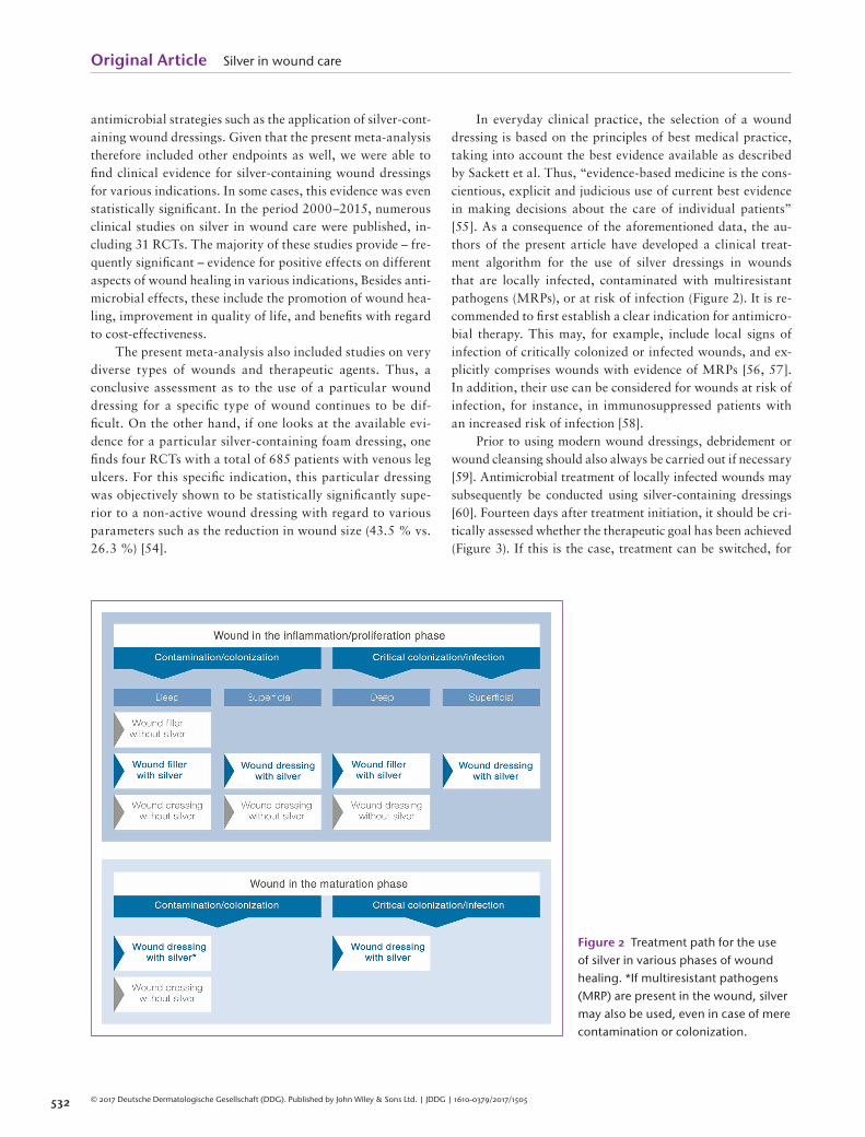
In everyday clinical practice, the selection of a wound dressing is based on the principles of best medical practice, taking into account the best evidence available as described by Sackett et al. Thus, “evidence-based medicine is the cons-cientious, explicit and judicious use of current best evidence in making decisions about the care of individual patients” [ 55 ] . As a consequence of the aforementioned data, the au-thors of the present article have developed a clinical treat-ment algorithm for the use of silver dressings in wounds that are locally infected, contaminated with multiresistant pathogens (MRPs), or at risk of infection (Figure 2 ). It is re-commended to fi rst establish a clear indication for antimicro-bial therapy. This may, for example, include local signs of infection of critically colonized or infected wounds, and ex-plicitly comprises wounds with evidence of MRPs [ 56, 57 ] . In addition, their use can be considered for wounds at risk of infection, for instance, in immunosuppressed patients with an increased risk of infection [ 58 ] .
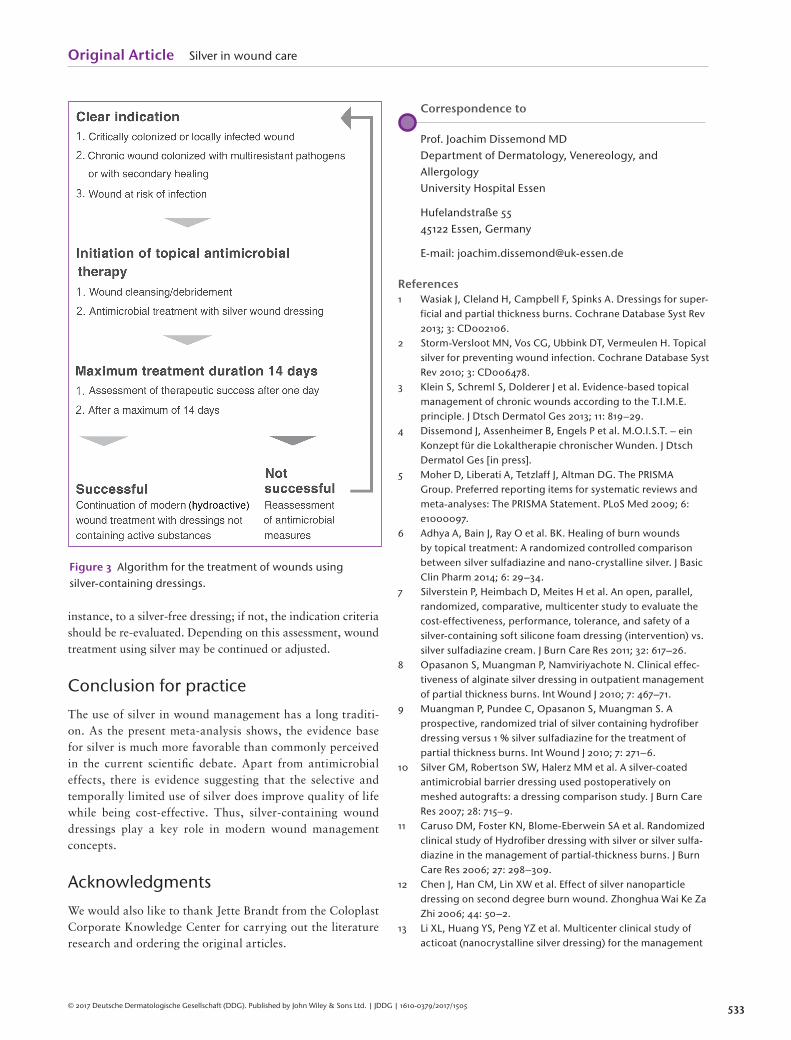
Prior to using modern wound dressings, debridement or wound cleansing should also always be carried out if necessary [ 59 ] . Antimicrobial treatment of locally infected wounds may subsequently be conducted using silver-containing dressings [ 60 ] . Fourteen days after treatment initiation, it should be cri-tically assessed whether the therapeutic goal has been achieved (Figure 3 ). If this is the case, treatment can be switched, for
Figure 2 Treatment path for the use of silver in various phases of wound healing. *If multiresistant pathogens (MRP) are present in the wound, silver may also be used, even in case of mere contamination or colonization.
Original Article Silver in wound care
533© 2017 Deutsche Dermatologische Gesellschaft (DDG). Published by John Wiley & Sons Ltd. | JDDG | 1610-0379/2017/1505
instance, to a silver-free dressing; if not, the indication criteria should be re-evaluated. Depending on this assessment, wound treatment using silver may be continued or adjusted.
Conclusion for practice
The use of silver in wound management has a long traditi-on. As the present meta-analysis shows, the evidence base for silver is much more favorable than commonly perceived in the current scientifi c debate. Apart from antimicrobial effects, there is evidence suggesting that the selective and temporally limited use of silver does improve quality of life while being cost-effective. Thus, silver-containing wound dressings play a key role in modern wound management concepts.
Acknowledgments
We would also like to thank Jette Brandt from the Coloplast Corporate Knowledge Center for carrying out the literature research and ordering the original articles.
Correspondence to
Prof. Joachim Dissemond MD Department of Dermatology, Venereology, and Allergology University Hospital Essen
Hufelandstraße 55 45122 Essen, Germany
E-mail: [email protected]
References 1 Wasiak J , Cleland H , Campbell F , Spinks A . Dressings for super-
ficial and partial thickness burns . Cochrane Database Syst Rev 2013 ; 3 : CD002106 .
2 Storm-Versloot MN , Vos CG , Ubbink DT , Vermeulen H . Topical silver for preventing wound infection . Cochrane Database Syst Rev 2010 ; 3 : CD006478 .
3 Klein S , Schreml S , Dolderer J et al. Evidence-based topical management of chronic wounds according to the T.I.M.E. principle . J Dtsch Dermatol Ges 2013 ; 11 : 819 – 29 .
4 Dissemond J , Assenheimer B , Engels P et al. M.O.I.S.T. – ein Konzept für die Lokaltherapie chronischer Wunden . J Dtsch Dermatol Ges [in press].
5 Moher D , Liberati A , Tetzlaff J , Altman DG . The PRISMA Group . Preferred reporting items for systematic reviews and meta-analyses: The PRISMA Statement . PLoS Med 2009 ; 6 : e1000097 .
6 Adhya A , Bain J , Ray O et al. BK. Healing of burn wounds by topical treatment: A randomized controlled comparison between silver sulfadiazine and nano-crystalline silver . J Basic Clin Pharm 2014 ; 6 : 29 – 34 .
7 Silverstein P , Heimbach D , Meites H et al. An open, parallel, randomized, comparative, multicenter study to evaluate the cost-effectiveness, performance, tolerance, and safety of a silver-containing soft silicone foam dressing (intervention) vs. silver sulfadiazine cream . J Burn Care Res 2011 ; 32 : 617 – 26 .
8 Opasanon S , Muangman P , Namviriyachote N . Clinical effec-tiveness of alginate silver dressing in outpatient management of partial thickness burns . Int Wound J 2010 ; 7 : 467 – 71 .
9 Muangman P , Pundee C , Opasanon S , Muangman S . A prospective, randomized trial of silver containing hydrofiber dressing versus 1 % silver sulfadiazine for the treatment of partial thickness burns . Int Wound J 2010 ; 7 : 271 – 6 .
10 Silver GM , Robertson SW , Halerz MM et al. A silver-coated antimicrobial barrier dressing used postoperatively on meshed autografts: a dressing comparison study . J Burn Care Res 2007 ; 28 : 715 – 9 .
11 Caruso DM , Foster KN , Blome-Eberwein SA et al. Randomized clinical study of Hydrofiber dressing with silver or silver sulfa-diazine in the management of partial-thickness burns . J Burn Care Res 2006 ; 27 : 298 – 309 .
12 Chen J , Han CM , Lin XW et al. Effect of silver nanoparticle dressing on second degree burn wound . Zhonghua Wai Ke Za Zhi 2006 ; 44 : 50 – 2 .
13 Li XL , Huang YS , Peng YZ et al. Multicenter clinical study of acticoat (nanocrystalline silver dressing) for the management
Figure 3 Algorithm for the treatment of wounds using silver-containing dressings.
Original Article Silver in wound care
534 © 2017 Deutsche Dermatologische Gesellschaft (DDG). Published by John Wiley & Sons Ltd. | JDDG | 1610-0379/2017/1505
of residual burn wounds . Zhonghua Shao Shang Za Zhi 2006 ; 22 : 15 – 8 .
14 Varas RP , O’Keeffe T , Namias N et al. A prospective, random-ized trial of Acticoat versus silver sulfadiazine in the treatment of partial-thickness burns: which method is less painful? J Burn Care Rehabil 2005 ; 26 : 344 – 7 .
15 Senet P , Bause R , Jørgensen B , Fogh K . Clinical efficacy of a silver-releasing foam dressing in venous leg ulcer healing: a randomised controlled trial . Int Wound J 2014 ; 11 : 649 – 55 .
16 Harding K , Gottrup F , Jawien A et al. A prospective, multi-centre, randomised, open label, parallel, comparative study to evaluate effects of AQUACEL ® Ag and Urgotul ® Silver dressing on healing of chronic venous leg ulcers . Int Wound J 2012 ; 9 : 285 – 94 .
17 Dimakakos EP , Katsenis K , Kalemikerakis J et al. Infected venous leg ulcers: management with silver-releasing foam dressing . Wounds 2009 ; 21 : 4 – 8 .
18 Lazareth I , Meaume S , Sigal-Grinberg ML et al. The role of a silver releasing lipido-colloid contact layer in venous leg ulcers presenting inflammatory signs suggesting heavy bacterial col-onization: Results of a randomized controlled study . Wounds 2008 ; 20 : 158 – 66 .
19 Münter KC , Beele H , Russell L et al. Effect of a sustained silver-releasing dressing on ulcers with delayed healing: the CON-TOP study . J Wound Care 2006 ; 15 : 199 – 206 .
20 Romanelli M , Price P . Health-related quality of life aspects after treatment with a foam dressing and a silver-containing foam dressing in chronic leg ulcers . J Am Acad Dermatol 2005 ; 52 : 21 .
21 Chuangsuwanich A , Charnsanti O , Lohsiriwat V et al. The effi-cacy of silver mesh dressing compared with silver sulfadiazine cream for the treatment of pressure ulcers . J Med Assoc Thai 2011 ; 94 : 559 – 65 .
22 Jude EB , Apelqvist J , Spraul M , Martini J . Silver Dressing Study Group . Prospective randomized controlled study of hydrofiber dressing containing ionic silver or calcium alginate dressings in non-ischaemic diabetic foot ulcers . Diabet Med 2007 ; 24 : 280 – 8 .
23 Russell L. The CONTOP multinational study: preliminary data from the UK arm . Wounds UK 2005 ; 1 : 44 – 54 .
24 Verdú Soriano J , Rueda López J , Martínez Cuervo F , Soldevilla Agreda J . Effects of an activated charcoal silver dressing on chronic wounds with no clinical signs of infection . J Wound Care 2004 ; 13 : 419 : 421 – 3 .
25 Beele H , Meuleneire F , Nahuys M , Percival SL . A prospective randomised open label study to evaluate the potential of a new silver alginate/carboxymethylcellulose antimicrobial wound dressing to promote wound healing . Int Wound J 2010 ; 7 : 262 – 70 .
26 Jørgensen B , Price P , Andersen KE et al. The silver-releasing foam dressing, Contreet Foam, promotes faster healing of critically colonised venous leg ulcers: a randomised, con-trolled trial . Int Wound J 2005 ; 2 : 64 – 73 .
27 Meaume S , Vallet D , Morere MN , Téot L . Evaluation of a silver-releasing hydroalginate dressing in chronic wounds with signs of local infection . J Wound Care 2005 ; 14 : 411 – 9 .
28 Eberlein T , Haemmerle G , Signer M et al. Comparison of PHMB-containing dressing and silver dressings in patients
with critically colonised or locally infected wounds . J Wound Care 2012 ; 21 : 12 – 20 .
29 Siegel HJ , Herrera DF , Gay J . Silver negative pressure dressing with vacuum-assisted closure of massive pelvic and extremity wounds . Clin Orthop Relat Res 2014 ; 472 : 830 – 5 .
30 Krieger BR , Davis DM , Sanchez JE et al. The use of silver nylon in preventing surgical site infections following colon and rectal surgery . Dis Colon Rectum 2011 ; 54 : 1014 – 9 .
31 Siah CJ , Yatim J . Efficacy of a total occlusive ionic silver- containing dressing combination in decreasing risk of surgical site infection: an RCT . J Wound Care 2011 ; 20 : 561 – 8 .
32 Bailey S , Carmean M , Cinat M et al. A randomized comparison study of Aquacel Ag and Glucan II as donor site dressings with regard to healing time, cosmesis, infection rate, and patient’s perceived pain: a pilot study . J Burn Care Res 2011 ; 32 : 627 – 32 .
33 Lohsiriwat V , Chuangsuwanich A . Comparison of the ionic silver-containing hydrofiber and paraffin gauze dressing on split-thickness skin graft donor sites . Ann Plast Surg 2009 ; 62 : 421 – 2 .
34 Jurczak F , Dugré T , Johnstone A et al. Aquacel Ag Surgical/Trauma Wound Study Group . Randomised clinical trial of hydrofiber dressing with silver versus povidone-iodine gauze in the management of open surgical and traumatic wounds . Int Wound J 2007 ; 4 : 66 – 76 .
35 Walker M , Metclaf D , Parsons D , Bowler P . A real-life clinical evaluation of a next-generation antimicrobial dressing on acute and chronic wounds . J Wound Care 2015 ; 24 : 11 – 22 .
36 Blome-Eberwein S , Johnson RM , Miller SF et al. Hydrofiber dressing with silver for the management of split-thickness donor sites: a randomized evaluation of two protocols of care . Burns 2010 ; 36 : 665 – 72 .
37 Miller CN , Newall N , Kapp SE et al. A randomized-controlled trial comparing cadexomer iodine and nanocrystalline silver on the healing of leg ulcers . Wound Repair Regen 2010 ; 18 : 359 – 67 .
38 Trial C , Darbas H , Lavigne JP et al. Assessment of the antimi-crobial effectiveness of a new silver alginate wound dressing: a RCT . J Wound Care 2010 ; 19 : 20 – 6 .
39 Carsin H , Wassermann D , Pannier M et al. A silver sulphad-iazine-impregnated lipidocolloid wound dressing to treat second-degree burns . J Wound Care 2004 ; 13 : 145 – 8 .
40 Dickinson JC , Culver CR , Baker JW . A prospective, randomized controlled trial comparing 3 dressing types following ster-notomy . Ostomy Wound Manage 2015 ; 61 : 42 – 9 .
41 Ozaki CK , Hamdan AD , Barshes NR et al. Prospective, randomized, multi-institutional clinical trial of a silver alginate dressing to reduce lower extremity vascular surgery wound complications . J Vasc Surg 2015 ; 61 : 419 – 27 .
42 Loh JV , Percival SL , Woods EJ et al. Silver resistance in MRSA isolated from wound and nasal sources in humans and animals . Int Wound J 2009 ; 6 : 32 – 8 .
43 Michaels JA , Campbell WB , King BM et al. A prospective randomised controlled trial and economic modelling of antimicrobial silver dressings versus non-adherent control dressings for venous leg ulcers: the VULCAN trial . Health Technol Assess 2009 ; 13 : 1 – 114 .
44 Landsdown AB , Williams A . Bacterial resistance to silver in wound care and medical devices . J Wound Care 2007 ; 16 : 15 – 9 .
Original Article Silver in wound care
535© 2017 Deutsche Dermatologische Gesellschaft (DDG). Published by John Wiley & Sons Ltd. | JDDG | 1610-0379/2017/1505
45 International consensus . Appropriate use of silver dressings in wounds . An expert working group consensus. London: Wounds International, 2012 , www.woundsinternational.com. Available on http://www.woundsinternational.com/media/issues/567/files/content_10381.pdf [Last accessed November 10, 2016].
46 http://www.werner-sellmer.de [Last accessed November 10, 2016].
47 Kostenko V , Lyczak J , Turner K , Martinuzzi RJ . Impact of silver-containing wound dressings on bacterial biofilm viability and susceptibility to antibiotics during prolonged treatment . Antimicrob Agents Chemother 2010 ; 54 : 5120 – 31 .
48 McCague A , Joe VC . A case of argyria and acute leukopenia associated with the use of an antimicrobial soft silicone foam dressing . J Burn Care Res 2016 ; 37 ( 5 ): e493 - 6 .
49 Wang XQ , Chang HE , Francis R et al. Silver deposits in cutane-ous burn scar tissue is a common phenomenon following ap-plication of a silver dressing . J Cutan Pathol 2009 ; 36 : 788 – 92 .
50 Lo SF , Hayter M , Chang CJ et al. A systematic review of silver-releasing dressings in the management of infected chronic wounds . J Clin Nurs 2008 ; 17 : 1973 – 85 .
51 Lo SF , Chang CJ , Hu WY et al. The effectiveness of silver-releasing dressings in the management of non-healing chronic wounds: a meta-analysis . J Clin Nurs 2009 ; 18 : 716 – 28 .
52 Health Quality Ontario . Management of chronic pressure ulcers: an evidence-based analysis . Ont Health Technol Assess Ser 2009 ; 9 : 1 – 203 .
53 Carter MJ , Tingley-Kelley K , Warriner RA . Silver treatments and silver-impregnated dressings for the healing of leg wounds and ulcers: a systematic review and meta-analysis . J Am Acad Dermatol 2010 ; 63 : 668 – 79 .
54 Leaper D , Münter C , Meaume S et al. The use of biatain Ag in hard-to-heal venous leg ulcers: meta-analysis of randomised controlled trials . PLoS One 2013 ; 8 : e67083 .
55 Sackett DL , Rosenberg WM , Gray JAM et al. Evidence based medicine: what it is and what it isn’t . BMJ 1996 ; 312 : 71 .
56 Dissemond J. Methicillin resistant Staphylococcus aureus (MRSA): Diagnostic, clinical relevance and therapy . J Dtsch Dermatol Ges 2009 ; 7 : 544 – 51 .
57 Schwarzkopf A , Dissemond J . Indications and practical implementation of microbiologic diagnostics in patients with chronic wounds . J Dtsch Dermatol Ges 2015 ; 13 : 203 – 9 .
58 Dissemond J , Assadian O , Gerber V et al. Classification of wounds at risk and their antimicrobial treatment with poli-hexanide: a practice-oriented expert recommendation . Skin Pharmacol Physiol 2011 ; 24 : 245 – 55 .
59 Strohal R , Dissemond J , Jordan O’Brien J et al. An updated overview and clarification of the principle role of debride-ment . J Wound Care 2013 ; 22 ( Suppl. ): 1 – 52 .
60 Dissemond J , Augustin M , Eming SA et al. Modern wound care – practical aspects of non-interventional topical treatment of patients with chronic wounds . J Dtsch Dermatol Ges 2014 ; 12 : 541 – 54 .


![Sed petrolgy[1]](https://img.pdfslide.us/doc/110x75/55c6215fbb61ebce338b4583/sed-petrolgy1.jpg)