Embed Size (px)
Citation preview

68
Medical Journal of Babylon Vol. 14- No. 1 : 68 – 82 , 2017
http://www.medicaljb.com ISSN 2312-6760©2015 University of Babylon
Original Research Article
Evidence For Complex Physiological Processes In The Enamel Organ of
The Rodent Mandibular Incisor Throughout Amelogenesis
Anas Falah Mahdee1 Ahmed Ghanim Alhelal
2* John Whitworth
1
Jane Eastham1 James Gillespie
1
1Newcastle University, School of Dental Sciences, ENGLAND
2 College of Dentistry, University of Babylon, IRAQ
*E-mail:[email protected]
Accepted 10 May, 2017 Abstract The process of tooth formation and development is complex involving many signalling pathways and
molecules. The enamel formation is a process controlled entirely by the enamel organ with many cell-cell
interaction and signalling. Although this process was studied extensively, the full understanding is still to be
achieved. Twenty dental pulps from rat mandibular incisor were dissected, fixed, frozen, sectioned, stained
with specific antibodies then carefully examined using fluorescence microscope. The basic findings were
cellular heterogeneity, presence of spherical vacuoles which may be blood vessels, and striking differential
expression of some very important signalling molecules antigen throughout the enamel organ at different
stages of development. This paper revealed some of the complexity associated with enamelogenesis and
proved that the previous description of enamel organ is very simplistic.
Key Words: enamel organ, amelogenesis, NaK-ATPase, NOS, Actin.
الخالصة . ان عممية تكوين و تطوير االسنان ىي عممية معقدة تشتمل عمى العديد من المسالك و الجزيئات التي تشارك في ارسال و استقبال االشارات
عمى الرغم انو . عممية تكوين طبقة الميناء تتم بالكامل عن طريق عضو تكوين الميناء عن طريق العديد من التفاعل واالشارات بين الخالياتم تجميع عشرون لب اسنان من السن . لكن الفيم الكامل لمعممية لم يتم الوصول اليو لحد االن, العممية قد تمت دراستيا عمى نطاق واسعىذه
الموجودات . المتألقصبغت بأجسام مضادة معينة ثم تم فحصيا بعناية بأستعمال المجير , تم تقطيعيا, جمدت, ثبتت, القاطع السفمي لمجرذاناكتشاف فجوات كروية التي قد تكون اوعية دموية و بيان تواجد بعض االجسام , مختمفةالبحث ىي اكتشاف خاليا متنوعة و ة من ىذااالساسي
ىذا البحث . في مختمف المراحل التطوريةقة الميناء وعضو تكوين الميناء والمضادة لجزيئات االشارة في اماكن دون اخرى عمى طول طب. غير وافيق لعضو تكوين الميناء مبسط جدا ومتعمق بعممية تكوين الميناء و اثبت ان الوصف السابيكشف بعض التعقيد ال
انزيم تصنيع اوكسيد , يتيبيزأ البوتاسيوم الصوديوم, عممية تكوين الميناء, عضو تكوين الميناء :الكممات المفتاحية
.االكتين, النتريك

Mahdee et al. MJB-2017
69
Introduction
dontogenesis is a highly-
organised series of events started
with the determination and
localisation of the future teeth with
distinct morphology and size. Advanced
signalling cross-talks between
mesenchymal cells and epithelium are
necessary for the initiation and
development of each tooth [1, 2].
Enamel organ is the part of the tooth
germ that are responsible for the secretion
and mineralising the enamel. It is
composed of external and internal enamel
epithelium, stellate reticulum and stratum
intermedium. Enamel and organic matrix
secretion and mineralisation is the
product of a highly specialised cells
called ameloblasts through a process
called amelogenesis [3, 4].
It is well documented that the
ameloblasts, in addition to the production
and mineralisation of enamel, are also
responsible for the elaboration of wide
range of proteins into the organic matrix
[5, 6]. Furthermore, ameloblasts are also
considered as the major coordinator for
the final rods and inter-rod architecture of
enamel [7], such architecture are
fundamental for enamel to withstand the
masticatory forces [8].
Moreover, it is also hypothesised that
ameloblasts rules the calcium and
phosphate ions movement during
amelogenesis [9, 10].
Amelogenesis can be divided into three
main phases, secretory in which the
enamel organic matrix is secreted with
low mineral contents. The second phase
shows enamel degradation and called
transition phase, whereas large amounts
of inorganic crystallites start to deposited
and dominate the developing enamel in
the third phase known as maturation
phase [11-13].
The resulted enamel is brittle, hard and
made mainly of inorganic hydroxyapatite
crystals with minor fraction of organic
matrix.
The current study utilized a contemporary
immunohistochemistry to carefully
examine the enamel organ of the rat
mandibular incisor to uncover some
complexity.
Materials and Methods
Twenty lower incisors were carefully
dissected from Wester male rats (300-
400mg) that had been freshly killed by
neck dislocation. The teeth were
immediately fixed with 4% para-
formaldehyde for 24 hours at 4°C, then
demineralized in 17% EDTA with pH
7.4, for 4-6 weeks at 4°C with constant
agitation [14]. After that the teeth were
thoroughly washed with PBS for 10
minutes, before transfer to graded sucrose
(10%, 20%, 30%) solutions for 24 hours
in each at 4°C for cryoprotection.
Samples were then placed in optimal
cutting solution (OCT) (Sakura Finetck
Europe B.V. Netherlands), snap frozen in
isopentane and liquid nitrogen, and stored
in -80°C. The frozen stored samples were
subsequently mounted on a chuck piece
in the cryostat chamber (Shandon,
cryotome FSE, Thermo Fisher scientific,
USA) at -25oC, sectioned longitudinally
at 7µm and placed on polysine coated
slides.
The Immunofluorescence staining
procedure was performed as follows. The
slides were washed with TBS, TBST, and
TBS for 5 minutes each using a 3D
rocking platform (Stewart Scientific, UK)
beforedividing them randomly into 3
staining groups (around 80 slides for each
group) according to the type of primary
antibody combination used. The first
group was stained with monoclonal anti-
vimentin structure protein (vim) (mouse
1:5000, Sigma) and monoclonal anti-
NaK-ATPase enzyme (rabbit 1:500,
Abcam), while the second group was also
stained with anti-vimentin but also with
polyclonal anti-neuronal nitric oxide
synthase enzyme (eNOS) (rabbit, 1:500,
Santa Cruz).The third group was stained
with NOS and monoclonal anti-α smooth
muscle actin (rabbit, 1:100, Abcam).The
primary antibody combination was
applied to each section before incubating
slides in a humid atmosphere at 4°C for
24 hours. The following day, the slides
O
Mahdee et al. MJB-2017
70
were washed in a three-stage cycle (TBS,
TBST, and TBS) for 20 minutes each.
The slides were then dried and placed in
the humidifier at room temperature
before applying the secondary antibody
which was selected in accordance to the
species of primary antibody that had been
used. Donkey anti-mouse/rabbitIgG
antibody conjugates, Alexa Fluor 488
(Molecular Probes®, Invitrogen) which
target the first primary antibody where
applied to the samples, incubated in the
humidifier for one hour, then washed
with (TBS, TBST, and TBS) for 20
minutes each. Subsequently, the second
secondary antibody donkey anti-
rabbitIgG antibody conjugates, Alexa
Fluor 594 (Molecular Probes®,
Invitrogen) which target the second
primary antibody was applied before
incubation in the humidifier for one hour.
Finally, the slides were washed with
(TBS, TBST, and TBS) for 20 minutes
each before applying Vectashield hard set
mounting medium with DAPI (nucleic
acid molecular probe stain) (Vector
Laboratories Inc, Burlingame) and
glycerol with PBS and left for two
minutes to set, then placing a glass cover
slip, and sealing with a nail varnish
around its margins.
Positive and negative control samples
were used. In the positive controls the
slides were incubated with PBS instead of
the primary antibodies before staining
with the secondary antibodies only.
While in the negative controls the slides
were incubated with PBS only as
explained in [15].
The stained slides were examined at X10,
X20, and X60 magnification with an
Olympus BX61 microscope using Alexa
Fluor 488 and 594 fluorochromes
detected via the microscope light source
and dichroic mirror to split excitation and
emission light wavelengths. Relevant
images were captured with a microscope-
mounted Olympus XM10 monochrome
camera and examined using Image J
software (Java- based image processing
program- National Institute of Health
(USA)). Approximately 80 slides were
examined to confirm the accuracy and
consistency of the staining technique and
to reveal constant staining phenomena
[16].
Results
The control samples did not exhibit any
specific fluorescent labelling. The
principle observations explored within
this paper are illustrated in Figure 1.
Panels A and B show the early and late
secretory stages of enamel formation.
Note that the boundary of the ameloblast
and enamel is indentified by the dotted
lines, as the ameloblasts, in this region,
do not stain with NaK-ATPase.In
contrast, immuno-reactivity to the NaK-
ATPase (NaK-ATPase-IR) was located
principally in the cells of the developing
stratum intermedium (si) and stellate
reticulum (sr).Panels C and D show the
early and late maturation stages of
enamel formation process respectively.
NaK-ATPase-IR was found within all
cells of the papillary layer. Immuno-
reactivity to nitric oxide synthase (NOS-
IR) was located more in cells of the
stratum intermedium (si) in the secretory
stages (E and F) but appeared in the early
maturation phase, of enamel formation, in
the ameloblasts and papillary layer. Note
that NOS-IR is concentrated in the basal
region of the ameloblasts in the late
maturation stage.
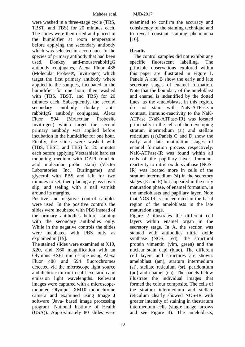
Figure 2 illustrates the different cell
layers within enamel organ in the
secretory stage. In A, the section was
stained with antibodies nitric oxide
synthase (NOS, red), the structural
protein vimentin (vim, green) and the
nuclear stain dapi (blue). The different
cell layers and structures are shown:
ameloblast (am), stratum intermedium
(si), stellate reticulum (sr), peridontium
(pd) and enamel (en). The panels below
illustrate the individual images that
formed the colour composite. The cells of
the stratum intermedium and stellate
reticulum clearly showed NOS-IR with
greater intensity of staining in thestratum
intermedium cells (single image, arrows
and see Figure 3). The ameloblasts,

Mahdee et al. MJB-2017
71
particularly in their apical region, also
showed NOS-IR. The nuclei of the
ameloblasts were located in the basal
region which may influence the apparent
distribution of NOS-IR. However, in
these cells the NOS-IR was less intense.
Interstitial cells of the peridontium were
vimentin immuno-reactive (vim-IR) and
can be seen to lie also between the
processes of the stellate reticulum cells.
This is more easily seen in the single
vimentin image (arrows). The enamel
appears to stain with the vimentin
antibody revealing the Tome’s processes
(tp) adjacent to the apical surface of the
ameloblast. It is not known, at this stage,
whether this staining is specific or non-
specific. Figure 2 B illustrates the
distribution of the enzyme NaK-ATPase.
The lower panels show the separate
images making the composite. The
primary cells demonstrating NaK-
ATPase-IR were in the stratum
intermedium and stellate reticulum. Note
the more intense staining in the stellate
reticulum cells (arrows, see also Figure
4).
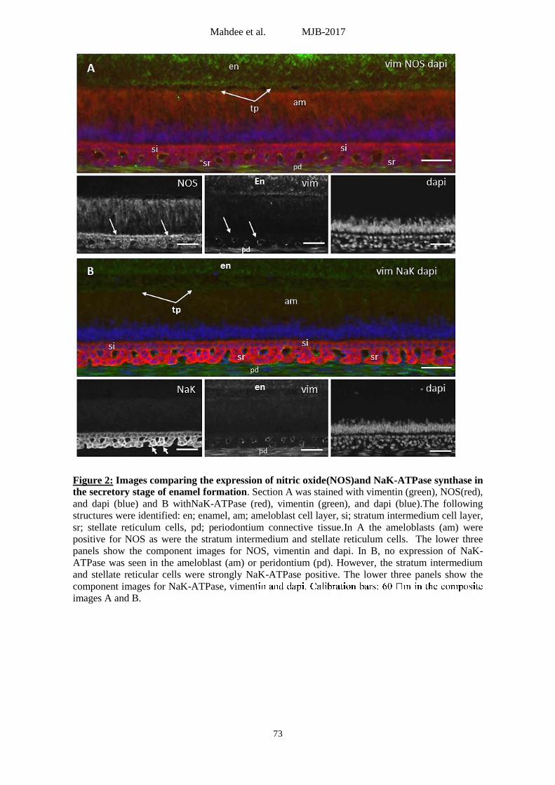
A more detailed illustration of the
distribution of NOS-IR in the secretory
stage is shown in Figure 3. Panel A
shows an over view, with two regions of
interest are identified as (a) and (b),
focusing on the ameloblast and stratum
intermedium respectively. In (a) dense
NOS-IR is seen in the apical region of the
ameloblast (arrows) and the relative
position of this staining to the cell
nucleus at the base of the cells is
apparent. In (b) an intense NOS-IR in the
stratum intermedium can be seen (see
arrow in the individual NOS image). The
vim-IR cells (green) of the peridontium
are seen in the combined image (b) as are
the NOS-IR cells of the stellate
reticulum. Note that vim-IR cells appear
to surround cell free spaces (vacuoles (v))
between the cell processes of the stellate
reticulum (see also Figure 7).
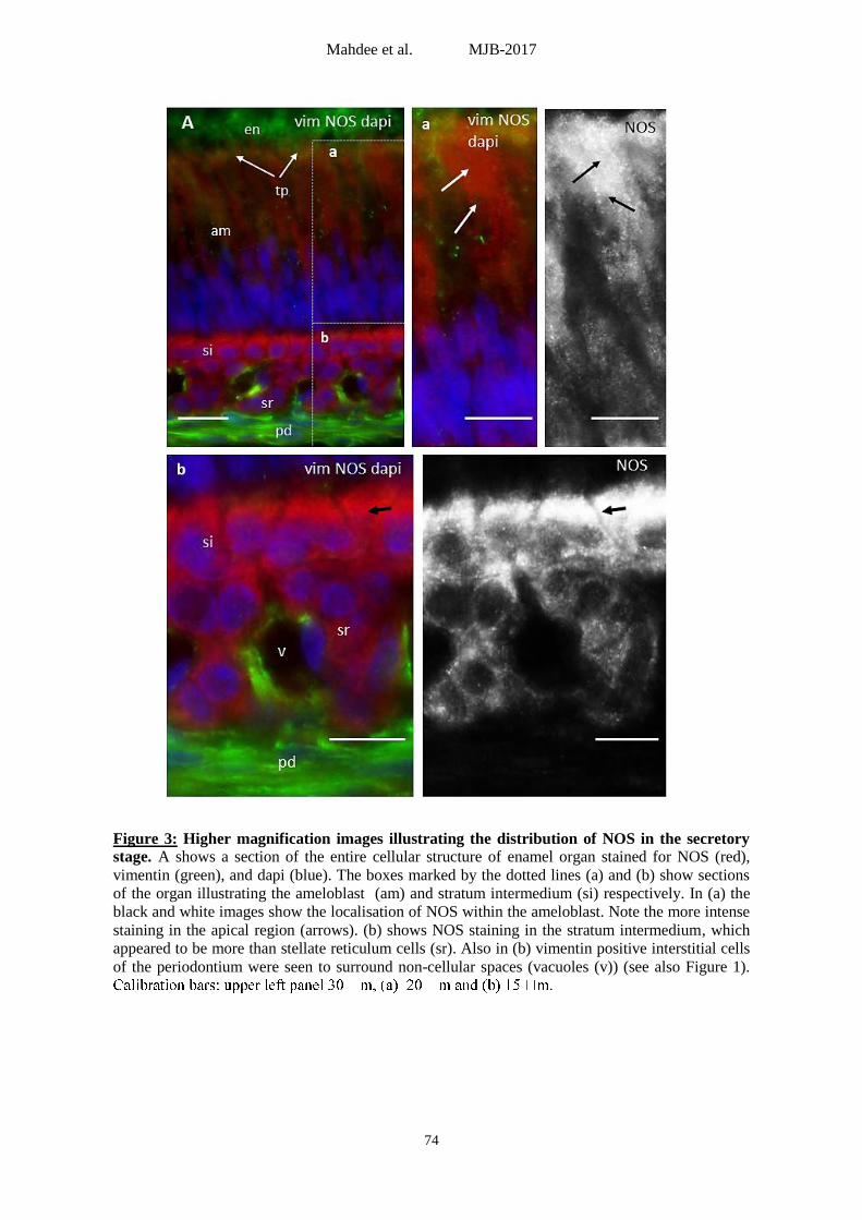
Figure 4 shows details of the distribution
of NaK-ATPase-IR in the secretory
region. As described earlier, there is
ahighly-localised expression of NaK-
ATPase-IR within the cells of the stellate
reticulum and stratum intermedium. The
spaces between the stellate reticular
processes that are surrounded by vim-IR
cells can also be seen in panel B (*).
Panel C, showing only NaK-ATPase-IR,
demonstrates the weak immuno-reactivity
in the stratum intermedium and stronger
signal in the stellate reticulum.
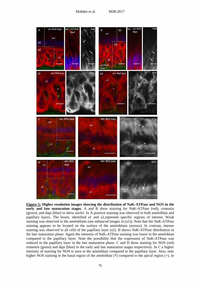
Figure 5 compares the staining patterns
for NaK-ATPase-IR and NOS-IR in the
early and late stages of the maturation
phase. A and B respectively show the
early and late matured enamel organ
expressed with NaK-ATPase-IR. Regions
of interests are identified. In A (a1) the
NaK-ATPase-IR in the ameloblasts is
enhanced to illustrate its presence
(arrows). In (a2) the loss of stratum
intermedium and the more uniform NaK-
ATPase-IR is illustrated in the papillary
cells. In B, the late phase, NaK-ATPase-
IR is seen in the ameloblasts and
papillary layer but the papillary staining
is less intense. In C and D the distribution
of NOS-IR is shown for the early and late
stages. There appears to be a reduction in
NOS-IR in the apical region of the
ameloblast in the late stage, but the
intense staining in the basal region
remains. In the papillary layer the NOS-
IR remains uniform.
Actin-IR is not found in the ameloblasts
or in the papillary layer. It is detected in
the peridontial region (Figure 7). Panel A
shows a low power overview
demonstrating actin-IR in the spaces
between the papillae and in a layer of
cells immediately below the papillae.
Panel B shows that the actin-IR appears
to surround the spaces between the
papillae giving the appearance of
vacuoles. Actin-IR filaments can be seen
to run between adjacent vacuoles
(arrows). At higher magnification (C),
there is a region immediately below the
papillae which contains cells that are not
actin-IR (black space). The nuclei of cells
in this region can clearly be identified (*).
Below this layer are cells that are actin-
IR. Again, the nuclei of these cells,
surrounded by actin-IR can be seen ().

Mahdee et al. MJB-2017
72
The expression of NaK-ATPase-IR in the
ameloblast cells during all stages of
enamel formation is illustrated in Figure
8. In panel A, it appears that there is no
NaK-ATPase-IR in ameloblast during
secretory stage. Panel B shows the
transitional zone where ameloblast cells
change from no NaK-ATPase-IR (+)
toward the apical direction (Ap) to more
NaK-ATPase-IR in the basal region of
ameloblast cells (*) toward the incisal
direction (Inc). Enhanced images of the
early and late matured ameloblast layer
are showed in panel C and D. Panels E
and F illustrate the incisal end of the
enamel organ where the ameloblasts and
other cellular constituent this organ are
reduced in size and number, with no
NaK-ATPase-IR and seem to be similar
to the adjacent periodontal cells.
\ Figure 1: The regional expression of NaK-ATPase and NOS in the enamel organ at different
stages of the enamel formation process. A illustrates the expression of NaK-ATPase in the outer
enamel organ cells in the early secretory stage (the dotted lines show the boundary of the
ameloblast which, in this region does not stain), B shows the secretory stage, C and D the early and
late maturation stages respectively. E-H show the expression of NOS. E illustrates the expression
of NaK-ATPase in the outer enamel organ cells in the early secretory stage, F the secretory stage,
G and H the early and late maturation stages respectively. Note the difference in sizes of the
stratum intermedium (si) and stellate reticulum (sr) between A and B (arrows) also between E
(arrows) and F.
Mahdee et al. MJB-2017
73
Figure 2: Images comparing the expression of nitric oxide(NOS)and NaK-ATPase synthase in
the secretory stage of enamel formation. Section A was stained with vimentin (green), NOS(red),
and dapi (blue) and B withNaK-ATPase (red), vimentin (green), and dapi (blue).The following
structures were identified: en; enamel, am; ameloblast cell layer, si; stratum intermedium cell layer,
sr; stellate reticulum cells, pd; periodontium connective tissue.In A the ameloblasts (am) were
positive for NOS as were the stratum intermedium and stellate reticulum cells. The lower three
panels show the component images for NOS, vimentin and dapi. In B, no expression of NaK-
ATPase was seen in the ameloblast (am) or peridontium (pd). However, the stratum intermedium
and stellate reticular cells were strongly NaK-ATPase positive. The lower three panels show the
component images for NaK-ATPase, vimen
images A and B.
Mahdee et al. MJB-2017
74
Figure 3: Higher magnification images illustrating the distribution of NOS in the secretory
stage. A shows a section of the entire cellular structure of enamel organ stained for NOS (red),
vimentin (green), and dapi (blue). The boxes marked by the dotted lines (a) and (b) show sections
of the organ illustrating the ameloblast (am) and stratum intermedium (si) respectively. In (a) the
black and white images show the localisation of NOS within the ameloblast. Note the more intense
staining in the apical region (arrows). (b) shows NOS staining in the stratum intermedium, which
appeared to be more than stellate reticulum cells (sr). Also in (b) vimentin positive interstitial cells
of the periodontium were seen to surround non-cellular spaces (vacuoles (v)) (see also Figure 1).
Mahdee et al. MJB-2017
75
Figure 4 :Cellular and non-cellular layers in the secretory stage of enamel formation. A shows
a low power image of a section stained with antibodies to vimentin (green), NaK-ATPase (red),
and dapi (blue).B is magnified region from panel A showing stratum intermedium, stellate
reticulum and periodontal connective tissue. Note, the presence of large vacuoles beneath the
stellate reticulum surrounded by nucleated vim+ periodontal interstitial cells (*). Cshows the
original single wavelength image illustrating the distribution of NaK-ATPase. Note, the higher
intensity of staining in the stellate reticular cells (white arrow) compared to the stratum
Mahdee et al. MJB-2017
76
Figure 5: Higher resolution images showing the distribution of NaK-ATPase and NOS in the
early and late maturation stages. A and B show staining for NaK-ATPase (red), vimentin
(green), and dapi (blue) to show nuclei. In A positive staining was observed in both ameloblast and
papillary layers. The boxes, identified a1 and a2,represent specific regions of interest. Weak
staining was observed in the ameloblasts (see enhanced images in (a1)). Note that the NaK-ATPase
staining appears to be located on the surface of the ameloblasts (arrows). In contrast, intense
staining was observed in all cells of the papillary layer (a2). B shows NaK-ATPase distribution in
the late maturation phase. Again the intensity of NaK-ATPase staining was lower in the ameloblast
compared to the papillary layer. Note the possibility that the expression of NaK-ATPase was
reduced in the papillary layer in the late maturation phase. C and D show staining for NOS (red)
vimentin (green) and dapi (blue) in the early and late maturation stages respectively. In C a higher
intensity of staining for NOS is seen in the ameloblast compared to the papillary layer. Also, note
higher NOS staining in the basal region of the ameloblast (*) compared to the apical region (+). In

Mahdee et al. MJB-2017
77
D, the late maturation phase, note the relatively higher NOS staining in the basal region of the
Figure 6: The distribution of actin filaments in the secretory stage of enamel formation. A
shows a low power image of a sections stained for NOS (green), actin (red) and dapi (blue). Note
the presence of actin filaments in the interstitial cells in regions of the peridontium (pd) and
surrounding the spaces between the papillae (v). B shows, at higher magnification, actin fibres
within the cells that form vacuoles (arrow) between the papillary cells. These are the vimentin+
cells described in Figure 3. C shows details of the peridontial region. Note (i) cells containing actin
filaments surrounding the vacuoles, (ii) cells immediately below the papillary layer that are not
actin-IR (note the nuclei of these cells (*)) and (iii) cells below the layer (ii) that are actin-IR (the
nuclei of these cells is shown ()). Note also the complex actin-IR within one of the vacuoles in
this section.

Mahdee et al. MJB-2017
78
Figure 7:The expression of NaK-ATPase in ameloblasts in different stages during the process
of enamel formation. A illustrates the sectretory ameloblast. Note the absence of any significant
NaK-ATPase in the ameloblasts. B shows a section illustrating the transitional zone. Ameloblasts
(am) towards the apical direction do not express NaK-ATPase (+) while ameloblasts towardsthe
incisal direction express NaK-ATPase (*)in the basal region of the cells. C and D show
respectively the early and late maturation stages. Note that the ameloblasts now express NaK-
ATPase and that the orientation of the cells changes: in the early stage the cells slope towards the
apical side,while toward the incisal direction in the late stage. E and F shows incisal end of the
enamel organ. m in A-D and
Mahdee et al. MJB-2017
79
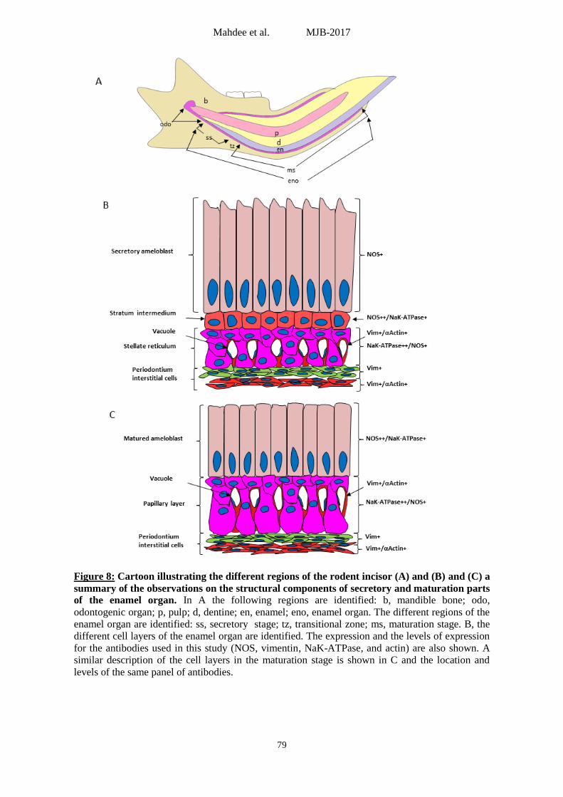
Figure 8: Cartoon illustrating the different regions of the rodent incisor (A) and (B) and (C) a
summary of the observations on the structural components of secretory and maturation parts
of the enamel organ. In A the following regions are identified: b, mandible bone; odo,
odontogenic organ; p, pulp; d, dentine; en, enamel; eno, enamel organ. The different regions of the
enamel organ are identified: ss, secretory stage; tz, transitional zone; ms, maturation stage. B, the
different cell layers of the enamel organ are identified. The expression and the levels of expression
for the antibodies used in this study (NOS, vimentin, NaK-ATPase, and actin) are also shown. A
similar description of the cell layers in the maturation stage is shown in C and the location and
levels of the same panel of antibodies.
Mahdee et al. MJB-2017
80
Discussion
In the current study, the dental pulp
from rat mandibular incisor has been used
as an experimental model for many
reasons. First it is readily available, well
established in the literature and share
many similarities and differences with
human dental pulp. One of the most
excellent features of the rat incisor dental
pulp is the continuous growth which
provide an excellent model to observe the
tooth development and all stages at the
same time and in one sample [17].
The immunohistochemical technique
utilized in this study is essential to
observe the various cellular and tissue
antigens. [18] hypothesized that cell
markers immunohistochemistry provide a
method to talk with cells, because it
grants a way of identification of the
histological architecture of the cells as
well as indicating the function if the
suitable antibodies used.
It is not logical and unsatisfactory to
consider the immunohistochemistry as a
descriptive method only, in fact the
invitro and in situ experiments can be
regarded as accurate pictures to in vivo
situations [18].
It is becoming progressively accepted
that the histological and functional
description of the enamel organ explained
in the introduction is too simplistic, for
example, ameloblasts have been found to
secrete various proteins not only during
the secretory phase but throughout their
life in variable magnitudes. The cellular
component of enamel organ is controlled
by many signalling molecules like
components of extracellular matrix and
growth factors to guide the process of
secretion and maturation of enamel.
NaK ATPase is an enzyme responsible
for pumping the Na ions to the
extracellular space and moving the K ions
intracellularly against their concentration
gradient in an active process (consumes
energy). This active transport help to
preserve the cell resting action potential
and monitor the cell size [19]. NaK
ATPase may also function as a signalling
molecule in many physiological
pathways, neurons and play a role in
regulating the intracellular calcium [20].
It has been found that NaK-ATPase-IR is
widely distributed in the dental pulp
including many cellular systems [21] and
found to play a crucial role in regulating
the transmembrane ionic transport and
balance.
Observation from the current study
showed that NaK-IR was found primarily
in the sub-ameloblast area at the secretory
stage with completely negative IR in the
ameloblasts area. In the maturation stage,
the ameloblasts start to show NaK-IR but
this expression was lost again at the end
of amelogenesis where the enamel organ
is nearly functionless. This variable
expression may reflect the activity of
different components of enamel organ at
each stage as NaK ATPase can be
regarded as marker for cell activity. It
may also provide a picture about which
cell layer consumes more energy and
works more and the late participation of
ameloblasts.It is apparent that the
amelobalst cells expressed NaK-ATPase-
IR (as seen in figure 5), but the more
interesting observation that these cells
change their orientation from apical to
incisal during early and late maturation
stages respectively.
Nitric oxide produced by nNOS (NOS1
used in the current study) can be regarded
as a neurotransmitter and signalling
molecule which helps in reducing the
smooth muscles and blood vessels tone
[22] and has a role in memory, learning
and neurogenesis [23].
Ameloblasts, throughout the enamel-
genesis, have been found to express
NOS-IR which provide a clue that
ameloblasts may have a signalling
function or play a role in controlling the
vascular tone. The sustainability of this
positive IR reflects that the related
function is continuous throughout
amelogenesis.
The coupled and continuous positive
expression of NaK ATPase and NOS in
the papillary layer provide an evidence
about the crucial contribution of these
layers at all stages of amelogenesis.
Mahdee et al. MJB-2017
81
One of the new findings in the current
study is the observation of a large
vacuoles beneath the stellate reticulum
surrounded by nucleated vim+
periodontal interstitial cells. In all
sections from all preparations these
structures were mainly spherical or ovoid.
No tubules were seen, suggesting that
these structures are primarily spherical.
The precise nature and function of these
circular structures, is unclear.
Additionally, cellular heterogeneity was
observed, where some cells show actin-
IR. Re-examination of Figures 3 and 4
shows clearly that both cell types in this
region, those that are actin-IR and those
that are not, are all vim-IR. Thus, there
are two populations of interstitial cells in
the periodontal space.
Collectively, these observations
demonstrate the expression of different
functional elements and processes in the
different cell layers of the enamel organ.
This must imply that such specialisations
underlie specific functional properties in
the different cell layers.
References 1. Mina, M. and E. Kollar, The induction of
odontogenesis in non-dental mesenchyme combined with early murine mandibular arch epithelium. Arch Oral Biol, 1987; 32(2): 123-127.
2. Thesleff I, et al., Eplthelial-Mesenchymal Signaling during Tooth Development. Connective tissue Res, 1995; 32(1-4): 9-15.
3. Beeman CS, JE. Kronmiller. Temporal distribution of endogenous retinoids in the embryonic mouse mandible. Arch oral biol, 1994; 39(9): 733-739.
4. Miller, N., Ten Cate's oral histology. Bri Dent J, 2012. 213 (4): 194-194.
5. Deutsch, D., Structure and function of enamel gene products. The Anatomical Record, 1989; 224 (2): 189-210.
6. Nanci A, CE. Smith. Development and calcification of enamel. Calcification in biological systems. CRC Press, Boca Raton, 1992; 313-343.
7. Warshawsky H, et al. The development of enamel structure in rat incisors as compared to the teeth of monkey and
man. The Anatomical Record, 1981; 200(4): 371-399.
8. Smith, C., Cellular and chemical events during enamel maturation. Critical Rev Oral Biol Med, 1998; 9(2): 128-161.
9. Bawden, J.W., Calcium transport during mineralization. The Anatomical Record, 1989; 224(2): 226-233.
10. Takano, Y., M. Crenshaw, and J. Bawden, Calcium movement in vivo and in vitro in secretory-stage enamel of rat incisors. Arch oral biol, 1992; 37(5): 377-383.
11. Paine, M.L., et al., Regulated gene expression dictates enamel structure and tooth function. Matrix Biol, 2001; 20(5): 273-292.
12. Robinson, C., et al., Matrix and mineral changes in developing enamel. J Dental Res, 1979; 58(2_suppl): 871-882.
13. Bronckers, A., et al., Degradation of hamster amelogenins during secretory stage enamel formation in organ culture. Matrix biology, 1995. 14(7): p. 533-541.
14. Cho, A., et al., A method for rapid demineralization of teeth and bones. The open Dent J, 2010; 4:223-229.
15. Mahdee A, et al., Complex cellular responses to tooth wear in rodent molar. Arch Oral Biol, 2016; 61: 106-114.
16. Gillespie JI, et al., Interstitial cells and cholinergic signalling in the outer muscle layers of the guinea-pig bladder. BJU Int, 2006; 97(2): 379-385.
17. Ohshima, H. and S. Yoshida, The relationship between odontoblasts and pulp capillaries in the process of enamel-and cementum-related dentin formation in rat incisors. Cell and tissue Res, 1992; 268(1):51-63.
18. Brandtzaeg, P., The increasing power of immunohistochemistry and immunocyto-chemistry. J immunol meth, 1998; 216 (1): p. 49-67.
19. Hall, J.E., Guyton and Hall textbook of medical physiology. 2015: Elsevier Health Sciences.
20. Howarth C, P. Gleeson, D. Attwell. Updated energy budgets for neural computation in the neocortex and cerebellum. J Cerebral Blood Flow Metabol, 2012; 32(7): 1222-1232.

Mahdee et al. MJB-2017
82
21. Duan, X., Ion Channels, Channelopathies, and Tooth Formation. J Dent Res, 2014; 93(2): p. 117-125.
22. Förstermann, U. and W.C. Sessa, Nitric oxide synthases: regulation and function. European heart J, 2012; 33(7): p. 829-837.
23. Zhou, L. and D.-Y. Zhu, Neuronal nitric oxide synthase: structure, subcellular localization, regulation, and clinical implications. Nitric Oxide, 2009; 20(4): 223-230.