Embed Size (px)
Citation preview
15Karbandi et al
Evidence Based Care Journal http://ebcj.mums.ac.ir/
Effect of Lung Manual
Hyperinflation (MHI) on
Oxygenation of Patients Following
Abdominal Surgery and T-Tube
Support
Javad Malekzadeh, Mahboube Yazdani, Alireza Sedaghat, Seyed Reza Mazlom, Alieh
Pasandideh khajebeyk
The online version of this article can be found at
http://ebcj.mums.ac.ir/article_7663.html
Evidence Based Care Journal 2016 06:63 originally published
online 01 October 2016
Online ISSN: 2008-370X
Address: Mashhad Nursing and Midwifery School, Ebn-e-Sina St., Mashhad, Iran P.O.Box: 9137913199 Tel.: (098 51) 38591511-294 Fax: (098 51) 38539775 Email: [email protected]
Evidence Based Care Journal, 6 (3): 55-6655
Downloaded from http://ebcj.mums.ac.ir/ at Mashhad University of Medical Sciences on October 01, 2016
Original Article
Effect of Lung Manual Hyperinflation (MHI) on
Oxygenation of Patients Following Abdominal Surgery
and T-Tube Support
Javad Malekzadeh1,*Mahboube Yazdani2, Alireza Sedaghat3, Seyed Reza Mazlom1, Alieh
Pasandideh khajebeyk2
Received: 05/09/2016
Accepted: 06/10/2016
Abstract
Background: Postoperative pulmonary complications (PPC) are of the major reasons for death.
Prolonged mechanical ventilation (PMV) and delayed extubation are leading to the incidence of more
seriously complications. The effect of hyperinflation has not been investigated in control of these
complications in patients who have been weaned from mechanical ventilation and are undergoing T-
tube support.
Aim: Investigation of MHI effect on oxygenation of patients following abdominal surgery and T-tube
support.
Method: This clinical trial was performed on 40 patients undergoing abdominal surgery and T-tube
support hospitalized in intensive care units of hospitals in Mashhad, Iran, in 2015-2016. The
participants were divided randomly into two experimental and control groups. In the experimental
group, MHI technique was performed using Mapleson circuit for three twenty-minute periods. The
control group received routine hospital care. The two groups were compared for PaO2, PaCO2 and
SpO2 before intervention, 5 and 20 minutes after intervention. Data were analyzed using SPSS
software.
Results: The mean age was 66.7±8.3 and 67.5±9.0 years in experimental and control groups,
respectively. In intergroup comparison using independent t-test, the mean PaCO2, PaO2 and SpO2 had
no significant differences in the experimental group before the intervention. However, the means
SpO2 and PaO2 at 5 and 20 minutes after intervention were significantly higher in the experimental
group (p<0.001) than the control group. The mean PaCO2 at 5 and 20 minutes after intervention
decreased significantly in the experimental group (p=0.03).
Implications for Practice: The results showed that the MHI technique by increasing oxygenation and
ventilation could improve lung function in the participants, resulting in shortening the duration of
mechanical ventilation, accelerating the process of extubation, and thus faster patient recovery.
Keywords: Lung Hyperinflation, Abdominal Surgery, Oxygenation, Postoperative Pulmonary
Complications
1. Evidence Based Care Research Centre, Instructor of Nursing, Instructor of Medical-Surgical Nursing, School of Nursing
and Midwifery, Mashhad University of Medical Sciences, Mashhad, Iran
2. MS in Nursing, School of Nursing and Midwifery, Mashhad University of Medical Sciences, Mashhad, Iran
3. Associate Professor of Anesthesiology and Critical Care, School of Medicine, Mashhad University of Medical Sciences,
Mashhad, Iran
Evidence Based Care Journal, 6 (3): 63-74
Malekzadeh et al. Effect of Hyperinflation on Oxygenation in Spontaneously Breathing Patients 56
Downloaded from http://ebcj.mums.ac.ir/ at Mashhad University of Medical Sciences on October 01, 2016
* Corresponding author, Email: [email protected]
Introduction Nowadays, surgery is considered as selective treatment for many patients. Often following surgery,
damage to vital body systems is inevitable, especially respiratory system; and instability of the
systems will be further by expanding the surgery (1). Postoperative pulmonary complications (PPC)
are of the major reasons for death, which are seen frequently after upper abdominal surgeries so that
have been reported in more than 75% of patients (3). Some of the most important postoperative
pulmonary complications are atelectasis, pneumonia and respiratory failure (2, 4). Disorders of gas
exchange and oxygenation in the postoperative period vary from a slight decline in PaO2 to life-
threatening hypoxemia (5). The incidence rate of hypoxemia after abdominal surgery has been
reported 30%-50% (6). The hypoxemia can lead to complications including postoperative delirium,
wound infections, ECG changes and even MI (5). Therefore, one of the major postoperative concerns
for patients is decreasing pulmonary complications and subsequent reduction of mortality. Mechanical
ventilation is one of the most widely used postoperative treatments, because it can ensure acceptable
gas exchanges within the lungs of the patients (7).
However, prolonged mechanical ventilation and hospitalization can increase hospital deaths by
causing problems such as VAP (ventilator-associated pneumonia), damage to the airways and
atelectasis (8). Extubation failure increases mortality rate up to 6 times. Thus improving lung function
and oxygenation have an important role in successful weaning and extubation and are of the most
important therapeutic targets in patients undergoing mechanical ventilation (9). The use of T-tube
during the process of weaning from mechanical ventilation is a commonly used and approved method.
It is used also in patients who have a tracheostomy and require oxygen therapy with no long-term
mechanical ventilation (10). Accordingly, any action that would be effective in the prevention and
treatment of pulmonary complications and the improvement of pulmonary ventilation and
oxygenation can lead to short term mechanical ventilation and successful weaning and can have an
important role in the recovery process (6).
A study on patients with abdominal surgery showed that any factor that causes dilation of the lungs
after surgery could prevent the occurrence of atelectasis and subsequent lung volume reduction and
improve its performance. Some of these methods are chest physiotherapy, training of breathing
exercises, deep cough, incentive spirometry and positive end-expiratory pressure (PEEP) as well as
the MHI (6).
A study that Margaret Mackay et al. (2005) conducted on open abdominal surgery in high-risk
patients showed that postoperative respiratory physiotherapy had no positive impact on the reduction
of pulmonary complications after open abdominal surgery (11). On the other hand, manual respiratory
physiotherapy in patients undergoing chest surgeries is limited due to multiple incisions in the chest,
bone fractures, instability of sternum and ribs (12). Pain after abdominal surgery has been identified
as one of the causative agents of respiratory muscle dysfunction (13). In addition, the pain prevents
deep breathing exercises in surgical patients (2). Incentive spirometry is a tool in which the patient
breathes deeply through a device with visual feedback view (14). It should be noted that perfect
consciousness and cooperation of the patient are essential for the efficiency of this method; such
cooperation of the patients is not possible with decrease of consciousness. A systematic study
conducted by Overend et al. in 2001 concerning the effect of incentive spirometry on postoperative
pulmonary complications determined that the evidence does not support the effectiveness of incentive
spirometry in preventing pulmonary complications after thoracic and upper abdominal surgeries (15).
The use of PEEP maintains alveolar distension capabilities at the end of exhalation and prevents
alveolar collapse and generally hypoventilation and subsequent atelectasis (16). The use of CPAP
compared with the conventional treatment for atelectasis control can reduce its level to half in patients
undergoing abdominal surgery (5), but its necessity is applying mechanical ventilation by patient.
Hemmes et al. (2014) examined the high and low levels of PEEP during general anesthesia in patients
undergoing abdominal surgery and showed that the incidence rate of pulmonary complications had no
significant difference between the two groups. On the other hand, a greater need for vasoactive drugs
was found in the group with high PEEP due to hypotension (17). MHI is a technique in which
breathing with slow inspiratory flow, further tidal volume, inspiratory pause and peak expiratory flow
(2, 18-21) can improve lung function, help to reopen of poorly ventilated alveoli, facilitate the
clearing and removal of secretions, improve oxygenation and gas exchange in the lungs (19, 20, 22-
Evidence Based Care Journal, 6 (3): 55-6657
Downloaded from http://ebcj.mums.ac.ir/ at Mashhad University of Medical Sciences on October 01, 2016
24). This method can be used in mechanically ventilated patients or in those who have artificial
airway but not supported by mechanical ventilation (Patients with spontaneous breathing). On the
other hand, there is no need for consciousness and cooperation of patient to achieve the mentioned
advantages. However, so far, all studies on the effects of the technique have been conducted on
patients supported by mechanical ventilation and there is no study to evaluate the effect of this
technique in patients who had successful weaning from mechanical ventilation and T-tube support.
Accordingly, the present study was conducted to assess the MHI effect on oxygenation of patients
following abdominal surgery and T-tube support.
Methods In this randomized controlled clinical trial, the participants were selected among the patients admitted
to the surgical intensive care units of Imam Reza and Ghaem hospitals in Mashhad, Iran, underwent
abdominal surgery and T-tube support during the seven months from September 2015 to March 2016.
The patients were enrolled in the study after explaining the project methods before starting the study
and obtaining informed consent from the conscious patient or their first-degree relatives in the
absence of consciousness. Given that, a similar study was not available, so the exact sample size was
determined with regard to the dependent variables, including 1- partial pressure of oxygen (PaO2) in
arterial blood 2- partial pressure of carbon dioxide (PaCO2) in arterial blood 3- arterial oxygen
saturation (SpO2) and using the formula of "determining sample size for comparing two independent
population means". The pilot study was performed on 20 patients (10 in the experimental group and
10 in control group) and the sample size was calculated for each of the three above index. The largest
sample size was related to arterial oxygen saturation, 20 patients in each group (40 in total), and the
same number were recruited. In addition, confidence level of 95% and test power of 80% were
considered to calculate the sample size.
Inclusion criteria were the patients undergoing abdominal surgery, weaned from mechanical
ventilation, age over 18 years, oxygen intake via T-tube for at least two hours, no pulmonary
pathologies (such as ARDS, acute lung edema, hemoptysis and pneumothorax), no cardiovascular
disorders (hemodynamic instability, such as MAP less than 60 or arrhythmia such as, VT, Af with fast
ventricular response and PSVT). Exclusion criteria included extubation criteria, need for repeated
surgery, mechanical ventilatory support and hemodynamic instability during the study. The subjects
were divided randomly into the two experimental (hyperinflation maneuver) and control (routine care)
groups using random numbers table.
Content validity was used for research unit selection form, demographic information form and
pulmonary function registration table. SOFA score and Richmond Agitation-Sedation Scale
(RASS) are international standard tools for assessing disease severity and GCSC-agitation; their
validity has been confirmed. It is worth mentioning that in this study contractually based on scores in
Richmond criteria, the patients were divided into three levels 1-conscious and relatively conscious, 2-
semi-coma and 3 - coma. The validity of the Gasometer (Gem PREMIER 3000) and Pulse Oximeter
(SAADAT ALBORZ B9) devices were approved, since they were for reputable companies. On the
other hand, Gasometer validity has been confirmed by studies to measure arterial blood gases (25).
The SOFA score and Richmond criteria are international standard tools, and their reliability has been
confirmed. The Gasometer and monitoring reliability were approved as internal calibration.
The researcher was trained on the proper implementation of hyperinflation maneuver under controlled
conditions and monitoring the volume and pressure in skill lab unit to enter the study environment,
and then the study began. At baseline, the researcher through interview and medical history collected
basic data of participants, including demographic and disease data. Studying on two experimental and
control groups was started with suction (without hyperoxygenation). Then the patient was placed in
supine position (without any intervention) for half an hour. The researcher collected arterial blood gas
samples from both groups, and arterial oxygen saturation was recorded using Pulse Oximeter.
After recording the basic data in the experimental group, the researcher applied the MHI technique for
three twenty-minute periods with twenty-minute rest intervals. Thus, in each twenty-minute period, 12
breaths with delivery volume as twice as tidal volume of spontaneous breaths were presented using
Mapleson C circuit that was connected to oxygen at a rate of 15 to 20 liters divided per minute
coordinated with patient breathing. How to breathe was as follows; 1.5-2 seconds inhaling along with
2 seconds inspiratory pause and with a quick release of bag during exhalation. The airway pressure
Malekzadeh et al. Effect of Hyperinflation on Oxygenation in Spontaneously Breathing Patients 58
Downloaded from http://ebcj.mums.ac.ir/ at Mashhad University of Medical Sciences on October 01, 2016
was monitored throughout the technique so that it did not exceed 40 cm water. The volumes given
during the technique were monitored using Exhalometer device that had been placed in Mapleson
circuit. At the end of the three periods, arterial blood gas samples were taken at 5 and 20 minutes after
intervention and arterial oxygen saturation levels were recorded using Pulse Oximeter. Data were
recorded in the control group at the same times without any intervention. Since the record of all data
and intervention actions were carried out by the researcher, so blinded record was not possible. In
addition, T-tube venturi in different patients had various FIO2 levels. However, each patient
underwent oxygen therapy by same venturi before and after intervention similar to during the
intervention. The data were recorded in the same FIO2 condition for each patient.
After ensuring the accuracy of data entry, data were analyzed using SPSS version 22 software.
Normal distribution of quantitative variables was assessed using the Kolmogorov-Smirnov and the
Shapiro-Wilk tests. To study the homogeneity of the two groups in terms of quantitative variables and
normal distribution, independent t-test and variables without normal distribution, Mann-Whitney test
were used. The homogeneity of qualitative variables were analyzed with chi-square, Fisher and Chi-
square exact tests. Two groups were compared in terms of the dependent variables with independent t-
test and in case of non-normal distribution with Mann-Whitney test. The intra-group comparison was
carried out by repeated measures ANOVA. The ANOVA test was used to investigate the relationship
between underlying and confounding variables between the two groups. Confidence interval of 95%
(α=5%) was considered in the tests and thus the significance of differences was reported at P<0.05.
Results
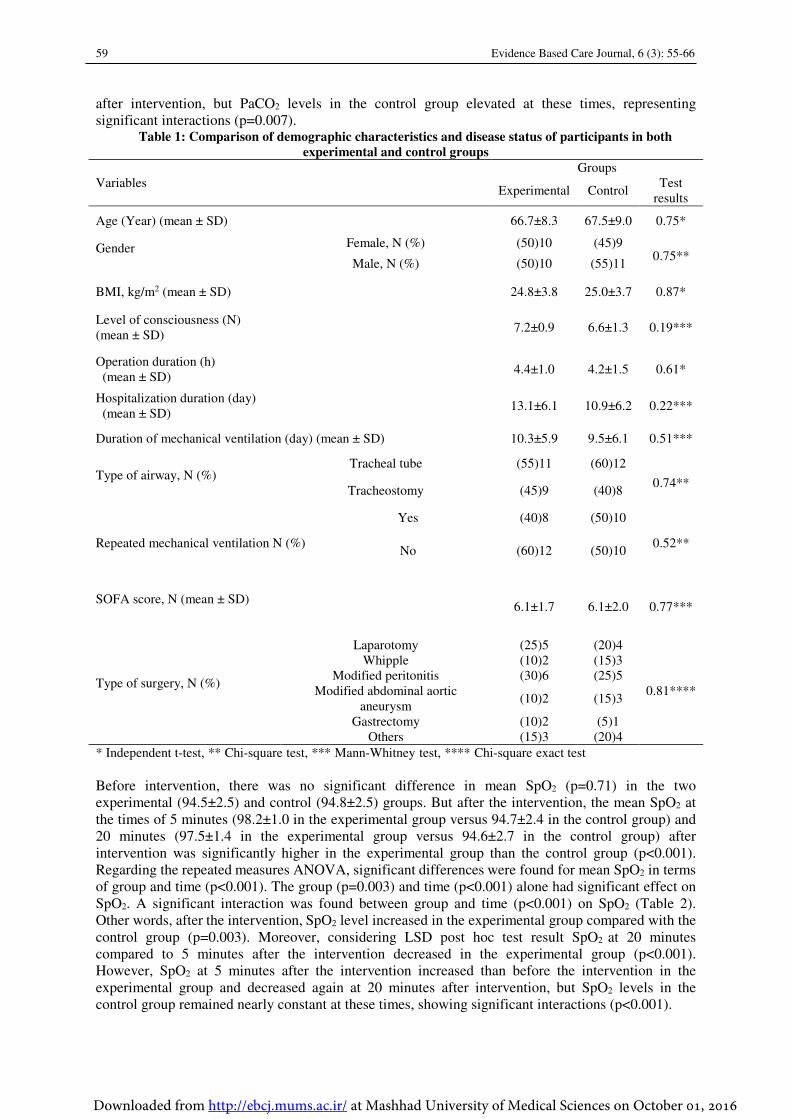
The mean age of subjects was 67.1±8.5 years with a range of 50 to 86 years (52.5% males). Table 1
shows the data related to patients. There were no significant differences in these variables between the
two groups (Table 1). It should be noted that participants in both groups were matched for atelectasis
(p=0.220).
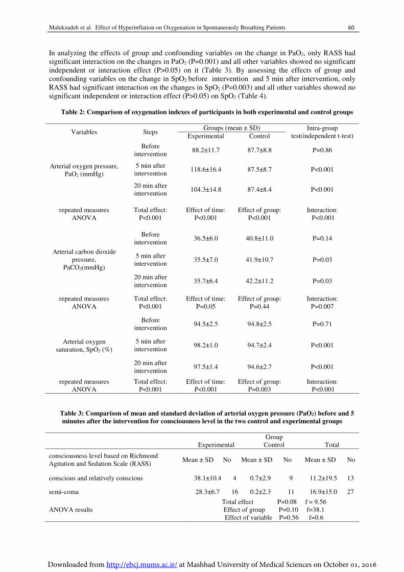
Before intervention using independent t-test, there was no significant difference in mean PaO2
(p=0.86) in the two experimental (88.2±11.7) and control (87.7±8.8) groups. But after the
intervention, the mean PaO2 at 5 minutes after intervention (118.6 ±16.4 in the experimental group
versus 87.5± 8.7 in the control group) and 20 minutes after intervention (104.3 ±14.8 in the
experimental group versus 87.4± 8.4 in the control group) significantly (p<0.001) was higher in the
experimental group compared with the control group. According to the repeated measures ANOVA,
significant differences were found for mean PaO2 in terms of group and time (p<0.001). The group
(p<0.001) and time (p<0.001) alone had significant effect on PaO2. A significant interaction was
observed between group and time (p<0.001) on PaO2 (Table 2). In other words, after the intervention,
PaO2 level increased in the experimental group compared with the control group (p<0.001). Also,
according to LSD post hoc test result PaO2 at 20 minutes compared to 5 minutes after the intervention
decreased in the experimental group (p<0.001). At all these steps, PaO2 remained almost constant at
the control group and showed no significant difference. However, PaO2 at 5 minutes after the
intervention increased than before the intervention in the experimental group and decreased again at
20 minutes after intervention, but PaO2 levels in the control group remained nearly constant at these
times, indicating significant interactions (p<0.001).
Before intervention using independent t-test, there was no significant difference in mean PaCO2
(p=0.14) in the two experimental (36.5± 6.0) and control (40.8±11.0) groups. But after the
intervention, the mean PaCO2 at the times of 5 minutes (35.5±7.0 in the experimental group versus
41.9±10.7 in the control group) and 20 minutes (35.7±6.4 in the experimental group versus 42.2±11.2
in the control group) after intervention was significantly lower in the experimental group than the
control group (p=0.03). Based on the repeated measures ANOVA, significant differences were found
for mean PaCO2 in terms of group and time (p <0.001), but the time (p=0.44) as well as the group (p=
0.051) alone had no significant effect on the PaCO2. In addition, group and time indicated significant
interaction (p=0.007) on the PaCO2 (Table 2). On the other hand, after the intervention, the PaCO2
level decreased in the experimental group and increased in the control group, but non-significant
(p=0.05). Also, the mean PaCO2 after 20 minutes showed no significant changes compared to 5
minutes after invention in both groups (p=0.44). However, PaCO2 at 5 minutes after the intervention
decreased than before the intervention in the experimental group and increased again at 20 minutes
Evidence Based Care Journal, 6 (3): 55-6659
Downloaded from http://ebcj.mums.ac.ir/ at Mashhad University of Medical Sciences on October 01, 2016
after intervention, but PaCO2 levels in the control group elevated at these times, representing
significant interactions (p=0.007). Table 1: Comparison of demographic characteristics and disease status of participants in both
experimental and control groups
Variables
Groups
Experimental Control Test
results
Age (Year) (mean ± SD) 66.7±8.3 67.5±9.0 0.75*
Gender
Female, N (%) (50)10 (45)9 0.75**
Male, N (%) (50)10 (55)11
BMI, kg/m2 (mean ± SD) 24.8±3.8 25.0±3.7 0.87*
Level of consciousness (N)
(mean ± SD) 7.2±0.9 6.6±1.3 0.19***
Operation duration (h)
(mean ± SD) 4.4±1.0 4.2±1.5 0.61*
Hospitalization duration (day)
(mean ± SD) 13.1±6.1 10.9±6.2 0.22***
Duration of mechanical ventilation (day) (mean ± SD) 10.3±5.9 9.5±6.1 0.51***
Type of airway, N (%)
Tracheal tube (55)11 (60)12
0.74** Tracheostomy (45)9 (40)8
Repeated mechanical ventilation N (%)
Yes (40)8 (50)10
0.52** No (60)12 (50)10
SOFA score, N (mean ± SD)
6.1±1.7 6.1±2.0 0.77***
Type of surgery, N (%)
Laparotomy (25)5 (20)4
0.81****
Whipple (10)2 (15)3
Modified peritonitis (30)6 (25)5
Modified abdominal aortic
aneurysm (10)2 (15)3
Gastrectomy (10)2 (5)1
Others (15)3 (20)4
* Independent t-test, ** Chi-square test, *** Mann-Whitney test, **** Chi-square exact test
Before intervention, there was no significant difference in mean SpO2 (p=0.71) in the two
experimental (94.5±2.5) and control (94.8±2.5) groups. But after the intervention, the mean SpO2 at
the times of 5 minutes (98.2±1.0 in the experimental group versus 94.7±2.4 in the control group) and
20 minutes (97.5±1.4 in the experimental group versus 94.6±2.7 in the control group) after
intervention was significantly higher in the experimental group than the control group (p<0.001).
Regarding the repeated measures ANOVA, significant differences were found for mean SpO2 in terms
of group and time (p<0.001). The group (p=0.003) and time (p<0.001) alone had significant effect on
SpO2. A significant interaction was found between group and time (p<0.001) on SpO2 (Table 2).
Other words, after the intervention, SpO2 level increased in the experimental group compared with the
control group (p=0.003). Moreover, considering LSD post hoc test result SpO2 at 20 minutes
compared to 5 minutes after the intervention decreased in the experimental group (p<0.001).
However, SpO2 at 5 minutes after the intervention increased than before the intervention in the
experimental group and decreased again at 20 minutes after intervention, but SpO2 levels in the
control group remained nearly constant at these times, showing significant interactions (p<0.001).
Malekzadeh et al. Effect of Hyperinflation on Oxygenation in Spontaneously Breathing Patients 60
Downloaded from http://ebcj.mums.ac.ir/ at Mashhad University of Medical Sciences on October 01, 2016
In analyzing the effects of group and confounding variables on the change in PaO2, only RASS had
significant interaction on the changes in PaO2 (P=0.001) and all other variables showed no significant
independent or interaction effect (P>0.05) on it (Table 3). By assessing the effects of group and
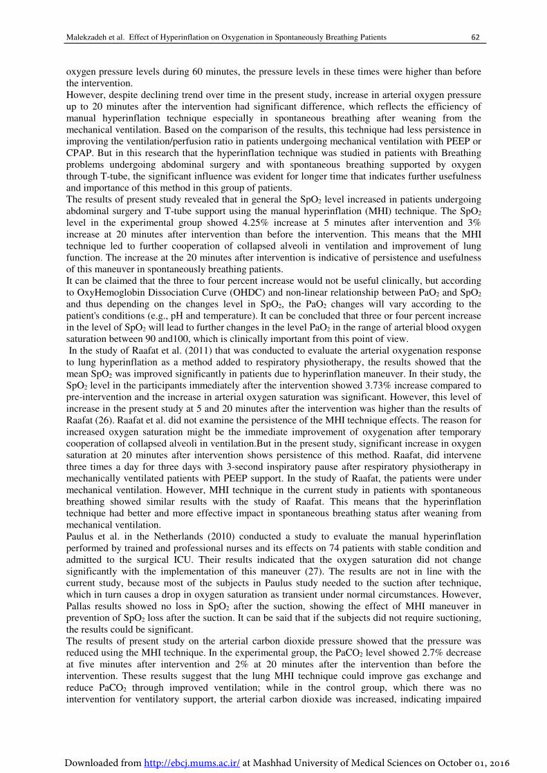
confounding variables on the change in SpO2 before intervention and 5 min after intervention, only
RASS had significant interaction on the changes in SpO2 (P=0.003) and all other variables showed no
significant independent or interaction effect (P>0.05) on SpO2 (Table 4).
Table 2: Comparison of oxygenation indexes of participants in both experimental and control groups
Variables Steps Groups (mean ± SD) Intra-group
test(independent t-test) Experimental Control
Arterial oxygen pressure,
PaO2 (mmHg)
Before
intervention 88.2±11.7 87.7±8.8 P=0.86
5 min after
intervention 118.6±16.4 87.5±8.7 P<0.001
20 min after
intervention 104.3±14.8 87.4±8.4 P<0.001
repeated measures
ANOVA
Total effect:
P<0.001
Effect of time:
P<0.001
Effect of group:
P<0.001
Interaction:
P<0.001
Arterial carbon dioxide
pressure,
PaCO2(mmHg)
Before
intervention 36.5±6.0 40.8±11.0 P=0.14
5 min after
intervention 35.5±7.0 41.9±10.7 P=0.03
20 min after
intervention 35.7±6.4 42.2±11.2 P=0.03
repeated measures
ANOVA
Total effect:
P<0.001
Effect of time:
P=0.05
Effect of group:
P=0.44
Interaction:
P=0.007
Arterial oxygen
saturation, SpO2 (%)
Before
intervention 94.5±2.5 94.8±2.5 P=0.71
5 min after
intervention 98.2±1.0 94.7±2.4 P<0.001
20 min after
intervention 97.5±1.4 94.6±2.7 P<0.001
repeated measures
ANOVA
Total effect:
P<0.001
Effect of time:
P<0.001
Effect of group:
P=0.003
Interaction:
P<0.001
Table 3: Comparison of mean and standard deviation of arterial oxygen pressure (PaO2) before and 5
minutes after the intervention for consciousness level in the two control and experimental groups
Group
Experimental Control Total
consciousness level based on Richmond
Agitation and Sedation Scale (RASS) Mean ± SD No Mean ± SD No Mean ± SD No
conscious and relatively conscious 38.1±10.4 4 0.7±2.9 9 11.2±19.5 13
semi-coma 28.3±6.7 16 0.2±2.3 11 16.9±15.0 27
ANOVA results
Total effect P=0.08 f-= 9.56
Effect of group P=0.10 f=38.1
Effect of variable P=0.56 f=0.6
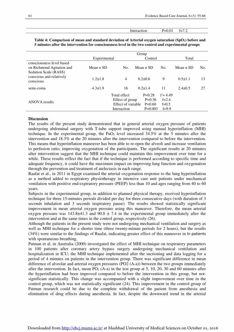
Evidence Based Care Journal, 6 (3): 55-6661
Downloaded from http://ebcj.mums.ac.ir/ at Mashhad University of Medical Sciences on October 01, 2016
Interaction P=0.01 f=7.2
Table 4: Comparison of mean and standard deviation of Arterial oxygen saturation (SpO2) before and
5 minutes after the intervention for consciousness level in the two control and experimental groups
Group
Experimental Control Total
consciousness level based
on Richmond Agitation and
Sedation Scale (RASS)
Mean ± SD No. Mean ± SD No. Mean ± SD No.
conscious and relatively
conscious 1.2±1.8 4 0.2±0.6 9 0.5±1.1 13
semi-coma 4.3±1.9 16 0.2±1.4 11 2.4±0.5 27
ANOVA results
Total effect P=0.28 f-= 4.49
Effect of group P=0.36 f=2.4
Effect of variable P=0.60 f=0.5
Interaction P=0.003 f=9.9
Discussion
The results of the present study demonstrated that in general arterial oxygen pressure of patients
undergoing abdominal surgery with T-tube support improved using manual hyperinflation (MHI)
technique. In the experimental group, the PaO2 level increased 34.5% at the 5 minutes after the
intervention and 18.3% at the 20 minutes after the intervention compared to before the intervention.
This means that hyperinflation maneuver has been able to re-open the alveoli and increase ventilation
to perfusion ratio, improving oxygenation of the participants. The significant results at 20 minutes
after intervention suggest that the MHI technique could maintain this improvement over time for a
while. These results reflect the fact that if the technique is performed according to specific time and
adequate frequency, it could have the maximum impact on improving lung function and oxygenation
through the prevention and treatment of atelectasis in each range.
Raafat et al., in 2011 in Egypt examined the arterial oxygenation response to the lung hyperinflation
as a method added to respiratory physiotherapy in intensive care unit patients under mechanical
ventilation with positive end-expiratory pressure (PEEP) less than 10 and ages ranging from 40 to 60
years.
Subjects in the experimental group, in addition to planned physical therapy, received hyperinflation
technique for three 15-minutes periods divided per day for three consecutive days (with duration of 3
seconds inhalation and 3 seconds inspiratory pause). The results showed statistically significant
improvement in mean arterial oxygen pressure using this maneuver. Therefore, the mean arterial
oxygen pressure was 143.8±41.3 and 90.8 ± 7.4 in the experimental group immediately after the
intervention and at the same times in the control group, respectively (26).
Although the patients in the present study were not undergoing mechanical ventilation and surgery as
well as MHI technique for a shorter time (three twenty-minute periods for 2 hours), but the results
(34%) were similar to the findings of Raafat, indicating greater effect of this maneuver in In patients
with spontaneous breathing.
Patman et al. in Australia (2000) investigated the effect of MHI technique on respiratory parameters
in 100 patients after coronary artery bypass surgery undergoing mechanical ventilation and
hospitalization in ICU; the MHI technique implemented after the suctioning and data logging for a
period of 4 minutes on patients in the intervention group. There was significant difference in mean
difference of alveolar and arterial oxygen pressures (PO2 (A-a)) between the two groups immediately
after the intervention. In fact, mean PO2 (A-a) in the test group at 5, 10, 20, 30 and 60 minutes after
the hyperinflation had been improved compared to before the intervention in this group, but not-
significant statistically. This change was accompanied with a slight improvement over time in the
control group, which was not statistically significant (24). This improvement in the control group of
Patman research could be due to the complete withdrawal of the patient from anesthesia and
elimination of drug effects during anesthesia. In fact, despite the downward trend in the arterial
Malekzadeh et al. Effect of Hyperinflation on Oxygenation in Spontaneously Breathing Patients 62
Downloaded from http://ebcj.mums.ac.ir/ at Mashhad University of Medical Sciences on October 01, 2016
oxygen pressure levels during 60 minutes, the pressure levels in these times were higher than before
the intervention.
However, despite declining trend over time in the present study, increase in arterial oxygen pressure
up to 20 minutes after the intervention had significant difference, which reflects the efficiency of
manual hyperinflation technique especially in spontaneous breathing after weaning from the
mechanical ventilation. Based on the comparison of the results, this technique had less persistence in
improving the ventilation/perfusion ratio in patients undergoing mechanical ventilation with PEEP or
CPAP. But in this research that the hyperinflation technique was studied in patients with Breathing
problems undergoing abdominal surgery and with spontaneous breathing supported by oxygen
through T-tube, the significant influence was evident for longer time that indicates further usefulness
and importance of this method in this group of patients.
The results of present study revealed that in general the SpO2 level increased in patients undergoing
abdominal surgery and T-tube support using the manual hyperinflation (MHI) technique. The SpO2
level in the experimental group showed 4.25% increase at 5 minutes after intervention and 3%
increase at 20 minutes after intervention than before the intervention. This means that the MHI
technique led to further cooperation of collapsed alveoli in ventilation and improvement of lung
function. The increase at the 20 minutes after intervention is indicative of persistence and usefulness
of this maneuver in spontaneously breathing patients.
It can be claimed that the three to four percent increase would not be useful clinically, but according
to OxyHemoglobin Dissociation Curve (OHDC) and non-linear relationship between PaO2 and SpO2
and thus depending on the changes level in SpO2, the PaO2 changes will vary according to the
patient's conditions (e.g., pH and temperature). It can be concluded that three or four percent increase
in the level of SpO2 will lead to further changes in the level PaO2 in the range of arterial blood oxygen
saturation between 90 and100, which is clinically important from this point of view.
In the study of Raafat et al. (2011) that was conducted to evaluate the arterial oxygenation response
to lung hyperinflation as a method added to respiratory physiotherapy, the results showed that the
mean SpO2 was improved significantly in patients due to hyperinflation maneuver. In their study, the
SpO2 level in the participants immediately after the intervention showed 3.73% increase compared to
pre-intervention and the increase in arterial oxygen saturation was significant. However, this level of
increase in the present study at 5 and 20 minutes after the intervention was higher than the results of
Raafat (26). Raafat et al. did not examine the persistence of the MHI technique effects. The reason for
increased oxygen saturation might be the immediate improvement of oxygenation after temporary
cooperation of collapsed alveoli in ventilation.But in the present study, significant increase in oxygen
saturation at 20 minutes after intervention shows persistence of this method. Raafat, did intervene
three times a day for three days with 3-second inspiratory pause after respiratory physiotherapy in
mechanically ventilated patients with PEEP support. In the study of Raafat, the patients were under
mechanical ventilation. However, MHI technique in the current study in patients with spontaneous
breathing showed similar results with the study of Raafat. This means that the hyperinflation
technique had better and more effective impact in spontaneous breathing status after weaning from
mechanical ventilation.
Paulus et al. in the Netherlands (2010) conducted a study to evaluate the manual hyperinflation
performed by trained and professional nurses and its effects on 74 patients with stable condition and
admitted to the surgical ICU. Their results indicated that the oxygen saturation did not change
significantly with the implementation of this maneuver (27). The results are not in line with the
current study, because most of the subjects in Paulus study needed to the suction after technique,
which in turn causes a drop in oxygen saturation as transient under normal circumstances. However,
Pallas results showed no loss in SpO2 after the suction, showing the effect of MHI maneuver in
prevention of SpO2 loss after the suction. It can be said that if the subjects did not require suctioning,
the results could be significant.
The results of present study on the arterial carbon dioxide pressure showed that the pressure was
reduced using the MHI technique. In the experimental group, the PaCO2 level showed 2.7% decrease
at five minutes after intervention and 2% at 20 minutes after the intervention than before the
intervention. These results suggest that the lung MHI technique could improve gas exchange and
reduce PaCO2 through improved ventilation; while in the control group, which there was no
intervention for ventilatory support, the arterial carbon dioxide was increased, indicating impaired
Evidence Based Care Journal, 6 (3): 55-6663
Downloaded from http://ebcj.mums.ac.ir/ at Mashhad University of Medical Sciences on October 01, 2016
ventilation and gas exchange after two hours than before. The results are clinically important in the
control group, even if there was no reduction in PaCO2 in the experimental group. Because according
to the results, continued spontaneous breathing and fatigue increased PaCO2 in the control group due
to impaired ventilation/perfusion ratio caused by fatigue and hypoventilation of the patients.
However, this technique prevented the occurrence of this disorder in the experimental group and
reduced the level of carbon dioxide partial pressure. In fact, hyperinflation technique in the
experimental group made more cooperation of alveolar ventilation and improved ventilation/perfusion
ratio, which lasted significantly up to 20 minutes after the intervention, probably due to a significant
reduction in the carbon dioxide pressure level at 5 minutes later resulting from hyperinflation
occurred during the technique. However, the reason for its significance after 20 minutes was the
improved persistency of the ventilation/perfusion ratio.
Ahmed et al. in India (2010) showed that the level of PaCO2 in the MHI group had 1.7% decrease
immediately after the intervention and 0.8% at 20 minutes after the intervention than before the
intervention. In addition, the decline was 1.4% in VHI group immediately and 20 minutes after the
intervention than before, but the difference between MHI and VHI was not statistically significant
(21). The reason for failing to reduce carbon dioxide pressure may be implementation of manual
hyperinflation in a group and ventilator hyperinflation in another one. They kept constant minute
volume due to the sedation of patients and reduced respiratory rate along with 1.5-fold increase in
tidal volume, which it can also be another reason for the lack of change in carbon dioxide pressure.
The study duration was 3 minutes.In the present study, hyperinflation maneuver was carried out in
accordance with the patient's breaths and double tidal volume for three twenty-minute periods.
Differences in the types of patients, duration of technique, number of maneuvers and tidal volume
were causing the effects of hyperinflation in the present intervention can lead to improve ventilation
and PaCO2 level so that the improvement was visible in statistical results.
According to the studies mentioned above, it was observed that the levels of persistency and
improved ventilation/perfusion ratio were lower in the short-term interventions and were observed for
a limited time. In contrast, long-term interventions led to improve ventilation/perfusion ratio and more
persistency of the improvements over time. These results suggest that the MHI technique should be
performed as programmed with adequate frequencies to achieve the maximum improvement and
effectiveness in patients.
The limitation of this study was a two-hour intervention that could interfere with nursing and
confounding factors such as suction and repositioning. Therefore, we tried to coordinate the
interventions with nursing as well as to do interventions at times with the lowest probability of
interference with nursing actions.
Implications for Practice
In the present study, we were able to improve oxygenation and ventilation in spontaneously breathing
patients using the MHI technique without the need to increase FiO2, apply high PEEP and PaCO2
retention. In fact, it can be concluded that this technique with the cooperation of collapsed alveoli in
ventilation and improvement of gas exchange can lead to significant improvement of
ventilation/perfusion ratio and thereby improve lung function in patients with respiratory problems.
As a blueprint for future research, it is recommended to evaluate the effect of this technique on
mechanical ventilation duration, other populations or the length of stay at the intensive care units.
Acknowledgments This article has been adopted from the M.A thesis approved by Mashhad University of Medical
Sciences, Iran (Code: 931 711, date: 6/07/2015) and Iranian Registry of Clinical Trials
(IRCT2015103024790N1) and funded by Research Deputy of Mashhad University of Medical
Sciences. Hereby, the research team expresses thanks and appreciations to respected authorities of
School of Nursing and Midwifery at Mashhad University of Medical Sciences as well as officials of
the surgical ICU at Imam Reza and Ghaem hospitals in Mashhad to provide the conditions for
research and sincere cooperation.
Conflict of interest The authors declare that there is no conflict of interest.
Malekzadeh et al. Effect of Hyperinflation on Oxygenation in Spontaneously Breathing Patients 64
Downloaded from http://ebcj.mums.ac.ir/ at Mashhad University of Medical Sciences on October 01, 2016
References
1. Shiri H,Nikravan M. Principles of Care in Cardiac Surgery.Tehran;Noor-e-Danesh.
2012.p340-71(persian).
2. Paulus F, Veelo DP, de Nijs SB, Beenen L, Bresser P, de Mol B, et al. Manual
Hyperinflation Partly Prevents Reductions of Functional Residual Capacity in Cardiac
Surgical Patients-A Randomized Controlled Trial. Crit Care. 2011;15(4):R187.
3. Thomas JA, McIntosh JM. Are Incentive Spirometry, Intermittent Positive Pressure
Breathing, and Deep Breathing Exercises Effective in the Prevention of Postoperative
Pulmonary Complications After Upper Abdominal Surgery? A Systematic Overview
and Meta-analysis. Phys Ther. 1994;74(1):3-10.
4. Kanat F, Golcuk A, Teke T, Golcuk M. Risk Factors for Postoperative Pulmonary
Complications in Upper Abdominal Surgery. ANZ J Surg. 2007;77(3):135-41.
5. Tusman G, Böhm SH, Warner DO, Sprung J. Atelectasis and Perioperative
Pulmonary Complications in High-risk Patients. Curr Opin Anaesthesiol.
2012;25(1):1-10.
6. Ferreyra GP, Baussano I, Squadrone V, Richiardi L, Marchiaro G, Del Sorbo L, et al.
Continuous Positive Airway Pressure for Treatment of Respiratory Complications
After Abdominal Surgery: A Systematic Review and Meta-analysis. Ann Surg.
2008;247(4):617-26.
7. Futier E, Godet T, Millot A, Constantin J-M, Jaber S, editors. Mechanical Ventilation
in Abdominal Surgery. Ann Fr Anesth Reanim; 2014: Elsevier.
8. Gnanapandithan K, Agarwal R, Aggarwal A, Gupta D. Weaning by Gradual Pressure
Support (PS) Reduction without an Initial Spontaneous Breathing Trial (SBT) Versus
PS-supported SBT: A Pilot Study. Rev Port Pneumol. 2011;17(6):244-52.
9. Varon, Joseph, and Pilar Acosta. Handbook of Critical and Intensive Care Medicine.
New York: Springer, 2010.p 1-418.
10. Matiæ I ,Majeriæ-Kogler V. Comparison of Pressure Support and T-tube Weaning
From Mechanical Ventilation: Randomized Prospective Study. Croat Med J.
2004;45(2):162-6.
11. Mackay MR, Ellis E, Johnston C. Randomised Clinical Trial of Physiotherapy After
Open Abdominal Surgery in High Risk Patients. Aust J Physiother. 2005;51(3):151-9.
12. Blattner C, Guaragna JC, Saadi E. Oxygenation and Static Compliance Is Improved
Immediately After Early Manual Hyperinflation Following Myocardial
Revascularisation: A Randomised Controlled Trial. Aust J Physiother.
2008;54(3):173-8.
13. Vassilakopoulos T, Mastora Z, Katsaounou P, Doukas G, Klimopoulos S, Roussos C,
et al. Contribution of Pain to Inspiratory Muscle Dysfunction After Upper Abdominal
Surgery: A Randomized Controlled Trial. Am J Respir Crit Care Med.
2000;161(4):1372-5.
14. Agostini P, Singh S. Incentive Spirometry Following thoracic Surgery: What Should
We Be Doing? Physiotherapy. 2009;95(2):76-82.
15. Overend TJ, Anderson CM, Lucy SD, Bhatia C, Jonsson BI, Timmermans C. The
Effect of Incentive Spirometry on Postoperative Pulmonary Complications: A
Systematic Review. CHEST J. 2001;120(3):971-8.
16. Santos LJd, Blattner CN, Micol CAB, Pinto FAM, Renon A, Pletsch R. Effects of
Manual Hyperinflation Maneuver Associated with Positive End Expiratory Pressure
in Patients within Coronary Artery Bypass Grafting. Rev Bras Ter Intensiva.
2010;22(1):40-6.
Evidence Based Care Journal, 6 (3): 55-6665
Downloaded from http://ebcj.mums.ac.ir/ at Mashhad University of Medical Sciences on October 01, 2016
17. Hemmes S, Gama dAM, Pelosi P, Schultz MJ. High Versus Low Positive End-
Expiratory Pressure During General Anaesthesia for Open Abdominal Surgery
(PROVHILO trial): A Multicentre Randomised Controlled Trial. J Lancet.
2014;384(9942):495-503.
18. Paulus F, Binnekade JM, Vroom MB, Schultz MJ. Benefits and Risks of Manual
Hyperinflation in Intubated and Mechanically Ventilated Intensive Care Unit Patients:
A Systematic Review. Crit Care. 2012;16(4):1-18.
19. Maa S-H, Hung T-J, Hsu K-H, Hsieh Y-I, Wang K-Y, Wang C-H, et al. Manual
Hyperinflation Improves Alveolar Recruitment in Difficult-to-Wean Patients. CHEST
Journal. 2005;128(4):2714-21.
20. Ortiz TdA, Forti G, Volpe MS, Carvalho CRR, Amato MBP, Tucci MR.
Experimental Study on the Efficiency and Safety of the Manual Hyperinflation
Maneuver as a Secretion Clearance Technique. J Bras Pneumol. 2013;39(2):205-13.
21. Ahmed F, Shafeeq AM, Moiz JA, Geelani MA. Comparison of Effects of Manual
Versus Ventilator Hyperinflation on Respiratory Compliance and Arterial Blood
Gases in Patients Undergoing Mitral Valve Replacement. Heart Lung.
2010;39(5):437-43.
22. Clini E, Ambrosino N. Early Physiotherapy in the Respiratory Intensive Care Unit.
Respiratory Medicine. 2005;99(9):1096-104.
23. Patman S, Jenkins S, Smith K. Manual Hyperinflation: Consistency and Modification
of the Technique by Physiotherapists. Physiother Res Int. 2001;6(2):106-17.
24. Patman S, Jenkins S, Stiller K. Manual Hyperinflation—Effects on Respiratory
Parameters. Physiother Res Int. 2000;5(3):157-71.
25. Mackenzie C, Shin B, McAslan T. Chest Physiotherapy: The Effect on Arterial
Oxygenation. Anesth Analg. 1978;57(1):28-30.
26. Raafat A, Elbasiouny HS. Arterial Oxygenation Response to Manual Hyperinflation
as an Added Procedure to Chest Physiotherapy in Critically Ill Mechanically
Ventilated Patients. Am J Sci. 2011;7 )12.( :585-90
27. Paulus F, Binnekade J, Vermeulen M, Vroom M, Schultz M. Manual Hyperinflation
Is Associated with a Low Rate of Adverse Events When Performed by Experienced
and Trained Nurses in Stable Critically Ill Patients--A Prospective Observational
Study. Minerva Anestesiol. 2010;76(12):1036-42.
Malekzadeh et al. Effect of Hyperinflation on Oxygenation in Spontaneously Breathing Patients 66
Downloaded from http://ebcj.mums.ac.ir/ at Mashhad University of Medical Sciences on October 01, 2016