Embed Size (px)
Citation preview
Everybody Loves Baby Chicks:A proposal for a demonstration on chick development at the
Museum of Science and Industry
Elisabeth MontegnaMSCOPE 1/27/06
I. Introduction
Chickens provide a unique opportunity to observe embryonic development. Because theylay eggs instead of giving live birth, one has easy access to embryos at many stages ofdevelopment. During the first four days of embryonic development, the chick heart isformed. By the third day, it is beating. Due to the size of the embryos, this is easily seeneven without a magnifying glass.
At the Museum of Science and Industry, one of the most popular exhibits is the chickhatchery. On any given day, you can see many, many people watching the chicks hatch.This exhibit is currently part of the Genetics exhibition. While the hatchery no doubtdraws many people into the genetics exhibit that would not normally go in, there does notseem to be a direct connection drawn between the hatchery and the subject of genetics.The hatchery sits between an exhibit on development and the demonstration area for thegenetics exhibition. Creating a demonstration centered on chick development will be away to more concretely link the hatchery to the exhibition in which it resides.
The purpose of the demonstration will be to explain that genes turn on and off to makeproteins that help create specific structures during development. The demonstration willfocus on heart development in the early embryo in particular because it is the first organto start forming, is easily seen in early embryos, and the majority of early term humanmiscarriages are caused by abnormal heart development (which will give the public away to relate the demonstration back to their own lives).
The target audience for this demonstration is people 10 and older. While this proposalfocuses on creating a demonstration within the genetics exhibition, one could alsoimagine the demonstration being carried out using a media cart near the prenataldevelopment exhibit, emphasizing some of the similarities and differences betweenhuman and chick early development, or could be part of a learning lab on embryology forhigh school students. Two types of demonstrations are described here, one with liveembryos and one with preserved specimens.
II. Materials and MethodsA. Live EmbryosCurrently, the museum has access to eggs for its hatchery. For a demonstration that isdone everyday, four freshly laid eggs will be obtained and kept in an incubator. The eggs
will be incubated from one to four days in the incubator. Each demonstration will usefour embryos, one each from embryonic days one through four. Embryos will beremoved from the shell by delicately cracking the eggs with a small sharp object thenremoving bits of shell using tweezers until the egg can be successfully poured into a Petridish. The embryos will remain in the Petri dish (covered) until the end of the day, andthen discarded in normal waste. If the museum does not wish to use it’s own eggs, it mayobtain them from an organic farmer.
B. Preserved Specimens and SlidesA set of six preserved chick embryos and slides of whole mounted early embryos ( chickembryos at 24 hours, 33 hours, 48 hours, 72 hours, and 96 hours) will be obtained fromCarolina (www2.Carolina.com).
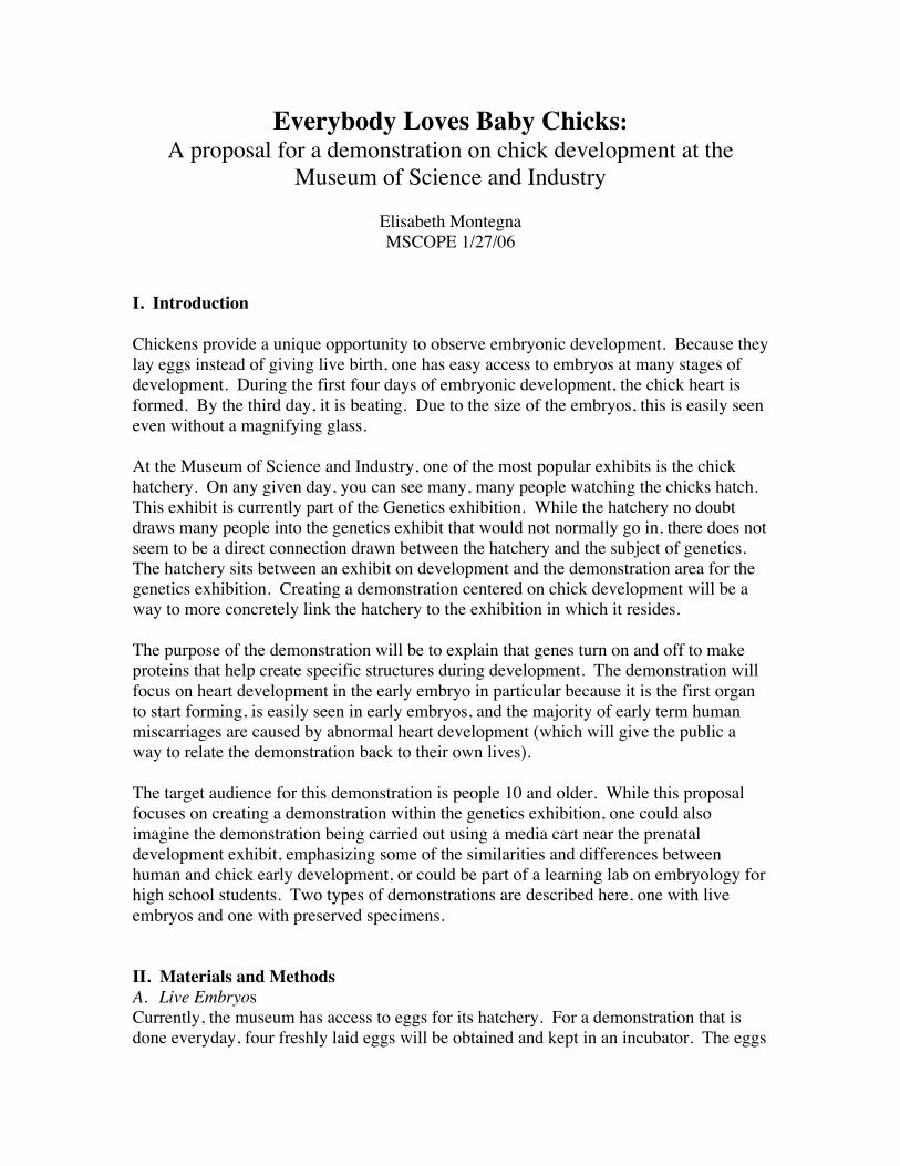
C. Observing Live EmbryosThe embryos will be presented to the public in covered Petri dishes. To see fine detail,the demonstrator will use a Proscope (www.x-tremegeek.com) connected to an ordinaryTV (Fig. 1). The Proscope is a digital microscope capable of 200X magnification thatconnects to any TV using an RCA feed.
D. Observing Preserved Specimens and Slides
The demonstrator will use the Proscope to show the slides of the early embryos to thepublic. The public may examine the preserved later stage embryos using magnifyingglasses.
E. Program
The demonstrator will start by explaining that DNA has genes that code for proteins thatcarry out the function of the gene. S/he will then explain the idea that genes turn on andoff to make certain structures in the body. The demonstrator will then briefly discuss
Figure 1. Proscope [1]
chick heart development, drawing parallels to human heart development. Thedemonstrator will then use the Proscope to show the audience the heart in each of the fourlive embryos or on the slide. Finally the public will be invited to come look at either thelive embryos or the preserved specimens using a magnifying glass or the Proscope.
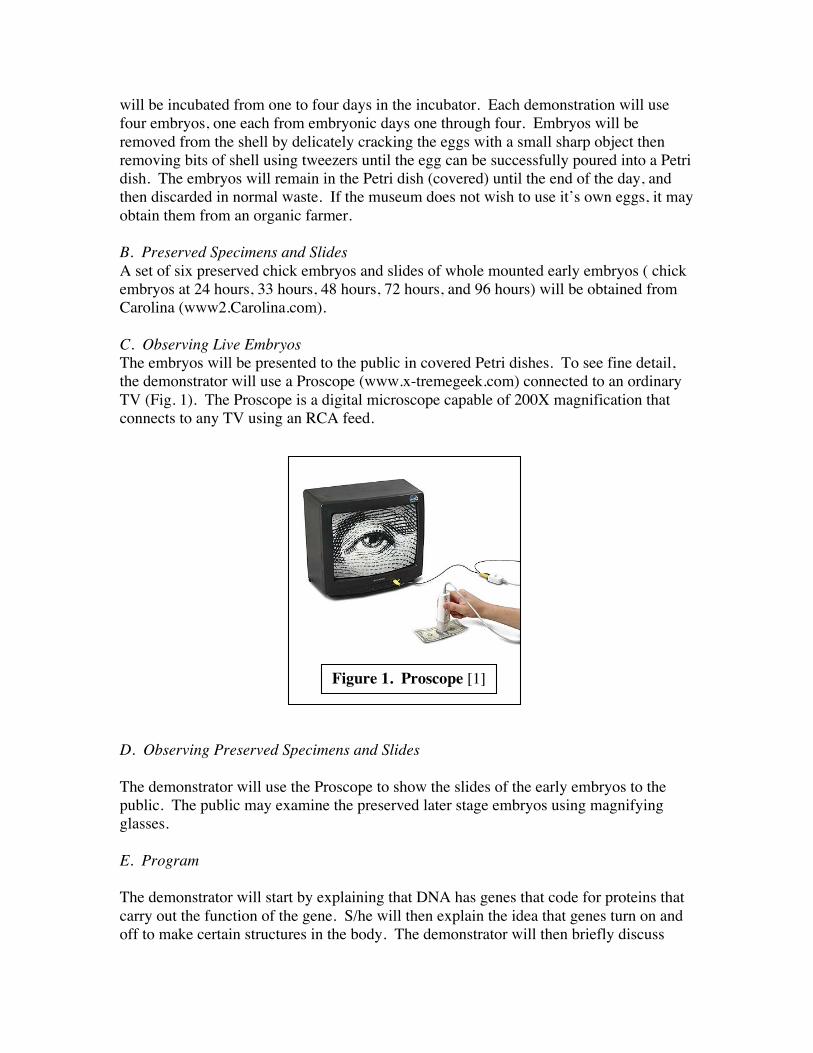
III. Budget
A. Both Demonstrations
Equipment CostProscope Tv, 200x handheld video microscope (X-
tremegeek.com #142-0866)$99.99
TV, 24” (Circuit City) $140-$180Megalens magnifying glasses, 10 (Carolina # 95-3807) $50.00 ($5.00 each)
Total $290-$330
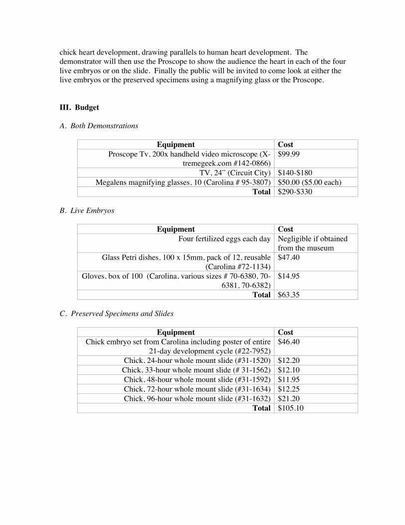
B. Live Embryos
Equipment CostFour fertilized eggs each day Negligible if obtained
from the museumGlass Petri dishes, 100 x 15mm, pack of 12, reusable
(Carolina #72-1134)$47.40
Gloves, box of 100 (Carolina, various sizes # 70-6380, 70-6381, 70-6382)
$14.95
Total $63.35
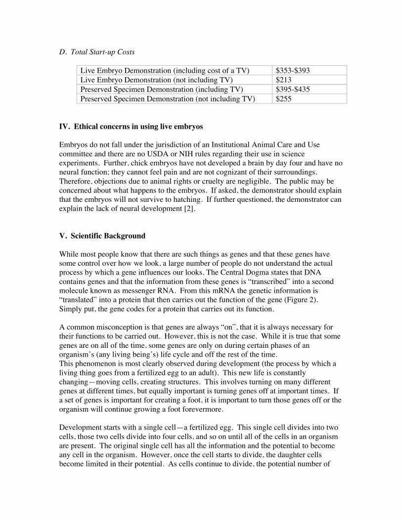
C. Preserved Specimens and Slides
Equipment CostChick embryo set from Carolina including poster of entire
21-day development cycle (#22-7952)$46.40
Chick, 24-hour whole mount slide (#31-1520) $12.20Chick, 33-hour whole mount slide (# 31-1562) $12.10Chick, 48-hour whole mount slide (#31-1592) $11.95Chick, 72-hour whole mount slide (#31-1634) $12.25Chick, 96-hour whole mount slide (#31-1632) $21.20
Total $105.10
D. Total Start-up Costs
Live Embryo Demonstration (including cost of a TV) $353-$393Live Embryo Demonstration (not including TV) $213Preserved Specimen Demonstration (including TV) $395-$435Preserved Specimen Demonstration (not including TV) $255
IV. Ethical concerns in using live embryos
Embryos do not fall under the jurisdiction of an Institutional Animal Care and Usecommittee and there are no USDA or NIH rules regarding their use in scienceexperiments. Further, chick embryos have not developed a brain by day four and have noneural function; they cannot feel pain and are not cognizant of their surroundings.Therefore, objections due to animal rights or cruelty are negligible. The public may beconcerned about what happens to the embryos. If asked, the demonstrator should explainthat the embryos will not survive to hatching. If further questioned, the demonstrator canexplain the lack of neural development [2].
V. Scientific Background
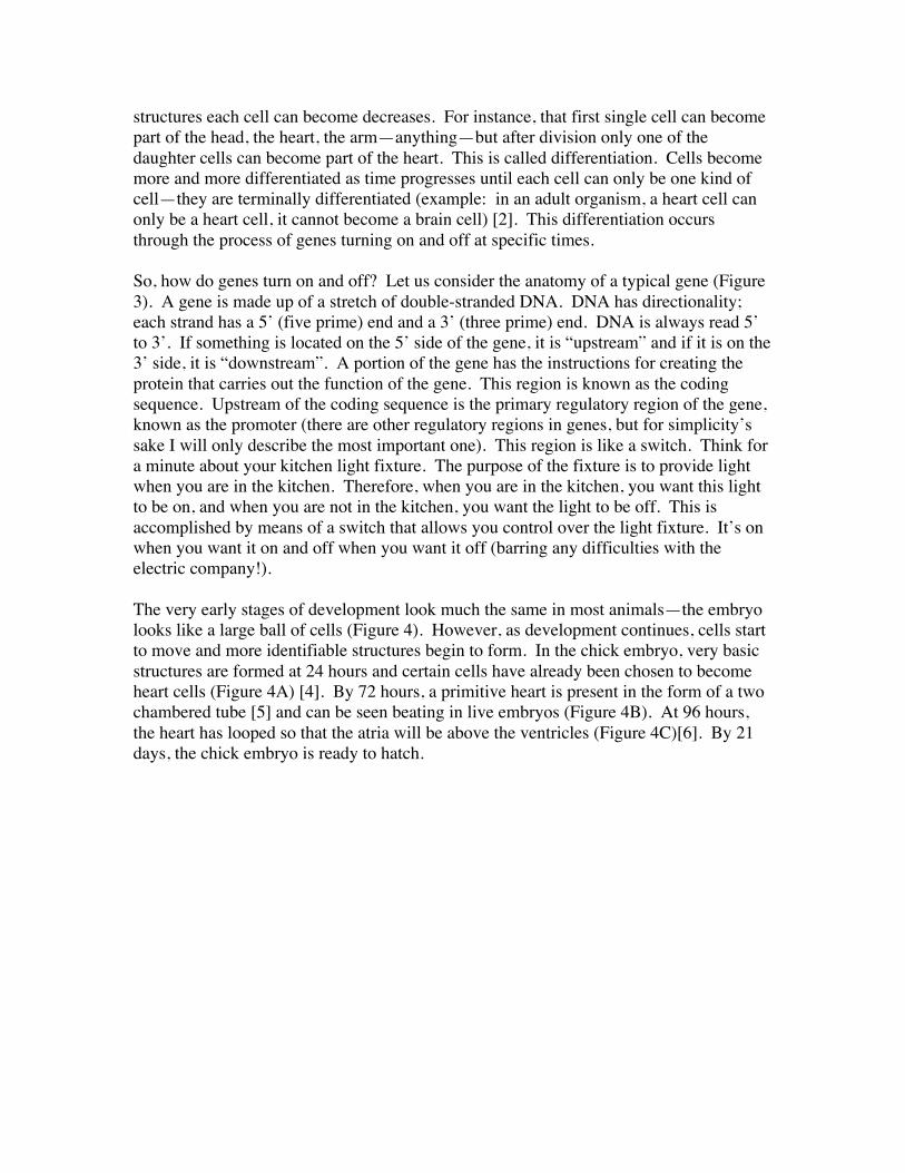
While most people know that there are such things as genes and that these genes havesome control over how we look, a large number of people do not understand the actualprocess by which a gene influences our looks. The Central Dogma states that DNAcontains genes and that the information from these genes is “transcribed” into a secondmolecule known as messenger RNA. From this mRNA the genetic information is“translated” into a protein that then carries out the function of the gene (Figure 2).Simply put, the gene codes for a protein that carries out its function.
A common misconception is that genes are always “on”, that it is always necessary fortheir functions to be carried out. However, this is not the case. While it is true that somegenes are on all of the time, some genes are only on during certain phases of anorganism’s (any living being’s) life cycle and off the rest of the time.This phenomenon is most clearly observed during development (the process by which aliving thing goes from a fertilized egg to an adult). This new life is constantlychanging—moving cells, creating structures. This involves turning on many differentgenes at different times, but equally important is turning genes off at important times. Ifa set of genes is important for creating a foot, it is important to turn those genes off or theorganism will continue growing a foot forevermore.
Development starts with a single cell—a fertilized egg. This single cell divides into twocells, those two cells divide into four cells, and so on until all of the cells in an organismare present. The original single cell has all the information and the potential to becomeany cell in the organism. However, once the cell starts to divide, the daughter cellsbecome limited in their potential. As cells continue to divide, the potential number of
structures each cell can become decreases. For instance, that first single cell can becomepart of the head, the heart, the arm—anything—but after division only one of thedaughter cells can become part of the heart. This is called differentiation. Cells becomemore and more differentiated as time progresses until each cell can only be one kind ofcell—they are terminally differentiated (example: in an adult organism, a heart cell canonly be a heart cell, it cannot become a brain cell) [2]. This differentiation occursthrough the process of genes turning on and off at specific times.
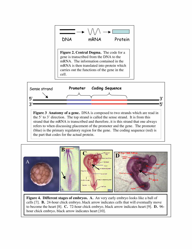
So, how do genes turn on and off? Let us consider the anatomy of a typical gene (Figure3). A gene is made up of a stretch of double-stranded DNA. DNA has directionality;each strand has a 5’ (five prime) end and a 3’ (three prime) end. DNA is always read 5’to 3’. If something is located on the 5’ side of the gene, it is “upstream” and if it is on the3’ side, it is “downstream”. A portion of the gene has the instructions for creating theprotein that carries out the function of the gene. This region is known as the codingsequence. Upstream of the coding sequence is the primary regulatory region of the gene,known as the promoter (there are other regulatory regions in genes, but for simplicity’ssake I will only describe the most important one). This region is like a switch. Think fora minute about your kitchen light fixture. The purpose of the fixture is to provide lightwhen you are in the kitchen. Therefore, when you are in the kitchen, you want this lightto be on, and when you are not in the kitchen, you want the light to be off. This isaccomplished by means of a switch that allows you control over the light fixture. It’s onwhen you want it on and off when you want it off (barring any difficulties with theelectric company!).
The very early stages of development look much the same in most animals—the embryolooks like a large ball of cells (Figure 4). However, as development continues, cells startto move and more identifiable structures begin to form. In the chick embryo, very basicstructures are formed at 24 hours and certain cells have already been chosen to becomeheart cells (Figure 4A) [4]. By 72 hours, a primitive heart is present in the form of a twochambered tube [5] and can be seen beating in live embryos (Figure 4B). At 96 hours,the heart has looped so that the atria will be above the ventricles (Figure 4C)[6]. By 21days, the chick embryo is ready to hatch.
DNA mRNA Protein
Figure 2. Central Dogma. The code for agene is transcribed from the DNA to themRNA. The information contained in themRNA is then translated into protein whichcarries out the functions of the gene in thecell.
A
B C D
Figure 4. Different stages of embryos. A. An very early embryo looks like a ball ofcells [7]. B. 24-hour chick embryo, black arrow indicates cells that will eventually moveto become the heart [8]. C. 72-hour chick embryo, black arrow indicates heart [9]. D. 96-hour chick embryo, black arrow indicates heart [10].
Promoter Coding Sequence
5’5’3’
3’
Figure 3 Anatomy of a gene. DNA is composed to two strands which are read inthe 5’ to 3’ direction. The top strand is called the sense strand. It is from thisstrand that the mRNA is transcribed and therefore, it is this strand that one alwaysrefers to when discussing placement of the promoter and the gene. The promoter(blue) is the primary regulatory region for the gene. The coding sequence (red) isthe part that codes for the actual protein.
Sense strand
VI. Assessment
A. Formative Evaluation
Two types of formative evaluations will be done on a prototype demonstration usingpreserved specimens.
1. ObservationVisitors will be observed during the demonstration to assess the general reaction of thepublic to the demonstration. Behaviors that will be watched:
1. Of the people who walk past an in-progress demonstration what percentagestops to watch? (Does the demonstration grab people’s attentions?)
1. What percentage of participants leave before the end of the demonstration? (Isthe demonstration too long or the language too complex?)
1. What percentage of participants asks questions? (How interested have theparticipants become?)
1. What is the overall mood of the crowd (intrigued, amused, bored, repulsed,etc.)?
After observing three demonstrations, adjustments to the demonstration will then bemade based on observations. For example, if a high percentage of people are leavingprior to the end of the demonstration, the demonstration might be shortened. If peoplegenerally appear to be bored, then the focus of the demonstration might be changed.
2. InterviewsOnce improvements on the prototype demonstration have been made, a revised version ofthe demonstration will be conducted for the public. Visitors will be interviewedimmediately following the demonstration. Questions that will be asked:
1. What did you like most about the demonstration? (What are we doing well?)1. What did you like least about the demonstration? (What can we improve upon?)1. What do you think the purpose of the demonstration was? (Are we meeting our
objectives?)1. Is there anything you would like to see added to the demonstration? (How can we
make this material more relevant to the participants?)1. Did you feel the demonstration was presented at a level you feel comfortable
with? (Are we presenting material that is too complicated?)
After interviewing 40 people, adjustments will be made to the demonstration based on theanswers to the above questions.
B. Summative Evaluation
When all adjustments have been made, a final, summative interview evaluation will bedone using the above interview questions.
VII. Training
Once the assessment has led to a format of the demonstration is successful, trainingmaterials will be developed to train museum employees to conduct the demonstration.These materials will include a powerpoint presentation, a short manual, and a list of bothpaper and online resources. The effectiveness of these materials will be assessed by firstusing them to train another MSCOPE participant to conduct the demonstration.Revisions will be made to the materials until the new demonstrator is capable ofconducting the demonstration well. When the training materials are in their completedforms, the first round of training for museum staff will be led by me.
VIII. Summary
I have proposed two demonstrations (one using live embryos, the other using preservedspecimens and slides) around the concept of chick developmental genetics, focusing onheart development. These demonstrations are designed to more closely link the chickhatchery to the genetics exhibition it resides in. The objective of the demonstrations is toexplain that the function of a gene is carried out by a protein and that genes are turned onand off during an organism’s development in order to make proteins that help createcertain structures such as the heart. The start-up costs for each of these demonstrationscould be under $300 if the institution already has a TV it is willing to use for theProscope. The only disposables are the gloves and the eggs in the case of the live embryodemonstration, therefore there will be very little cost to the museum after the initialinvestment in equipment and specimens.
In developing the program for the demonstration, two types of summative evaluations(observations and interviews) will be done on a prototype demonstration. After theprogram has been finalized, a final summative evaluation using interviews will beconducted. Training materials will be developed and evaluated by other MSCOPEparticipants with the first training session for museum staff led by me.
Finally, while this demonstration will work very well in the genetics demonstration area,the demonstration could also be reworked and shown by the human prenatal exhibit, orcould become part of a learning lab on embryology for high school students. Regardlessof the venue, adding this demonstration will improve the general public’s understandingof how genetics influences development and will have a positive impact on theexperience of museum visitors.
Notes and References
1. Image taken from www.x-tremegeek.com.2. The Exploratorium in San Francisco has a permanent display using live chick
embryos. I was able to speak to one of the people in charge of the exhibit at a recentconference. The Exploratorium does not receive any complaints about the embryosfrom the public, though once in a while someone writes a letter asking what happensto the embryos. On a personal note, when I visited the exhibit, a woman and herdaughter were looking at the embryos. When the daughter asked about the babychicks hatching, the mother replied that she didn’t think these chicks would behatching. Neither appeared very concerned about this. They were, however,fascinated by the beating hearts of the embryos.
3. This is what makes stem cells so valuable. They are cells that have not yet becometerminally differentiated and therefore can still become several different kinds ofcells. This also explains why people are so eager to get them from embryos. Whileadults have a few certain kinds of stem cells (bone marrow cells that make bloodcells, for example), embryos are stuffed full of them and the earlier the embryo, theless differentiated the cells are.
4. Gilbert, Scott F. Developmental Biology 6th ed. 2000. Sinauer Associates, Inc. p. 472-475
5. Ibid6. Ibid7. Image taken from
www.learner.org/channel/courses/biology/images/archive/textbook/1982_tb.jpg.
8. Image taken fromwww.uoguelph.ca/zoology/devobio/24hrchck/images/24cktb01.gif
9. Image taken from www.uoguelph.ca/zoology/devobio/210labs/72hrwm.GIF10. Image taken from
www.umanitoba.ca/faculties/science/biological_sciences/lab14/images/chick96.jpeg.