Embed Size (px)
Citation preview


Welcome to EMBL · IBEC Winter Conference on ENGINEERING MULTICELLULAR SYSTEMSRecent breakthroughs in stem cell biology, organ-on-chip assays, 3D Bioprinting, and cell mechanobiology have revolutionized our ability to design and assemble multicellular living systems, from organoids to embryos.
This conference will focus on how engineering multicellular living systems is boosting our understanding of tissue and organ function, with applications in disease modelling, drug screening, and tissue engineering.
Organizing Committee:
James Sharpe, EMBL · Chair Xavier Trepat, IBEC · Chair Miki Ebisuya, EMBL Nuria Montserrat, IBEC Josep Samitier, IBEC Vikas Trivedi, EMBL

4 EMBL · IBEC Winter Conference on ENGINEERING MULTICELLULAR SYSTEMS
EMBL BarcelonaScientists at EMBL Barcelona explore how tissues and organs function and develop, in health and disease.
We combine a number of themes and approaches to achieve this:
• The development and use of 3D in vitro models, (organoids, vascular systems, and others) where the dynamic structure and function of multicellular systems is more accessible to measurement and manipulation than in vivo.
• Quantitative approaches and computational modelling, to create more a rigorous understanding and predictions of the dynamics of multicellular systems.
• 3D and 4D mesoscopic imaging tailored towards tissues, organs and organoids. We develop and improve these state-of-the-art technologies, as well as using them for our research.
• An engineering and synthetic philosophy, in which building and creating new biological models is a key part of understanding and controlling them.
Using these approaches, EMBL Barcelona studies a variety of multicellular questions:
• Organogenesis – how a complex mammalian organ (the limb bud) develops, grows and organises itself during development.
• Scaling – how dynamic patterning processes change their relative speeds and sizes, across different tissues and species.
• Self-organisation – how collectives of cells spontaneously break-symmetry and cooperate to organise themselves into functional tissues.
• Vasculogenesis – how blood vessels organise themselves into networks, and how this can be harnessed for improved tissue engineering and disease modelling.
• Vascular diseases – how genetic or infectious diseases interact with vascular system to cause pathologies, such as malaria.

EMBL · IBEC Winter Conference on ENGINEERING MULTICELLULAR SYSTEMS 5
Institute for Bioengineering of Catalonia (IBEC)The Institute for Bioengineering of Catalonia (IBEC) is a leading-edge multidisciplinary research centre based in Barcelona that conducts research at the frontiers of basic and life sciences linked with engineering to generate new knowledge and applications to enhance health and quality of life.
IBEC creates wealth by putting together biophysics, cell engineering, nanomedicine, biomaterials, tissue engineering and the applications of information technology to health.
IBECisanon-profit-makingfoundationsetupin2005bytheDepartmentsofHealthand Innovation, Universities and Enterprise of the Government of Catalonia, the University of Barcelona and the Technical University of Catalonia.
AtIBEC,frontierresearchiscombinedwithspecifictransfertargetstoproducenewapplied technologies to be used in life and health sciences. We have the versatility to generate cutting-edge research and, at the same time, work with clinicians and industry to develop new diagnostic or treatment systems. The model envisaged by IBEC is inspired by a creative, innovative new ecosystem based on interaction between research experts in different enabling technologies (nano-bio-info-cogno) to generate new knowledge and engineering solutions in health technology.
The knowledge that exists in IBEC is placed at the service of science and society to progress in three major research programmes:
• Bioengineeringforfuturemedicine, with the aim of developing technology that goes beyond the existing paradigm of medical care in hospital to incorporate new areas such as personalize medicine, tailoring diagnostic and therapies to the individual, optopharmacology, diagnosis and therapies based on mechanobiology and nanomedicine.
• Bioengineeringforactiveageing, with the aim of developing care and technology and improve the quality of life of an increasingly older population. Assisted living technologies such as mobile health solutions, including home-based devices and services for remote monitoring, consultation and diagnosis are developed..
• Bioengineeringforregenerativetherapies, with the aim of developing regenerative technologies to allow the creation of implants able to bring about the regeneration of damaged tissues or organs and to develop cell therapies.
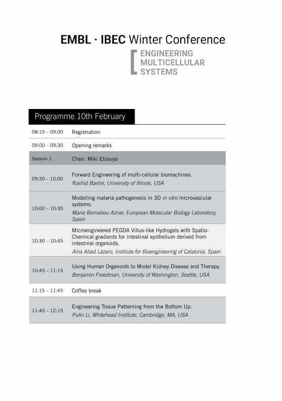
08:15 – 09:00 Registration
09:00 – 09:30 Opening remarks
Session 1 Chair: Miki Ebisuya
09:30 – 10:00Forward Engineering of multi-cellular biomachines.
Rashid Bashir, University of Illinois, USA
10:00 – 10:30
Modelling malaria pathogenesis in 3D in vitro microvascular systems.
Maria Bernabeu Aznar, European Molecular Biology Laboratory, Spain
10:30 – 10:45
Microengineered PEGDA Villus-like Hydrogels with Spatio-Chemical gradients for intestinal epithelium derived from intestinal organoids.
Aina Abad Lázaro, Institute for Bioengineering of Catalonia, Spain
10:45 – 11:15Using Human Organoids to Model Kidney Disease and Therapy.
Benjamin Freedman, University of Washington, Seattle, USA
11:15 – 11:45 Coffee break
11:45 – 12:15Engineering Tissue Patterning from the Bottom Up.
Pulin Li, Whitehead Institute, Cambridge, MA, USA
Programme10thFebruary

EMBL · IBEC Winter Conference on ENGINEERING MULTICELLULAR SYSTEMS 7
12:15 – 12:30
Morphogenesis is stressful – Elastic properties of folding cell sheets.
Stephanie Hoehn, University of Cambridge, UK
12:30 – 13:00
Mechanics of the intestinal crypt.
Xavier Trepat, Institute for Bioengineering of Catalonia (IBEC), Spain
13:00 – 13:20 Group photo
13:20 – 14:50 Lunch and Poster session
Session 2 Chair: Nuria Montserrat
14:50 – 15:20
Gastruloids: A Self-engineered system for the study of early mammalian development.
Alfonso Martínez Arias, University of Cambridge, UK
15:20 – 15:35
Three-dimensional culture of pancreas progenitors differentiated from mouse embryonic stem cells.
Shlomit Edri, Technion Israel Institute of Technology, Israel
15:35 – 16:00 Coffee break
16:00 – 16:30
How is Ethics Relevant for the Engineering of Multicellular Systems?
Insoo Hyun, PhD Professor, Department of Bioethics, Harvard Medical School
16:30 – 16:45
Collective Cell Movement and Cell State Transitions in Gastruloids Specify the Formation of Endoderm.
Ali Hashmi, Institut de Biologie du Développement de Marseille (IBDM), France
16:45 – 17:15
Self-organization of stem cells into embryos: A window on early mammalian development.
Magdalena Zernicka-Goetz, Department of Physiology, Cambridge University

Session 3 Chair: Josep Samitier
09:00 – 9:30Models of neurological disease.
Roger D. Kamm, Massachusetts Institute of Technology, USA
9:30 – 10:00
Engineering approaches for human pluripotent stem cells derived kidney organoids: emulating tissue features through bioengineering design
Núria Montserrat, Institute for Bioengineering of Catalonia, Spain
10:00 – 10:15
Mechanics and active cell behaviours contribute to self-organization of mesenchymal cells in the limb”.
Xavier Diego, EMBL Barcelona, Spain
10:15 – 10:45From haploid stem cells to blood vessel engineering.
Josef Penninger, University of British Columbia, Canada
10:45 – 11:15 Coffee Break
Session 4 Chair: James Sharpe
11:15 – 11:45Vascular Integration towards improved disease modelling.
Kristina Haase, European Molecular Biology Laboratory, Spain
11:45 – 12:00
A 3D Morphogenetic Model of Organogenesis in a Human Genetic Context.
Aimal Khankhel, University of California, Santa Barbara, USA
12:00 – 12:30
Engineering Organoid Development.
Matthias Lütolf, École Polytechnique Fédérale de Lausanne, Switzerland
Programme11thFebruary
EMBL · IBEC Winter Conference on ENGINEERING MULTICELLULAR SYSTEMS 9
12:30 – 12:45Induction of Synthetic Tissue Folding.
Guillermo Martínez Ara, EMBL Barcelona, Spain
12:45 – 13:00
Onset and Patterning Rules of Mesendoderm and Definitive Endoderm in Embryoid Bodies.
Iftach Nachman, Tel Aviv University, Israel
13:00 – 14:30 Lunch and Poster session
Session 5 Chair: Vikas Trivedi
14:30 – 14:50 Talk sponsored by Zeiss
14:50 – 15:20
Recreating Kidney Organogenesis in vitro with Human Pluripotent Stem Cells.
Ryuji Morizane, Harvard Stem Cell Institute, Cambridge, MA, USA
15:20 – 15:35
Tension heterogeneity instructs morphogenesis and fate specification during heart development.
Rashmi Priya, Max Planck Institute for Heart and Lung Research, Germany
15:35 – 16:00 Coffee break
16:00 – 16:30Mechanoregulation of thrombus formation in the bloodstream.
Hongxia Fu, University of Washington, Seattle, USA
16:30 – 16:45
A novel mathematical law to understand 3D self-organization in epithelial tubes.
Pedro Gómez-Gálvez, Instituto de Biomedicina de Sevilla (IBiS), Spain.
16:45 – 17:15Programming self-organizing tissues.
Wendell Lim, University of California, San Francisco, USA
17:15 – 17:45
Synthetic Embryology: A new window on mammalian development.
Eric D Siggia, The Rockefeller University, New York, USA

Session 6 Chair: Xavier Trepat
09:00 – 9:303D Bioprinting for in vitro Tissue Engineering.
Wei Sun, Tsinghua University, China
9:30 – 9:45
Contribution of stromal cells to the formation and stabalisation of blood vessels.
Negar Shahreza, University College London (UCL), UK
9:45 – 10:15
Modeling epithelial cells-fibroblasts interactions in gut homeostasis and pre-invasive cancers.
Danijela Vignjevic, Institut Curie, Paris, France
10:15 – 11:30 Coffee Break and poster session
11:30 – 12:00
Building and Breaking Tissues to Understand Development and Disease.
Zev Gartner, University of California, San Francisco, USA
12:00 – 12:15
Shear-induced crystallization drives precise patterning of hair cells in the mammalian inner ear.
David Sprinzak, Tel Aviv University, Israel
12:15 – 12:45Feedback, Dynamics and the Control of Size and Scale.
Arthur Lander, University of California, Irvine, USA
12:45 – 13:00 Awards ceremony
13:00 – 13:15 Closing remarks and farewell
Programme12thFebruary

EMBL · IBEC Winter Conference on ENGINEERING MULTICELLULAR SYSTEMS 11


Keynote Lectures

14 EMBL · IBEC Winter Conference on ENGINEERING MULTICELLULAR SYSTEMS
Monday10thFebruary·9:30
ForwardEngineeringofmulti-cellularbiomachines Rashid Bashir, University of Illinois, USA
The integration of living cells with 3D printed soft scaffolds can enable the realization of cellular machines for a range of applications in engineering and medicine. In this talk, I will present our group’s efforts towards developing such centimeter scale biological robots that actuated by skeletal muscles cells and our efforts to integrate neural control in these biological machines. These machines are controlled via electrical or optogenetic signals and demonstrated improved healing after a damage when exercised via optical stimulation. We also explore their lifetime and degradation in performance due to breakdown of the matrix that contains the cells. We will also present an approach to form functional in vitro neural tissue mimic (NTM) of different shapesusingstemcells,afibrinmatrix,and3Dprintedmolds.
We used murine-derived embryonic stem cells for optimizing cell-seeding protocols, characterization of the resulting internal structure of the construct, and remodeling of the extracellular matrix, as well as validation of electrophysiological activity. We also characterized the effects of optogenetic stimulation during neural differentiation, by showing morphological changes in network formation. These cellular systems present many opportunities in the next decade and beyond with potential applications in drug delivery, power generation, and other biomimetic systems. As these cellular machines increase in capabilities, exhibit emergent behavior, and potentially reveal the ability for self-assembly and self-repair, important questions can also arise about the ethical implications of this work.
EMBL · IBEC Winter Conference on ENGINEERING MULTICELLULAR SYSTEMS 15
Rashid Bashir, University of Illinois, USA
Rashid Bashir is Dean of Engineering, the Grainger Distinguished Chair in Engineering and Professor of Bioengineering at the University of Illinois at Urbana-Champaign. Previously, he was the Executive Associate Dean at the Carle-Illinois College of Medicine (2017 – present), the Abel Bliss Professor of Engineering, Head of Department of Bioengineering (2013 – 2017), and Director of the Micro and Nanotechnology Laboratory
(a campus-wide clean room research facility) (2007 – 2013). Prior to joining UIUC, he was at Purdue University (1998 – 2007) with faculty appointments in Electrical and Computer Engineering, and Bioengineering. From 1992 to 1998 he worked at National Semiconductor Corporation in Santa Clara, CA as Sr. Engineering Manager.
He graduated with a PhD in Electrical Engineering from Purdue University in 1992. He has authored or co-authored over 250 journal papers, over 200 conference papers and conference abstracts, and over 120 invited talks, and has been granted 49 patents. He received the NSF Faculty Early Career Award, the 2012 IEEE EMBS Technical Achievement Award, and the Pritzker Distinguished Lectureship Award from BMES in 2018. He is a fellow of IEEE, AIMBE, AAAS, BMES, IAMBE, RSC, APS, and NAI. He has been involved in 3 startups that have licensed his technologies. He was part of the core founding team and co-chair of the curriculum committee for the Carle Illinois College of Medicine, the world’s first engineering based College of Medicine at the University of Illinois at Urbana-Champaign.
His research group is interested in developing new technologies for precision and personalized medicine, and 3D bio-fabrication of cellular systems. Using bionanotechnology, BioMEMS, and lab on chip, he is working at the interface of biology and engineering from the molecular to the tissue scale, and aiming to make an impact on grand challenges in health and medicine including cancer, sepsis, and others.
In addition to leading his own research group, he was the PI on an NSF IGERT on Cellular and Molecular Mechanics and Bionanotechnology and PI on an NIH Training Grant on Cancer Nanotechnology. He is also co-PI on a recently funded National Research Traineeship (NRT) from NSF. He is also Associate Director and UIUC site lead on an NSF Science and Technology Center on Emergent Behavior of Integrated Cellular Systems (with MIT, Georgia Tech, and other partners).
16 EMBL · IBEC Winter Conference on ENGINEERING MULTICELLULAR SYSTEMS
Modelling malaria pathogenesis in 3D in vitro microvascular systems Maria Bernabeu, European Molecular Biology Laboratory, Spain
CaitlinHoward2, Celina Gunnarsson2, Christopher K. Arakawa2, Maria Vishnyakova3,ColeA.DeForest2, Joseph D. Smith3, Ying Zheng2
2 Department of Bioengineering, University of Washington, Seattle, WA, USA3 Seattle Children´s Research Institute, Seattle, WA, USA
Severemalariacarriesmortalityratesof15–20%,despitetreatmentwitheffectiveand fast-acting antimalarials. Plasmodium falciparum sequestration in the brain microvasculatureisthemainhallmarkofseveremalaria.However,themechanismsofparasite sequestration are only partially understood due to the lack of a rodent malaria model that mimics P. falciparum sequestration. We have recently engineered two new in vitro 3D microvascular models to better understand P. falciparum-host interactions. Thefirstmodelincludesa3Dbrainmicrovesselmodelthatdisplaysvesselsof100µmofdiameterarrangedina13x13 grid. This generates a wide range of wall shear stress within the same device. By using this system, we have shown that P. falciparum clonal lines, expressing different cytoadhesion ligands, respond differently to endothelium activation. In our second model, we have developed a robust method tobuild3Dendothelializedcapillary-sizevessels(5–10µm).
Perfusion of wild-type parasites or those lacking either cytoadhesion ligands or membrane-stiffening knobs showed different spatiotemporal patterns of sequestration. These results reveal that while molecular interactions are the most important requisite to achieve microvascular obstruction, infected red blood cell reduced deformability plays a secondary role. Altogether, our systems represent a new platform to study the biomechanical and biological determinants of blood disorders that affect the microvasculature.
Monday10thFebruary·10:00
EMBL · IBEC Winter Conference on ENGINEERING MULTICELLULAR SYSTEMS 17
Maria Bernabeu, European Molecular Biology Laboratory, Spain
Maria Bernabeu received her PhD in 2013 from University of Barcelona where she studied the biology of the malaria parasite. She later moved to Seattle Children’s Research Institute to study severe malaria pathogenesis. While in Seattle, she also worked at the Department of Bioengineering at the University of Washington, where she developed engineered models of brain microvessels. She recently has joint EMBL
Barcelona as a Group Leader and her group focuses on the study of cerebral malaria pathogenesis.
Her lab uses a combination of bioengineering, computational and experimental tools to better understand cerebral malaria pathogenesis. By developing an in vitro 3D human blood-brain barrier model, they aim to understand how parasite and host factors interact to cause endothelial and perivascular damage. With this system, they will model the pathogenic mechanisms and signalling pathways that lead to BBB breakdown in patients with cerebral malaria.

18 EMBL · IBEC Winter Conference on ENGINEERING MULTICELLULAR SYSTEMS
UsingHumanOrganoidstoModelKidneyDisease and TherapyBenjaminFreedman,UniversityofWashington,Seattle,USA
A series of discoveries has recently revealed the potential of human pluripotent stem cells to differentiate into the renal lineage, culminating in the generation of kidney organoids with nephron-like segments in vitro. These beautiful structures contain diverse cell lineages and are capable of modeling complex disorders, such as polycystic kidney disease, glomerulosclerosis, and cystinosis. When transplanted in vivo, kidney organois exhibit potential for maturation. Could these lead to the 'holy grail'ofimmunocompatibleregeneration?Askidneyorganoidsapproachtheirfiveyearbirthday,Dr.Freedmanwilllookbackatthebeginningsofthefield,reflectuponrecent progress, and engage in a conversation about its future.
Monday10thFebruary·10:45
EMBL · IBEC Winter Conference on ENGINEERING MULTICELLULAR SYSTEMS 19
BenjaminFreedman,UniversityofWashington, Seattle, USA
Dr. Benjamin Freedman is an Assistant Professor of Medicine at the University of Washington School of Medicine, Division of Nephrology. He is also a member of the Kidney Research Institute and the Institute for Stem Cell and Regenerative Medicine. Dr. Freedman received his Ph.D. in Cell and Developmental Biology in 2009 from the University of California at Berkeley.
He performed postdoctoral studies in the Renal Division of Brigham and Women’s Hospital, at Harvard Medical School. Dr. Freedman is currently performing biomedical research using human pluripotent stem cells to model kidney disease pathophysiology and develop new therapies.

20 EMBL · IBEC Winter Conference on ENGINEERING MULTICELLULAR SYSTEMS
Engineering Tissue Patterning from the Bottom UpPulin Li, Whitehead Institute, Cambridge, MA, USA
Howgenes,operatinginindividualcells,generatecoordinatedmulticellularbehavior is a fundamental question in biology. Applying a bottom-up approach, we quantitatively analyzed how tissue patterning dynamics and precision arise from the underlying genetic interactions. Morphogens, forming concentration gradients in space, set the blueprint for tissue patterning. By reconstituting morphogen gradients in vitro, re-wiring genetic interactions, and using quantitative time-lapse imaging and mathematical modeling, we revealed how architectural features of morphogen pathways could improve patterning robustness.
Our ongoing work expands the morphogen platform to address the question of evolvability: how does the "same" morphogen pattern tissues of drastically different lengthscales? The ability to isolate patterning from concurrent developmental processes and to quantitatively analyze the patterning behavior under different biophysical parameters offers a new way to uncover developmental design principles and engineer multicellular patterning.
Monday10thFebruary·11:45
EMBL · IBEC Winter Conference on ENGINEERING MULTICELLULAR SYSTEMS 21
Pulin Li, Whitehead Institute, Cambridge, MA, USA
Pulin Li is a member of the Whitehead Institute for Biomedical Research, and an assistant professor of Biology at Massachusetts Institute of Technology. Her lab is interested in quantitatively understanding how genetic circuits create multicellular behavior in both natural and synthetically engineered systems. She obtained her B.S. at Peking University in China, and Ph.D. in Chemical Biology at Harvard University.
Her Ph.D. work used chemical genetic tools to control and understand the behavior of hematopoietic stem cells, under the mentorship of Dr. Leonard Zon. She then joined Dr. Michael Elowitz's group at California Institute of Technology to study developmental tissue patterning using synthetic and systems biology approaches.

22 EMBL · IBEC Winter Conference on ENGINEERING MULTICELLULAR SYSTEMS
Mechanics of the intestinal cryptXavier Trepat, Institute for Bioengineering of Catalonia (IBEC), Spain
Theintestinalepitheliumisahighlydynamictissuethatself-renewsevery3–5days.Epithelial self-renewal is achieved through the action of stem cells that reside at the bottom of highly curved invaginations called crypts, where they coexist with secretory Paneth cells in a loose checker-board spatial pattern. To maintain homeostasis, stem cells constantly divide, giving rise to new cells that leave the niche, proliferate in the crypt neck, differentiate, and migrate to the tip of dome-like protrusions called villi, where they are extruded into the intestinal lumen. Each of these processes involves mechanical forces that have not yet been accessed experimentally.
Hereweuseintestinalorganoidsasamodelsystemtoprovidemapsoftheforcesthatcells generate as they divide, differentiate, fold and migrate. Our data suggests that the crypt is a mechanical unit that folds through apical constriction. Apical tension reachesthefirsttransientamplifyingcells,whichtransmittheforcestothesubstrate,thus mechanically compartmentalizing the niche. We show, further, that stem cell division perturbs local tractions abruptly, but does not affect the overall force pattern at the crypt. Instead, cells leave the crypt through collective migration under tension, rather than being expelled by pressure as previously thought.
Monday10thFebruary·12:30

EMBL · IBEC Winter Conference on ENGINEERING MULTICELLULAR SYSTEMS 23
Xavier Trepat, Institute for Bioengineering of Catalonia (IBEC), Spain
Xavier Trepat received a BSc in Physics in 2000 and a BSc in Engineering in 2001. In 2004 he obtained his PhD from the Medical School at the University of Barcelona. He then joined the Program in Molecular and Integrative Physiological Sciences at Harvard University as a postdoctoral researcher. In 2008 he became a "Ramón y Cajal" researcher at the University of Barcelona and in January 2011 an ICREA Research
Professor at the Institute for Bioengineering of Catalonia (IBEC).
He is Group Leader of the Integrative Cell and Tissue Dynamics research line at IBEC. In 2015 he won the Banc de Sabadell Award for Biomedical Research. In 2018 he was elected EMBO Member. He has been awarded with 3 grants from the European Research Council.
24 EMBL · IBEC Winter Conference on ENGINEERING MULTICELLULAR SYSTEMS
Gastruloids: a Self-engineered system for the study of early mammalian developmentAlfonso Martinez Arias, University of Cambridge, UK
Whensmall,specifiednumbersofPluripotentStemCells(PSCs)areplacedindefinedcultureconditionstheyaggregateandinitiateasequenceofpatternforming events that mimic processes that take place in the embryo: they undergo symmetrybreaking,gastrulationlikemovements,axialspecificationandgermlayerorganization. Over time, they assemble a spatially organize a genetic blueprint of the organism. In the case of mouse, gastruloids can be cultured for up to seven daystoreachastagecomparabletoE9.0inthemouseembryo.Wehaverecentlyextrapolated the system to human PSCs.
Gastruloids exhibit differences with mammalian embryos, most notably despite the spatial organization of gene expression they exhibit limited morphogenesis. This discrepancy suggests the existence of convergent but different modules involvedinmammaliandevelopment.Ishallbediscussingspecificexamplesandthe implications that these observations have for the theoretical and practical understanding of developmental events in mammals.
References:
1. Turner, D. et al. (2017) Anteroposterior polarity and elongation in the absence of extraembryonic tissues and spatialy organized signaling in Gastruloids, mammalian embryonic organoids. Development 144, 3894-3906
2. Van den Brink, S. et al. (2014) Symmetry breaking, germ layer specification and axial organisation in aggregates of mouse ES cells. Development 141, 4231-4242.
3. Beccari et al. (2018) Multiaxial self organization properties of mouse embryonic stem cells gastruloids. Nature 562, 272-276.
Monday10thFebruary·14:50
EMBL · IBEC Winter Conference on ENGINEERING MULTICELLULAR SYSTEMS 25
Alfonso Martinez Arias, University of Cambridge, UK
Alfonso Martinez-Arias studied Biology at the Universidad Complutense in Madrid (Spain). After graduating in 1977, he obtained a Fullbright scholarship to study in the US and in 1978 he went to the Department of Biophysics of the University of Chicago, Chicago (USA). In 1983 he moved on to do a postdoc with Peter Lawrence at the MRC Lab of Molecular Biology in Cambridge, UK.
In 1987 he was awarded a Wellcome Senior Fellowship which he held until 2002 when he became a member of the University of Cambridge and since 2003, he is Professor of Developmental Mechanics. During this time, first in the department of Zoology and since 2000 in the Department of Genetics he has pursued his interests in the logic of animal development.
26 EMBL · IBEC Winter Conference on ENGINEERING MULTICELLULAR SYSTEMS
HowisEthicsRelevantfortheEngineeringofMulticellular Systems?InsooHyun,HarvardMedicalSchool,Boston,MA,USA
Research into engineered multicellular systems is progressing at a rapid pace. Some observersmayassumethatethicalconsiderationsforthisnewfieldamountonlytoregulatory issues concerning safety and/or informed consent concerns related to cell donors or patient recipients of investigational multicellular products. While these issues are important, they do not exhaust all the ways in which ethical considerations can affect the speed and direction of much of this research.
This talk will explain how a focus on bioengineering ethics can help facilitate rapid yet socially responsible progress in the engineering of multicellular systems for innovative research and clinical translation. A collaborative version of bioengineering ethics will beofferedthatbringsresearchersandethiciststogethertohelpdefineanethicalapproach that can be responsive to both the science and the ethical uncertainties that itmaygenerate.Recentscientificexampleswillhelpillustratethewaysinwhichethicsremains relevant for the future development of multicellular systems engineering.
Monday10thFebruary·16:00

EMBL · IBEC Winter Conference on ENGINEERING MULTICELLULAR SYSTEMS 27
InsooHyun,HarvardMedicalSchool,Boston, MA, USA
Insoo Hyun is Professor of Bioethics and Philosophy at Case Western Reserve University School of Medicine and Faculty Member in the Center for Bioethics at Harvard Medical School. As a Fulbright Scholar and Hastings Center Fellow, Dr. Hyun’s interests include ethical and policy issues in stem cell research and new biotechnologies.
Currently, Dr. Hyun is the Principal Investigator of a BRAIN Initiative-funded project exploring the ethical issues surrounding human brain organoid research, in collaboration with leading scientists at Harvard and Stanford. He is the Co-Principal Investigator, along with colleagues at the Hastings Center, of an NIH grant identifying ways to improve the oversight of stem cell-based human-animal chimera research. And he is the Principal Investigator of a Greenwall Foundation project seeking to formulate a new bioengineering ethics framework for research involving the use of multi-cellular engineered living systems derived from human cells. This Greenwall project is in collaboration with scientists at Harvard, MIT, and the University of Michigan.
Dr. Hyun has been involved for many years with the ISSCR (International Society for Stem Cell Research), for which he has helped draft all of the ISSCR’s international research guidelines and has served as their Chair of the Ethics and Public Policy Committee. He now serves as a member of the Neuroethics Subgroup of the BRAIN 2.0 Working Group of Advisory Committee to the Director, NIH.
Dr. Hyun received his BA and MA in Philosophy with Honors in Ethics in Society from Stanford University and his PhD in Philosophy from Brown University. He has been interviewed frequently on National Public Radio and has served on national commissions for the Institute of Medicine and the National Academy of Sciences in Washington D.C. Dr. Hyun is a regular contributor to Nature, Science, Cell Stem Cell, The Hastings Center Report, among many other journals. His book Bioethics and the Future of Stem Cell Research was published by Cambridge University Press in 2013.

28 EMBL · IBEC Winter Conference on ENGINEERING MULTICELLULAR SYSTEMS
Self-organization of stem cells into embryos: A window on early mammalian development Magdalena Zernicka-Goetz, Department of Physiology, Cambridge University
Embryonic development is orchestrated by robust and complex regulatory mechanisms acting at different scales of organization. In vivo studies are particularly challenging for mammals after implantation, owing to the small size and inaccessibility of the embryo. The generation of stem cell models of the embryo represents a powerful system with which to dissect this complexity. Control of geometry, modulation of the physical environment, and priming with chemical signals reveal the intrinsic capacity of embryonic stem cells to make patterns. Adding the stem cells for the extraembryonic lineages generates three-dimensional models that are more autonomous from the environment and recapitulate many features of the pre- and postimplantation mouse embryo, including gastrulation.
Monday10thFebruary·16:45
EMBL · IBEC Winter Conference on ENGINEERING MULTICELLULAR SYSTEMS 29
Magdalena Zernicka-Goetz, Department of Physiology, Cambridge University
Magdalena carried out her Ph.D. at the University of Warsaw, Poland, under supervision of Andrzej Tarkowski. She came to Cambridge in 1995 to join Martin Evans group with the long-term aim of studying the mechanisms of regulative nature of development and spatial patterning in the mouse embryo. In 1997 she was awarded a Senior Research Fellowship from the Lister Institute to start her independent group at the
Wellcome Trust/Cancer Research UK Gurdon Institute in Cambridge.
In 2001 she became a Wellcome Senior Research Fellow. In 2010, she became a Professor of Mammalian Development and Stem Cell Biology. In 1993 she received a Promising Young Scientist Prize from Foundation for Polish Science, in 2001, Young Investigator Award from EMBO, in 2007 she was elected to EMBO membership and in 2013 she became Fellow of British Academy of Medical Science.
30 EMBL · IBEC Winter Conference on ENGINEERING MULTICELLULAR SYSTEMS
Models of neurological disease Roger D Kamm, Massachusetts Institute of Technology, USA
Many of the most debilitating and life-threatening diseases are associated with the central nervous system. Some, such as neurodegenerative diseases, predominately afflictouragingpopulation.YetotherssuchasbraincancersandALSareprevalentat all ages. In this presentation, models will be presented that attempt to recapitulate certain aspects of the central or peripheral nervous system, both to probe the disease process, and as a platform to screen for new therapeutics.
Fourexampleswillbepresented.First,amodelforthehealthyblood-brainbarrierand neurovascular unit have been developed in order to capture the essential aspects associated with these diseases. Second, the blood-brain barrier model is used to study metastasis of cancers to the brain, as well as primary glioblastoma. In the third part of the talk, a model for the healthy and diseased neuromuscular junction will be presentedasasteptowarddevelopingtherapeuticstrategiesfortreatingALS.Finally,a model for cerebral amyloid angiopathy, often associated with Alzheimer’s disease, will be discussed as a disease model and for its drug screening potential.
Tuesday11thFebruary·09:00
EMBL · IBEC Winter Conference on ENGINEERING MULTICELLULAR SYSTEMS 31
Roger D Kamm, Massachusetts Institute of Technology, USA
Kamm is currently the Cecil and Ida Green Distinguished Professor of Biological and Mechanical Engineering at MIT, where he has served on the faculty since 1978. Kamm has long been instrumental in developing research activities at the interface of biology and mechanics, formerly in cell and molecular mechanics, and now in engineered living systems. Current interests are in developing models of healthy
and diseased organ function using microfluidic technologies, with a focus on vascularization.
Kamm has fostered biomechanics as Chair of the US National Committee on Biomechanics (2006-2009) and of the World Council on Biomechanics (2006-2010). Kamm currently directs the NSF Science and Technology Center on Emergent Behaviors of Integrated Cellular Systems. He is the 2010 recipient of the ASME Lissner Medal (American Society of Mechanical Engineering) and the 2015 recipient of the Huiskes Medal (European Society of Biomechanics), both for lifetime achievements, and is the inaugural recipient of the ASME Nerem Medal for mentoring and education. He was elected to the National Academy of Medicine in 2010. Kamm is co-founder of two companies, Cardiovascular Technologies and AIM Biotech, a manufacturer of microfluidic systems for 3D culture.
32 EMBL · IBEC Winter Conference on ENGINEERING MULTICELLULAR SYSTEMS
Engineering approaches for human pluripotent stem cells derived kidney organoids: emulating tissue features through bioengineering design Nuria Montserrat, Institute for Bioengineering of Catalonia (IBEC), Spain
The generation of human pluripotent stem cells (hPSCs) derived organoids is one ofthebiggestscientificadvancesinregenerativemedicine.Recently,wehavedemonstrated that by lengthening the time that hPSCs are exposed to a three-dimensionalmicroenvironmentinthepresenceofdefinedrenalinductivesignals,weare able to generate kidney organoids that transcriptomically match second-trimester humanfetalkidneys.Furthermore,wehaverecentlydevelopedatransplantationmethod that utilizes the chick chorioallantoic membrane (CAM). In our hands, this approach created a soft in vivo microenvironment that promotes the growth and differentiation of implanted kidney organoids, as well as providing a vascular component.
Through bioengineering, we have mimicked the stiffness of the chick CAM by fabricating compliant hydrogels. This approach resulted in the acceleration of kidney organoid formation proving that mechanical cues are determinant for the generation of hPSC-renal progenitor cells and kidney organoids. Overall, we will discuss how these preliminaryfindingsareadvancingourresearchtowardstheapplicationofdifferentbioengineering strategies (i.e., including 3D bioprinting and tissue engineering) for kidney organoid generation and human disease modeling.
Tuesday11thFebruary·09:30

EMBL · IBEC Winter Conference on ENGINEERING MULTICELLULAR SYSTEMS 33
Nuria Montserrat, Institute for Bioengineering of Catalonia (IBEC), Spain
Núria Montserrat is ICREA research professor since January 2019 and group leader of the “Pluripotency for organ regeneration” group at IBEC. She received BSc in Biology in 2006 at University of Barcelona and PostDoc at Fundaçao per a Ciência e Tecnología (Portugal).
In 2008 she moved to the Center of Regenerative Medicine in Barcelona (CMRB) as an associate-
researcher, supported by a Juan de la Cierva fellowship. In 2014 she was awarded with the European Research Council Starting Grant aiming at studying kidney development. and disease using hiPSC-derived kidney organoids.
34 EMBL · IBEC Winter Conference on ENGINEERING MULTICELLULAR SYSTEMS
FromhaploidstemcellstobloodvesselengineeringJosef Penninger, University of British Columbia, Canada
We have previously generated murine stem cells with a single set of chromosomes, termed haploid ES cells. Using such cells we have been able to rapidly mine essential biological pathway and to use revertible mutagenesis to identify novel mediators of angiogenesis. Recently we have expanded our work to engineer human blood vessel organoids that can be transplanted into mice to establish a fully human vascular tree. We have used this system to model the pathogenesis of diabetes vasculopathies.
Tuesday11thFebruary·10:15

EMBL · IBEC Winter Conference on ENGINEERING MULTICELLULAR SYSTEMS 35
Josef Penninger, University of British Columbia, Canada
Josef Penninger, MD was formerly a lead researcher at the Amgen Research Institute in Toronto. Since 2002 Josef Penninger was the founding and scientific director of the newly established Institute of Molecular Biotechnology (IMBA) of the Austrian Academy of Sciences in Vienna, Austria. In 2018 he accepted the appointment as Director of the Life Sciences Institute (LSI) at the University of British Columbia (UBC) in Canada.
Major achievements include pioneering insights into the molecular basis of osteoporosis and breast cancer, as well as the study of metastatic spread. Josef Penninger’s major awards include the Descartes Prize, the Wittgenstein Prize of the Austrian Federal Government, the Ernst Jung Prize for medical excellence, an AAAS Award, the Innovator Award from Era of Hope/U.S. Department of Defense and a second ERC Advanced grant.
36 EMBL · IBEC Winter Conference on ENGINEERING MULTICELLULAR SYSTEMS
Vascular Integration towards improved disease modellingKristinaHaase,EuropeanMolecularBiologyLaboratory,Spain
The development of ever more complex in vitro models has increased drastically over thelasttwodecades.Now,in2020,weexpectthat3Dmodelsshouldmaketheleapfromthebenchtothebedside.However,alackofadoptionthusfarresultsfromtheinability of these models to accurately predict clinical outcomes. Despite advances in patient-specificiPSCandgene-editingtechnologies,thelackoffunctionalvasculatureis hypothesized as a likely key factor in the limited predictability of current in vitro models. Given its role in regulating oxygen tension, nutrient delivery and immune cell interactions, functional vasculature is required for the development and long-term maintenance of normal tissue physiology. To overcome this limitation, we develop disease-specificmodelsintegratingmicrovasculatureonmillimetre-scalechips.
This talk will provide an overview of several vascularized models (placental, ischemic, and tumor) where we exploit the self-assembly process of primary endothelial and combined stromal cells. Generating perfusable microvessels in a tissue-dependent contextallowsustoinvestigatespecificcell-cellinteractionsandtheeffectsofmechanicalcues,suchasluminalflow,ontheregulationofmicrovesselmorphologyand permeability. In addition, drug dissemination studies can be performed and examined in the context of perfused microvessels. These physiologically relevant systemsholdsignificantpromisefordevelopmentintomoreaccuratepreclinicalmodels – a major focus of our new lab.
Tuesday11thFebruary·11:15

EMBL · IBEC Winter Conference on ENGINEERING MULTICELLULAR SYSTEMS 37
KristinaHaase,EuropeanMolecularBiologyLaboratory, Spain
Kristina Haase is a mechanical engineer by training. She holds a Masters degree in Mechanical Engineering and a Ph.D. in Biophysics from University of Ottawa.
Her post-doctoral work was carried out in the bio-engineering lab of Prof. Roger Kamm at MIT in Massachusetts, USA.
Since October 2019 she has started a new lab at EMBL Barcelona, focusing on tissue engineering and disease-specific modeling.

38 EMBL · IBEC Winter Conference on ENGINEERING MULTICELLULAR SYSTEMS
Engineering Organoid DevelopmentMatthiasLütolf,ÉcolePolytechniqueFédéraledeLausanne(EPFL),Switzerland
Organoids form through poorly understood morphogenetic processes in which initially homogeneous ensembles of stem cells spontaneously self-organize in suspension or within permissive three-dimensional extracellular matrices. Yet, theabsenceofvirtuallyanypredefinedpatterninginfluencessuchasmorphogengradients or mechanical cues results in an extensive heterogeneity. Moreover, the current mismatch in shape, size and lifespan between native organs and their in vitro counterparts hinders their even wider applicability. In this talk I will discuss some of our ongoing efforts in developing next-generation organoids that are assembled by guiding cell-intrinsic self-patterning through engineered stem cell microenvironments.
Tuesday11thFebruary·12:00

EMBL · IBEC Winter Conference on ENGINEERING MULTICELLULAR SYSTEMS 39
Matthias Lütolf, École Polytechnique FédéraledeLausanne(EPFL),Switzerland
Professor Matthias Lutolf is Director of the Laboratory of Stem Cell Bioengineering at Ecole Polytechnique Fédérale de Lausanne (EPFL), Switzerland. His highly innovative and cross-disciplinary research program is focused on the development of bio- and tissue-engineering strategies for improving organoid culture and enabling its translation to real-life applications.

40 EMBL · IBEC Winter Conference on ENGINEERING MULTICELLULAR SYSTEMS
Recreating Kidney Organogenesis in vitro with HumanPluripotentStemCellsRyujiMorizane,HarvardStemCellInstitute,Cambridge,MA,USA
Wehavedevelopedanefficient,chemicallydefinedprotocolfordifferentiatinghumanpluripotent stem cells into multipotent nephron progenitor cells (NPCs) that can form kidney organoids. By recapitulating metanephric kidney development in vitro wegenerateSIX2+SALL1+WT1+PAX2+NPCswith80–90%efficiencywithin8–9days of differentiation. NPCs form kidney organoids containing epithelial nephron-likestructuresexpressingmarkersofpodocytes,proximaltubules,loopsofHenleand distal nephrons in an organized, continuous arrangement that resembles the nephron in vivo.Theorganoidsexpressgenesreflectingmanytransportersseenin adult metanephric-derived kidney, enabling assessment of transporter-mediated drugnephrotoxicity.StromalcellsarealsogeneratedwiththepresenceofPDGFRb+fibroblasts/pericytes,andCD31+endothelialcells.Thiskidneydifferentiationsystemcan be used to study mechanisms of human kidney development.
Repetitiveinjurytotubularcellscausesinterstitialfibroblastexpansionwithcharacteristicsofmyofibroblasts,indicatingkidneyorganoidscanbeusedtomodelkidneyfibrosisin vitro. Polycystic kidney disease (PKD) patient-derived organoids exhibitcysticphenotypes.Hencethegeneratedkidneyorganoidsareeffectivetoolsto study genetic disorders of the kidney as well as mechanisms of kidney injury and fibrosis.Microphysiologicalplatformsin vitro facilitate kidney organoid vascularization and maturation, which may lead to the development of functional bioengineered kidneys in the future.
Tuesday11thFebruary·14:50
EMBL · IBEC Winter Conference on ENGINEERING MULTICELLULAR SYSTEMS 41
RyujiMorizane,HarvardStemCellInstitute,Cambridge, MA, USA
Ryuji is currently a Principal Investigator at Brigham and Women’s Hospital, Harvard Medical School. He has pioneered research in stem cell differentiation and kidney organoids. Ryuji directs research groups focused on kidney regenerative medicine, genome editing in stem cells, and kidney disease modelling. His research also extends to simulation of kidney microenvironment
using organ-on-chip systems. At the Wyss, Ryuji is collaborating with Professor Jennifer Lewis aiming to create functional vascularized kidney tissues in vitro.
Ryuji’s research has been recognized internationally, and he has received various awards including the Research Excellence Award at Discover Brigham in 2015 and 2016 and Career Development Award at Brigham and Women’s Hospital in 2016. He is a recipient of funding from the Uehara Memorial Foundation, Japan Society for the Promotion of Science, ReproCELL, Harvard Stem Cell Institute, and the Diabetic Complications Consortium.
42 EMBL · IBEC Winter Conference on ENGINEERING MULTICELLULAR SYSTEMS
Mechanoregulation of thrombus formation in the bloodstreamHongxiaFu,UniversityofWashington,Seattle,WA,USA
A thrombus is like a double-edged sword in blood vessels: on the positive side, it can stop bleeding at sites of injury, but on the negative side, it can cause vascular occlusion in many cardiovascular diseases, such as thrombosis, stroke, and sepsis. Endothelial cells (ECs) on the interior surface of blood vessels play an important role in regulating thrombus formation. We are interested in how a blood protein called von Willebrandfactor(VWF),whichissynthesizedbyECs,mediatesplateletadhesionandaggregation on the blood vessel wall and controls the initiation and development of thrombi in bloodstream.
Combingstemcells,microfluidics,andsinglemoleculebiophysicstools,ourstudieshaveunveiledanovelmechanoregulatorymechanismforVWFtobelocallyactivatedbyhydrodynamicforceinducedbybloodflowduringhemorrhageorindiseasedvessels, but rapidly deactivated downstream in normal blood circulation.
Tuesday11thFebruary·16:00
EMBL · IBEC Winter Conference on ENGINEERING MULTICELLULAR SYSTEMS 43
HongxiaFu,UniversityofWashington,Seattle, WA, USA
Hongxia Fu obtained her Ph.D. at the National University of Singapore, where she studied DNA biomechanics using single-molecule force manipulation tools and theoretical models. She continued her postdoctoral research training in biophysics and mechanobiology at the National University of Singapore and Harvard Medical School, where her research focused on developing and applying tools
combining single-molecule fluorescence imaging and force manipulation techniques, microfluidics, and cell biology to study protein functions under force.
Currently she is an Assistant Professor in the Division of Hematology, University of Washington (UW) School of Medicine, the Institute for Stem Cell and Regenerative Medicine, the Department of Bioengineering at UW, and Bloodworks Northwest Research Institute in Seattle, USA. Her current research focuses on understanding the mechanisms of mechanosensory proteins in blood clotting, studying related blood diseases, such as thrombosis and bleeding disorders, based on vascular, molecular, and single-molecule models of health and disease, and seeking new methodologies for therapeutics testing and treatment of blood and circulatory system disorders.
44 EMBL · IBEC Winter Conference on ENGINEERING MULTICELLULAR SYSTEMS
Programming self-organizing tissuesWendellLim,UniversityofCalifornia,SanFrancisco,USA
Satoshi Toda, Jonathan Brunger, Adam Stevens, Wesley McKeithan, Pilar Lopez
Multicellular organisms have evolved cell-cell communication programs that specify the robust self-organization of diverse body-plans and tissues. It is remarkable that genetically encoded programs can so compactly store the information to construct macroscopictissuesandorganisms.Althoughwehaveidentifiedmanycommonthemes seen in diverse developmental programs, many fundamental questions remain unclear.
What are the minimal components required for multi-cellular self-organization, and how might metazoan multi-cellularity have arisen? Can we understand the language ofself-organizationsufficientlytobeabletogeneticallyprogramtheformationofnew types of tissues, or to drive developmental programs in response to novel inputs and niches (i.e. for regenerative medicine etc.)? We have been developing a set of orthogonalizedmolecularpartsthatfacilitatetheconstructdefined,user-designedcell-cell interaction networks. Using these components (including orthogonal juxtacrine signals, paracrine signals, and adhesion/assembly systems) we have begun exploring how to program the formation of simple synthetic tissues from the bottom-up, as well as to modulate and control natural developmental programs.
Tuesday11thFebruary·16:45
EMBL · IBEC Winter Conference on ENGINEERING MULTICELLULAR SYSTEMS 45
Wendell Lim, University of California, San Francisco,USA
Wendell A.Lim obtained his Ph.D. in Biochemistry&Biophysics in 1991 at the Massachusetts Institute of Technology and his Postdoc in 1996 at Yale University. He is currently Professor & Chair, Department of Cellular and Molecular Pharmacology at UCSF and Director of UCSF Center for Systems & Synthetic Biology.
His general scientific interests are in understanding how genetically encoded molecular programs can yield the remarkable behaviors observed in biological organisms, at multiple scales. His research career began as a biophysical chemist and structural biologist studying problems such as the evolutionary optimization of enzymes, how protein structure is encoded in sequence, and the determinants of protein-protein interaction specificity. Now,his research has gradually shifted towards utilizing this mechanistic understanding of molecules as a foundation to study how systems of interacting molecules assemble to yield cellular or organismal signaling behaviors – complex behaviors in both space and time. His lab is interested in both the fundamental principles governing these molecular programs, as well as the way such programs have evolved.

46 EMBL · IBEC Winter Conference on ENGINEERING MULTICELLULAR SYSTEMS
Synthetic Embryology: A new window on mammalian development Eric D. Siggia, The Rockefeller University, New York, USA
The embryo evolved to make a fetus and thus multiple modes of regulation conspire toensurearobustoutcome.Thismakesthetaskofquantifyingthepathwaysdefiningthemammalianembryoparticularlydifficult.Embryonicstemcells(ESC)giverisetoallcellsofthebodyproper.Wehaveshownseveralyearsago,howmerelyconfininghuman ESC to two dimensional patterns, causes the cells to recapitulate the spatial patterning seen in the mouse embryo at the onset of gastrulation. We have dissected the cascade of secreted factors and the location of receptors driven by apical-basal polarity responsible for the patterns.
Apredictionforhowmorphogensaretargetedinthemousewasrecentlyconfirmedby another group. Our assay can be extended to three dimensions and the model epiblast shown to spontaneously break symmetry and form a primitive streak. A second layer of extraembryonic like cells adds additional realism and new interactions. Synthetic systems allow one to peal back the layers of regulation that make embryonic development so robust. They are easy to manipulate and suggest targeted experiments to pursue in-vivo.
Tuesday11thFebruary·17:15
EMBL · IBEC Winter Conference on ENGINEERING MULTICELLULAR SYSTEMS 47
Eric D. Siggia, The Rockefeller University, New York, USA
Dr Siggia was trained as a physicist and worked in the areas of statistical mechanics of phase transitions, quantum magnetism, fluid mechanics and nonlinear dynamics. He moved to Rockefeller University from Cornell in 1998, and converted to biology.
He collaborated on projects in the areas of protein trafficking, bioinformatics of gene regulation, evolution
of antibiotic resistance, cell cycle in yeast. In the last decade he worked with Ali Brivanlou and others on dynamics of vertebrate signaling pathways and synthetic embryology using human embryonic stem cells.
48 EMBL · IBEC Winter Conference on ENGINEERING MULTICELLULAR SYSTEMS
3D Bioprinting for in vitro Tissue EngineeringWei Sun, Tsinghua University, China
3D Bio-Printing uses living cells as building blocks to fabricate in vitro biological models.Theprinted3Dtissuemodelshavebeenwidelyappliedtothefieldofregenerative medicine, disease study, drug discovery and drug toxicity testing. This presentation will report our recent study on printing cells for construction of in vitro tissue models, disease models, tumor models and micro-organ chips with application to cancer and drug toxicity study. An overview of 3D Bioprinting on in vitro tissue engineering will be given. Enabling techniques for cell printing to construct 3D biological models will be introduced.
Examples of 3D printing in vitro tissue models for: 1) cervical tumor in vitro with tumor morphology, MMP and genes expressions, chemo-resistance, and epithelial-mesenchymaltransitionsstudy;2)micro-liver-organfordrugmetabolismstudy;and3) printing hepatocytes for drug hepatotoxicity study; and 4) printing cell models for renalfiltrationandnephrotoxicitytest.Comparisonofbiologicaldataderivedfrom3Dprintedmodelswith2Dplanarpetri-dishesmodelswillbeconducted.Discussionsonchallenges and opportunities of 3D Bio-Printing model as alternative tissue models for drug development and testing will also be presented.
Wednesday12thFebruary·09:00
EMBL · IBEC Winter Conference on ENGINEERING MULTICELLULAR SYSTEMS 49
Wei Sun, Tsinghua University, China
Dr. Wei Sun is Albert Soffa Chair Professor of Mechanical Engineering, Drexel University, and Professor and Director of Biomanufacturing Research Center, Tsinghua University, Beijing, China. Dr. Sun’s research has been on Biofabrication, Cell Printing and Tissue Engineering. His research has been sponsored by the US National Science Foundation (NSF), Defense Advanced Research Projects Agency (DARPA), National Aeronautics and Space Administration (NASA), Chinese
Ministry of Science and Technology (MOST) and Chinese Ministry of Education (MoE).
Dr. Sun has published 160+ journal papers, with 9500+ SCI citations, 45 issued patents, and conducted 360+ invited presentations in the field of his research. Dr. Sun is the Founding President for International Society of Biofabrication (2010-2014), and the Founding Editor-in-Chief for international journal Biofabrication (2009-present).
Dr. Sun received Award of Distinguished Visiting Fellow from the Royal Academy of Engineering in UK (2018), the Senior Investigator Award from International Society of Biofabrication (2017), MII / Fralin Visiting Scholar Award from Virginia Tech (2015), Outstanding Research Award, College of Engineering, Drexel University (2009), William Mong Fellow Award, the University of Hong Kong (2008) and Ralph R. Teetor Educational Award, International Society for Automotive Engineers (2003).

50 EMBL · IBEC Winter Conference on ENGINEERING MULTICELLULAR SYSTEMS
Modelingepithelialcells-fibroblastsinteractions in gut homeostasis and pre-invasive cancersDanijelaVignjevic,InstitutCurie,Paris,France
Fibroblastsareoneofthemostabundantcelltypesinthestromaofmanytissues.They produce the extracellular matrix (ECM) and factors that modify its biochemical and physical properties. They also secrete growth factors and cytokines that affect proliferation, migration, and survival of neighboring cells. As such, they play an essential role in gut organogenesis, homeostasis, and tumor invasion.
We have developed a microstructured device that allows the study of the interaction offibroblastswiththeepithelialcells.Basedona3DcollagenIscaffold,thedevicehas the typical topography of the mouse gut, an array of villi surrounded by crypts. Rigidifyingthescaffoldbycross-linkingcollagenfiberswiththreosepreservesitscytocompatibilityandenablestheincorporationoffibroblastsreproducingthegut stromal compartment. Mouse organoids deposited into crypts, open up and epithelize the scaffold, generating a polarized monolayer containing proliferative and differentiated cells. Applying physical strain to the epithelium maintains its long-term integrity.Usingthisdevice,wefoundthatprimaryintestinalfibroblastsarerequiredtostimulatetheefficientepithelializationofthescaffoldwhilemaintainingtheapicobasalpolarity of the epithelial cells.
Incancer,fibroblastssurroundingatumoraregenerallycalledcancer-associatedfibroblasts(CAFs).Attheearlystageoftumorprogression,beforetheonsetofinvasion,cancercellsandCAFsaresegregated.CAFsaccumulateatthetumorperiphery,formingacontinuousandcohesivelayerthatsurroundsthetumor.Howisthis organization of early-stage tumors achieved it is not clear. Using hollow alginate capsules,wefoundthatCAFsdonotspontaneouslyenvelopcancercells.Instead,confinement,thebuildupofcompressivestress,andreorganizationofthefibronectinnetworkwerenecessarytoinducefibroblastsspreadingovertheaggregatesoftumor cells. We propose that the compressive stress generated by the tumor growth representsamechanismbywhichCAFsenwrapthetumor.
Wednesday12thFebruary·09:45

EMBL · IBEC Winter Conference on ENGINEERING MULTICELLULAR SYSTEMS 51
DanijelaVignjevic,InstitutCurie,Paris,France
Danijela Matic Vignjevic was trained as a molecular biologist at the University of Belgrade, Serbia, and University of Wisconsin-Madison, US. She did her Ph.D. in cell biology, working on the role of the actin cytoskeleton in cell migration in the lab of Gary Borisy at Northwestern University, Chicago, US. She then did a post-doc in the lab of Daniel Louvard at Institut Curie, working on mouse models for colon cancer metastasis as a HFSP fellow.
After being recruited as an INSERM researcher, she continued working on cell migration-related questions. She started her independent team in 2013 when she got interested in how epithelial cells interact with their microenvironment (focusing on ECM and fibroblasts) in homeostasis and during cancer invasion. Her research strategy combines molecular and cell biology techniques with live-cell imaging using different model systems such as 2D and 3D in vitro cell cultures, tissue slices cultured ex vivo, and different transgenic mouse models.
She is the recipient of ERC Starting grant (2013-2017) and Consolidator grant (2018-2023), and she received several awards such as “Grand Prix” in Cancer research, Foundation Simone et Cino del Luca, and Dandrimont-Benicourt, French Academy of Science.

52 EMBL · IBEC Winter Conference on ENGINEERING MULTICELLULAR SYSTEMS
Building and Breaking Tissues to Understand Development and DiseaseZevGartner,UniversityofCalifornia,SanFrancisco,USA
Cells are living materials – their physical properties are not static but change dynamically in response to the environment. This property of cells as materials gives them the capacity to self-organize into complex three dimensional structures. Self-organization is critical to tissue developmental and repair, and a better understanding of how tissues self-organize will generate new strategies to slow the breakdown of tissue structure that contributes to the initiation and progression of diseases like breast cancer. In this presentation I will discuss our efforts to understand the physical mechanisms used by primary human epithelial cells to self-organize into a bilayered mammary epithelium. I will then describe how this program of self-organization becomes dysregulated by the activation of breast cancer driver genes contributing to disease progression.
Wednesday12thFebruary·11:30

EMBL · IBEC Winter Conference on ENGINEERING MULTICELLULAR SYSTEMS 53
Zev Gartner, University of California, San Francisco,USA
Professor Zev Gartner obtained his Ph.D. in Chemical Biology in 2004 at Harvard University and his PostDoc in 2008 at University of California. He is Professor, Department of Pharmaceutical Chemistry, UCSF and Co-director, Center for Cellular Construction.
His lab is working to understand how cells assemble into multicellular tissues, how the structure of tissues
controls the behaviour of individual cells, and how changes to tissue structure drive the progression of diseases like cancer. Toward these goals, they build, perturb, and model human tissues in vitro using techniques from the chemical, engineering, physical and biological sciences.
54 EMBL · IBEC Winter Conference on ENGINEERING MULTICELLULAR SYSTEMS
Feedback,DynamicsandtheControlofSizeand ScaleArthur D. Lander, University of California, Irvine, USA
Animal development is a remarkable feat of engineering, capable of reaching precise endpoints in the face of massive internal and external unreliability. That precision isreflectedinthesizesofmacroscopicstructures(tissuesandorgans),thespatialpatterns of cell behavior within those structures, and the scaling relationships that couplepatternandsize.Onrareoccasionsreliableoutcomesaretheresultoffinely-tuned initial conditions, but far more often they depend on feedback processes involving morphogens and other intercellular signals.
Understanding how such feedback circuits are constructed is one of the oldest and most pressing goals of developmental biology. I will discuss recent experimental and modeling results on the role of feedback in pattern scaling in Drosophila, and its relationship to the control of size.
Wednesday12thFebruary·12:15

EMBL · IBEC Winter Conference on ENGINEERING MULTICELLULAR SYSTEMS 55
Arthur D. Lander, University of California, Irvine, USA
Arthur Lander obtained his Ph.D. in Neuroscience in1985 at University of California and his PostDoc in 1987 at Columbia University College of Physicians & Surgeons, New York, NY.
Arthur Lander is the Donald Bren Professor of Developmental and Cell Biology and holds joint appointments in the Departments of Biomedical
Engineering, and Logic & Philosophy of Science. He serves on the editorial board of BMC Biology, and is a member of the American Society for Clinical Investigation, a fellow of the American Association for the Advancement of Science, and a member of the Science Board of the Sante Fe Institute. He holds visiting professor appointments at National Taiwan University and the University of Tsukuba (Japan).
Research in the Lander lab is focused on the Systems Biology of Development and Disease, and deals with topics in Developmental Biology, Cell Biology, Mathematical/Computational Biology, Glycobiology, Neurobiology, Cancer Biology and Engineering.


Short talks
58 EMBL · IBEC Winter Conference on ENGINEERING MULTICELLULAR SYSTEMS
MicroengineeredPEGDAVillus-likeHydrogelswithSpatio-Chemical Gradients for Intestinal Epithelium Derived from Intestinal Organoids
Gizem Altay1, AinaAbad-Lazaro1, Emilio J. Gualda2, María García-Díaz1, Núria Torras1,JordiFolch1,SébastienTosi3, Vanesa Férnandez1, Eduard Batlle4,5,6, Pablo Alvarez-Loza3, Elena Martínez1,7,8
1 Biomimetic Systems for Cell Engineering Laboratory, Institute for Bioengineering of Catalonia (IBEC), The Barcelona Institute of Science and Technology (BIST), Baldiri Reixac 15-21, 08028 Barcelona Spain
2 SLN Research Facility, The Institute of Photonic Sciences (ICFO), The Barcelona Institute of Science and Technology (BIST), Castelldefels 08860, Barcelona, Spain
3 Advanced Digital Microscopy Core Facility (ADMCF), Institute for Research in Biomedicine (IRB Barcelona), The Barcelona Institute of Science and Technology (BIST), Baldiri Reixac 10-12, Barcelona 08028, Spain
4 Colorectal Cancer Laboratory, Institute for Research in Biomedicine (IRB Barcelona), The Barcelona Institute of Science and Technology (BIST), Baldiri Reixac 10-12, Barcelona 08028, Spain
5 Centro de Investigación Biomédica en Red de Cáncer (CIBERONC), Barcelona, Spain6 ICREA, Passeig Lluís Companys 23, 08010 Barcelona, Spain7 Centro de Investigación Biomédica en Red en Bioingeniería, Biomateriales y Nanomedicina (CIBER-BBN), Av. Monforte de Lemos 3-5, Pabellón 11, Planta 0, 28029 Madrid, Spain
8 Department of Electronics and Biomedical Engineering, University of Barcelona (UB), Martí i Franquès 1, Barcelona 08028, Spain
Thesmallintestinalepitheliumisformedbyfinger-likeprotrusionscalledvilliandinvaginations called crypts. The intestinal epithelium cell turnover relies on intestinal stem cells (ISCs) located at the crypt base that divide giving rise to proliferative cells that migrate up along the villi while differentiating into mature epithelium. Differentiated cells die at the tips of the villi and exfoliate into the lumen. This homeostasisistightlycontrolledbybiomoleculargradientsofEGF,WntandBMPsignalling pathways formed along the crypt-villus axis [1]. The development of three-dimensional (3D) in vitro culture methods has made possible the use of primary ISCs to create organoids1. One of the main drawbacks of intestinal organoids is their 3D closed geometry which hinders the access to the organoid lumen limiting their use in many applications. Therefore, there is a need for engineering culture platforms that overcome this limitation and provide physiologically cellular microenvironment combining all key features of the intestinal epithelium: 3D architecture, distinct stem/proliferative and differentiated cell types, and gradients of ISCs niche biomolecules.
We fabricated poly(ethylene) glycol diacrylate (PEGDA) based 3D villus-like scaffold byasimplephotolithographictechnique[2].WegeneratedISCsnichebiomolecules
Monday10thFebruary·10:30

EMBL · IBEC Winter Conference on ENGINEERING MULTICELLULAR SYSTEMS 59
gradients through the microstructured hydrogels, based on the free diffusion of the factors from a source to a sink chamber. We performed in silico models to simulate thesespatio-chemicalgradients.Forthecharacterization,wedesignedandfabricatedafluidicchipallocatingthehydrogel.WeusedfluorescentlylabelledproteinsandLight-sheetfluorescencemicroscopytovisualizethegradients.WefunctionalizedthescaffoldswithcollagentypeIbyEDC/NHSchemistryafterco-polymerizingPEGDAwith acrylic acid. We seeded cells derived from intestinal organoids on the scaffolds bearing the gradients of ISCs niche biomolecules.
Using in silico models and microscopic characterization, we demonstrated that thesteadystategradientprofilescouldbeobtainedinoursystembyperiodicallyreplenishing the media in the source and the sink chambers. We could obtain full coverage of the scaffold surface, bearing the biomolecular gradients, by primary intestinal epithelial cells. As a proof of concept, we demonstrated that different ISCs nichebiomoleculargradientsprofilesandcompositionaffecttheproportionandpositioning of the different intestinal organoid-derived cell types along the vertical axis of our scaffold.
We believe our 3D in vitro intestinal model with physiologically relevant physico-chemical characteristics will allow for a thorough in vitro analysis of the intestinal epithelial cells’ biology under physiological and pathological conditions.
1. Sato T et al. Growing self-organizing mini-guts from a single intestinal stem cell: mechanism and applications. Science. 2013;340(6137):1190-1194.
2. Castaño AG, et al. Dynamic photopolymerization produces complex microstructures on hydrogels in a moldless approach to generate a 3D intestinal tissue model. Biofabrication. 2019;11.
60 EMBL · IBEC Winter Conference on ENGINEERING MULTICELLULAR SYSTEMS
Morphogenesis is stressful – Elastic properties of folding cell sheetsStephanieS.M.H.Höhn,PierreA.Haas,KyriacosC.LeptosandRaymond E. Goldstein
Department of Applied Mathematics and Theoretical Physics, University of Cambridge, UK
Living tissues are intelligent materials that can change their mechanical properties while they develop. In spite of extensive studies in multiple model organisms we are only just beginning to understand these dynamic properties and their role in tissue development. Although many tissues are known to exhibit visco-elastic properties, it is unclear which properties dominate three-dimensional shape changes of cellular monolayers, such as epithelia.
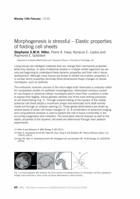
The embryonic inversion process in the micro-algal order Volvocales is uniquely suited for comparative studies on epithelial morphogenesis. Volvocalean embryos consist of cup-shaped or spherical cellular monolayers which invert their curvature in order toexposetheirflagella.Volvoxglobatorexhibitsoneofthemoststrikingprocessesofcellsheetfolding(Fig.1):Throughinwardsfoldingattheequatoroftheinitiallyspherical cell sheet adopts a mushroom shape and eventually turns itself entirely inside-out through an anterior opening [1]. These global deformations are driven by severalwavesofactivecellshapechanges[2,3].Acombinationofadvancedimagingand computational analyses is used to explore the role of tissue contractility in the occurring invagination and involution. The associated internal stresses as well as the elastic properties of the dynamic cell sheet are determined through laser ablation experiments.
[1] Höhn S and Hallmann A. BMC Biology 9, 89 (2011).
[2] Höhn S, Honerkamp-Smith AR, Haas PA, Khuc Trong P, and Goldstein RE. Physical Review Letters 114, 178101 (2015).
[3] Haas PA, Höhn S, Honerkamp-Smith AR, Kirkegaard JB, and Goldstein RE. PLOS Biology 16, e2005536 (2018).
Fig.1(a)Volvoxglobatorwithembryos;(b)Cross-sectionofinvertingembryo;(c)cellshapes,redline:cytoplasmicbridges (cell-connections); blue: nuclei; (d) tissue deformations in early inversion.
Monday10thFebruary·12:15
EMBL · IBEC Winter Conference on ENGINEERING MULTICELLULAR SYSTEMS 61
Three-dimensional culture of pancreas progenitors differentiated from mouse embryonic stem cellsShlomitEdri1, Ariel Szklanny1, Ben Kaplan1,FrancescaSpagnoli2 and Shulamit Levenberg1
1 Department of Biomedical Engineering, Technion – Israel Institute of Technology, Haifa, Israel2 Centre for Stem Cells & Regenerative Medicine, King's College London, UK
Over the last few years the establishment of three-dimensional (3D) cell culture methods allow embryonic stem cells (ESCs), induced pluripotent stem cells (iPSCs) or stem/progenitor cells to recapitulate many aspects of their differentiation programs and development in vitro.Specifically,ESCscanbecoaxedintospecificstructuresresembling in vivo tissues and organs, like eye caps, intestine, forebrain and liver, which termed organoids. The ability of cells to aggregate in this way has been referred as self-organization. With the great potential the organoids hold, they have limitations: they are small and lack mechanical support and vasculature.
In our study we focus on differentiation mouse ESCs to pancreas identity. We developedarobustandefficientdifferentiationprotocolofadherentmouseESCstopancreas progenitors, which we aggregated to pancreatic organoids. To supply the mechanicalsupportfortheaggregates,wecreated3Dspatiallydefinedhighlyporouspolymeric scaffolds using a 3D printing technique. Seeding pancreas endothelial cells and mesenchymal support cells on this scaffold, allowed the formation of vessel-like networks inside the entire scaffold. Utilizing the self-organization of the organoids and the scaffold support, we integrate pancreas organoids and vessel-like networks in the polymeric scaffold, mimicking the complex structure of the developing pancreas, which in future will enable the functional maturation upon implantation.
Our study provides a new approach that combines the inherent self-organization of ESCs, engineered vessel networks and mechanical support to better resemble the developingpancreas,whichhasenormouspotentialinthefieldsofregenerativemedicine and developmental biology.
Monday10thFebruary·15:20
62 EMBL · IBEC Winter Conference on ENGINEERING MULTICELLULAR SYSTEMS
Collective Cell Movement and Cell State Transitions in Gastruloids Specify the FormationofEndodermAliHashmi1, Pierre Perrin1, Sham Tlili1, Alfonso Martinez Arias2, Pierre-FrançoisLenne1
1 Aix Marseille University, CNRS, IBDM, Turing Centre for Living Systems, Marseille, France2 Department of Genetics, University of Cambridge, UK
In recent years, “gastruloids” have emerged as an alternative model system to dissect the complex intricacies of gastrulation and early morphogenesis. By utilizing a smaller number of embryonic stem cells (ESC) for gastruloids it is now possible to investigate the earliest symmetry breaking events and the formation of germ layers in a controlled fashion.
In this study we generated small aggregates from mouse ESC, primed to behave more epithelial-likeinpresenceofFGF,tostudytheemergenceoftheendoderm.Despitethe absence of an extraembryonic tissue, the aggregates that are given a Chiron pulse (a Wnt agonist) grow and elongate to attain a teardrop morphology with the emergence of a tip after 4 days. We found that the tip contains a group of cells that are endodermal-like as shown by the expression of various markers. We used imaging onfixedandlivegastruloidstodissectthemechanismsleadingtotheformationofthisregion and focused on E-cadherin (E-cad) dynamics in relation with the transcription factors T/Brachury (T/Bra) and Sox17, which are markers of mesendoderm and endoderm, respectively.
The distribution of E-cad, which is initially homogeneous, exhibits temporal inhomogeneities, that exacerbate after the Chiron pulse. At the macroscopic scale, the elongation of the aggregates is preceded by a polarized expression of E-cad and T/Bra. A large population of cells that express both E-cad and T/Bra lose E-cad expression over time, all the while retaining a small fraction of pluripotent Ecad cells that do not express T/Bra to begin with. This cell-state switching and maintenance of local pluripotency lead to the formation of small islands of E-cad rich cells that are surrounded by cells that are expressing T/Bra. The islands move in a directional manner to the region of the aggregate which forms the tip. The E-cad islands eventually combine to form a single cluster wherein the cells commit towards an endodermfate,markedbyexpressionofSox17andFoxA2.
Our data suggest that the endoderm can emerge from epithelial-like cells, without cycles of epithelial-to-mesenchymal transition (EMT) and the reverse transition (MET).
Monday10thFebruary·16:30
EMBL · IBEC Winter Conference on ENGINEERING MULTICELLULAR SYSTEMS 63
Mechanics and active cell behaviours contribute to self-organization of mesenchymal cells in the limbXavierDiego1*, A. Malandrino1*,H.Cardona1*, X. Trepat2,3, J. Sharpe1,3
1 EMBL Barcelona, Spain2 Institucio Catalana de Recerca i Estudis Avançats, Barcelona, Spain3 Institute for Bioengineering of Catalonia, Spain
* Equal contributions
A reaction-diffusion mechanism is believed to govern the formation of digits during development.However,mechanicsandactivecellbehaviorscouldalsohavearolein this process, an hypothesis that has been proposed before but has not been investigated systematically.
Thus, we decided to test the importance of mechanics for self-organization by culturingmesenchymalcellsfrommouselimbsonsubstratesofdifferentrigidities.Forthefirsttime,wedemonstratethatpurelymechanicalpropertiesclearlyinfluencetheemergent patterns.
Combiningliveimagingandtractionforcemicroscopywehavequantifiedtheevolutionofcellforces,velocitiesanddensitiesduringthefirstthreedaysofthepatterningprocess.
Our observations suggest that the mechanism of self-organization in the limb mesenchyme requires feedback between a reaction-diffusion program and the mechanical and migratory state of cells, which is dynamically modulated over time. Computational modeling and transcriptomics allows us to characterize the physical and molecular nature of this feedback.
Tuesday11thFebruary·10:00
64 EMBL · IBEC Winter Conference on ENGINEERING MULTICELLULAR SYSTEMS
A 3D Morphogenetic Model of Organogenesis inaHumanGeneticContextEyal Karzbrun1,3, AimalH.Khankhel2,HeitorC.Megale3, Sebastian J. Streichan2,3
1 Kavli Institute for Theoretical Physics, University of California, Santa Barbara, Santa Barbara, CA, USA2 Biomolecular Science and Engineering Program, University of California, Santa Barbara, Santa Barbara, CA, USA
3 Department of Physics, University of California, Santa Barbara, Santa Barbara, CA, USA
Duringembryogenesis,tissuelayersundergomorphogeneticflowsthatrearrangeandfoldtheembryoanditsorgansintospecificshapes.Whilecharacteristicsignalingpathwaysandmorphogeneticfeatureshavebeenidentifiedusinganimalmodels,extrapolating this information to human systems is challenging. Moreover, mammalian embryosexhibitspecies-specificdifferencesinmorphology,andmanymolecularmarkershavenotbeenconfirmed.Herewereportanovelin vitro technique to produce large arrays of 3D pluripotent epithelial cultures composed of human pluripotent stem cells (hPSCs).
Using this technique, we are able to robustly control initial conditions such as shape, cell number, and cell density for the generation of organoids. We exploit this control and under subsequent differentiation obtain reproducible fate-patterning in our 3D organoids. Upon differentiation, we observe spontaneous symmetry breaking and morphologicalchanges.Time-lapseimagingrevealstissuefoldingandstratification,driven by cellular rearrangements, which is a key motif of organogenesis. Our reproducible on-chip approach establishes a new model system to study organ development and enables opportunities to direct morphogenetic systems.
Tuesday11thFebruary·11:45
EMBL · IBEC Winter Conference on ENGINEERING MULTICELLULAR SYSTEMS 65
InductionofSyntheticTissueFoldingGuillermoMartínezAra1, Nuria Taberner Carretero2, Mami Takayama, Casandra Villava Robles1, Miki Ebisuya1
1 EMBL Barcelona, Spain2 Riken, Kobe, Japan
During the last years, several developmental processes such as patterning and cell differentiation have been reconstituted in vitro to improve our understanding of them.However,3Dtissueshapeisstillmainlyachievedthroughtissueengineeringmethods, based on the use of structures that will direct cells to acquire different tissuemorphologies.However,initialchangesintissueshapewillconditionthefollowing developmental stages, and therefore are essential for the development offunctionaltissuesandorgans.Forthisreason,weaimtoreconstitute3Dmorphogenetic processes in vitro.
Howcancell-levelprocessesprovokechangesintissueshapeduringdevelopment?Many essential genes need to be expressed for changes in tissue shape to occur. Someofthesegeneshavealsobeenobservedtobesufficienttocausechangesincellshape,proliferationormigration.Tofindouttowhatextenteachoftheseproteinsand cellular events can drive morphogenesis, we use a minimal setting in which we study how epithelial tissues acquire new shapes.
In our latest work, we induce apical constriction in a sheet of MDCK (Madin-Darby Canine Kidney) cells set on a soft substrate, to test which types of tissue deformation can be provoked by this process. This change in cell shape (from cuboidal to trapezoidal) is induced through the expression of one single protein: Shroom3, and it issufficienttocausecurvatureofepithelialcoloniesandeventuallyfolding.Tofurthertest how different patterns of apical constriction can create different 3D shapes, we have developed an optogenetic version of the protein.
In this setting in which unknown forces and interactions with other tissues are minimized, we plan to analyze which are the necessary conditions to achieve different tissue shapes. Our main focus is now to investigate how size and initial shape of cell colonies can affect these deformations.
Tuesday11thFebruary·12:30

66 EMBL · IBEC Winter Conference on ENGINEERING MULTICELLULAR SYSTEMS
Onset and Patterning Rules of Mesendoderm andDefinitiveEndoderminEmbryoidBodiesNaor Sagy1, Maayan Pour1, Shaked Slovin1, Abhishek S. Kumar2, Gaya Savyon1, Maya Allalouf1, Jonathan Boxman1, Alexander Meissner2, IftachNachman1
1 School of Neurobiology, Biochemistry and Biophysics, Tel Aviv University, Tel Aviv, Israel2 Department of Genome Regulation, Max Planck Institute for Molecular Genetics, Berlin, Germany
In vitro models of early mammalian development, such as embryoid bodies (EBs) and gastruloids, provide accessible systems for the study of basic rules shaping cell fate and patterning during embryogenesis. Using high-throughput live 3D imaging, we study two early differentiation decisions in mESC-derived EBs and tetraploid-complementation embryos: the formation of primitive streak and mesendoderm progenitors(usingBra-GFP),andthedownstreamemergenceofdefinitiveendoderm(DE),usingSox17-RFP.
Wefindthatmechanical,biochemicalandneighboringcellcuespredictoraffectthe positioning of a primitive streak-like locus, determining the AP axis. Bra onset is biasedbyEBcontactpointswithsurfaces,canbemaneuveredtoaspecificlocus,twoloci, or to an isotropic peripheral pattern. The EB can integrate separate mechanical andbiochemicalsignalsources,resultinginasinglelocus.Interestingly,Foxa2onsetprovides a predictive earlier symmetry-breaking event, explained by a local Wnt gradient.
In contrast, DE cells arise from within the mesendoderm population in a temporally-synchronized, yet spatially stochastic “salt-and-pepper” pattern. This is followed by aself-sortingphaseoftheSox17+cells,leadingtoaggregationorlumenogenesisasthe in-vitro counterpart to intercalation in the embryo. Self-sorting correlates with up-regulation of E-cadherin, and is not essential for DE differentiation or proliferation. We findthatasmallsubpopulationofBra-highcellsarecommittedtotheirSox17+fateindependent of external Wnt signal.
The remarkably different strategies demonstrated in these two fate decisions may be explained in the context of embryo dynamics.
Sagy et. al., Development 146 (20), dev181917, 2019
Pour et. al., bioRxiv, 728642, 2019
Tuesday11thFebruary·12:45
EMBL · IBEC Winter Conference on ENGINEERING MULTICELLULAR SYSTEMS 67
Tension heterogeneity instructs morphogenesisandfatespecificationduringheart development
RashmiPriya, Srinivas Allanki, Alessandra Gentile, Shivani Mansingh,Hans-MartinMaischeinandDidierStainier
Department of Developmental Genetics, Max Planck Institute for Heart and Lung Research, Bad Nauheim, Germany.
Embryogenesis entails generation of diverse cell fates and emergence of complex morphogenetic patterns. A key question remains as to how morphogenesis and mechanics contribute to cell fate decisions in a complex and growing organ. During cardiac development, the myocardial wall transforms from a monolayer to an intricate topological structure consisting of two distinct types of cardiomyocytes (CMs): outer compact and inner trabecular layer CMs. This process of cardiac trabeculation is crucial for cardiac function as aberrations lead to congenital cardiomyopathies and embryoniclethality.Yet,themechanismsunderlyingtheemergenceandspecificationoftrabecularCMsremainunknown.Usingthezebrafishheartincombinationwithhigh-resolution quantitative microscopy, in vivo measurements of tension/subcellular dynamics, genetic mosaic tools and embryological interventions, we now report that contractility couples morphogenesis and cell fate to ensure robust self-organization of CMs into compact versus trabecular layer.
Proliferation induced crowding triggers symmetry breaking by generating local differences in cellular contractility. These effects lead to stochastic delamination of CMs from the outer compact layer to seed the inner trabecular layer. By manipulating contractility at the single cell-level, we show that reducing contractility abrogates delamination while inducing contractility augments delamination, and strikingly, inducingcontractilityissufficienttodrivedelaminationevenintheabsenceofcriticaltrabeculationsignalslikeNrg/Erbb2orblood-flow.Further,usingcontrolledperturbationstodecouplemechanicalcuesfrombiochemicalsignaling,wefindthatmechanicalcuesdriveCMfatespecification.Inducingtensionheterogeneity(andtherebyCMdelamination)bymanipulationofcelldensityorcontractilityissufficientto generate differential Notch activity as well as apicobasal polarity. Overall, our study reveals how form and function emerge as a collective product of individual cell behaviors, and argues for system-level approach integrating mechanics with regulatory circuits for a cogent understanding of multicellular organization in vivo.
Tuesday11thFebruary·15:20

68 EMBL · IBEC Winter Conference on ENGINEERING MULTICELLULAR SYSTEMS
A novel mathematical law to understand 3D self-organization in epithelial tubes PedroGómez-Gálvez1,2,*, Pablo Vicente-Munuera1,2,*, Samira Anbari3,*, Antonio Tagua1,2,*, Carmen Gordillo-Vázquez1,2, Ana M. Palacios1,2, Antonio Velasco1, Carlos Capitán-Agudo1, Clara Grima5, Valentina Annese1,2, Rafael Robles5, Alberto Márquez5, Javier Buceta3,4, Luis M. Escudero1,2
1 Instituto de Biomedicina de Sevilla (IBiS), Hospital Universitario Virgen del Rocío/CSIC/Universidad de Sevilla and Departamento de Biología Celular, Universidad de Sevilla. 41013 Seville, Spain.
2 Biomedical Network Research Centre on Neurodegenerative Diseases (CIBERNED), Madrid, Spain. 3 Chemical and Biomolecular Engineering Department, Lehigh University. Bethlehem, PA 18018, USA.4 Bioengineering Department, Lehigh University. Bethlehem, PA 18018, USA.5 Departamento de Matemática Aplicada I, Universidad de Sevilla. 41012 Seville, Spain.
* These authors contributed equally to this work.
The analysis of epithelial organization as tiles on planar surfaces has been essential to theunderstandingofanimaldevelopment.However,realisticinsightintoorganogenesisrequires a three-dimensional perspective able to deal also with tissue bending. In that regard, apico-basal cell intercalations (scutoids) have been shown as a key factor to optimize energy expenditure in folding epithelia. Yet, the consequences of this new morphogenetic paradigm remain uncharacterized. Until now, two mathematical principles have been widely used to investigate morphogenesis quantitatively: Euler´s formula and Lewis’ law.
Theimportanceoftheselawsresidesintwofundamentalaspects.First,theirapplicabilityto different tissues from different organisms and, therefore, their character as driving developmental principles. Second, their biological implications since they connect geometrical traits with key functionality features such as patterning, fate establishment, cell division, and tissue growth. Importantly, the certainty of these principles in 3D hasbeentakenforgranted.However,ourrecentdiscoveryofscutoidsquestionstheirtruthfulness. We have found a relationship between the 3D topology of epithelia and the inherent geometrical and physical constraints of tissues. We will show how the scutoids presence implies a breakdown of Euler’s principle in 3D and will present what wehavecalledthe“Flintstones’law”:thethicknessandcurvatureofepithelialtubesarelogarithmically related to the cellular connectivity of the tissue via energetic cues. The existence of this novel, and universal, law is supported by data and technical advances from four different disciplines: computed-aided image analysis of 3D bended tissues; computational models imitating the 3D cellular packing; mathematical analyses of cellularorganizationandastochasticbiophysicalmodelabletopredictthe“Flintstones’law” based on the energetic cost needed to increase the number of 3D cells contacts.
In conclusion, our research shows how mathematical and physical principles, arranged in the form of simple rules, underlie the emergence of functionally complex developmental structures.
Tuesday11thFebruary·16:30
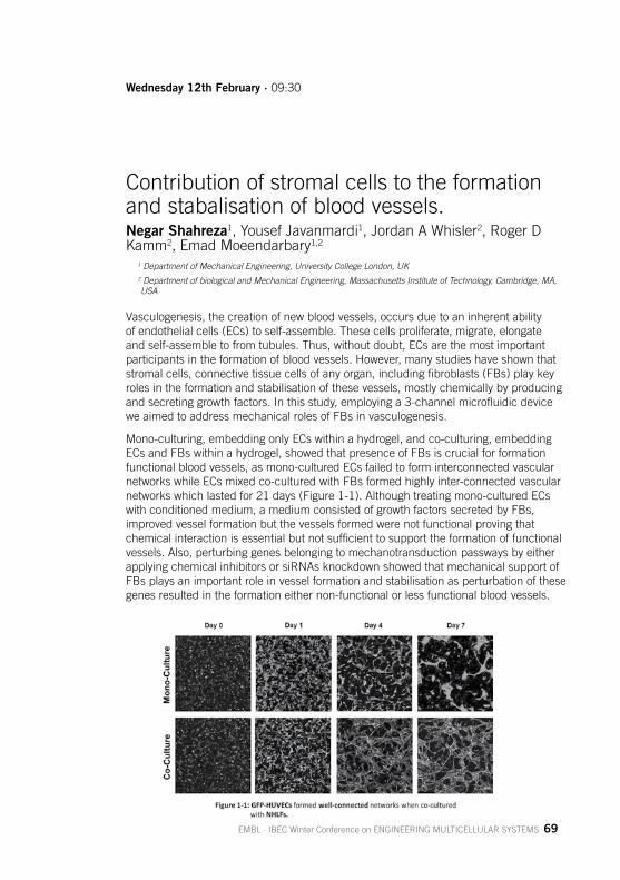
EMBL · IBEC Winter Conference on ENGINEERING MULTICELLULAR SYSTEMS 69
Contribution of stromal cells to the formation and stabalisation of blood vessels.NegarShahreza1, Yousef Javanmardi1, Jordan A Whisler2, Roger D Kamm2, Emad Moeendarbary1,2
1 Department of Mechanical Engineering, University College London, UK2 Department of biological and Mechanical Engineering, Massachusetts Institute of Technology, Cambridge, MA, USA
Vasculogenesis, the creation of new blood vessels, occurs due to an inherent ability of endothelial cells (ECs) to self-assemble. These cells proliferate, migrate, elongate and self-assemble to from tubules. Thus, without doubt, ECs are the most important participantsintheformationofbloodvessels.However,manystudieshaveshownthatstromalcells,connectivetissuecellsofanyorgan,includingfibroblasts(FBs)playkeyroles in the formation and stabilisation of these vessels, mostly chemically by producing andsecretinggrowthfactors.Inthisstudy,employinga3-channelmicrofluidicdeviceweaimedtoaddressmechanicalrolesofFBsinvasculogenesis.
Mono-culturing, embedding only ECs within a hydrogel, and co-culturing, embedding ECsandFBswithinahydrogel,showedthatpresenceofFBsiscrucialforformationfunctional blood vessels, as mono-cultured ECs failed to form interconnected vascular networkswhileECsmixedco-culturedwithFBsformedhighlyinter-connectedvascularnetworkswhichlastedfor21days(Figure1-1).Althoughtreatingmono-culturedECswithconditionedmedium,amediumconsistedofgrowthfactorssecretedbyFBs,improved vessel formation but the vessels formed were not functional proving that chemicalinteractionisessentialbutnotsufficienttosupporttheformationoffunctionalvessels. Also, perturbing genes belonging to mechanotransduction passways by either applying chemical inhibitors or siRNAs knockdown showed that mechanical support of FBsplaysanimportantroleinvesselformationandstabilisationasperturbationofthesegenes resulted in the formation either non-functional or less functional blood vessels.
Wednesday12thFebruary·09:30

70 EMBL · IBEC Winter Conference on ENGINEERING MULTICELLULAR SYSTEMS
Shear-induced crystallization drives precise patterning of hair cells in the mammalian inner ear Roie Cohen1,2, Liat Amir-Zilberstein1,#,MichaHersch3,4, Shiran Woland1, Shahar Taiber1,5,FumioMatsuzaki6, Sven Bergmann3,4,7, Karen B. Avraham5, and DavidSprinzak1
1 School of Neurobiology, Biochemistry and Biophysics, George S. Wise Faculty of Life Sciences, Tel Aviv University, Tel Aviv 6997801, Israel
2 Raymond and Beverly Sackler School of Physics and Astronomy, Faculty of Exact Sciences, Tel Aviv University, Tel Aviv 6997801, Israel
3 Department of Computational Biology, University of Lausanne, 1015 Lausanne, Switzerland4 Swiss Institute of Bioinformatics, Lausanne 1015, Switzerland5 Department of Human Molecular Genetics and Biochemistry, Sackler Faculty of Medicine and Sagol School of Neuroscience, Tel Aviv University, Tel Aviv 6997801, Israel
6 Laboratory of Cell Asymmetry, RIKEN Center for Biosystems Dynamics Research, Kobe 650-0047, Japan7 Department of Integrative Biomedical Sciences, University of Cape Town, Cape Town, South Africa
Precise periodic organization of cells is required for the function of many organs and tissues. It is often unclear, however, how such precise patterns emerge during development. The mammalian hearing organ, the organ of Corti, consists of a remarkably organized pattern of four rows of hair cells interspersed by non-sensory supportingcells.Thischeckerboard-likepatternofHCsandSCsemergesfromadisordered epithelium over several days, yet the transition to an ordered cellular pattern is not well understood. Using time-lapse imaging of mouse cochlear explants andmathematicalmodeling,weshowthathaircellsrearrangegraduallyover2-3daysthrough a tissue-wide shear motion that coordinates intercalation and delamination events to achieve precision patterning.
A mathematical model, where tissue morphology is described in terms of the mechanical forces that act on cells and cellular junctions, suggests that global shear andlocalrepulsionforcesonhaircellsaresufficienttodrivethetissueintothefinalcheckerboard-likepattern.Ourfindingssuggestthatmechanicalforcesdrivethe transition from disordered to ordered cellular patterns in a process strikingly analogous to the process of shear-induced crystallization in physics.
Wednesday12thFebruary·12:00

EMBL · IBEC Winter Conference on ENGINEERING MULTICELLULAR SYSTEMS 71
PlatformsforFastandGentle3DSuperresolution MicroscopyGerry Sexton Ph.D, 3D Microscopy Specialist, Zeiss
Our life sciences research can be demanding, and if you are involved in neuroscience, cancer research or other cell- or organism-based disciplines, you’ll often need confocal microscopy data for your work. Emerging technologies open up innovativewaysofthinkingandallowyoutoaskaltogethernewscientificquestions,deeply affecting your imaging experiments. To monitor life as undisturbed as possible requires low labeling density for your biological models – for example, 3D cell culture, spheroids, organoids or even whole organisms – and this calls for 3D live cell imaging that combines optical sectioning with low phototoxicity and high speed.
ThenewLSM9FamilywithAiryscan2istheidealconfocalmicroscopefor4Dimaging. The entire beam path is optimized for simultaneous spectral detection of multipleweaklabelswiththehighestlightefficiency.AddthenewMultiplexmodeforAiryscan2togetmoreimagingoptions,with4-8xmoresignal-to-noiseratio(SNR)foryourexperiments.Youcannowchoosetheperfectsetuptogentlyimagelargerfieldsof view with superresolution in shorter acquisition times than ever before.
Your life sciences research often requires you to measure, quantify and understand thefinestdetailsandsub-cellularstructuresofyoursample.Youmaybeworkingwithtissue,bacteria,organoids,neurons,livingorfixed-cellsandmanydifferentlabels.
By the other hand, Elyra 7 with Lattice SIM takes you beyond the diffraction limit of conventional microscopy to image your samples with superresolution. You examine thefastestprocessesinlivingsamples–inlargefieldsofview,in3D,overlongtimeperiods, and with multiple colors. The new Lattice SIM technology of your Elyra 7 brings structured illumination microscopy (SIM) to a new level. Groundbreaking light efficiencygivesyougentlesuperresolutionimagingwithincrediblyhighspeed–at255fpsyouwillgetyourdatafasterthaneverbefore.Elyra7letsyoucombineLatticeSIM with single molecule localization microscopy (SMLM) for techniques such as PALM, dSTORM and PAINT. You can now choose freely among your labels when imagingwithresolutionsdownto20nmlaterally.Highpowerlaserlinesallowyoutoimage your sample with ease, from green to far red.
Tuesday11thFebruary·14:30 TalksponsoredbyZeiss
72 EMBL · IBEC Winter Conference on ENGINEERING MULTICELLULAR SYSTEMS
EMBL · IBEC Winter Conference on ENGINEERING MULTICELLULAR SYSTEMS 73
Posters Session

N Name Affiliation Title
1Vanesa Fernández Majada
Institute for Bioengineering of Catalonia (IBEC)
Myofibroblast-derived mechanical cues direct Epithelial Migration
2 Elena GarretaInstitute for Bioengineering of Catalonia (IBEC)
Efficient Generation of Human Pluripotent Stem Cell-derived Kidney Constructs by 3D Bioprinting of cell-laden Kidney Specific Bioinks
3 Tom GoldeInstitute for Bioengineering of Catalonia (IBEC)
The role of intermediate filaments in stress resistance in 3D epithelial structures
4Carmen M. Gordillo Vázquez
Universidad de Sevilla Natural variation in 3D tissue organization
5 Judith GuaschInstitute of Materials Science of Barcelona (ICMAB-CSIC)
Steps towards Mimicking the Lymph Nodes to Enhance T Cell Expansion
6Maria Guillot-Ferriols
Biomaterials and Nanomedicine (CIBER-BBN)
Elastin-like recombinamers as poly(vinylidene fluoride) membrane coatings, a new approach for mesenchymal stem cell culture and differentiation
7 Ju Yeon Han EMBL BarcelonaTo what extent is digit patterning a Turing system?
8Mathieu Hautefeuille
Universidad Nacional Autónoma de México
Fabrication of biphasic microstructures for viscoelastic cell culture substrates
9Mathieu Hautefeuille
Universidad Nacional Autónoma de México
Impact of the ECM stiffness and geometry in the collective dynamics of epithelial and mesenchymal lung cells
PosterSession10thFebruary
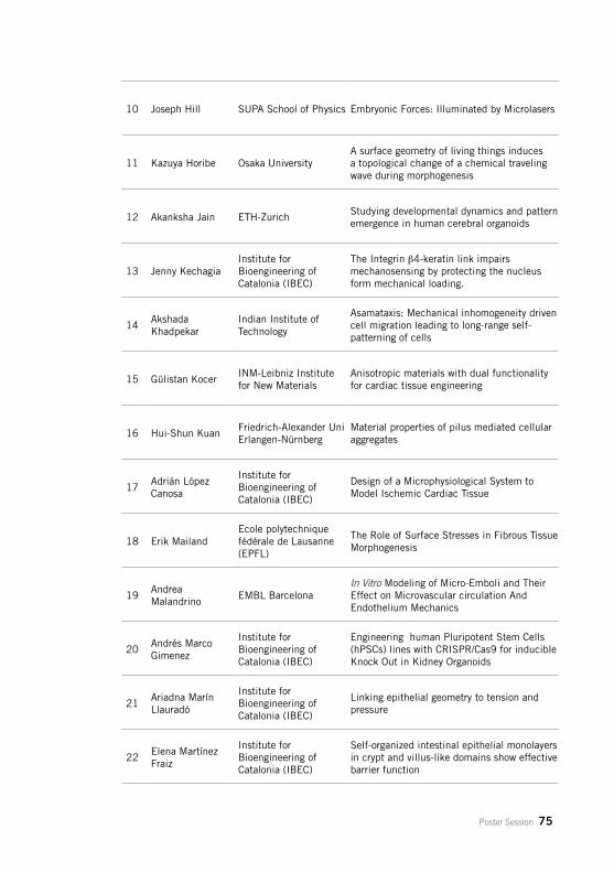
Poster Session 75
10 Joseph Hill SUPA School of Physics Embryonic Forces: Illuminated by Microlasers
11 Kazuya Horibe Osaka UniversityA surface geometry of living things induces a topological change of a chemical traveling wave during morphogenesis
12 Akanksha Jain ETH-ZurichStudying developmental dynamics and pattern emergence in human cerebral organoids
13 Jenny KechagiaInstitute for Bioengineering of Catalonia (IBEC)
The Integrin b4-keratin link impairs mechanosensing by protecting the nucleus form mechanical loading.
14Akshada Khadpekar
Indian Institute of Technology
Asamataxis: Mechanical inhomogeneity driven cell migration leading to long-range self-patterning of cells
15 Gülistan KocerINM-Leibniz Institute for New Materials
Anisotropic materials with dual functionality for cardiac tissue engineering
16 Hui-Shun KuanFriedrich-Alexander Uni Erlangen-Nürnberg
Material properties of pilus mediated cellular aggregates
17Adrián López Canosa
Institute for Bioengineering of Catalonia (IBEC)
Design of a Microphysiological System to Model Ischemic Cardiac Tissue
18 Erik MailandEcole polytechnique fédérale de Lausanne (EPFL)
The Role of Surface Stresses in Fibrous Tissue Morphogenesis
19Andrea Malandrino
EMBL BarcelonaIn Vitro Modeling of Micro-Emboli and Their Effect on Microvascular circulation And Endothelium Mechanics
20Andrés Marco Gimenez
Institute for Bioengineering of Catalonia (IBEC)
Engineering human Pluripotent Stem Cells (hPSCs) lines with CRISPR/Cas9 for inducible Knock Out in Kidney Organoids
21Ariadna Marín Llauradó
Institute for Bioengineering of Catalonia (IBEC)
Linking epithelial geometry to tension and pressure
22Elena Martínez Fraiz
Institute for Bioengineering of Catalonia (IBEC)
Self-organized intestinal epithelial monolayers in crypt and villus-like domains show effective barrier function
76 Poster Session
23 Marina Matsumiya EMBL BarcelonaRecapitulating the somitogenesis in vitro to identify novel causative genes for congenital bone diseases
24Antoni Matyjaszkiewicz
EMBL BarcelonaLIMBNET: online image-based computational modelling of limb development
25 Torsten Müeller Bruker Nano GmbHQuantitative characterization of adhesion and cytomechnics of living cells on biomaterials and tissues
26 Jose MunozUniversitat Politècnica de Catalunya (UPC)
Stability and oscillatory evolution of Drosophila Central Nervous System depends on tissue rigidity and adhesive contributions
27Anastasiia Mykuliak
Tampere UniversityIn Vitro Vascular Networks for Bone Tissue Engineering
28 Erik Noetzel-ReissInstitute of Complex Systems Biomechanics (ICS-7)
Physical and chemical basement membrane disruption modes synergistically promote the invasiveness of MCF10A breast acini.
29 David Oriola EMBL BarcelonaInferring the network topology driving mesoderm differentiation from single cell time-course data
30 Adam OuzeriUniversitat Politècnica de Catalunya (UPC)
Upscaling active gel models of the actin cortex to epithelial mechanics
31Raghavendra Palankar
University Medicine Greifswald
Platelet and Thrombus Biomechanics: The Need for Vasculature-on-Chip
32 Patricia PradoInstitute for Bioengineering of Catalonia (IBEC)
Differentiation of Human Pluripotent Stem Cells into Ureteric Bud-like Cells and Assessment of their Renal Potential by the Use of ex vivo Kidney Reconstruction Assays
33 Bernad RaquelInstitute for Research in Biomedicine (IRB Barcelona)
CDK8/19 inhibition stabilizes human naive pluripotency
34 Laura RijnsEindhoven University of Technology
Biofunctionalized Hydrogels based on Benzene-1,3,5-tricarboxamides (BTAs) for Kidney Regeneration
PosterSession11thFebruary
N Name Affiliation Title
35 Alicia Roig MerinoGerman Cancer Research Center (DKFZ)
SMARt engineering: Generation of isogenic Stem Cells and transgenic models utilising a non-integrative and autonomously replicating DNA vector system
36 William RomanInstituto de Medicina Molecular
Generation of an in vitro neuromuscular junction at the cellular resolution
37 Aurelien Roux University of GenevaTopological defects organize morphogenetic stresses
38 Gerard RubíInstitute for Bioengineering of Catalonia (IBEC)
Development of an in vitro three-dimensional colorectal tumor model for drug screening
39 Christian SchröterUniversity of Copenhagen
Generation and maintenance of robust cell fate proportions by FGF/ERK signaling
40 Lucia SelfaInstitute for Bioengineering of Catalonia (IBEC)
Studying Wilms’ Tumor 1 (WT1) function in human kidney development and disease using human pluripotent stem cells-derived organoids and genome editing
41 Yvenn Sermeus KU LeuvenIndividual cell based modeling of limb bud outgrowth to predict shape and mechanical properties
42 Tiziano SerraAO Research Institute Davos
A sound-induced technology for spatial orchestration of functional large-scale microvascular networks
43 Shayan Shamipour IST AustriaBulk Actin Dynamics Drive Phase Segregation in Zebrafish Oocytes

78 Poster Session
44 Apeksha Shapeti KU Leuven
Quantifying invasion and 3D traction forces during multicellular angiogenic sprouting upon CCM-2 loss shows a ROCK-dependent increase in contractility and invasiveness
45Aleksandra Slabikova
Ryazan State Medical University
Examination of proliferative activity and cell viability during neurogenic differentiation from iPSCs in 3D culture of cerebral organoids
46 Aki Stubb University of TurkuSuperresolution architecture of cornerstone focal adhesions in human pluripotent stem cells.
47Alejandro Torres-Sánchez
The Francis Crick Institute
Interacting Cellular Meshes: A new theoretical and computational framework to investigate tissue reorganisation during morphogenesis
48 Manuela UrbanGerman Cancer Research Center (DKFZ)
Non-viral episomal S/MAR DNA vectors for the persistent genetic engineering of hiPSCs and their progenies
49Johnick Van Sprang
Eindhoven University of Technology
Supramolecular Hydrogels For Kidney Organoid Development
50 Jef Vangheel KU LeuvenExploring the Cell Jamming Phase Diagram Using a 3D Deformable Cell Model
51 Chiara VenturiniUniversitat Politècnica de Catalunya (UPC)
A generalized clutch model to explain cell adhesion mechanics
52 Tacker Vivek Global Health InstituteLung-on-a-chip microphysiological systems for studies of host-pathogen interactions in Tuberculosis
53 Sara WatsonUniversity College London
Development of an in vitro microfluidic model of thymocyte extravasation
54 Justina YeungThe Francis Crick Institute
Spatial Regulation of Proneural Gene Expression in the Hindbrain
55 Ayushi AgrawalUniversity College London
Integrated in vitro model of tumour vasculogenesis and cancer cell intravasation
56 Enrico AlmiciInstitute for Bioengineering of Catalonia (IBEC)
Engineered cell-derived matrices as 3D tumor stroma models

Poster Session 79
57 Krisztina Arato EMBL BarcelonaA Study on Actomyosin Influencing the Formation of Spatial Asymmetries in Early Embryonic Organoids
58 Melis AsalIzmir Biomedicine and Genome Center
Lacrimal Gland on Chip Derived from Induced Pluripotent Stem Cells
59 Jordina BalaguerInstitute for Bioengineering of Catalonia (IBEC)
Magnetic bead-based immunosensing platform for in-situ detection of secreted cytokines in response to oligonucleotide treatment
60 Natalia BasalovaLomonosov Moscow State University
Decellularized multicellular fibroblast sheets for mimicking the profibrotic microenvironment in vitro
61 Sara Bonavia Université Paris DiderotAn in vitro system for the mouse Epiblast to investigate the establishment og the anterposterior polarity
62 Carlo BrighiItalian Institute of Technology
Modeling Fragile X syndrome with iPSC-derived neurons in 2D and 3D tissue culture conditions.
63 Isabel CalvoInstitute for Research in Biomedicine (IRB Barcelona)
Visualization of Mediator complexes in naive and primed embryonic pluripotent cells
64 Ignasi CasanellasInstitute for Bioengineering of Catalonia (IBEC)
Cell-matrix nanoscale adherence continually modulates intercellular communication in morphogenesis
65Sandra Clara-Trujillo
Biomedical Research Networking Center on Bioengineering, Biomaterials and Nanomedicine (CIBER-BBN)
From 2D to 3D cell culture with biomimetic microgels
66 Laura CluaInstitute for Bioengineering of Catalonia (IBEC)
Micro-Spheroids For b-like Cell Encapsulation
67 Mar Cóndor KU LeuvenCellular force generation during sprouting angiogenesis
68 James Cotterell EMBL BarcelonaA Local, Self-Organizing Reaction-Diffusion Model Can Explain Somite Patterning in Embryos.
PosterSession12thFebruary
N Name Affiliation Title
69 Ross CowieUniversity of St Andrews
Quantifying biomechanical stresses in 3D cellular environments via micro-droplet laser spectroscopy
70 Eleni DalakaUniversity of St Andrews
Measurement of invadopodia forces in 2D and 3D environments
71Giovanni Dalmasso
EMBL-BarcelonaThe role of SOX9 in vasculature re-modelling: an in vitro study
72 Malik DawiUniversitat Politècnica de Catalunya (UPC)
Vertex Modeling and Three-dimensional Tissue Reorganization
73 Simone de JongEindhoven University of Technology
The Introduction of Fc-Fusion Bioactive Proteins in Supramolecular Biomaterials
74 Rocky Diegmiller Princeton UniversityCell and Nuclear Growth Dynamics in Developing Drosophila Germline Cysts
75 Camille Douillet PoietisMorphogenesis control of dermal tissues produced by Laser-Assisted Bioprinting (LAB)
76 Arthur Douillet LaTIM & PoietisAn Agent-Based Modeling Framework To Predict Bioprinted Tissue Morphogenesis
77Aleksey Emelin Ryazan
State Medical University
Efficiency evaluation of iPSCs neurogenic and myogenic differentiation in organoids composition
Poster Session 81
78 Ismail EsUniversity College London
Formation of tumor spheroid models using 3D-printed micromolds
79 Elena GarretaInstitute for Bioengineering of Catalonia (IBEC)
Substrate Stiffness Impacts the Differentiation of Kidney Organoids Derived from Human Pluripotent Stem Cells
80 Gemma Gabriel IMB-CNM Organ-on-chip monitoring
81 Maria GalloInstitute for Bioengineering of Catalonia (IBEC)
Developing kidney-specific bioinks for human pluripotent stem cells 3D Bioprinting into kidney organoids
82 Jad SalehInstitut Jacques Monod
Université Paris Diderot/CRNS
Mechanical impact on stem cell niche morphogenesis

PLATINUM Sponsor:
GOLD Sponsor:
SILVER Sponsor:
With the support of: