Embed Size (px)
Citation preview
IntroductionThe diagnosis of dental caries is an integral component of
a clinician’s practice. During intra-oral examinations, dentists
attempt to discern whether or not lesions are cavitated or non-
cavitated, active or arrested, and to estimate the depth of a lesion,
questioning whether it is limited to the enamel, entering the
dentin, or even approximating the pulp. These considerations
ultimately will lead to a treatment plan that includes preventive
intervention, monitoring, sealing, or providing restorative treat-
ment for carious lesions.1,2
For years, visual/tactile examinations served as the principal
diagnostic methods for occlusal caries detection.3 As research
progressed, the notion that excessive probing with an explorer
can lead to premature cavitation of the enamel encouraged
dentists to put down their explorers and rely solely on visual
detection.4 Unfortunately, as studies have shown, visual detec-
tion lacks high sensitivity.5 Radiographic examination is a useful
adjunct to the detection of occlusal caries. As studies have indi-
cated, occlusal caries must be quite large and well into the dentin
in order to be diagnosed using bitewing radiographs.6,7
Newer diagnostic methods have been introduced to the den-
tal community, and several optical methods of caries detection
are available to clinicians. These devices direct a particular
wavelength at tooth structure and detect light emissions from the
tooth tissue that are indicative of intact tissue or decay. The
Quantitative Light Fluorescent device QLF™ (Inspektor Re-
search Systems BV, Amsterdam, The Netherlands) uses a wave-
length of light that induces fluorescence in dentin. Healthy
enamel allows this fluorescent light to pass through with mini-
mal scattering. Enamel white spot lesions, areas having subsur-
face areas of demineralization, will scatter the fluorescent light
and appear in QLF images as dark areas with low fluorescence.8
Quantitative changes in QLF fluorescence correlate well with the
magnitude of enamel mineral loss. This device’s accompanying
1
Abstract• Objective:The goal of this study was to perform an in vitro evaluation of the Spectra™, a new caries detector that uses light- induced
fluorescence of healthy tooth structure and bacterial pigments to optically detect caries. The Spectra generates a storable color map
image of examined tooth surfaces which shows areas of enamel and dentin caries. In this study, Spectra readings of occlusal surfaces
were compared to clinical, radiographic, and histological assessments of caries.
• Methods: Two examiners evaluated 41 extracted molars. The teeth were radiographed and then visually assessed. The International
Caries Detection and Assessment System (ICDAS) was used to classify the extent of caries. The teeth were then sectioned and
assigned a histological score based on the extension of caries into enamel or dentin.
• Results: Teeth lacking radiographic caries had a mean Spectra reading of 1.5. Teeth having radiographic caries had a mean Spectra
reading of 2.0. This difference was statistically significant. In general, higher ICDAS scores were associated with higher Spectra
readings. Teeth with histologically evident deep dentin caries had significantly higher Spectra readings than intact teeth or teeth
with superficial enamel demineralization. Spectra assessment of occlusal caries agrees with clinical and radiographic methods.
• Conclusion: Spectra images illustrate the full spectrum of caries severity, from enamel demineralization to dentin decay. The Spectra
is a promising technology for the diagnosis and for monitoring the progression of occlusal caries.
(J Clin Dent 2012;23:1–6)
In Vitro Evaluation of the Spectra™ Early Caries Detection SystemMaria Graye, DMD
Pedodontist in Private Practice
Hamilton, NJ, USA
Kenneth Markowitz, DDS, MSD
Assistant Professor, Department of Oral Biology
UMDNJ-New Jersey Dental School
Newark, NJ, USA
Maxine Strickland, DMD, MPH
Clinical Assistant Professor, Department of Diagnostic Sciences
UMDNJ-New Jersey Dental School
Newark, NJ, USA
Gerald Guzy, DDS Mary Burke, DMD Milton Houpt DDS, PhD
Clinical Assistant Professor Clinical Assistant Professor Professor and Chair
Department of Pediatric Dentistry
UMDNJ-New Jersey Dental School
Newark, NJ, USA
2 The Journal of Clinical Dentistry Vol. XXIII, No. 1
Following extraction, teeth were stored in 1% phenol and de-
brided of adherent soft and hard tissues. Any residual debris
found to be adhering to the occlusal surfaces of the teeth was re-
moved gently with a dental probe. Two examiners (MG and KM)
independently evaluated the teeth using three caries detection
methods: clinical-visual examination, radiographic examination,
and the Spectra device.
For the clinical examination, teeth were dried and evaluated sep-
arately by the two examiners using standard dental operatory il-
lumination. The International Caries Detection and Assessment
System (ICDAS)12 was used to score all teeth. ICDAS consists of
the following scores: 0: Sound tooth surface; 1: Pits and fissures
showing opacity with prolonged drying; 2: Distinct visual change
in enamel when wet, carious opacity wider than the natural fossa;
3: Localized enamel breakdown because of caries with no visible
dentin or underlying shadow; 4: Underlying dark shadow from
dentin with or without localized enamel breakdown; 5: Distinct
cavity with visible dentin; and 6: Extensive distinct cavity with vis-
ible dentin. In the rare cases where the two examiners differed in
their ICDAS score, the disagreement was reconciled by joint
examination. The teeth were then photographed with a digital
camera (Canon USA, Lake Success, NY, USA).
Radiographs were prepared of each tooth. The teeth were
placed on standard size #2 films with the lingual surface facing
the film, and a radiograph of each tooth was obtained using a per-
pendicular angle of the cone to the film. The examiners evalu-
ated the radiographs on a light box in a darkened room. After
individually analyzing the films, the examiners assigned each
tooth a “Yes” or “No,” indicating the presence or absence of
radio graphic decay.
The teeth were then examined using the Spectra handpiece and
accompanying software. The Spectra unit’s 10 mm distance
spacer was rested on the occlusal surface of each molar, the
overhead fluorescent light was turned off, and an image for each
tooth was collected. After collection of an image, the analysis
was performed using the Spectra’s Visix® software, which pro-
duced both numerical and color ratings of regions of tooth struc-
ture using the following color coded and numerical scale: Green:
0–1.0–sound enamel; Blue: 1.0–1.5–initial enamel decalcifica-
tion; Red: 1.5–2.0–enamel caries up to the DEJ; Orange:
2.0–2.5–dentin caries; and Yellow: 2.5 or higher–deep dentin
caries. A training and calibration exercise was held where the two
examiners obtained Spectra readings from a group of 12 teeth,
that included teeth in various ICDAS categories. By using a
uniform method of positioning the Spectra handpiece with
respect to the tooth, the two examiners were able to generate
equivalent color maps of the teeth with peak Spectra readings
which disagreed by no more than 0.2.
Following visual, radiographic, and Spectra analysis, the teeth
were sectioned perpendicular to the long axis of the tooth using
a slow speed saw (Isomet®, Buehler LTD., Lake Bluff, IL, USA)
with diamond blade and water lubrication. Cuts were made to a
depth that exposed the base of the occlusal pits and fissures. A
histological evaluation of each tooth was performed to verify the
presence of caries, and the depth/extent of enamel and dentin
involvement. The following histological scale was used: H0–no
whitening of enamel and no dentin staining; HE1
–cut surface
software allows changes in lesion status to be tracked over time.
The QLF has been used in clinical studies to assess the remin -
eralization of white spot lesions.8
The DIAGNOdent® (KaVo, Biberach, Germany) uses a laser
to induce fluorescence in bacterial pigments such as porphyrins,
that are deposited into carious dentin.9 This instrument has a
0–99 numerical display and generates an audible tone. Numerous
studies have been conducted analyzing both the repeatability and
validity of this technique. The DIAGNOdent detects dentin caries,
but unlike the QLF, does not detect enamel demineralization.1,10,11
In 2008, a new caries detection device, the Spectra™ (Air
Techniques, Melville, NY, USA), was introduced. The Spectra
unit emits a blue-violet light at a wavelength of 405 nm, in turn
causing the porphyrins deposited by caries-producing bacteria to
fluoresce. This aspect of the Spectra is similar to DIAGNOdent
detection. The Spectra also differentiates fluorescence from
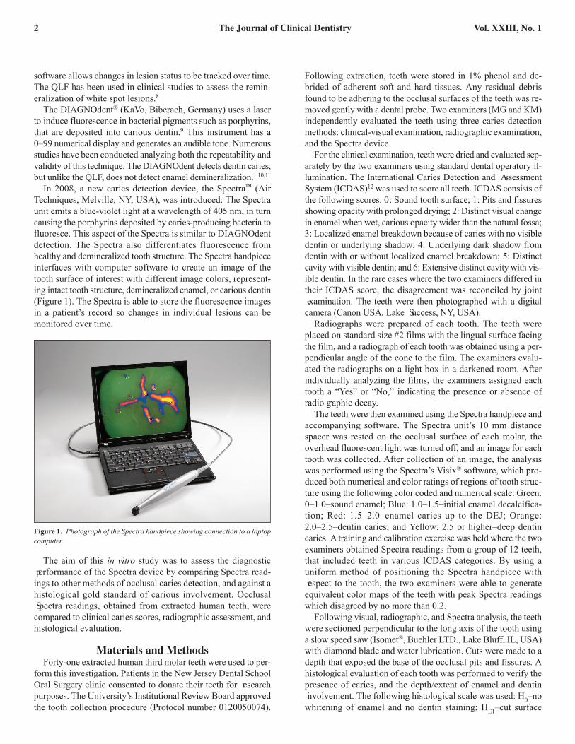
healthy and demineralized tooth structure. The Spectra handpiece
interfaces with computer software to create an image of the
tooth surface of interest with different image colors, represent-
ing intact tooth structure, demineralized enamel, or carious dentin
(Figure 1). The Spectra is able to store the fluorescence images
in a patient’s record so changes in individual lesions can be
monitored over time.
The aim of this in vitro study was to assess the diagnostic
performance of the Spectra device by comparing Spectra read-
ings to other methods of occlusal caries detection, and against a
histological gold standard of carious involvement. Occlusal
Spectra readings, obtained from extracted human teeth, were
compared to clinical caries scores, radiographic assessment, and
histological evaluation.
Materials and MethodsForty-one extracted human third molar teeth were used to per-
form this investigation. Patients in the New Jersey Dental School
Oral Surgery clinic consented to donate their teeth for research
purposes. The University’s Institutional Review Board approved
the tooth collection procedure (Protocol number 0120050074).
Figure 1. Photograph of the Spectra handpiece showing connection to a laptop
computer.

Vol. XXIII, No. 1 The Journal of Clinical Dentistry 3
reveals small white patches within the enamel; HE2
–cut surface
reveals extensive white patches within the enamel; HDI
–cut
surface reveals white discoloration of enamel and dark discolor -
ation of dentin; and HD2
–cut surface reveals areas of white
enamel and extensive dark discoloration of dentin. The two
examiners performed a joint examination in uncommon cases
where their histological scores differed.
Data AnalysisSpectra readings were expressed as mean ± standard deviation.
A two-tailed Student’s t-test was performed to determine if
Spectra readings were significantly different between teeth judged
to have radiographic caries and radiographically intact teeth. In
order to determine whether teeth in different ICDAS and histo-
logical categories had significantly different Spectra readings, an
ANOVA with a pair-wise Tukey-Kramer HSD test was performed
using the SPSS (SAS Institute Inc., Cary, NC, USA) software
package. Statistical significance was set at the p < 0.05 level.
The sensitivity and specificity of the various diagnostic meth-
ods was calculated in order to determine how the techniques
compared to each other.13 The sensitivity is the proportion of
teeth where the test correctly made the diagnosis, and the speci-
ficity is the proportion of teeth where the test correctly identified
teeth not having the caries diagnosis. In calculating the sensitivity
and specificity of the various diagnostic methods, the correctness
of each tooth’s radiographic, clinical, or Spectra assessment was
determined using that tooth’s histological evaluation as a gold
standard means of determining whether the occlusal surface was
intact, had areas of enamel demineralization, or dentin caries. For
the purpose of the sensitivity and specificity calculations, teeth
with histological scores of HE1
or HE2
were classified together as
having enamel lesions. Teeth with histological scores of HD1
or
HD2
were classified together as having dentin caries.
ResultsAll radiographs were analyzed by both examiners, and recorded
as either “Yes,” having radiographic decay or “No,” containing
no radiographic evidence of decay. The examiners were in agree-
ment for the analysis of the 41 teeth, with thirty-two teeth
displaying no radiographic indications of decay and nine teeth
having radiographic decay. For Spectra readings, the “No” radio -
graphic caries group had a mean value of 1.5 ± 0.4, and the
“Yes” radiographic caries group had a mean value of 2.0 ± 0.2;
the difference between these values was significant (p < 0.001).
Next, the relationship between Spectra readings and the
ICDAS scores was determined for the 41 teeth used in the study.
There were seven teeth with a score of 0, nine rated as ICDAS
1, 12 assigned a score of 2, 10 given a score of 3, and three rated
above 3. In Figure 2A, the occlusal view of an ICDAS 2 tooth
is presented. The Spectra map for the same tooth is shown in
Figure 2B. The blue areas at the periphery of the fissures repre-
sent superficial enamel caries, the red are deep enamel caries, and
the orange spots are areas of dentin caries. The cut surface of this
tooth is shown in Figure 2C. The dark areas observed on the cut
surface confirm the presence of dentin caries. Comparison of
Figures 2B and C indicates that the Spectra correctly identified
areas of dentin caries.
The mean Spectra values were determined for the teeth in
each ICDAS category (Figure 3). An ANOVA with a Tukey’s
post hoc test was used to determine if the instrument readings
from the various ICDAS groups differed significantly from one
another. Teeth with higher ICDAS scores generally had higher
Spectra readings. For the Spectra readings, ICDAS 0 teeth did not
differ significantly from ICDAS 1 teeth, but did have signifi-
cantly lower Spectra readings than teeth with ICDAS scores of
2 or greater. The Spectra readings obtained from ICDAS 1 teeth
did not differ from readings obtained from ICDAS 2 teeth, but
were significantly lower than the Spectra readings obtained from
teeth with ICDAS scores ≥ 3. Spectra readings from teeth with
an ICDAS score of 2 did not differ significantly from teeth in
other ICDAS categories, except ICDAS 0 as noted. Teeth with
ICDAS scores of 3 had higher Spectra readings than ICDAS 0
or 1 teeth, but did not significantly differ from teeth in other
categories. The small number of teeth with ICDAS scores greater
than 3 had significantly higher Spectra readings than ICDAS 0
or 1 teeth, but did not differ significantly from other ICDAS
categories.
The histological extent of caries was evaluated by examina-
tion of the cut occlusal surface (Table I). For the four teeth that
were histologically identified as being without caries, lacking
subsurface discoloration of the enamel or dentin, the mean
Spectra reading of 1.0 ± 0.7 is consistent with sound tooth struc-
ture. For the 15 teeth with small demineralized white areas in the
deep fissures, the mean Spectra reading of 1.2 ± 0.6 indicates
early enamel demineralization. The three teeth with extensive
white patches limited to the enamel had a mean Spectra reading
Figure 2. (A) Photograph of a molar assessed as ICDAS 2 (distinct color
changes, wider than natural fissures). (B) Spectra image of the same tooth as is
shown in 2A. Blue areas indicate enamel demineralization, red area indicates
deep enamel decay, and orange area indicates dentin decay. (C) Photograph of
the same tooth as shown in Figures 2A and B following removal of the occlusal
surface. Dark areas confirm the presence of dentin decay.
A B C
Figure 3. Peak occlusal Spectra readings (± standard deviations) obtained
from molars whose caries status was classified using the ICDAS system. Teeth
with higher ICDAS scores had higher Spectra readings.
4 The Journal of Clinical Dentistry Vol. XXIII, No. 1
of 1.6 ± 0.6, indicating deep enamel caries. For the three teeth
in the histological group identified as small dark patches within
the dentin, the mean Spectra reading of 1.5 ± 0.2 incorrectly iden-
tified the lesion as deep enamel caries. The cut surface of 16 teeth
had extensive dark patches in dentin. These teeth had a mean
Spectra reading of 2.0 ± 0.2, indicating dentin caries. Teeth with
deep dentin caries had significantly different Spectra readings
than caries-free teeth and teeth with superficial enamel caries.
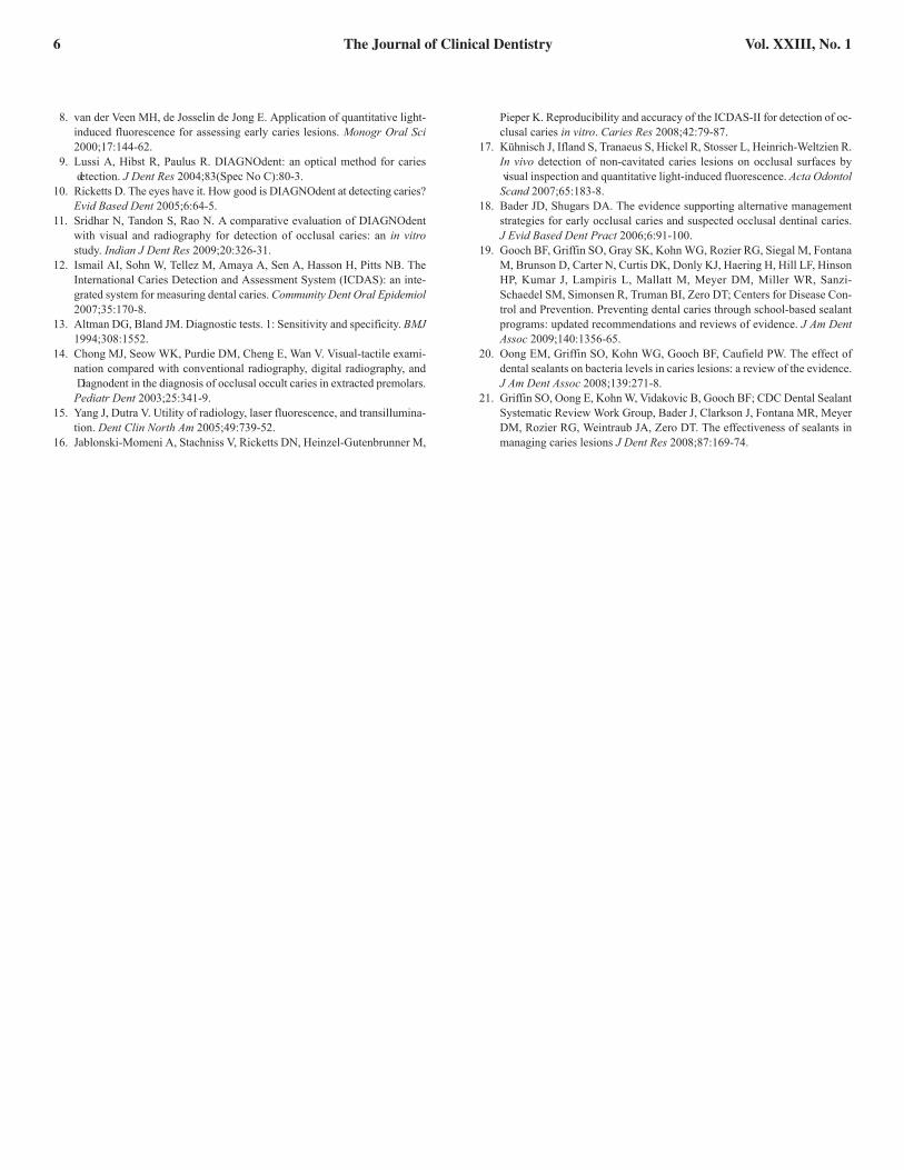
Figures 4A and B show photographs and Spectra images obtained
from the cut surface of an ICDAS 2 tooth, histologically classi-
fied as having dentin caries. The Spectra image clearly highlights
areas of demineralized enamel and caries involved dentin on the
cut surfaces.
Using each tooth’s histological caries assessment as a gold
standard, the sensitivity and specificity of each diagnostic method
was calculated (Table II). The sensitivity of the radiographs in
detecting dentin caries was 0.47, indicating that radiographs de-
tected dentin caries in less than half the teeth in which they were
present. The specificity of the radiographs was 1, indicating that
radiographic evidence of occlusal caries was never observed in
teeth that lacked dentin caries. Compared to radiographs, the
Spectra showed superior sensitivity (0.57) at detecting dentin
caries. For this determination, a Spectra score of 2 was the cut-
off for dentin caries. The specificity of the Spectra in detecting
dentin caries (0.9) was lower than 1.0, indicating some instances
of the Spectra detecting dentin caries when dentin lesions were
not present histologically. ICDAS code 3 denotes a visually de-
tectable cavitation. The sensitivity of cavitation as a clinical
sign of dentin caries was 0.68, and the specificity 1.0 indicates
that the visual finding of small cavitaion is a good indicator of
dentin caries. Using Spectra readings of 1–2 as an indication of
enamel demineralization, the Spectra’s sensitivity in detecting
enamel demineralization was 0.78, and the specificity was 0.52.
Of the 11 teeth incorrectly diagnosed by the Spectra as having
enamel caries, eight had dentin caries, indicating that these teeth
were under-diagnosed as having enamel lesions. ICDAS scores
of 1 and 2 denote visible enamel color changes with intact sur-
faces. Using these clinical criteria to detect enamel demineral-
ization, the sensitivity of ICDAS in detecting enamel caries was
0.72, and the specificity was 0.65.
DiscussionThe aim of this laboratory study was to assess the usefulness
of the Spectra in the diagnosis of occlusal carious lesions by com-
paring the performance of this device to other diagnostic tech-
niques. Each tooth’s histological score was used as the gold
standard determination of the tooth’s caries status. The low sen-
sitivity and high specificity of the radiographs observed in this
study agree with previous results demonstrating that radiographic
diagnosis of occlusal caries is restricted to large lesions.14 Sig-
nificantly higher Spectra readings were observed in teeth that had
radiographic occlusal caries. The Spectra demonstrated superior
sensitivity at detecting dentin caries than radiographs. None of
the teeth used in this study had lesions that were not clinically
apparent but observed radiographically. Clinical studies are re-
quired to assess the performance of caries detectors in the diag-
nosis of hidden caries.15
The ICDAS classification system grades carious lesion severity
according to visual criteria.16 The 41 molars used in this study
were evaluated by both examiners and separated into different
visual categories based upon the ICDAS scores. Generally higher
Spectra readings were measured from teeth with higher ICDAS
scores. These results indicate that the readings obtained with this
caries detector agree with the researchers’ visual examinations.
The groups of teeth identified histologically as being intact,
having superficial and deep enamel demineralization, and hav-
ing deep dentin caries all had mean Spectra readings consistent
with those classifications. The small group of teeth histologically
classified as having early dentin caries was diagnosed by the
Spectra as having lesions restricted to enamel, indicating that the
Spectra under-diagnosed these lesions.
Comparing the sensitivity and specificity of the Spectra to the
clinical evaluation scores indicates that the Spectra reliably
detected dentin caries with a superior sensitivity to radiographic
diagnosis. In small occlusal lesions extending into dentin, the
fissures are lined with demineralized white opaque enamel. This
tissue scatters light and makes it difficult for caries detectors to
sense dentinal pigmentation. This characteristic of occlusal le-
Table I
Spectra Readings ± Standard Deviations for Teeth
with Different Histological Scores
Mean Peak
Histological Score Number of Teeth Spectra Reading ± SD
H01 4 1.0 ± 0.7
He1 15 1.2 ± 0.6
He2 3 1.6 ± 0.6
Hd1 3 1.5 ± 0.2
Hd2 16 2.0 ± 0.2
Table II
Sensitivity and Specificity of Spectra Measurements
Compared to Other Methods of Caries Detection
Diagnostic Parameter Sensitivity Specificity
Radiographic detection of dentin caries 0.47 1.00
Spectra detection of dentin caries (Spectra ≥ 2) 0.57 0.90
Clinical detection of dentin caries (ICDAS ≥ 3) 0.68 1.00
Spectra detection of enamel caries (Spectra 1–1.9) 0.78 0.52
Clinical detection of enamel caries (ICDAS 1 or 2) 0.72 0.65
Figure 4. Cut surface photograph (A) and corresponding Spectra images (B) of
a tooth having small occlusal caries (ICDAS 2). Spectra image of cut surfaces
indicates areas of demineralized enamel shown by blue and red, as well as dark
areas of carious dentin.
A B

Vol. XXIII, No. 1 The Journal of Clinical Dentistry 5
sions makes longitudinal monitoring very important, since small
dentinal lesions can be obscured by overlying enamel changes.
An important objective of caries examinations is the accurate
detection of occlusal lesions which extend into dentin, since
these lesions are typically managed with some form of surgi-
cal/restorative treatment. Using the manufacture’s Spectra cut-
off score of 2.0 as the criteria for dentin caries, the sensitivity of
the Spectra was higher than that of radiographic detection, but
lower than that of clinical examination. We are currently per-
forming further in vitro evaluations where an individual tooth’s
Spectra scores are compared to their histological score. Using this
data we may be able to propose revised Spectra cutoff scores for
dentin caries that would have higher sensitivity and comparable
specificity to the currently recommended cutoff scores.
These results are noteworthy in establishing the utility of the
Spectra in documenting the presence of occlusal caries that is re-
stricted to the enamel. The sensitivity of the Spectra, with regard
to enamel lesions, is higher than that of the clinical examination.
This is important, since the anatomy of the occlusal surface
makes detection of these early carious changes difficult. Future
studies will examine the ability of the Spectra device to detect
artificial enamel caries induced with a lactic acid gel.
The current study indicates that when used with proper visual
and radiographic examination, Spectra is an effective adjunctive
means of analyzing the occlusal surface of molars, and is well
suited to detecting early stages of the carious process. The Spectra
shares with the DIAGNOdent the ability to detect caries de-
rived pigment in dentin. In contrast to the DIAGNOdent, the
Spectra is able to detect areas of enamel demineralization as
well as dentin decay. This enables this device to create a con-
tinuous scale that represents the full extent of the caries process.
This is done through both numerical values and color graphic
representations. A clinical study by Kühnisch and coworkers
demonstrated that the QLF is like the Spectra, effective at de-
tecting early occlusal lesions. Despite these encouraging results,
these authors concluded that the time-consuming nature of the
QLF’s image capture and data analysis made this device im-
practical for widespread clinical use.17
The results of this study show that caries assessment by the
Spectra and visual examination generally agree. This may pro-
voke the question as to why use these new technologies if visual
inspection alone is effective. For clinicians, the goal is to educate
patients and parents. The Spectra provides color images and
numbers to show patients the severity and extent of lesions. The
Spectra allows clinicians to save an image as well as numerical
data, and re-evaluate the same area at recall examinations. Clin-
icians can use the Spectra to monitor lesion progression or
regression over time. The ability of the Spectra to image caries
on cut tooth structure suggests that this device may also aid
clinicians in determining the completeness of caries excavation.
In this in vitro study, the researchers were able to evaluate the
extracted molars using ideal conditions, outside of the oral cav-
ity. Clinical studies are needed to confirm the correlation between
various commonly used methods of caries detection and Spec-
tra readings within patient populations. These studies should
also examine the reproducibility of Spectra measurements taken
under clinical conditions.
Diagnosis of occlusal caries remains a subjective matter.1,16
Spectra and other caries detectors can provide quantitative in-
formation on difficult-to-diagnose lesions. The Spectra can detect
enamel decay, provide insight to the depth of a carious lesion, and
perform with more sensitivity than radiography. Unfortunately,
few methods of caries detection allow the examiner to determine
if the caries process is active, and likely to progress with time.
When dentists detect early occlusal lesions, the objective
should be to utilize therapies that will result in the remineral-
ization or long-term arrest of the caries process.18 Restorative
treatment is usually reserved for cavitated surfaces.19 The abil-
ity of the Spectra to detect and document non-cavitated occusal
lesions indicates that this device can be useful in identifying pa-
tients and teeth in need of preventive interventions, as well as
treatments designed to arrest lesion progression. Although the
ability of oral hygiene practices to remineralize occclusal sur-
faces is unclear, non-cavitated occlusal lesions can be managed
by placing sealants and periodic monitoring.20,21 Future studies
will examine the ability of the Spectra to assess caries on sealed
teeth. It is hoped that new diagnostic technologies such as the
Spectra can facilitate the dentist’s ability to document and com-
municate information concerning caries risk to patients and
parents.
Conclusions• The Spectra is able to detect caries over a broad range, from
enamel demineralization to deep dentin caries.
• Although clinical criteria such as ICDAS are reliable methods
of caries detection, caries detectors could be an adjunctive
method, providing quantitative analysis and, in the case of
Spectra, photographic documentation of difficult-to-
diagnose early occlusal lesions. The Spectra can also allow
lesions to be monitored longitudinally.
Acknowledgment: The authors would like to thank Air Techniques for donating
the Spectra device, for providing valuable technical assistance, and for supporting
the publication of this paper.
For further correspondence with the authors of this paper,
contact Dr. Kenneth Markowitz—[email protected].
References1. Pereira AC, Eggertsson H, Martinez-Mier EA, Mialhe FL, Eckert GJ, Zero
DT. Validity of caries detection on occlusal surfaces and treatment decisions
based on results from multiple caries-detection methods. Eur J Oral Sci
2009;117:51-7.
2. Featherstone JD. The continuum of dental caries—evidence for a dynamic
disease process. J Dent Res 2004;83(Spec No C):39-42.
3. Ismail AI. Visual and visuo-tactile detection of dental caries. J Dent Res
2004;83(Spec No C):56-66.
4. Stookey G. Should a dental explorer be used to probe suspected carious
lesions? No—use of an explorer can lead to misdiagnosis and disrupt
re mineralization. J Am Dent Assoc 2005;136:1527, 1529, 1531.
5. Weerheijm KL, van Amerongen WE, Eggink CO. The clinical diagnosis of
occlusal caries: a problem. ASDC J Dent Child 1989;56:196-200.
6. Wenzel A, Fejerskov O, Kidd E, Joyston-Bechal S, Groeneveld A. Depth of
occlusal caries assessed clinically, by conventional film radiographs, and by
digitized, processed radiographs. Caries Res 1990;24:327-33.
7. Ricketts DN, Ekstrand KR, Martignon S, Alatsaris M, Nugent Z. Accuracy
and reproducibility of conventional radiographic assessment and subtraction
radiography in detecting demineralization in occlusal surfaces. Caries Res
2007;41:121-8.
6 The Journal of Clinical Dentistry Vol. XXIII, No. 1
8. van der Veen MH, de Josselin de Jong E. Application of quantitative light-
induced fluorescence for assessing early caries lesions. Monogr Oral Sci
2000;17:144-62.
9. Lussi A, Hibst R, Paulus R. DIAGNOdent: an optical method for caries
detection. J Dent Res 2004;83(Spec No C):80-3.
10. Ricketts D. The eyes have it. How good is DIAGNOdent at detecting caries?
Evid Based Dent 2005;6:64-5.
11. Sridhar N, Tandon S, Rao N. A comparative evaluation of DIAGNOdent
with visual and radiography for detection of occlusal caries: an in vitro
study. Indian J Dent Res 2009;20:326-31.
12. Ismail AI, Sohn W, Tellez M, Amaya A, Sen A, Hasson H, Pitts NB. The
International Caries Detection and Assessment System (ICDAS): an inte-
grated system for measuring dental caries. Community Dent Oral Epidemiol
2007;35:170-8.
13. Altman DG, Bland JM. Diagnostic tests. 1: Sensitivity and specificity. BMJ
1994;308:1552.
14. Chong MJ, Seow WK, Purdie DM, Cheng E, Wan V. Visual-tactile exami-
nation compared with conventional radiography, digital radiography, and
Diagnodent in the diagnosis of occlusal occult caries in extracted premolars.
Pediatr Dent 2003;25:341-9.
15. Yang J, Dutra V. Utility of radiology, laser fluorescence, and transillumina-
tion. Dent Clin North Am 2005;49:739-52.
16. Jablonski-Momeni A, Stachniss V, Ricketts DN, Heinzel-Gutenbrunner M,
Pieper K. Reproducibility and accuracy of the ICDAS-II for detection of oc-
clusal caries in vitro. Caries Res 2008;42:79-87.
17. Kühnisch J, Ifland S, Tranaeus S, Hickel R, Stosser L, Heinrich-Weltzien R.
In vivo detection of non-cavitated caries lesions on occlusal surfaces by
visual inspection and quantitative light-induced fluorescence. Acta Odontol
Scand 2007;65:183-8.
18. Bader JD, Shugars DA. The evidence supporting alternative management
strategies for early occlusal caries and suspected occlusal dentinal caries.
J Evid Based Dent Pract 2006;6:91-100.
19. Gooch BF, Griffin SO, Gray SK, Kohn WG, Rozier RG, Siegal M, Fontana
M, Brunson D, Carter N, Curtis DK, Donly KJ, Haering H, Hill LF, Hinson
HP, Kumar J, Lampiris L, Mallatt M, Meyer DM, Miller WR, Sanzi-
Schaedel SM, Simonsen R, Truman BI, Zero DT; Centers for Disease Con-
trol and Prevention. Preventing dental caries through school-based sealant
programs: updated recommendations and reviews of evidence. J Am Dent
Assoc 2009;140:1356-65.
20. Oong EM, Griffin SO, Kohn WG, Gooch BF, Caufield PW. The effect of
dental sealants on bacteria levels in caries lesions: a review of the evidence.
J Am Dent Assoc 2008;139:271-8.
21. Griffin SO, Oong E, Kohn W, Vidakovic B, Gooch BF; CDC Dental Sealant
Systematic Review Work Group, Bader J, Clarkson J, Fontana MR, Meyer
DM, Rozier RG, Weintraub JA, Zero DT. The effectiveness of sealants in
managing caries lesions J Dent Res 2008;87:169-74.