Embed Size (px)
Citation preview
Evaluation of the porcine intestinal collagen layer asa biomaterial
Ginger A. Abraham, James Murray, Kristen Billiar, Susan J. SullivanOrganogenesis Inc., 150 Dan Road, Canton, Massachusetts 02021
Received 8 October 1999; accepted 29 December 1999
Abstract: The submucosal layer of the small intestine hasbeen investigated as a source of collagenous tissue with thepotential to be used as a biomaterial because of its inherentstrength and biocompatibility. In this study we utilized anovel method for processing the tissue to generate an acel-lular intestinal collagen layer (ICL). This nondetergent, non-enzymatic chemical cleaning protocol removes cells and cel-lular debris without damaging the native collagen structure.Multilayer laminates of ICL crosslinked with a water-solublecarbodiimide (EDC) were evaluated as a tissue repair mate-
rial in a rabbit abdominal hernia model. The ICL laminatesprovided the requisite physical properties and did not leadto adhesion formation. No immune response to the porcinecollagen was detectable, and this material did not show anycalcification in either the rabbit model or in the juvenile ratmodel. © 2000 John Wiley & Sons, Inc. J Biomed Mater Res,51, 442–452, 2000.
Key words: collagen biomaterial; porcine submucosa; soft-tissue repair
INTRODUCTION
The clinical need for strong, biocompatible materi-als that encourage integration while minimizing ad-verse tissue reactions, such as adhesion formation, isapparent. Yet, the design of optimal surgical repairmaterials to reinforce or replace soft tissues remainsproblematic. In the repair of abdominal wall defectsand hernias, for example, conventional prosthetic ma-terials such as knitted polypropylene mesh sufferfrom a number of complications, including mesh ex-trusion, bowel adherence, fistula formation, woundinfection, and seroma development.1–3 Despite theirlimitations, these surgical repair materials are re-quired in order to reduce the tension on the woundwhen the defect is large and/or the abdominal tissuelacks sufficient strength for primary closure.3 These“tension-free” methods using prosthetic materialshave reduced hernia recurrence rates substan-tially.1,2,4,5 Problems with permanent repair materialshave lead to the development of synthetic resorbablematerials that are not associated with a continuingforeign-body reaction. Ideal degradable materialswould persist long enough in the patient to act as a
scaffold for appropriate host tissue deposition buteventually would be replaced.Thus far, synthetic degradable materials have not pro-vided sufficient strength during the degradation pro-cess.6,7 In both degradable and nondegradable syn-thetic materials, the suture line remains a weak areadue to the lack of tissue ingrowth at the periphery ofthe patch, which can lead to re-herniation.1,8
Natural collagenous materials are being investi-gated for surgical repair because of their inherent lowantigenicity and their ability to integrate with sur-rounding tissue.8–11 Degradable collagenous materialshave shown potential but may lose strength in vivo ifthey are not adequately crosslinked.11,12 The submu-cosal layer of the small intestine, a thin collagenousconnective tissue, has been used with promising re-sults for experimental abdominal wall repair.13,14 Thistissue also has been evaluated as a biomaterial for avariety of other tissue repair or replacement applica-tions, including tendon15 and bladder16,17 repair. Theintestinal submucosa appears to possess the requisitebiocompatibility and strength for surgical repair andcan be remodeled by the host tissue.18
In the above applications, the submucosal layer wasisolated by manually scraping off the mucosal andmuscular layers of porcine small intestines. However,this method generates a tissue containing variableamounts of cellular debris, glycosaminoglycans andlipids. While it is difficult to predict the impact of this
Correspondence to: S. J. Sullivan; e-mail: [email protected]
© 2000 John Wiley & Sons, Inc.

material heterogeneity for all applications, the vari-ability does influence the fabrication of constructs, andresidual cellular components have been implicated asnodes for calcification.31 Consequently we have devel-oped a method for generating an acellular layer ofintestinal collagen from the porcine submucosa with-out compromising the native collagen structure. Inthis report the preparation and characterization of thisporcine intestinal collagen material are described, andthe potential utility of this material is evaluated in arabbit abdominal hernia repair model.
MATERIALS AND METHODS
ICL preparation
The intestinal collagen layer (ICL) was prepared from theintestines of large (at least 205 kg) swine from a closed herd(Parsons Farm, Hadley, Massachusetts). The small intestineswere harvested (Adams’ Farm, Athol, Massachusetts) andshipped on ice to our facility (Organogenesis Inc., Canton,Massachusetts). The cleaning processes were initiatedwithin 4 h of harvest. The mesenteric and outer membranelayers manually were removed from the small intestine. Theintestines then were processed mechanically through a se-ries of rollers (Ernest Bitterling Model M34, Nottingham,England) under a constant flow of hot water (40°C) to re-move the inner mucosal and outer muscular layers. Aftermechanical cleaning, the intestine was slit longitudinally be-tween the lymphatic vessels and cut into 15-cm sections thatwere processed through a series of chemical cleaning steps.The v/v ratio of each chemical cleaning solution to tissuewas 100:1, and the incubations all were carried out at ambi-ent room temperature. The tissue was first incubated forabout 16 h in a solution of 100 mM of ethylenediaminetet-raacetic acid (EDTA) in 10 mM of sodium hydroxide(NaOH) with a pH of 11–12. The second incubation in 1M ofhydrochloric acid (HCl) in 1M of sodium chloride (NaCl) atpH 0 to 1 was carried out for 6–8 h. This was followed by anincubation in 1M of sodium chloride and 10 mM of phos-phate-buffered saline (PBS) at pH 7–7.4 for 16 h and then a2-h incubation in 10 mM of PBS at pH 7–7.4. Finally, thetissue was rinsed in sterile water at pH 5.8–7.0 for at least 2h. The resulting ICL material then was packaged for storageat −80°C and thawed as needed. All lots of material weretested for sterility and residual endotoxin levels prior to use.
ICL characterization
Following mechanical and chemical cleaning, 5 mm sec-tions of ICL were processed for histology and stained withhematoxylin and eosin to determine the extent of cellular
remnants and to assess the structure of the collagen fibers.The collagen amount and type were determined by measur-ing hydroxyproline content and by SDS-PAGE, as describedpreviously.19,20 Residual glycosaminoglycans (GAGs) wereassayed following digestion of the ICL with papain (SigmaChemical Co., St. Louis, Missouri). GAGs were extractedusing DOWEX cation exchange resin (Sigma Chemical Co.)and separated on cellulose acetate sheets by electrophoresis.The bands were stained with alcian blue (Sigma ChemicalCo.), cut out, dissolved in DMSO, and their absorbance mea-sured on a spectrophotometer at 678 nm. The DNA concen-tration was determined using a DNA/RNA isolation kit (US73750, Amersham Life Sciences, Cleveland, Ohio), followingthe standard protocol. The lipid content of dry ICL was de-termined by methylene chloride extraction. Briefly, ICL wasdried and the percent of the solids was determined on aCEM analyzer (model AVC-80, CEM Corp., Matthews,North Carolina). Methylene chloride was added to theround-bottom flask of a Soxhlet distiller/condenser (KontesBrand, Fisher Scientific, Pittsburgh, Pennsylvania) andplaced in a heated water bath. The ICL was placed in aporous thimble above which the organic solvent condensedand extracted the lipids from the ICL. After boiling off thecondensed methylene chloride, the weight and percent ofextracted material were determined. The thermal stability ofthe ICL was measured with a differential scanning calorim-eter (model DSC12E, Mettler Toledo Co., Highstown, NewJersey) before and after chemical cleaning. Samples weigh-ing 5–10 mg were cut from the ICL, sealed in 20-mL alumi-num crucibles, and heated from 35° to 100°C at a rate of10°C/min. Subsequently, Tonset, the temperature associatedwith the onset of the denaturation peak, was determinedfrom the thermogram using analysis software (DSC-MettlerToledo TA 89A).
Laminate construction
ICL laminates were constructed by placing the desirednumber of ICL sheets on top of each other with the layersaligned parallel to the long axis of the intestines. The lami-nates were dried and crosslinked with ethyl-3(3-dimethylamino) propyl carbodiimide (EDC) at the indicatedconcentration. The ICL laminates were rinsed in 0.1% per-acetic acid, vacuum-sealed into hermetic packaging, and ter-minally sterilized by gamma irradiation (25–35 kGy).
Laminate characterization
The thermal stability, suture retention, strength, and stiff-ness of the patch material were measured before and aftergamma sterilization to characterize the physical propertiesand determine the effects of irradiation. The thermal stabil-ity was measured as described above. The following testswere performed on moist samples using a servohydraulic
443PORCINE COLLAGEN LAYER AS BIOMATERIAL
material testing machine (MiniBionix 858, MTS Systems,Minneapolis, Minnesota) using TestWare SX and TestStar2software. The suture retention was measured using a rect-angular test sample 12.5 mm wide by 50 mm long. Eachsample was held in the lower grip and threaded with a 5-0Surgilene (polypropylene) suture 2 mm from its edge. Thetwo ends of the suture were attached to the upper grip andpulled at a constant rate of 125 mm/min. The suture reten-tion was defined as the peak force obtained during this pro-cedure. The stiffness and strength were obtained from con-stant strain rate (0.03 s−1) uniaxial tensile failure tests on12.5-mm wide strips of the ICL constructs. The gauge length(∼28 mm) was defined at a tare load of 0.5 g; the direction ofloading was perpendicular to the long axis of the intestines.The load was normalized to sample width (tension) but notthickness (stress) since the in vivo loading is in the plane ofthe tissue and must be supported by the graft regardless ofthe thickness of the material. The stiffness was defined as themaximum slope of the tension (force/width) versus strain (Dlength/gauge length) curve. The strength was defined as themaximum tension (N/cm).
Rabbit hernia model
The ICL laminates used for hernia repair were crosslinkedwith either 100 mM of EDC in 50% acetone/50% deionizedwater or with 1 mM of EDC in 2-[N-Morpholino]ethanesul-fonic acid (MES) buffer (Pierce Biochemical, Rockford, Illi-nois). New Zealand white rabbits (4.0 kg) were used for thein vivo studies. The animals were caged and kept on a 12-hlight/dark schedule at ∼22°C. All procedures were per-formed at the Tufts New England Medical Center (Boston,Massachusetts) in compliance with the Animal Care and UseCommittee (ACUC) guidelines and in accordance with theNIH guidelines for the use and care of laboratory animals(NIH Publication #85-23 Rev. 1985). Prior to surgery, theanimals were medicated with 3.0 cc of xylazine and 1.0 cc ofacepromazine. (Vedco Inc., St. Joseph, Missouri). Generalanesthesia was induced by an intramuscular injection of 40mg/kg of Ketamine (Phoenix Pharmaceuticals Inc., St. Jo-seph, Missouri). After depilation of the surgical site, the areawas wiped down with 70% isopropanol and iodine. A full-thickness defect approximately 5 cm long was made in theanterior abdominal wall through the center of the rectus ab-dominis muscle and the underlying peritoneum. The ICLpatch was trimmed to the size of the defect and attached tothe rectus sheath with an uninterrupted, nonresorbable 3-0Prolene suture. The skin was closed with 3-0 Dexon and 3-0Ethibond sutures. All suture material was obtained fromRoboz Surgical (Rockville, Maryland).
At the end of the indicated implant period, anesthesia wasinduced, as described above. After examination for anygross reaction or re-herniation of the defect, the abdomenwas opened to expose the peritoneal surface of the patch.Adhesions were graded using the standard scale in which ascore of 0 indicates no adhesions, 1 represents thin and filmyadhesions easily separable by blunt dissection, 2 indicates
firmly attached adhesions requiring aggressive blunt ormild sharp dissection, and a score of 3 indicates adhesionsintegrated with viscera that require aggressive sharp dissec-tion.21,22 The grafts also were evaluated for tissue integra-tion, surface vascularization, fibrous capsule formation, andinflammation. Immediately following gross examination,the animal was euthanized with potassium chloride. A full-thickness section of host tissue with the implanted patch wasremoved and fixed with 10% formalin and then processedfor evaluation. A median strip, including the patch, horizon-tal to the rectus abdominis muscle of approximately 1 cm inwidth was stained with hematoxylin and eosin (H&E) forhistologic analysis of cell infiltration, inflammatory re-sponse, and collagen morphology or with Alizarin red toidentify any sites of calcification.
Immunologic analysis
A 10-mL sample of whole blood was collected from eachanimal at the time of explant to assay for antibodies to theporcine collagen. Porcine Type I collagen (WAKO Co.,China) was coated onto Nunc Maxisorb 96-well plates (100mL per well at a concentration of 1 mg/mL) overnight at 4°C.The plates were then blocked using 250 mL per well of 3%fish gelatin (Sigma Chemical Co.) for 2 h at 37°C. The plateswere rinsed with PBS and 50 mL of rabbit serum was added.Purified rabbit anti-porcine collagen (Biodesign, Ken-nebunk, Maine) and normal rabbit serum were used as posi-tive and negative controls, respectively. After incubation at37°C for 2 h, the plates were washed four times with PBS +0.1% Tween-20. Fifty mL of biotinylated goat anti-rabbit Ig(Jackson Immunoresearch, West Grove, Pennsylvania) wereadded to each well at a final concentration of 1 mg/mL.Plates were incubated at 37°C for 1 h and then washed, asabove. Fifty mL of streptavidin–alkaline phosphatase (Jack-son Immunoresearch) diluted 1:2000 then were added toeach well, incubated for 30 min at 37°C, and washed. Plateswere developed using 100 mL of a 1-mg/mL solution ofp-nitrophenylphosphate, and antibody binding was deter-mined using a SpectroMax 250 plate reader (Molecular Dy-namics, Sunnyvale, California). The sensitivity of antibodydetection is 1-3 nanograms/mL, which is approximatelytwice background.
Mechanical characterization of explants
Uniaxial failure tests were performed on three 180-day-old explants and three corresponding control rabbit abdomi-nal specimens. From each explant, a 19 to 23-mm wide stripapproximately 50 mm in length was cut parallel to the pre-dominant muscle fiber direction. The sample consisted ofpatch, incorporation region, and abdominal tissue. A controlsample was cut parallel to the patch sample from the hostabdominal tissue. The patch portion of each specimen was
444 ABRAHAM ET AL.
placed in the upper grip, and the abdominal muscle wasplaced in the lower grip. The grip-to-grip spacing was ap-proximately 25 mm at a tare load of 0.5 g. For the controltissues both grips held abdominal muscle. All specimenswere tested at room temperature and kept moist with iso-tonic saline spray. The samples were loaded in uniaxial ten-sion at a strain rate of 0.03 s−1 until failure. As with thepre-implanted material, strength (N/cm) rather than ulti-mate tensile stress (N/cm2) was reported because the im-plant and attachment region must withstand the forces ex-perienced by the abdominal wall regardless of the thicknessof the material. Stiffness of the explants was not reportedsince the deformation of the host tissue and the attachmentregion cannot be separated from deformation of the patchmaterial itself. Similarly, the explant integration strength isnot a direct measure of the strength of the remaining graftmaterial since it was not gripped from both sides.
Rat calcification model
A one-layer ICL sheet was prepared using sterile tech-nique, crosslinked with 1 mM of EDC in deionized water for16 h, and terminally sterilized by gamma irradiation (25–35kGy). Four-week-old Sprague–Dawley rats were used forthe in vivo analysis; the studies were carried out at Pine AcreRabbitry/Farm (Norton, Massachusetts) in compliance withACUC and NIH guidelines for the use and care of laboratoryanimals (NIH Publication #85-23 Rev. 1985). Prior to surgeryeach animal was anesthetized with ketamine and xylazine.A full-thickness incision was created in the skin on the ven-trum. A 1-cm2 piece of ICL was folded and inserted subcu-taneously between the skin and the rectus abdominis muscles.The ICL was then stapled to the fascia and the wound wasclosed with staples and dressed with a sterile bandage. Us-ing the same protocol, bovine heart valves fixed with 0.625%glutaraldehyde for 3 days were implanted as the positivecontrol. At 7 and 28 days postsurgery, the animals wereanesthetized, as previously described. The ICL and valveswere explanted, fixed in 10% formalin, and processed forhistologic evaluation. Calcification was assessed by Alizarinred staining.
RESULTS
ICL preparation and characterization
In our early studies exploring the use of this collag-enous biomaterial in soft tissue repair and vascularprosthetics, the submucosal layer was obtained fromthe porcine small intestine after mechanical cleaning,using a customized, commercial gut-cleaning ma-chine. We demonstrated that a six-layer laminatecrosslinked with EDC, a water-soluble carbodiimide,
and sterilized with peracetic acid was effective as ahernia repair patch in the rabbit model. The resultingbiomaterial, GraftPatch™, received 510K clearancefrom the FDA for use in surgical repair.23 Althoughthese studies were promising, it became clear that thematerial obtained after mechanical cleaning was het-erogeneous. The amount of residual cellular materialin the submucosal layer varied between preparationsfrom different donor pigs as well as within differentsections of the small intestine. In particular, mechani-cal cleaning was not adequate for removing the endo-thelial cells in the vasculature of the tissue. Conse-quently, a chemical cleaning process was needed thatwould remove cells and cellular debris while main-taining the native collagen structure. To minimizedamage to the collagen, we developed a nonenzy-matic, nondetergent procedure for removing noncol-lagenous components of the tissue while at the sametime controlling the swelling of the collagen fibers.
The first step in this process uses an alkaline chelat-ing solution to saponify lipids and disrupt the base-ment membrane components of the cellular materialthat remains attached after mechanical cleaning. Al-though EDTA or EGTA [ethylenebis(oxyethylenitrilo)-tetraacetic acid] can be used, EDTA in NaOH at a pHof 11–12 was the most effective. The second incubationused an acidic salt solution; hydrochloric acid (HCl),acetic acid, or sulfuric acid at concentrations of 0.5 to2M with NaCl added at concentrations of 0.1 to 2.0Mto control swelling of the tissue. HCl is preferred forthis step because it breaks up DNA into fragments offewer than six base pairs. The next step involves ex-posure to a salt solution at physiologic pH (7.0 to 7.4).This solution helps neutralize the material and mini-mize swelling. We have found that the minimal effec-tive amount for the chemical cleaning of the tissue is aratio of 100:1 v/v of solution to tissue and that agita-tion of the solutions improves penetration of thechemicals. The required incubation times vary withthe thickness of the tissue and were established atroom temperature although the temperature canrange from 4.0° to 45.0°C. The final step uses injection-grade water to rinse the tissue free of the cleaningagents so that the endotoxin levels can be measured.
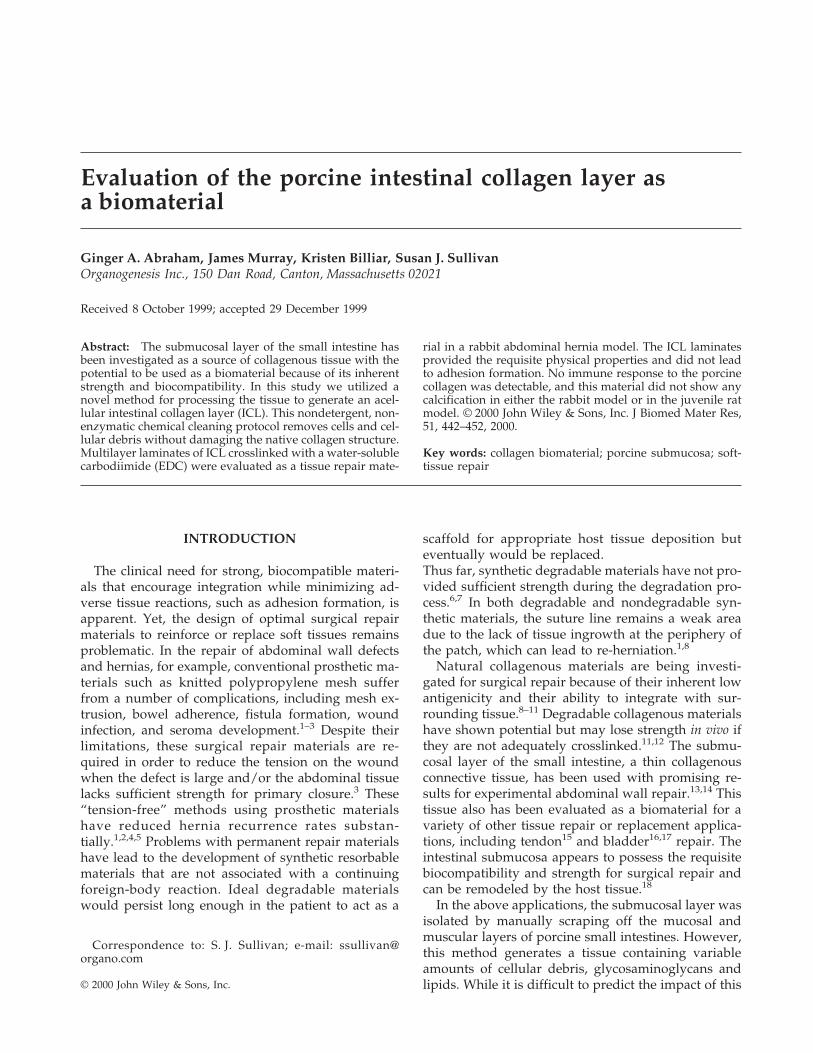
The efficacy of this chemical cleaning process is il-lustrated in Figure 1. The photomicrograph of tissuesections stained with H&E shows the presence of re-sidual cellular material after mechanical processing[Fig. 1(A)], which was removed by chemical cleaning[Fig. 1(B)] without visible damage to the collagen fi-bers. Biochemical analysis of the ICL obtained afterthis chemical processing confirmed that the tissue waspredominantly Type I collagen with less than 0.7%lipids and undetectable levels of GAGs (<0.6%) andDNA (<0.1 ng/mL). In addition to the morphologicand biochemical analysis of the ICL, thermal stability
445PORCINE COLLAGEN LAYER AS BIOMATERIAL
was assessed to evaluate the effect of the material pro-cessing. The denaturation onset temperatures were68.4° ± 0.4°C before and 68.3° ± 1.3°C after chemicalcleaning. This lack of alteration of the thermal stabilitywas consistent with the native collagen triple helicalstructure not having been disrupted by the tissue pro-cessing.
In vivo function of ICL laminates
Having developed a process that could generate anacellular collagen layer without damaging the colla-gen fibers, the ICL material was tested for in vivo func-tion in the rabbit abdominal hernia model. This studywas designed to determine if the chemical processingchanged the in vivo performance of the GraftPatch™laminate. In the first set of experiments, a six-layerlaminate crosslinked with 100 mM of EDC in 50% ac-
etone was tested for implant periods ranging from 1 to6 months, with three rabbits evaluated at each timepoint (30, 60, 99, 180 days). Full-thickness abdominalwall defects were created and then repaired, as de-scribed in the Materials and Methods section.
All animals underwent an uneventful postoperativecourse, with no swelling, herniation, or inflammationat the repair site of the abdominal wall. As shown in
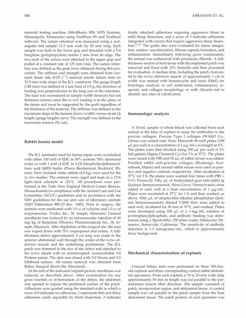
Figure 2. Gross morphology of the ICL hernia repairpatches. Six-layer ICL laminates crosslinked with 100 mM ofEDC were used to repair defects created in the rabbit ab-dominal wall. The rabbits were evaluated after 1, 2, 3, and 6months (n = 3 at each time point). No adverse reaction orseroma development was observed in any of the animals.(A) The patches were well integrated and had developed amesothelial-like covering within 1 month. At 1 month, someneovascularization was observed. After (B) 3 months and(C) 6 months, the morphologies were similar, with moreextensive vascularization on the visceral surface of thepatch.
Figure 1. The effect of chemical cleaning on porcine sub-mucosal tissue. The submucosal layer of the porcine smallintestine was evaluated using hematoxylin and eosin (H&E)staining after (A) mechanical cleaning and after (B) bothmechanical and chemical cleaning. In the absence of chemi-cal cleaning, cells and cellular debris can be seen throughoutthe tissue. However, no residual cellular material is seenafter the chemical cleaning procedure. Original magnifica-tions, ×20.
446 ABRAHAM ET AL.
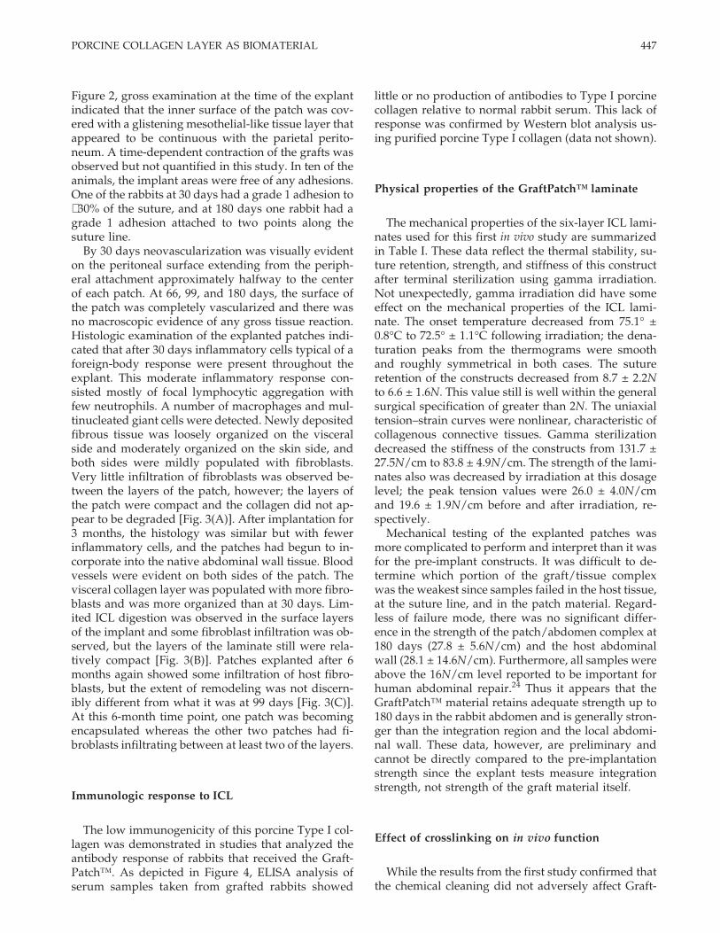
Figure 2, gross examination at the time of the explantindicated that the inner surface of the patch was cov-ered with a glistening mesothelial-like tissue layer thatappeared to be continuous with the parietal perito-neum. A time-dependent contraction of the grafts wasobserved but not quantified in this study. In ten of theanimals, the implant areas were free of any adhesions.One of the rabbits at 30 days had a grade 1 adhesion to∼30% of the suture, and at 180 days one rabbit had agrade 1 adhesion attached to two points along thesuture line.
By 30 days neovascularization was visually evidenton the peritoneal surface extending from the periph-eral attachment approximately halfway to the centerof each patch. At 66, 99, and 180 days, the surface ofthe patch was completely vascularized and there wasno macroscopic evidence of any gross tissue reaction.Histologic examination of the explanted patches indi-cated that after 30 days inflammatory cells typical of aforeign-body response were present throughout theexplant. This moderate inflammatory response con-sisted mostly of focal lymphocytic aggregation withfew neutrophils. A number of macrophages and mul-tinucleated giant cells were detected. Newly depositedfibrous tissue was loosely organized on the visceralside and moderately organized on the skin side, andboth sides were mildly populated with fibroblasts.Very little infiltration of fibroblasts was observed be-tween the layers of the patch, however; the layers ofthe patch were compact and the collagen did not ap-pear to be degraded [Fig. 3(A)]. After implantation for3 months, the histology was similar but with fewerinflammatory cells, and the patches had begun to in-corporate into the native abdominal wall tissue. Bloodvessels were evident on both sides of the patch. Thevisceral collagen layer was populated with more fibro-blasts and was more organized than at 30 days. Lim-ited ICL digestion was observed in the surface layersof the implant and some fibroblast infiltration was ob-served, but the layers of the laminate still were rela-tively compact [Fig. 3(B)]. Patches explanted after 6months again showed some infiltration of host fibro-blasts, but the extent of remodeling was not discern-ibly different from what it was at 99 days [Fig. 3(C)].At this 6-month time point, one patch was becomingencapsulated whereas the other two patches had fi-broblasts infiltrating between at least two of the layers.
Immunologic response to ICL
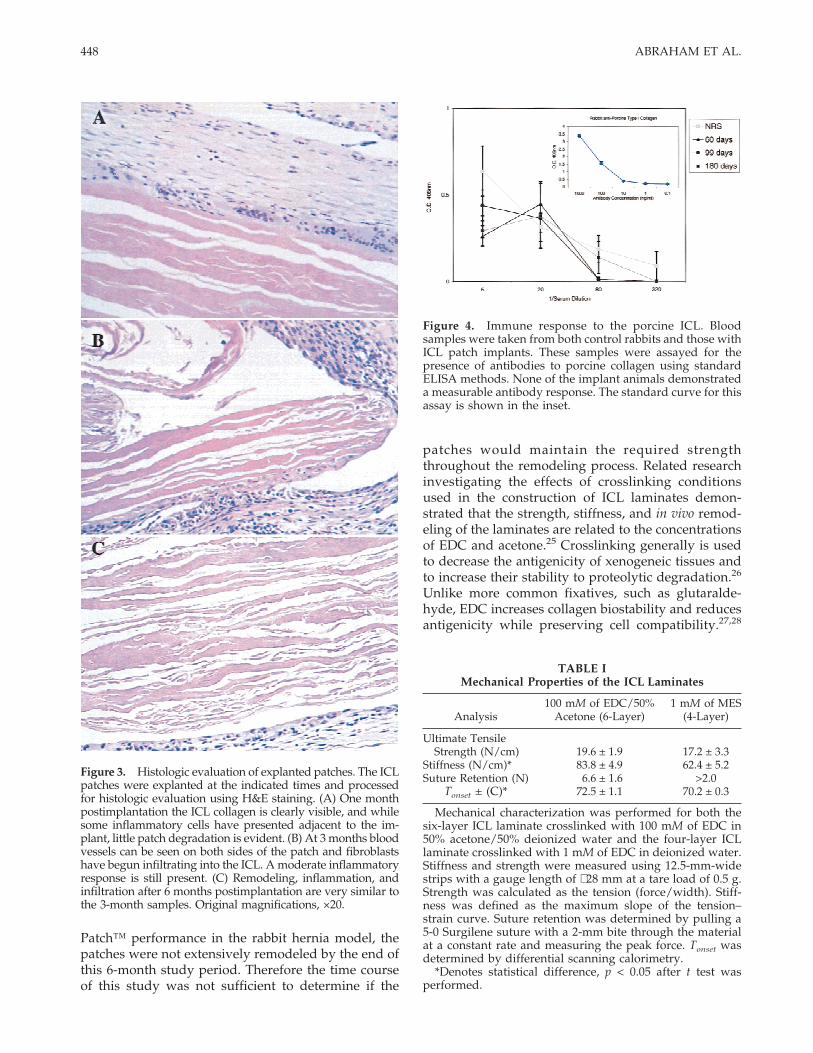
The low immunogenicity of this porcine Type I col-lagen was demonstrated in studies that analyzed theantibody response of rabbits that received the Graft-Patch™. As depicted in Figure 4, ELISA analysis ofserum samples taken from grafted rabbits showed
little or no production of antibodies to Type I porcinecollagen relative to normal rabbit serum. This lack ofresponse was confirmed by Western blot analysis us-ing purified porcine Type I collagen (data not shown).
Physical properties of the GraftPatch™ laminate
The mechanical properties of the six-layer ICL lami-nates used for this first in vivo study are summarizedin Table I. These data reflect the thermal stability, su-ture retention, strength, and stiffness of this constructafter terminal sterilization using gamma irradiation.Not unexpectedly, gamma irradiation did have someeffect on the mechanical properties of the ICL lami-nate. The onset temperature decreased from 75.1° ±0.8°C to 72.5° ± 1.1°C following irradiation; the dena-turation peaks from the thermograms were smoothand roughly symmetrical in both cases. The sutureretention of the constructs decreased from 8.7 ± 2.2Nto 6.6 ± 1.6N. This value still is well within the generalsurgical specification of greater than 2N. The uniaxialtension–strain curves were nonlinear, characteristic ofcollagenous connective tissues. Gamma sterilizationdecreased the stiffness of the constructs from 131.7 ±27.5N/cm to 83.8 ± 4.9N/cm. The strength of the lami-nates also was decreased by irradiation at this dosagelevel; the peak tension values were 26.0 ± 4.0N/cmand 19.6 ± 1.9N/cm before and after irradiation, re-spectively.
Mechanical testing of the explanted patches wasmore complicated to perform and interpret than it wasfor the pre-implant constructs. It was difficult to de-termine which portion of the graft/tissue complexwas the weakest since samples failed in the host tissue,at the suture line, and in the patch material. Regard-less of failure mode, there was no significant differ-ence in the strength of the patch/abdomen complex at180 days (27.8 ± 5.6N/cm) and the host abdominalwall (28.1 ± 14.6N/cm). Furthermore, all samples wereabove the 16N/cm level reported to be important forhuman abdominal repair.24 Thus it appears that theGraftPatch™ material retains adequate strength up to180 days in the rabbit abdomen and is generally stron-ger than the integration region and the local abdomi-nal wall. These data, however, are preliminary andcannot be directly compared to the pre-implantationstrength since the explant tests measure integrationstrength, not strength of the graft material itself.
Effect of crosslinking on in vivo function
While the results from the first study confirmed thatthe chemical cleaning did not adversely affect Graft-
447PORCINE COLLAGEN LAYER AS BIOMATERIAL
Patch™ performance in the rabbit hernia model, thepatches were not extensively remodeled by the end ofthis 6-month study period. Therefore the time courseof this study was not sufficient to determine if the
patches would maintain the required strengththroughout the remodeling process. Related researchinvestigating the effects of crosslinking conditionsused in the construction of ICL laminates demon-strated that the strength, stiffness, and in vivo remod-eling of the laminates are related to the concentrationsof EDC and acetone.25 Crosslinking generally is usedto decrease the antigenicity of xenogeneic tissues andto increase their stability to proteolytic degradation.26
Unlike more common fixatives, such as glutaralde-hyde, EDC increases collagen biostability and reducesantigenicity while preserving cell compatibility.27,28
TABLE IMechanical Properties of the ICL Laminates
Analysis100 mM of EDC/50%
Acetone (6-Layer)1 mM of MES
(4-Layer)
Ultimate TensileStrength (N/cm) 19.6 ± 1.9 17.2 ± 3.3
Stiffness (N/cm)* 83.8 ± 4.9 62.4 ± 5.2Suture Retention (N) 6.6 ± 1.6 >2.0
Tonset ± (C)* 72.5 ± 1.1 70.2 ± 0.3
Mechanical characterization was performed for both thesix-layer ICL laminate crosslinked with 100 mM of EDC in50% acetone/50% deionized water and the four-layer ICLlaminate crosslinked with 1 mM of EDC in deionized water.Stiffness and strength were measured using 12.5-mm-widestrips with a gauge length of ∼28 mm at a tare load of 0.5 g.Strength was calculated as the tension (force/width). Stiff-ness was defined as the maximum slope of the tension–strain curve. Suture retention was determined by pulling a5-0 Surgilene suture with a 2-mm bite through the materialat a constant rate and measuring the peak force. Tonset wasdetermined by differential scanning calorimetry.
*Denotes statistical difference, p < 0.05 after t test wasperformed.
Figure 3. Histologic evaluation of explanted patches. The ICLpatches were explanted at the indicated times and processedfor histologic evaluation using H&E staining. (A) One monthpostimplantation the ICL collagen is clearly visible, and whilesome inflammatory cells have presented adjacent to the im-plant, little patch degradation is evident. (B) At 3 months bloodvessels can be seen on both sides of the patch and fibroblastshave begun infiltrating into the ICL. A moderate inflammatoryresponse is still present. (C) Remodeling, inflammation, andinfiltration after 6 months postimplantation are very similar tothe 3-month samples. Original magnifications, ×20.
Figure 4. Immune response to the porcine ICL. Bloodsamples were taken from both control rabbits and those withICL patch implants. These samples were assayed for thepresence of antibodies to porcine collagen using standardELISA methods. None of the implant animals demonstrateda measurable antibody response. The standard curve for thisassay is shown in the inset.
448 ABRAHAM ET AL.
However, the acellular nature of the ICL decreases theneed for crosslinking to minimize the antigenicity ofresidual cellular material.
Based on the results from these previous crosslink-ing studies, the second in vivo study using the rabbithernia model evaluated the performance of four-layerlaminates crosslinked with 1 mM of EDC in deionizedwater without acetone. The physical properties ofthese constructs are shown in Table I. As expectedwith less stringent crosslinking conditions, the Tonset
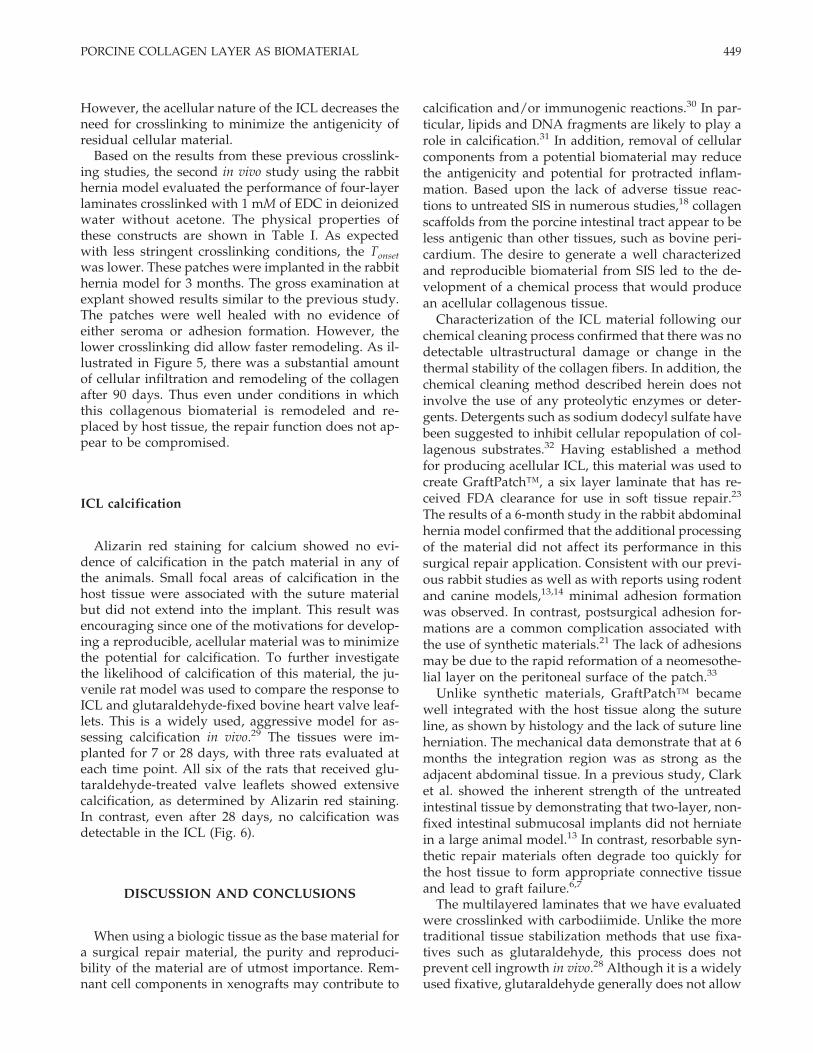
was lower. These patches were implanted in the rabbithernia model for 3 months. The gross examination atexplant showed results similar to the previous study.The patches were well healed with no evidence ofeither seroma or adhesion formation. However, thelower crosslinking did allow faster remodeling. As il-lustrated in Figure 5, there was a substantial amountof cellular infiltration and remodeling of the collagenafter 90 days. Thus even under conditions in whichthis collagenous biomaterial is remodeled and re-placed by host tissue, the repair function does not ap-pear to be compromised.
ICL calcification
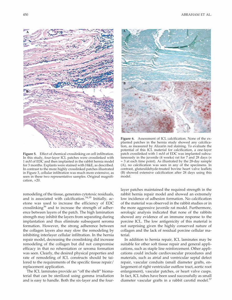
Alizarin red staining for calcium showed no evi-dence of calcification in the patch material in any ofthe animals. Small focal areas of calcification in thehost tissue were associated with the suture materialbut did not extend into the implant. This result wasencouraging since one of the motivations for develop-ing a reproducible, acellular material was to minimizethe potential for calcification. To further investigatethe likelihood of calcification of this material, the ju-venile rat model was used to compare the response toICL and glutaraldehyde-fixed bovine heart valve leaf-lets. This is a widely used, aggressive model for as-sessing calcification in vivo.29 The tissues were im-planted for 7 or 28 days, with three rats evaluated ateach time point. All six of the rats that received glu-taraldehyde-treated valve leaflets showed extensivecalcification, as determined by Alizarin red staining.In contrast, even after 28 days, no calcification wasdetectable in the ICL (Fig. 6).
DISCUSSION AND CONCLUSIONS
When using a biologic tissue as the base material fora surgical repair material, the purity and reproduci-bility of the material are of utmost importance. Rem-nant cell components in xenografts may contribute to
calcification and/or immunogenic reactions.30 In par-ticular, lipids and DNA fragments are likely to play arole in calcification.31 In addition, removal of cellularcomponents from a potential biomaterial may reducethe antigenicity and potential for protracted inflam-mation. Based upon the lack of adverse tissue reac-tions to untreated SIS in numerous studies,18 collagenscaffolds from the porcine intestinal tract appear to beless antigenic than other tissues, such as bovine peri-cardium. The desire to generate a well characterizedand reproducible biomaterial from SIS led to the de-velopment of a chemical process that would producean acellular collagenous tissue.
Characterization of the ICL material following ourchemical cleaning process confirmed that there was nodetectable ultrastructural damage or change in thethermal stability of the collagen fibers. In addition, thechemical cleaning method described herein does notinvolve the use of any proteolytic enzymes or deter-gents. Detergents such as sodium dodecyl sulfate havebeen suggested to inhibit cellular repopulation of col-lagenous substrates.32 Having established a methodfor producing acellular ICL, this material was used tocreate GraftPatch™, a six layer laminate that has re-ceived FDA clearance for use in soft tissue repair.23
The results of a 6-month study in the rabbit abdominalhernia model confirmed that the additional processingof the material did not affect its performance in thissurgical repair application. Consistent with our previ-ous rabbit studies as well as with reports using rodentand canine models,13,14 minimal adhesion formationwas observed. In contrast, postsurgical adhesion for-mations are a common complication associated withthe use of synthetic materials.21 The lack of adhesionsmay be due to the rapid reformation of a neomesothe-lial layer on the peritoneal surface of the patch.33
Unlike synthetic materials, GraftPatch™ becamewell integrated with the host tissue along the sutureline, as shown by histology and the lack of suture lineherniation. The mechanical data demonstrate that at 6months the integration region was as strong as theadjacent abdominal tissue. In a previous study, Clarket al. showed the inherent strength of the untreatedintestinal tissue by demonstrating that two-layer, non-fixed intestinal submucosal implants did not herniatein a large animal model.13 In contrast, resorbable syn-thetic repair materials often degrade too quickly forthe host tissue to form appropriate connective tissueand lead to graft failure.6,7
The multilayered laminates that we have evaluatedwere crosslinked with carbodiimide. Unlike the moretraditional tissue stabilization methods that use fixa-tives such as glutaraldehyde, this process does notprevent cell ingrowth in vivo.28 Although it is a widelyused fixative, glutaraldehyde generally does not allow
449PORCINE COLLAGEN LAYER AS BIOMATERIAL
remodeling of the tissue, generates cytotoxic residuals,and is associated with calcification.34,35 Initially, ac-etone was used to increase the efficiency of EDCcrosslinking36 and to increase the strength of adher-ence between layers of the patch. The high laminationstrength may inhibit the layers from separating duringimplantation and thus eliminate subsequent seromaformation. However, the strong adherence betweenthe collagen layers also may slow the remodeling byinhibiting interlayer cellular infiltration. In the herniarepair model, decreasing the crosslinking did increaseremodeling of the collagen but did not compromiseefficacy in that no reherniation or seroma formationwas seen. Clearly, the desired physical properties andrate of remodeling of ICL constructs should be tai-lored to the requirements of the specific tissue repair/replacement application.
The ICL laminates provide an “off the shelf” bioma-terial that can be sterilized using gamma irradiationand is easy to handle. Both the six-layer and the four-
layer patches maintained the required strength in therabbit hernia repair model and showed an extremelylow incidence of adhesion formation. No calcificationof the material was observed in the rabbit studies or inthe more aggressive juvenile rat model. Furthermore,serologic analysis indicated that none of the rabbitsshowed any evidence of an immune response to theporcine ICL. The low antigenicity of this material isnot surprising given the highly conserved nature ofcollagen and the lack of residual porcine cellular ma-terial.
In addition to hernia repair, ICL laminates may besuitable for other soft tissue repair and general appli-cations, such as staple line reinforcement. Other appli-cations could include cardiovascular procedures andmaterials, such as atrial and ventricular septal defectrepair, vascular conduits (small diameter grafts, en-largement of right ventricular outflow tract, aortic rootenlargement), vascular patches, or heart valve cusps.In fact, ICL tubes have been used successfully as smalldiameter vascular grafts in a rabbit carotid model.37
Figure 5. Effect of chemical crosslinking on cell infiltration.In this study, four-layer ICL patches were crosslinked with1 mM of EDC and then implanted in the rabbit hernia modelfor 3 months. Explants were stained with H&E, as described.In contrast to the more highly crosslinked patches illustratedin Figure 3, cellular infiltration was much more extensive, asseen in these two representative samples. Original magnifi-cation, ×20.
Figure 6. Assessment of ICL calcification. None of the ex-planted patches in the hernia study showed any calcifica-tion, as measured by Alizarin red staining. To evaluate thepotential of this ICL material for calcification, a one-layerpatch crosslinked with 1 mM of EDC was implanted subcu-taneously in the juvenile (4 weeks) rat for 7 and 28 days (n= 3 at each time point). As illustrated by the 28-day sample(A), no calcification was seen in any of the specimens. Incontrast, glutaraldehyde-treated bovine heart valve leaflets(B) showed extensive calcification after 28 days using thismodel.
450 ABRAHAM ET AL.
Although each application will have specific mechani-cal and biologic requirements, its biocompatibility,mechanical strength, and low adhesion formationmake ICL a promising surgical repair material.
We would like to thank Dr. Joseph Laning for the immu-nologic analyses, Jeff Crews, Reuben Chevere, and JonathanThayer for assistance with the histologic evaluation and cal-cification studies, and Dr. Nathaniel Bachrach for insightfuldiscussion during the preparation of this manuscript. Wealso appreciate the efforts of Dr. Raymond Connolly (TuftsNew England Medical Center), who provided the herniarepair model.
References
1. Molloy RG, Moran KT, Waldron RP, Brady MP, Kirwan WO.Massive incisional hernia: Abdominal wall replacement withMarlex mesh. Br J Surg 1991;78:242–244.
2. Lewis RT. Knitted polypropylene (Marlex) mesh in the repairof incisional hernias. Can J Surg 1984;27:155–157.
3. Larson GM, Vandertoll DJ. Approaches to repair of ventralhernia and full-thickness losses of the abdominal wall. SurgClin N Am 1984;64:335–349.
4. Liakakos T, Karanikas I, Panagiotidis H, Dendrinos S. Use ofMarlex mesh in the repair of recurrent incisional hernia. [Seecomments.] Br J Surg 1994;81:248–249.
5. Leber GE, Garb JL, Alexander AI, Reed WP. Long-term com-plications associated with prosthetic repair of incisional her-nias. Arch Surg 1998;133:378–382.
6. Tyrell J, Silberman H, Chandrasoma P, Niland J, Shull J. Ab-sorbable versus permanent mesh in abdominal operations.Surg Gynecol Obstet 1989;168:227–232.
7. Lamb JP, Vitale T, Kaminski DL. Comparative evaluation ofsynthetic meshes used for abdominal wall replacement. Sur-gery 1983;93:643–648.
8. Jenkins SD, Klamer TW, Parteka JJ, Condon RE. A comparisonof prosthetic materials used to repair abdominal wall defects.Surgery 1983;94:392–398.
9. James NL, Poole–Warren LA, Schindhelm K, Millthorpe BK,Mitchell RM, Mitchell RE, Howlett CR. Comparative evalua-tion of treated bovine pericardium as a xenograft for herniarepair. Biomaterials 1991;12:801–809.
10. Matsumoto H, Oguchi Y, Miyake Y, Masuda Y, Masada S,Kuno Y, Shibahara I, Takashima K, Yamane H, Yamagata S,Noishiki Y, Yamane Y. The use of epoxy patch grafts for therepair of experimentally-created diaphragmatic defects indogs. J Vet Med Sci 1996;58:685–687.
11. van der Laan JS, Lopez GP, van Wachem PB, Nieuwenhuis P,Ratner BD, Bleichrodt RP, Schakenraad JM. TFE-plasma poly-merized dermal sheep collagen for the repair of abdominalwall defects. Int J Artif Org 1991;14:661–666.
12. van Wachem PB, van Luyn MJ, Olde Damink LH, Dijkstra PJ,Feijen J, Nieuwenhuis P. Tissue regenerating capacity of car-bodiimide-crosslinked dermal sheep collagen during repair ofthe abdominal wall. Int J Artif Org 1994;17:230–239.
13. Clarke KM, Lantz GC, Salisbury SK, Badylak SF, Hiles MC,Voytik SL. Intestine submucosa and polypropylene mesh forabdominal wall repair in dogs. J Surg Res 1996;60:107–114.
14. Prevel CD, Eppley BL, Summerlin DJ, Jackson JR, McCarty M,
Badylak SF. Small intestinal submucosa: Use in repair of ro-dent abdominal wall defects. Ann Plast Surg 1995;35:374–380.
15. Badylak SF, Tullius R, Kokini K, Shelbourne KD, Klootwyk T,Voytik SL, Kraine MR, Simmons C. The use of xenogeneicsmall intestinal submucosa as a biomaterial for Achilles tendonrepair in a dog model. J Biomed Mater Res 1995;29:977–985.
16. Kropp BP, Sawyer BD, Shannon HE, Rippy MK, Badylak SF,Adams MC, Keating MA, Rink RC, Thor KB. Characterizationof small intestinal submucosa regenerated canine detrusor: As-sessment of reinnervation, in vitro compliance and contractil-ity. J Urol 1996;156:599–607.
17. Badylak S, Kropp B, McPherson T, Liang H, Snyder P. Smallintestinal submucosa: A rapidly resorbed bioscaffold for aug-mentation cytoplasty in a dog model. Tissue Eng 1998;4:379–387.
18. Badylak SF. Small intestinal submucosa (SIS): A biomaterialconducive to smart tissue remodeling. In: Bell E, editor. TissueEngineering: Current Perspectives. Cambridge, Massachusetts:Burkhauser Publishers; 1993. p 179–189.
19. Hayashi T, Nagai Y. Separation of the alpha chains of type Iand III collagens by SDS-polyacrylamide gel electrophoresis. JBiochem (Tokyo) 1979;86:453–459.
20. Woessner JF. Determination of hydroxyproline in tissue andprotein samples containing small proportions of this aminoacid. Arch Biochem Biophys 1961;93:440–447.
21. Bellon JM, Contreras LA, Bujan J, Carrera–San Martin A. Theuse of biomaterials in the repair of abdominal wall defects: Acomparative study between polypropylene meshes (Marlex)and a new polytetrafluoroethylene prosthesis (Dual Mesh). JBiomater Appl 1997;12:121–135.
22. Meddings RN, Carachi R, Gorham S, French DA. A new bio-prosthesis in large abdominal wall defects. J Pediatr Surg 1993;28:660–663.
23. Organogenesis Inc. 510(k) Premarket Notification. File#K970561; 1996.
24. Klinge U, Conze J, Klosterhalfen B, Limberg W, Obolenski B,Ottinger AP, Schumpelick V. Changes in abdominal wall me-chanics after mesh implantation. Experimental changes inmesh stability. [In German.] Langenbecks Arch Chir 1996;381:323–332.
25. Billiar K, Murray J, Laude D, Abraham G, Bachrach N. Effectsof carbodiimide crosslinking conditions on the physical prop-erties of intestinal submucosa. Biomaterials. To appear.
26. Khor E. Methods for the treatment of collagenous tissues forbioprostheses. Biomaterials 1997;18:95–105.
27. Kemp PD, Cavallaro JF, Hastings DN. Effects of carbodiimidecrosslinking and load environment on the remodeling of col-lagen scaffolds. Tissue Eng 1995;1:71–79.
28. Hardin–Young J, Carr RM, Downing GJ, Condon KD, TerminPL. Modification of native collagen reduces antigenicity butpreserves cell compatibility. Biotech Bioeng 1996;49:675–682.
29. Mako WJ, Vesely I. In vivo and in vitro models of calcificationin porcine aortic valve cusps. J Heart Valve Dis 1997;6:316–323.
30. Courtman DW, Pereira CA, Kashef V, McCromb D, Lee JM,Wilson GJ. Development of a pericardial acellular matrix bio-material: Biochemical and mechanical effects of cell extraction.J Biomed Mater Res 1994;28:655–666.
31. Jorge–Herrero E, Getierrez MP, Castillo–Olivares JL. Calcifica-tion of soft tissue employed in the construction of heart valveprostheses: Study of different chemical treatments. Biomateri-als 1991;12:249–252.
32. Wilson GJ, Courtman DW, Klement P, Lee JM, Yeger H. Acel-lular matrix: A biomaterials approach for coronary artery by-pass and heart valve replacement. Ann Thorac Surg 1995;60:S353–S358.
33. Law N, Ellis H. Adhesion formation and peritoneal healing onprosthetic materials. Clin Mater 1988;3:95–101.
451PORCINE COLLAGEN LAYER AS BIOMATERIAL

34. Golomb G, Schoen FJ, Smith MS, Linden J, Dixon M, Levy RJ.The role of glutaraldehyde-induced crosslinks in calcificationof bovine pericardium used in cardiac valve prostheses. Am JPathol 1987;127:122–130.
35. van Wachem PB, van Luyn MJ, Olde Damink LH, Dijkstra PJ,Feijen J, Nieuwenhuis P. Biocompatibility and tissue regener-ating capacity of crosslinked dermal sheep collagen. J BiomedMater Res 1994;28:353–363.
36. Choi YS, Hong SR, Lee YM, Song KW, Park MH, Nam YS.Study on gelatin-containing artificial skin: Preparation andcharacteristics of novel gelatin–alginate sponge. Biomaterials1999;20:409–417.
37. Huynh T, Abraham G, Murray J, Brockbank K, Hagen P-O,Sullivan S. Remodeling of an acellular collagen graft into aphysiologically responsive neovessel. Nature Biotechnol 1999;17:1083–1086.
452 ABRAHAM ET AL.