Embed Size (px)
Citation preview

EVALUATION OF THE DENTAL EFFECTS OF LARYNGOPHARYNGEAL REFLUX
by
John A Chamberlain III, DMD LT,DC, USN
A thesis submitted to the Faculty of the Prosthodontic Graduate Program
Naval Postgraduate Dental School Uniformed Services University of the Health Sciences
in pa11ial fulfillment of the requirements for the degree of Master of Science
in Oral Biology
March 2015
Naval Postgraduate Dental School Uniformed Services University of the Health Sciences
Bethesda, Maryland
CERTIFICATE OF APPROVAL
MASTER'S THESIS
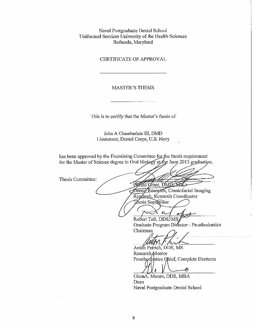
This is to certify that the Master's thesis of
John A Chamberlain III, DMD Lieutenant, Dental Corps, U.S. Navy
has been approved by the Examining Committee fo · the thesis requirement fortheMasterofSciencedegreeinOralBio ya eJune2013 gra uati n.
Thesis Connnittee:
ert Taft, DDS, MS Graduate Program Director - Prosthodontics
Chai~LL Antcf!1Petrich, DDS, MS Research Prosth d
GlennA. Munro, DDS, MBA Dean Naval Postgraduate Dental School
ii

The author hereby certifies that the use of any copyrighted material in the thesis manuscript titled:
EVALUATION OF THE DENTAL EFFECTS OF LARYNGOPHARYNGEAL REFLUX
is appropriately acknowledged and, beyond brief excerpts, is with the permission of the copyright owner.
~~~ Author Prosthodontics Graduate Program Naval Postgraduate Dental School (DATE)
NAVAL POSTGRADUATE DENTAL SCHOOL John A Chamberlain Ill, DMD
2015
This thesis may not be re-printed without the expressed written permission of the author.
iii

ABSTRACT
The association between dental erosion and gastroesophageal reflux (GERD) is
well known and documented in the literature. Laryngopharyngeal reflux (LPR) is a lesser
known clinical disorder that can mimic this destruction of hard tissues in the oral cavity.
Although these processes are related, each features differences in symptomology, clinical
findings, pathophysiology and treatment options. Because LPR typically does not
present with classic GERD symptoms such as heartburn or acid reflux, patients typically
do not seek treatment for the condition which would lead to a diagnosis. Like GERD,
LPR also presents with a number of potential medical complications, the most severe
being laryngeal carcinoma. LPR is referred to as silent reflux in the literature but
because of its association with cancer in the upper airway spaces, it is considered a
silent killer.
iv

TABLE OF CONTENTS
LIST OF FIGURES ........................................................................................................... vi
LIST OF ABBREVIATIONS ................................................................................................... vii
CHAPTER
I. INTRODUCTION .................................................................................. 1
II. ANATOMY AND FUNCTION ................................................................ 2
111. GASTROESOPHAGEAL REFLUX ........................................................... .5
IV. LARYNGOPHARYNGEAL REFLUX ....................................................... 10
V. DENTAL MANIFESTATOINS ................................................................ 14
STAGES OF EROSION ............................................................................ 17
OTHER DENTAL FINDINGS ................................................................... 19
VI. MEDICAL COMPLICATIONS ................................................................ 21
VI. DIAGNOSIS .......................................................................................... 23
VI. TREATMENT ....................................................................................... 27
VI. CONCLUSIONS .................................................................................... 33
REFERENCES ...................................................................................................................... 35
v
LIST OF FIGURES
Page
Figure 1: Comparison of GERD and LPR 13
Figure 2: Early Erosion 17
Figure 3: Intermediate Erosion 18
Figure 4: Advanced Erosion 19
Figure 5: Amalgam Island 20
Figure 6: Barrett esophagus 22
Figure 7: 48 Hour pH Testing 25
- All figures created by the thesis author with the exception of figure 6, courtesy of Janet C. Shaw MD, Department of GI/Hepatic Pathology, Joint Pathology Center.
vi

LIST OF ABBREVIATIONS
GER ....................................................... Gastroesophageal Reflux
GERD ....................................................... Gastroesophageal Reflux Disease
hCG ....................................................... Human Chorionic Gonadotropin
LES ....................................................... Lower Esophageal Sphincter
LPR ....................................................... Laryngopharyngeal Reflux
PPI ...................................................... .
sec ...................................................... .
UES
Proton Pump Inhibitor
Squamous Cell Carcinoma
Upper Esophageal Sphincter
vii

viii
INTRODUCTION
Most clinicians are familiar with the process of dental erosion and the resulting
destruction of hard tissues within the oral cavity. This phenomenon has been attributed
to a number of possible causes but is ultimately the result of intrinsic or extrinsic acids
which dissolve the hard tooth surfaces over time. One of the leading causes of erosion
in some patients can be attributed to gastroesophageal reflux disease (GERO), a clinical
disorder which is caused by chronic esophageal acid exposure. A related but lesser
known condition is laryngopharyngeal reflux (LPR), which is also known as
extraesophageal reflux disease, extra-esophageal GERO, silent erosive esophagitis, silent
GERO or silent reflux in the literature. Although these processes share certain
similarities, the etiology is different which results in different clinical presentations. LPR
is similar to GERO in that it allows endogenous stomach acids to enter the upper
airways, but does usually not present with the usual symptoms which would cause a
patient to seek treatment. As such it can go undiagnosed and result in a variety of
dental and medical complications. Of particular concern are the potential associations
with Barrett esophagus and laryngeal carcinoma.
1

ANATOMY AND FUNCTION
As a review, the esophagus is a 25 cm hollow fibromuscular tube that allows the
passage of foods and liquids from the pharynx to the stomach. Each end is bound by a
muscular ring, or sphincter, which serve to mitigate the backflow of gastric contents into
the esophagus, pharynx and upper airways. The upper end of the esophagus is bound
by the upper esophageal sphincter (UES) which measures 2-4 cm in length. This
sphincter is composed of striated or skeletal muscle but is not under conscious control;
instead it is triggered by the swallowing reflex. The primary function of the UES is to
protect the upper airway spaces from retrograde movement of stomach contents
(Wilson & Heading, 1993, pp. 357-372).
The lower end of the esophagus is bound by the lower esophageal sphincter
(LES) which measures 2.5-3.5 cm in length. Unlike the UES, the LES is composed of
smooth muscle and involves muscles of both the diaphragm and folds of the stomach.
The LES is not a true anatomical sphincter but is a physiological sphincter that is under
involuntary control by the sympathetic trunk and the vagas nerve. The primary function
of the LES is to minimize movement of stomach contents back into the esophagus but,
due to variable basal pressure, backflow of gastric contents commonly occurs after
meals. This is typically cleared by swallowing and peristalsis within the esophagus
(Johnson & DeMeester, 1974). It is important to note that the esophagus is lined with
squamous epithelium but there is an abrupt change at the gastro-esophageal junction,
to simple columnar epithelial cells with gastric glands and pits (Meyer, Austin, Brady, &
2
Castell, 1986). This squamo-columnar junction is of particular importance in patients
with acid reflux disease as it is ground zero for the development of Barrett esophagus, a ·
pre-malignant condition that is associated with esophageal adenocarcinoma (Naini,
Chak, Ali, & Odze, 2014).
As a system, the esophagus has several functions. The first is to allow the
passage for solids and liquids from the pharynx to the stomach in a synchronized
manner. The second function is clearance; in the event of blockage or regurgitation,
muscular peristalsis moves this material from the esophagus and into the stomach. The
third function is the prevention of gastroesophageal reflux due to the esophageal
sphincters which work as physical barriers to the retrograde movement of stomach
contents to the esophagus and upper airway spaces (Bartlett D. , 2006). During
deglutition or swallowing, food or liquids pass from the mouth, to the pharynx and into
the esophagus, while shunting the epiglottis. This occurs in three phases, the first of
which is the oral phase which involves the intake, chewing and lubrication of food. This
is a voluntary process and involves the muscles of facial expression, muscles of
mastication, musculature of the tongue, and parasympathetic innervation to the salivary
glands which are supplied via cranial nerves V3, VII, IX and XII. The second stage is the
pharyngeal phase and is an involuntary reflex. This begins with the closure of the
nasopharynx by the tensor palatini and levator palatini, and approximation of the
pharyngeal walls by cranial nerves V3, IX and X. Next, the oropharynx is closed by the
palatoglossus, intrinsic muscles of the tongue, and styloglossus via cranial nerves 9, 10 &
12. Finally, the larynx is closed by adduction of the vocal cords by cranial nerve X, which
3

temporarily closes the epiglottis and prevents the aspiration of food. This is concluded
with peristalsis via the pharyngeal constrictor muscles which forces the bolus through a
relaxed upper esophageal sphincter and into the esophagus. The third and final phase is
the esophageal phase and is also under involuntary neuromuscular control via the Vagas
nerve (Buthpitiya, Stroud, & Russell, 1987). This involves the process of peristalsis, the
simultaneous mesial constriction and distal relaxation which drives the bolus towards
the stomach at a rate of 2-5 cm/sin healthy patients (Richter JE, Blackwell, Nelson Ill,
Castell, & Castell, 1987). This process is concluded by relaxation of the lower
esophageal sphincter which allows the bolus to enter the stomach (Logemann, 2014).
4

GASTROESOPHAGEAL REFLUX
Because the LES is not a true or tight anatomic sphincter, retrograde movement
of the gastric contents into the distal esophagus can occur under certain conditions.
This is called gastroesophageal reflux (GER) and is based on the Latin word refluere,
which means to backflow (Kaufman, 2002 ). This is considered a normal physiologic
function and is due to transient relaxation of the lower esophageal sphincter which
exhibits variable basal pressure based on muscular input via the Vagas nerve (Dent, et
al., 1980). Gastroesophageal reflux typically occurs postprandial, or after meals, for
about an hour a day, but up to 50 GER episodes per day is considered within the normal
range (Demeester, Johnson, Joseph, Toscano, Hall, & Skinner, 1976; Postma, 2000). A
GER episode is diagnosed when esophageal pH drops below 4.0 for at least 30 seconds.
This pH is critical because cell death and mucosa! injury occurs below this point (Orr,
2005).
Fortunately, there are a number of protective mechanisms which prevent
mucosa! injury in the majority of healthy patients. These include salivary production
and bicarbonate buffering, esophageal peristalsis, and the squamous mucosa! barrier
(Orr, 2003; Pedersen, Bardow, Jensen, & Nauntofte, 2002). Because the squamous
epithelium in the esophagus lacks the protective lining as that found in the stomach,
chronic acid reflux can result in mucosa I damage and sensory nerve stimulation of the
esophageal tissues. This is likely the result of direct contact with refluxed acid which
liberates cytokines that stimulate epithelial sensory nerve endings (Chandrasoma,
5

2010). The end result is usually symptoms of esophagitis including heartburn and acid
regurgitation which are diagnostic for a clinical disorder called gastroesophageal reflux
disease {GERD). Although GERO can exhibit extra-esophageal symptoms, the presence
of heartburn and/ or acid regurgitation at least once a week for the last 3 months are
considered essential in diagnosis of the condition {Choi, Jung, Song, Shim, & Jung, 2013).
Other typical symptoms may include a sour taste due to low pH levels of the stomach
acids, as well as burping and/or belching. Patients may also report atypical symptoms
which are largely extraesophageal due to mesial migration of endogenous stomach
acids. These may include chest pain, sinusitis, globus, chronic cough, asthma-like
symptoms, dysphagia, odoynophagia, water brash, coarse or hoarse voice, as well as
nausea and/or vomiting (Rabine & Nostrant, 2009; Fass, Achem, Harding, Mittal, &
Quigley, 2004; Heidelbaugh, Gill, Van Harrison, & Nostrant, 2008).
GERO can occur at any time during the day but is typically exacerbated when in a
supine position. In fact, it is estimated that up to 79% of patients exhibit nighttime
GERO symptoms {Shaker, Castell, Schoenfeld, & Spechler, 2003). Salivary flow follows a
daily circadian rhythm but is greatest during waking hours, with an average of 25 to 60
swallows per hour. During sleep, the swallow reflex exhibits a fraction of that amount
and may be as little as 2 to 9 times for the entire night (Thie, Kato, Bader, Montplaisir, &
Lavigne, 2002 ). Because salivary flow rate is reduced during sleep, esophageal
clearance and salivary buffering are also diminished, allowing the proximal migration of
gastric contents. As such, patients will often wake with typical, as well as
extraesophageal, symptoms (Ranjitkar S, 2012). Furthermore, many GERO patients
6
exhibit esophageal dysmotility and prolonged clearance rates which serve to exacerbate
the condition (Postma, Tomek, Belafsky, & Kaufman, 2001).
As for the pathophysiology of GERD, there are a number of possible causes, but
the most common reasons are insufficient function of the LES which allows stomach
contents to enter the esophagus, and impaired motility which results in prolonged
esophageal clearance of these substances (Kaufman, 1991; Postma, Tomek, Belafsky, &
Kaufman, 2001). These gastric contents may include liquid or gaseous acid vapors, as
well as pepsin, a proteolytic enzyme produced by chief cells in the stomach that is
responsible for initial protein catabolism (Rabine & Nostrant, 2009; Dunn, 2002).
Esophageal injury is likely the result of the synergistic effects of pepsin and the acid
which activates it. In fact, studies have shown that esophageal damage is proportional
to the amount of pepsin present and that acid at a pH of 2.3 was ineffective without the
enzyme. (Goldberg, Dodds, Gee, Montgomery, & Zboralske, 1969). At higher
concentrations, hydrogen ions are highly corrosive and damaging to the apical surface of
the squamous epithelium. The overall mechanism of damage is penetration of the
epithelium by acids at low pH, then proteolysis of type IV collagen by pepsin which
disrupts the basement membrane of the squamous epithelial cells (Kresina & Miller,
1979; Ingber & Folkman, 1989; Bardhan, Strugala, & Dettmar, 2012).
Epidemiology studies indicate that GERD shows increasing prevalence
worldwide, especially in Western countries. In North America, the prevalence of
symptomatic GERD is estimated at 18-28% of the population. In South America this
7

estimate is 23%, and in Europe it ranges from 9-26%. In comparison, Asian countries
show a much lower prevalence with 2.5%-8% in East Asia, and 9%-33% in the Middle
East (El-Serag, 2007; El-Serag, Sweet, Winchester, & Dent, 2014). In terms of cost of
direct health care, GERD had the highest annual direct cost among digestive diseases in
the late 1990s with an annual direct cost of $9.3 billion dollars (Sandler, et al., 2002). By
2004, this cost had escalated with an estimated direct cost of $12.1 billion (Rubenstein
& Chen, 2014). Although there are a number of theories that attempt to explain this
increase, the literature is inconclusive. Changes in diet or lifestyle are often attributed
to an increase in GERD but the level of evidence is low. In contrast, there is a definite
relationship between GERD symptoms and obesity with associated increased body mass
index, waist circumference and weight gain (Corley & Kubo, 2006; Hampel, Abraham, &
El-Serag, 2005). This finding correlates to the number of Americans who are overweight
or obese which has tripled over the last two decades (Mokdad, et al., 2001; Ogden,
Carroll, Curtin, McDowell, Tabak, & Flegal, 2006; Sturm, 2002).
There are a number of generally accepted risk factors for reflux, the first are
conditions that result in incompetence of the lower esophageal sphincter. These can
include alcohol, nicotine, caffeine, or hiatal hernia, the protrusion (or herniation) of the
upper part of the stomach into the thorax through a tear or weakness in the diaphragm.
In addition, certain medications such as calcium channel blockers or anticholinergic
agents have been shown to cause LES incompetence (Allen M., Mellow, Robinson, &
Orr, 1987). Second are conditions which increase gastric volume such as eating a large
meal or intestinal blockage which result in slowed gastrointestinal motility. A common
8

misconception is that the speed of eating results in greater risk of reflux has been shown
to have no scientific basis (Valitova, Bayrak~1, & Bor, 2013). Third include foods which
act as potent refluxogenic agents such as raw onions or spicy meals, or meals that delay
gastric emptying such as foods high in fats (Allen M., Mellow, Robinson, & Orr, The
American Journal of Gastroenterology; Mushref & Srinivasan, 2013). Fourth are
conditions which increase intra-abdominal pressure such as obesity, pregnancy or
straining during weight lifting (Dodds, Dent, & Hogan, 1982). Finally, there are a
number of other conditions that may have a role in the development of reflux and
include sex, age, buffering capacity of the saliva, and infection of Helicobocter pylori
(Sonnenberg & El-Serag, 1999; Nozu & Komiyama, 2008; Wang, Tu, Chuang, Yu, Cheng,
& Hsu, 2010; Cho, et al., 2011).
9
LARYNGOPHARYNGEAL REFLUX
Laryngopharyngeal reflux (LPR) is similar to GERO but is a separate clinical
disorder that goes by a number of names in the literature including extraesophageal
reflux disease, non-acidic gastric reflux, extra-esophageal GERO, silent erosive
esophagitis, silent GERO or silent reflux. Like GERD, LPR involves the reflux of
endogenous stomach acids and into the esophagus through the normal physiologic
process of gastroesophageal reflux through the lower esophageal sphincter. But unlike
GERO, this typically extends into the upper airway spaces including the larynx, pharynx
and oral cavity (Koufman, 2002 ). The result is a number of differences in
symptomology, clinical findings, pathophysiology and treatment options. While
heartburn and acid reflux are often considered the sine qua non of GERO, these
symptoms. are often absent in patients with LPR. In fact, literature suggests that
heartburn is present in 6-40% of patients with LPR (Koufman, Belafsky, Bach, Daniel, &
Postma, 2002; Ossakow, Elta, Colturi, Bogdasarian, & Nostrant, 1987).
This difference is likely attributed to the fact that LPR patients typically exhibit
reflux symptoms during the day, when salivary flow and esophageal clearance are at
their peak (Belafsky & Postma, 2003). These patients usually exhibit both normal
esophageal motility and salivary buffering capacity so vagal nerve stimulation which can
result in heartburn or esophagitis is typically not seen. For this reason, patients with
LPR often do not seek treatment and can go undiagnosed for the condition (Postma,
Tomek, Belafsky, & Koufman, 2001). As for reported symptoms seen in LPR patients,
10
these are mainly extraesophageal and are found in the head and neck regions. The
most common include dysphonia, chronic postnasal drip, sinusitis, sore throat with pain
and/or dysphagia, hoarseness, globus pharyngeus, chronic cough, Eustachian tube
dysfunction, and asthma-like symptoms with shortness of breath (Belafsky, Postma,
Amin, & Koufman, 2002). Clinical findings are another key difference between GERD
and LPR. While the majority of patients with GERD exhibit esophagitis, only 12-18% of
patients with LPR show endoscopic evidence. Of these, another 7% exhibited signs of
Barrett esophagus, while the remaining 81% presented with normal esophageal
epithelial tissues upon biopsy (Koufman, 1991; Koufman, Belafsky, Bach, Daniel, &
Postma, 2002 ).
The pathophysiology of LPR is also different than GERO and is typically attributed
to a defect or dysfunction of the upper esophageal sphincter which allows gastric
contents to enter the upper airways (Ocak, Kubat, & Yorulmaz, 2015 ). While the
esophagus features a number of protective mechanisms which prevent injury to the
mucosa, the laryngeal and pharyngeal structures do not and are more susceptible to
damage from reflux of stomach contents {Ossakow, Elta, Colturi, Bogdasarian, &
Nostrant, 1987). Esophageal mucosa exhibits a critical pH of 4.0, the point at which cell
injury occurs. In comparison, damage to laryngeal epithelium occurs at a pH of 5.0
which makes it ten times more susceptible to acid attacks (Axford, et al., 2001;
Johnston, et al., 2003; Lillemoe, Johnson, & Harmon, 1982). Moreover, laryngeal
epithelium is up to 100 times more susceptible to pepsin damage than esophageal
11
tissues, and pepsin is thought to play a key role in the pathogenesis of
laryngopharyngeal reflux (Kaufman, 2002 ; Samuels & Johnston, 2009).
While pepsin exhibits maximum activity at a pH of 2.0, it is inactivated at a pH of
6.5 and stable until a pH of 8.0 at which point it becomes denatured. In comparison, the
laryngopharyngeal tissues exhibit a pH of 6.4 and, following a reflux event, pepsin in
these areas becomes enzymatically inactivated. Subsequent reflux episodes, however,
can result in reactivation due to a decrease in pH. This is thought to result in laryngeal
cell damage due to proteolytic activity which disrupts cellular cohesion (Johnston,
Dettmar, Bishwokarma, Lively, & Kaufman, 2007). Over time, pepsin is internalized by
laryngeal epithelial cells in a process called receptor-mediated endocytosis and stored
within the cell for up to 24 hours. This mechanism may explain why many patients with
laryngopharyngeal reflux are not as responsive to acid suppression therapy as those
with gastroesophageal reflux disease (Johnston, Wells, Blumin, Toohill, & Merati, 2007).
Although LPR and GERD exhibit differences in symptomology, clinical findings and
pathophysiology, they do share a number of risk factors as discussed previously. Table 1
outlines the primary differences between gastroesophageal reflux and
laryngopharyngeal reflux.
12

GERD LPR
Heartburn and/or acid reflux Yes (diagnostic) Uncommon
Etiology LES UES
Esophageal Motility Impaired Normal
Pattern Anytime Daytime
Critical pH 4.0 5.0
Damage Esophageal Extraesophageal
Table 1: Comparison ofGERD and LPR
13
DENTAL MANIFESTATIONS
As a review, there are four proposed types of tooth wear which can be classified
into three causal categories. The first is mechanical loss and includes both attrition and
abrasion. The second is chemical loss which includes erosion, the chemical dissolution
of tooth material without bacterial involvement (Pindborg, 1970). The third is
biomechanical loss which includes the theory of abfraction to explain non-carious
cervical lesions (Grippo, 1991). Each mechanism presents with unique clinical
characteristics but literature suggests that tooth wear is multifactorial. Although
independent in origin, erosion typically works synergistically with abrasion and attrition
as it enhances physical wear (Curtis, Jayanetti, Chu, & Staninec, 2011). In fact, current
literature disputes the theory ofabfraction and attributes the presence to non-carious
cervical lesions to a combination of erosion and toothbrush abrasion (Bartlett & Shah,
2006). Lab studies have shown that toothbrush abrasion has been shown to result in
50% greater loss of tooth structure during acid exposure than brushing alone
(Eisenburger, Shellis, & Addy, 2003). Because erosion is a multifactorial process, it can
be difficult for the clinician to recognize. As a consequence, it is often misdiagnosed and
can result in restorative treatment which can compromise the vitality of the tooth
(Cardoso, Canabarro, & Myers, 2000).
As for the etiology of erosion, there are a variety of exogenous and endogenous
sources that must be ruled out when treating a potential patient for erosion. Extrinsic
erosion is the result of exogenous acids found in the environment, most commonly
14

acidic foods, beverages and snacks. This can include behavioral causes such as citrus
sucking, fruit mulling, soda swishing, and eating pickled foods {S~vik, Skudutyte-Rysstad,
Tveit, Sandvik, & Mulic, 2015). Environmental causes are numerous and can include
industries that result in high acid vapor levels such as the manufacture of textiles,
batteries, fertilizers, rubber, or drugs such as methamphetamines {Cate Bruggen, 1968}.
Professional swimmers are also at risk due to highly chlorinated swimming pools, which,
when combined with water, forms hydrochloric acid (Geurtsen, 2000; Centerwall,
Armstrong, Funkhouser, & Elzay, 1986}. Intrinsic erosion is the result of endogenous
stomach acids and is typically caused by vomiting, regurgitation, reflux or rumination
{Mahoney & Kilpatrick, 2003}.
Dental erosion is considered a normal physiological process that occurs
throughout life and is considered the most common cause of tooth wear in the Western
world. When this process results in excessive and rapid loss of tissues, however, this is
considered a pathological condition {Paryag & Rafeek, 2014}. The literature shows a
strong correlation between GERD and dental erosion, with a median prevalence of 24%
in a large range of age groups. When viewed by age demographics, adults exhibited a
range of 21-83%, while children demonstrated 14-87% {Pace, Pallotta, Tonini, Vakil, &
Bianchi Porro, 2008}. Although tooth erosion due to reflux was first reported in 1933,
there are still barriers to successful management (Bodecker, 1933}. Because it is
typically a slowly-progressing phenomenon, many patients are unaware of the
destruction and typically do not present with a chief complaint of "erosion". Moreover,
there is a general lack of emphasis in dental curricula or continuing education programs
15

and the average dentist may be unaware of strategies for early diagnosis and treatment.
Finally, there is a lack of incentive for the clinician who is paid by the insurance
companies for restorative procedures, and not for conservative management (Curtis,
Jayanetti, Chu, & Staninec, 2011).
When viewed at the surface of the tooth, acid advancement first strips the
protective dental pellicle and then displaces the saliva which has a lower surface tension
and contact angle (Ranjitkar S, 2012). The pH of gastric acid can vary between 1.5 and
3.5, but is still much lower than the critical pH of hydroxyapatite and fluoroapatite
which are generally accepted at 5.5 and 4.5, respectively (Kutsch, Chaiyabutr, & Milicich,
2013) (Jones, Lekkas, Hunt, Mcintyre, & Rafir, 2002). This results in demineralization and
the loss of calcium and phosphate ions from the mineral surface. This is an irreversible
process and over time, forms dished-out lesions on the enamel surface (Ranjitkar S,
2012). Continued acid exposure not only results in clinically visible defects, but results
in a significant reduction in microhardness which makes the softened surface more
prone to mechanical wear from abrasion or attrition (Maupome, Diez-de-Bonilla, Torres
Villasenor, Andrade-Delgado, & Castano, 1998). While some sources suggest that acid
reflux promotes the growth of Streptococcus mutans and results in an increase in dental
caries, others suggest a lack of a relationship between them (Munoz, et al., 2003;
Schroeder, 1995).
16

Stages of Erosion
Clinical signs of early or mild erosion begin with an initial loss of tooth luster that
is soon followed with a smooth glazed appearance as the enamel is lost {Eccles &
Jenkins, 1974). Eventually, morphologic features tend to disappear including
developmental ridges, flattening of convex surfaces, and a change in color may be noted
due to the dentin showing through the thin, translucent enamel shell. The end result is
a melted appearance with slightly concave defects visible on some surfaces as seen in
image 1 (Lussi, Dental erosion clinical diagnosis and case history taking, 1996).
Figure 1: Early Erosion
Clinical signs of intermediate erosion include more exaggerated rounding of the
cusp and groove morphology, concavities or cupping on the occlusal or incisal surfaces,
and loss of surface enamel with exposed dentin surfaces and tooth sensitivity as shown
in figure 2. Continued erosion results in the formation of reactionary and reparative
17

dentin which obturate the dentinal tubules and result in discoloration and sclerotic
dentin (Ganss, Lussi, & Schlueter, 2014).
Figure 2: Intermediate Erosion
Clinical signs of advanced or severe erosion include gross destruction of the hard
tissues which results in compromised esthetics and function, and a possible loss of
occlusal vertical dimension. Furthermore, if erosive wear exceeds the reparative
capacity of the pulpal tissues, frank, or near, pulp exposure can occur. This may result in
endodontic complications such as sensitivity, pain, pulpal inflammation and necrosis
(Jaeggi & Lussi, 2006). In fact, endodontic sequelae are estimated to occur in 10% of
patients with significant erosive wear (Sivasithamparam, Harbrow, Vinczer, & Young,
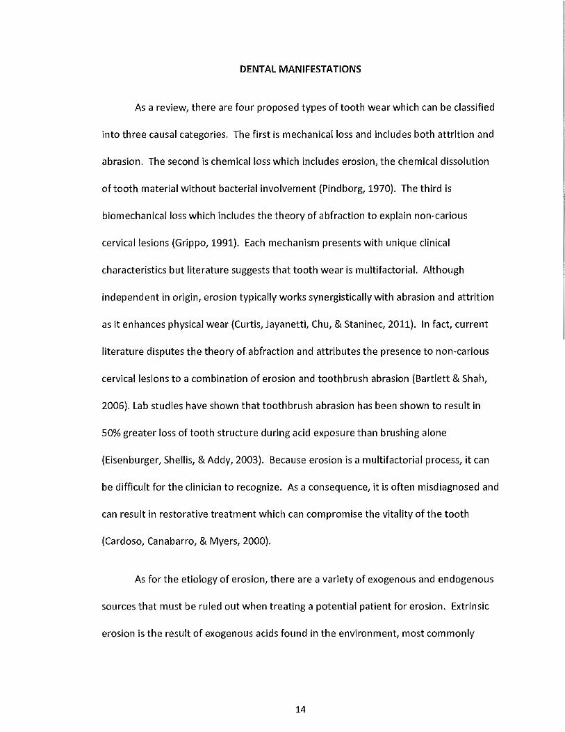
2003). Figure 3 demonstrates evidence of root canal therapy on the remaining maxillary
dentition in a patient that presented with undiagnosed reflux disease.
18
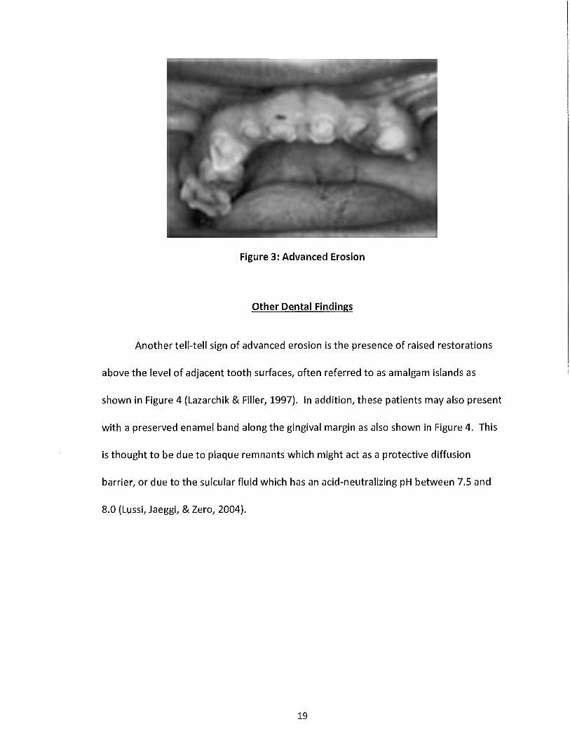
Figure 3: Advanced Erosion
Other Dental Findings
Another tell-tell sign of advanced erosion is the presence of raised restorations
above the level of adjacent tooth surfaces, often referred to as amalgam islands as
shown in Figure 4 (Lazarchik & Filler, 1997). In addition, these patients may also present
with a preserved enamel band along the gingival margin as also shown in Figure 4. This
is thought to be due to plaque remnants which might act as a protective diffusion
barrier, or due to the sulcular fluid which has an acid-neutralizing pH between 7.5 and
8.0 (Lussi, Jaeggi, & Zero, 2004).
19

Figure 4: Amalgam Island
Furthermore, oral mucosal lesions may result from acid and pepsin exposure and
can result in dry oral mucosa with a keratotic appearance of the gingival tissues (Rabine
& Nostrant, 2009). Histologic studies of palatal mucosa in reflux patients has found a
greater prevalence of epithelial atrophy in reflux patients as compared to control
subjects. Other findings include deepened epithelial crests in the connective tissues and
a high incidence of fibroblasts (Silva, Dam ante, Stipp, Tolentino, Carlotto, & Fleury,
2001). Furthermore, there is some evidence that burning mouth syndrome might be
associated with reflux (Gurvits & Tan, 2013). Like the laryngeal mucosa, damage to the
oral mucosa may be primarily mediated by pepsin and dependent on pH within the oral
cavity (Ocak, Kubat, & Yorulmaz, 2015; Jiang, et al., 2011).
20

MEDICAL COMPLICATIONS
In addition to dental manifestations, there are a number of potential medical
complications in patients with reflux disease which can include otitis media, constriction
of the larynx with scarring of the vocal cords, erosive esophagitis with narrowing as the
result of scar formation and the development of peptic ulcers (Bardhan, Strugala, &
Dettmar, 2012}. In addition, patients with LPR may exhibit persistent laryngopharyngitis
and recurrent pneumonia due to the aspiration of gastric contents (Fass & Dickman,
2006}. A particular concern of patients with reflux is the development of Barrett
esophagus which occurs in the distal esophagus as shown in Figure 5. This is thought to '
be the result of the body's reaction to chronic exposure of gastric contents and results
in metaplasia of the normal stratified squamous epithelium to simple columnar
epithelium with goblet cells as found in the intestinal tract. This is characterized by
aneuploidy or an abnormal number of chromosomes and is considered a pre-malignant
condition as it is the strongest risk factor for esophageal adenocarcinoma (Prasad,
Bansal, Sharma, & Wang, 2010; Rustgi & Sun, 2009}. Furthermore, up to 25% of patients
with Barrett esophagus and 40% of all esophageal adenocarcinomas occur in patients
with or without minimal previous reflux symptoms which gives cause for alarm in
patients with silent reflux (Gerson, Shetler, & Triadafilopoulos, 2002; Lagergren,
Bergstrom, Lindgren, & Nyren, 1999}.
21

Figure 5: Barrett esophagus exhibiting simple columnar epithelium with goblet cells
More serious, but less frequent, findings in patients with LPR include vocal cord
nodules, posterior laryngitis, laryngospasms, subglottic stenosis, arytenoid fixation, and
laryngeal cancer (Metz, Childs, Ruiz, & Weinstein, 1997; Little, Koufman, Kohut, &
Marshall, 1985). Although the relationship between LPR and laryngeal squamous cell
carcinoma {SCC} has never been proven, pepsin is thought to be a significant risk factor
in the development of cancer (Ward & Hanson, 1988). Studies show that pepsin
promotes the proliferation of epithelial cell cultures with associated gene expression
changes that result in neoplasia (Johnston, Yan, Hoekzema, Stoner, Blum in, & Bock,
2012}.
22

DIAGNOSIS
Although the clinician may suspect erosion due to reflux, the actual diagnosis
must be done by the patient's physician based on the medical history and exam. A
presumptive diagnosis of GERD is often made by symptomology alone, or by placing the
patient on antacids or a proton pump inhibitor (PPI} test, where the patient is
prescribed a proton pump inhibitor for a short period of time to see if it resolves gastric
symptoms (Sweis, Fox, Anggiansah, & Wong, 2011}. In the majority of patients, this is
considered sufficient for diagnosis of GERD but if the diagnosis is uncertain or the
patient presents with atypical symptoms such as those seen in LPR, the physician may
refer to a gastroenterologist for more invasive tests. These may include endoscopy,
barium swallow or pH monitoring to evaluate function and presentation. Endoscopy is
useful in the detection of erosive esophagitis, as well as the presence of Barrett
esophagus or hiatal hernia. It is often used to sample for the presence of Helicobacter
pylori (Wenner, Johnsson, Johansson, & Oberg, 2007}. When a structural abnormality
such as stricture, hiatal hernia, ulceration or erosion is suspected, a barium esophagram,
or upper GI series, is often useful in diagnosis. Although this test has proven practical in
patients that exhibit a high level of reflux, it demonstrates insufficient sensitivity and
specificity to reveal mild irritation as seen in patients with abnormal degrees of acid
reflux such as that seen in LPR (Johnston, Troshinsky, Castell, & Castell, 1996}.
As for clinical diagnostic tests, impedance pH testing is considered the gold
standard and is very useful in determination of daily reflux levels and patterns in
23
patients with reflux disease (Spencer, 1969). Historically, this has been done with a
transnasal catheter which involves surgical placement of a small catheter through the
nose and embedding it into the distal esophagus. Once in place, a sensor detects acid
pH levels and sends this information to a computer which records the findings over a 24
hour period. Due to the wired catheter, this procedure is inconvenient to the patient
and limits daily activities. More recently, wireless pH probes have gained in use and are
typically implanted in the distal esophagus and transmit information to a small receiver
via radiotelemetry. This not only minimizes disruption to the patient's daily activities,
but offers greater sensitivity and specificity than traditional transnasal pH testing (Sweis,
Fox, Anggiansah, & Wong, 2011).
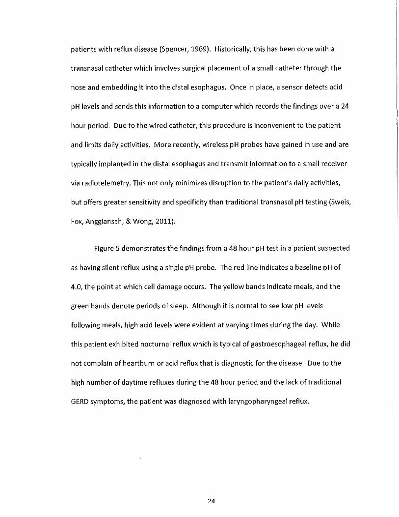
Figure 5 demonstrates the findings from a 48 hour pH test in a patient suspected
as having silent reflux using a single pH probe. The red line indicates a baseline pH of
4.0, the point at which cell damage occurs. The yellow bands indicate meals, and the
green bands denote periods of sleep. Although it is normal to see low pH levels
following meals, high acid levels were evident at varying times during the day. While
this patient exhibited nocturnal reflux which is typical of gastroesophageal reflux, he did
not complain of heartburn or acid reflux that is diagnostic for the disease. Due to the
high number of daytime refluxes during the 48 hour period and the lack of traditional
GERD symptoms, the patient was diagnosed with laryngopharyngeal reflux.
24

'pt((aplvtt:M~ndJY,..fun• 10.~U 1M:4tPl-t •W1dt1U•hy.Junt l:l.~J3i 1M:4;P_M ti-ur~tt~n: 4t hO mlnO S-
...
~\iO grrz~~~-~--~_-~,.,~R
Ut1' F.JIDn".:JI)
t.Jf;I f.Jl{V<"ilD'
Svf•-l fil(V~(ID
V<> f.JI lr.l'OlJ
Uni E/11/iilll
llf:o! f_jjff;-f,113
f.•_:pt-·j Uj l!;-1)1)
1-'ol fili'll[ll'.I
•=f'f =y-tqt I ! I
""" l'Jl(.1•11)13
l1 [") f'JJ(\o;:\)l:l
21 f1(1 r~llr((ll:)
l_j.-'j lH) (Jllf~•)IJ
n:<:.i f~llf.:1)\1
16 :>O Ullf.?iHl
:i'lfl\l ~)jl'l~\)l:l
t·S OD r»1tao1:J
Figure 5: 48 Hour pH Testing
l:O(Q
I~;:)
(iLY.J
\l!-.'.:•)
!)~•)
17(>'/ Qt~;)
(.-'J~)
In comparison to GERD, diagnosis of LPR is more challenging due to the presence
of atypical symptoms which typically do not respond to empirical therapy with proton
pump inhibitors. In fact, less than half of patients with LPR experience full resolution of
symptoms in a four month period (Belafsky, Postma, & Kaufman, 2001). While single-
probe pH testing is often used in the diagnosis of LPR, studies have shown it lacks the
sensitivity needed for an accurate diagnosis. In one study of conventional ambulatory
24 hour pH monitoring, false negative results were reported in 15-30% of patients with
LPR (Ocak, Kubat, & Yorulmaz, 2015 ). The current gold standard in the diagnosis of LPR
is 24-hour double probe pH monitoring which was first used to document
extraesophageal reflux in 1986 (Kaufman, 1991). Unlike a single-probe test, this method
utilizes two pH probes, one at the proximal end of the esophagus, and the other at the
distal end (Postma, Belafsky, Aviv, & Kaufman, 2002).
25

Although endoscopy, pH monitoring and barium swallow radiographs are highly
diagnostic, each involves a certain amount of risk to the patient, from surgical
complications to radiation exposure. More importantly, none of these tests correlate
oral findings such as erosion to the reflux process so are of limited diagnostic use to the
dentist. In the past, litmus paper has been used as a minimally invasive method to
check for the presence of acid in the oral cavity. Although this is a simple test which
lacks strong sensitivity or specificity, it could indicate the need for further evaluation of
potential reflux disorders (James & Ewer, 1999). More recently, immunoserologic
pepsin detection tests have gained in popularity as a cost effective and minimally
invasive diagnostic method. These tests are similar to an over-the-counter pregnancy
test, but instead of testing for human chorionic gonadotropin (hCG), these tests
measure the presence of pepsin in the saliva. The results can typically be read within 10
minutes and exhibit high sensitivity and specificity, making this a potentially useful
diagnostic tool in patients that don't present with classic GERO symptoms such as LPR
(Ocak, Kubat, & Yorulmaz, 2015; Knight, Lively, Johnston, Dettmar, & Kaufman, 2005).
26

TREATMENT
Treatment of GERD typically begins with dietary changes that restrict spicy or
acidic foods, and beverages such as alcohol, caffeine, chocolate and carbonated drinks.
Although diet has been shown to precipitate acid secretion in some patients, there is
little supporting evidence that dietary intake results in clinical manifestations of GERD
(Festi, et al., 2009). Regardless, this first-line treatment usually includes portion control
and moderation of even mildly acidic foods such as fatty meats and dairy products. In
addition, a well-balanced diet with neutral-to-alkaline foods which neutralize stomach
acid and inactivate pepsin is often recommended to the patient. For patients that
exhibit nighttime reflux, eating meals 2-3 hours before bedtime and walking
immediately after meals may help with symptoms (Kahrilas, et al., 2008).
Suggested lifestyle modifications for obese patients typically include an
enhanced exercise regimen in order to reduce body weight and intra-abdominal
pressure. While moderate exercise is shown to be efficacious, strenuous exercise may
induce GERD symptoms (Pandolfino, Bianchi, Lee, Hirano, & Kahrilas, 2004; Ravi, Stuart,
Byrne, & Reynolds, 2005). Another common recommendation to patients with
nocturnal reflux is to elevate the head of the bed or to support the head using pillows.
While this may work for some patients, there is a low level of evidence to support it
(Hamilton, Boisen, Yamamoto, Wagner, & Reichelderfer, 1988). In comparison,
literature shows a definite relationship between increased body mass index and GERD
symptoms {Corley & Kubo, 2006; Hampel, Abraham, & El-Se rag, 2005). Of the many
27
dietary and lifestyle recommendations made for patients with GERD, only weight loss
shows a moderate level of evidence in the literature {Kaltenbach, Crockett, & Gerson,
2006).
As for pharmacologic therapy, H2 receptor blockers and proton pump inhibitors
are typically used to treat reflux disease by inhibiting acid secretion. Of the two, PP ls
are the most efficacious and exhibit the highest level of evidence for use in treatment of
GERD symptoms {Katz, Gerson, & Vela, 2013). GERD is often diagnosed by placing the
patient on antacids or a proton pump inhibitor (PPI) test, where the patient is
prescribed a proton pump inhibitor for a short period of time to see if it resolves gastric
symptoms {Sweis, Fox, Anggiansah, & Wong, 2011). There are two major treatment
options and the first is called a step-up approach. This begins with an over the counter
H2 receptor blocker and if the condition fails to improve, therapy is stepped up to a
more powerful proton-pump inhibitor. The other model is called a step-down approach
and is opposite as it begins with the more potent drug. When the patient has been
symptom free for a period of time, they are stepped down to a less powerful
medication. For patients with moderate-to-severe reflux disease, many physicians favor
the second approach (lnadomi, et al., 2001; Sonnenberg, lnadomi, & Becker, 1999). In
patients with severe reflux, surgical correction is sometimes performed when medical
therapy has failed. There are a number of procedures which are used but the most
severe is the Nissen or complete fundoplication. In this method, the upper part of the
stomach is wrapped, or plicated, around the lower end of the esophagus and sutured in
place. This serves to reinforce the closing function of the lower esophageal sphincter
28

(Abbas, et al., 2004). Although this is considered a safe and effective procedure, it can
result in side effects such as gas and bloating (Waring, 1999).
In comparison to GERD, treatment of LPR is much more difficult as empirical
therapy with proton pump inhibitors is not as effective. In those patients, dosage is
often inadequate, and/or the duration of treatment is insufficient (Kaufman, Aviv,
Casiano, & Shaw, 2002). Despite this fact, PP ls are considered superior to H2 receptor
blockers in patients with moderate to severe LPR. In fact, histamine receptor
antagonists provide benefit to only about 50% of the LPR patients that respond to
treatment (Scott & Gelhot, 1999; Klinkenberg-Knol & Meuwissen, 1989; Smith, Gavey,
Nwokolo, & Pounder, 1990; Wilder-Smith, Ernst, Gennoni, Zeyen, Halter, & Merki,
1990). Regardless, LPR symptoms resolve slowly and fewer than half of patients treated
with PPls exhibit full recovery after 4 months of treatment (Belafsky, Postma, &
Kaufman, 2001). LPR can be classified into three groups; minor, major and life
threatening as based on symptomology and findings (Postma, Johnson, & Kaufman,
2002). Treatment options vary from conservative to aggressive, depending on the·
classification. According to the literature, diet and lifestyle modifications show limited
efficacy with a few exceptions. In one study, it was found that chewing sugarless gum
consistently increases esophageal and pharyngeal pH, making it a non-invasive
adjunctive therapy to laryngopharyngeal reflux (Smoak & Kaufman, 2001). Although the
level of evidence is low for changes in diet when treating GERD, a strict low-acid diet has
been shown to have therapeutic benefits in the treatment of PPl-resistant LPR. Because
29

pepsin is active up to a pH of 6.5, a virtually acid free diet demonstrates 95%
improvement of symptoms in test patients (Kaufman, 2011).
In regards to dental management, tooth wear is multifactorial and patients
exhibiting erosion must be correctly assessed along with the actual etiology of wear
(Litonjua, Andreana, Bush, & Cohen, 2003). This is typically followed by dietary
counseling and/or referral to the patient's physician or to a gastroenterologist for
further evaluation. Patient education should include oral hygiene instructions and may
include the recommendation not to brush immediately after eating acidic foods, or after
a reflux episode as the tooth is more susceptible to abrasion (Eisen burger, Shellis, &
Addy, 2003). Before considering dental treatment, it is important that the patient be
correctly managed by his or her physician prior to beginning any restorative work. Thus,
the medical condition should be treated before oral rehabilitation. Barrier
considerations should include the disuse of abrasive toothpastes and some sort of tray
or night guard (Curtis, Jayanetti, Chu, & Staninec, 2011). Fluoride products are also
indicated to facilitate remineralization, and products such as gum containing calcium
phosphate serve to enhance mineral precipitation of eroded enamel (Prestes, Souza,
Comar, Salomao, Rios, & Magalhaes, 2013).
Materials selection is also important when considering restorations in a patient
with erosion from reflux disease. Some authors advocate composite resin as the gold
standard in cases of mild erosion due to its retention and enamel margin sealing
(Broliato, et al., 2008). Unfortunately, in cases with moderate-to-severe erosive tooth
30
loss, the presence of sclerotic dentin results in lower bond strengths (Tay & Pashley,
2004). In these cases, full coverage restorations are often indicated to restore lost tooth
structure and function which may require adjunctive treatment such as root canal
therapy and/or crown lengthening (Curtis, Jayanetti, Chu, & Staninec, 2011). When
choosing a luting agent, glass ionomer cements are often chosen due to their adhesive
capacity and ease of use. Unfortunately, they exhibit erosion rates that are only slightly
lower than enamel or dentin (Eisenburger, Addy, & Rossbach, 2003). A similar
comparison between resin modified glass ionomer, polyacid-modified resin composite
(compomer) and resin composite found that resin composite cements exhibit the least
amount of leakage and may be preferable due to their inherent low solubility over time
(Yap, Lim, & Neo, 1995).
Placement of preparation finish lines is yet another consideration when using full
coverage restorations. In the past, anecdotal reports advocated the placement of
subgingival finish lines in an effort to minimize further erosive wear. The most prevalent
theory is that the sulcular fluid has a pH between 7.5 and 8.0 and works to neutralize
acids, although there is nothing in the literature to support this practice. While
subgingival placement of finish lines may mitigate further erosion, this can result in
periodontal issues as well as promote the incidence of new caries (Charbeneau G, 1975;
Silness, 1970; Newcomb, 1974). In one of the classic articles on margin placement, it
was determined that subgingival margins should be generally be avoided except for
certain situations such as esthetic demands, removal of caries, subgingival tooth
fracture, coverage of an existing subgingival restoration, to gain coronal crown length,
31

or to provide for a more favorable restoration contour (Becker & Kaldahl, 2005}.
Regardless of philosophy, the best treatment for the dentist is to ensure the patient's
reflux is controlled with the appropriate referrals and medication to address the disease in the
first place.
32

CONCLUSIONS
In conclusion, laryngopharyngeal reflux (LPR) is similar to gastroesophageal
reflux disease (GERD) in that it allows endogenous stomach acids to enter the upper
airways, but does usually not present with the usual symptoms which would cause a
patient to seek treatment. As such it can go undiagnosed and result in a variety of dental
and medical complications. Because of the association with Barrett's esophagus,
esophageal and laryngeal carcinoma, this disease has the potential to be considered a
silent killer.
33

REFERENCES
Abbas, A., Deschamps, C., Cassivi, S., Allen, M., Nichols, F., Miller, D., et al. (2004). The role of
laparoscopic fundoplication in Barrett's esophagus. Annals of Thoracic Surgery, 77(2),
393-396.
Allen, M., Mellow, M., Robinson, M., & Orr, W. (1987). Comparison of calcium channel blocking
agents and an anticholinergic agent on oesophageal function. Alimentary Pharmacology
& Therapeutics, 1(2), 1S3-159.
Allen, M., Mellow, M., Robinson, M., & Orr, W. (The American Journal of Gastroenterology). The
effect of raw onions on acid reflux and reflux symptoms. 85(4), 377-380.
Axford, S., Sharp, N., Ross, P., Pearson, J., Dettmar, P., Panelli, M., et al. (2001). Cell biology of
laryngeal epithelial defenses in health and disease: preliminary studies. The Annals of
Otology, Rhinology, and Laryngology, 110(12), 1099-1108.
Bardhan, K., Strugala, V., & Dettmar, P. (2012). Reflux Revisited: Advancing the Role of Pepsin.
The International Journal of Otolaryngology, 1-13.
Bartlett, D. (2006). Intrinsic Causes of Erosion. Jn Bartlett, & A. Lussi (Ed.), Dental Erosion From
Diagnosis to Therapy (Vol. 20, pp. 119-139). Basel, Switzerland: Karger.
Bartlett, D., & Shah, P. (2006). A critical review of non-carious cervical (wear) lesions and the
role of abfraction, erosion, and abrasion. Journal of Dental Research, 85(4), 306-312.
Bauman, N., Sandler, A., Schmidt, C., & Maher, J. (1994). Reflex laryngospasm induced by
stimulation of distal esophageal afferents. The Laryngoscope, 104, 209-214.
Belafsky, P., & Postma, G. (2003). The laryngeal and esophageal manifestations of Sjogren's
syndrome. Current Rheumatology Reports, 5(4), 297-303.
Belafsky, P., Postma, G., & Koufman, J. (2001). Laryngopharyngeal reflux symptoms improve
before changes in physical findings. The Laryngoscope, 111(6), 979-981.
Belafsky, P., Postma, G., Amin, M., & Kaufman, J. (2002). Symptoms and findings of
laryngopharyngeal reflux. Ear, Nase & Throat Journal, 81(9), 10-13.
Bodecker, C. (1933). Dental erosion: its possible causes and treatment. Dental Cosmas, 75,
1056-1062.
Broliato, G., Volcato, D., Reston, E., Kramer, P., Marquezan, M., Ruzzarin, F., et al. (2008).
Esthetic and functional dental rehabilitation in a patient with gastroesophageal reflux.
Quintessence International, 39(2), 131-137.
34

Buthpitiya, A., Stroud, D., & Russell, C. (1987). Pharyngeal pump and esophageal transit. .
Digestive Diseases and Sciences, 32(11), 1244-1248.
Cardoso, A., Canabarro, S., & Myers, S. {2000). Dental erosion: diagnostic-based noninvasive
treatment. Practical Periodontics and Aesthetic Dentistry, 12(2), 223-228.
Cate Bruggen, H.J. {1968). Dental Erosion in Industry. British Journal of Industrial Medicine, 25(4), 249-266.
Centerwall, B., Armstrong, C., Funkhouser, G., & Elzay, R. (1986). Erosion of dental enamel
among competitive swimmers in gas-chlorinated swimming pools. American Journal of Epidemiology, 123(4), 641-647.
Chandrasoma, P. (2010). The pathology of gastroesophageal reflux disease. In P. Chandrasoma,
& T. DeMeester, GERO: Reflux to Esophageal Adenocarcinoma (pp. 276-291). Salt Lake
City, Utah: Academic Press.
Cho, J., Kim, H., Ko, G., Woo, M., Moon, C., Kim, Y., et al. (2011). Old age and male sex are
associated with increased risk of asymptomatic erosive esophagitis: analysis of data
from local health examinations by the Korean National Health Insurance Corporation.
Journal of Gastroenterology and Hepatology, 26(6), 1034-1038.
Choi, J., Jung, H., Song, E., Shim, K., & Jung, S. {2013). Determinants of symptoms in
gastroesophageal reflux disease: nonerosive reflux disease, symptomatic, and silent
erosive reflux disease. European Journal of Gastroenterology & Hepatology, 25(7), 764-
771.
Corley, D., & Kubo, A. {2006 ). Body mass index and gastroesophageal reflux disease: a
systematic review and meta-analysis. The American Journal of Gastroenterology, 101(11), 2619-2628.
Curtis, D., Jayanetti, J., Chu, R., & Staninec, M. {2011). Decision-making in the management of
the patient with dental erosion. Journal of the California Dental Association, 39(4), 259-
265.
Dean, B., Aguilar, D., Johnson, L., McGuigan, J., Orr, W., Fass, R., et al. {2008). Night-time and
daytime atypical manifestations of gastro-oesophageal reflux disease: frequency,
severity and impact on health-related quality of life. Alimentary Pharmacology and Therapeutics, 27(4), 327-337.
Demeester, T., Johnson, L., Joseph, G., Toscano, M., Hall, A., & Skinner, D. ( 1976). Patterns of
gastroesophageal reflux in health and disease. Annals of Surgery, 184(4), 459-470.
35

Dent, J., Dodds, W., Friedman, R., Sekiguchi, T., Hogan, W., Arndorfer, R., et al. (1980).
Mechanism of gastroesophageal reflux in recumbent asymptomatic human subjects.
The Journal of Clinical Investigation, 65(2), 256-267.
Dodds, W., Dent, J., & Hogan, W. ( 1982). Mechanisms of gastroesophageal reflux in patients
with reflux esophagitis. The New England Journal of Medicine, 307(25), 1547-1552.
Dunn, B. (2002). Structure and mechanism of the pepsin-like family of aspartic peptidases.
Chemical Reviews, 102(12), 4431-4458.
Eccles, J., & Jenkins, W. (1974). Dental erosion and diet. Journal of Dentistry, 2(4), 153-159.
Eisenburger, M., Addy, M., & Rossbach, A. (2003). Acidic solubility of luting cements. Journal of Dentistry, 137-142.
Eisenburger, M., Shellis, R., &Addy, M. (2003). Comparative studyofwearofenamel induced by
alternating and simultaneous combinations of abrasion and erosion in vitro .. Caries Research, 37(6), 450-455.
El-Se rag, H. (2007). Time trends of gastroesophageal reflux disease: a systematic review. Clinical Gastraenteralogy and Hepatology, 5(1), 17-26.
El-Serag, H., Sweet, S., Winchester, C., & Dent, J. (2014). Update on the epidemiology of gastro
oesophageal reflux disease: a systematic review. Gut, 63(6), 871-880.
Fass, R., & Dickman, R. (2006). Clinical consequences of silent gastroesophageal reflux disease.
Current Gastroenteralogy Reports, 8(3), 195-201.
Fass, R., Achem, S., Harding, S., Mittal, R., & Quigley, E. (2004). Review article: supra
oesophageal manifestations of gastro-oesophageal reflux disease and the role of night
time gastro-oesophageal reflux. Fass R, Achem SR, Harding S, Mittal RK, Quigley E {2004). Review article: supra-oesophAlimentary Pharmacology and Therapeutics, 20(9),
26-38.
Festi, D., Scaiol, i. E., Baldi, F., Vestito, A., Pasqui, F., DBiase, A., et al. (2009). Body weight,
lifestyle, dietary habits and gastroesophageal reflux disease. World journal of gastraenterology, 15(4), 1690-1701.
Ganss, C., Lussi, A., & Schlueter, N. (2014). The histological features and physical properties of
eroded dental hard tissues. Monographs in Oral Science, 25, 99-107.
Gerson, L., Shetler, K., & Triadafilopoulos, G. (2002). Prevalence of Barrett's esophagus in
asymptomatic individuals. Gastraenteralogy, 123(2), 461-467.
Geurtsen, W. (2000). Rapid general dental erosion by gas-chlorinated swimming pool water.
Review of the literature and case report. American Journal of Dentistry, 13(6), 291-293.
36

Goldberg, H., Dodds, W., Gee, S., Montgomery, C., & Zboralske, F. {1969). Role of acid and
pepsin in acute experimental esophagitis. Gastroenterology, 56(2), 223-230.
Grippo, J. {1991). Abfractions: a new classification of hard tissue lesions of teeth. Journal of Esthetic Dentistry, 3(1), 14-19.
Gurvits, G., & Tan, A. (2013). Burning mouth syndrome. World Journal of Gastroenterology, 19(5), 665-672.
Hamilton, J., Boisen, R., Yamamoto, D., Wagner, J., & Reichelderfer, M. (1988). Sleeping on a
wedge diminishes exposure of the esophagus to refluxed acid. Digestive Diseases and Sciences, 33(5), 518-522.
Hampel, H., Abraham, N., & El-Serag, H. {2005). Meta-analysis: obesity and the risk for
gastroesophageal reflux disease and its complications. Annals of Internal Medicine, 143(3), 199-211.
Health, N. I. {1994). Digestive diseases in the United States: Epidemiology and Impact. Bethesda:
National Institutes of Health.
Heidelbaugh, J., Gill, A., Van Harrison, R., & Nostrant, T. {2008). Atypical presentations of
gastroesophageal reflux disease. American Family Physician, 78(4), 483-488.
lnadomi, J., Jamal, R., Murata, G., Hoffman, R., Lavezo, L., Vigil, J., et al. {2001). Step-down
management of gastroesophageal reflux disease. Gastroenterology, 121(5), 1095-1100.
Ingber, D., & Folkman, J. (1989). How does extracellular matrix control capillary morphogenesis?
Cell, 58(5), 803-805.
Jaeggi, T., & Lussi, A. {2006). Prevalence, incidence and distribution of erosion. In A. Lussi, Dental Erosion-from diagnosis to therapy (Vol. 20, pp. 9-16). Bern, Switzerland: Karger.
James, M., & Ewer, A. (1999). Acid oro-pharyngeal secretions can predict gastro-oesophageal
reflux in preterm infants. European Journal of Pediatrics, 158(5), 371-374.
Jiang, A., Liang, M., Su, Z., Chai, L., Lei, W., Wang, Z., et al. (2011). lmmunohistochemical
detection of pepsin in laryngeal mucosa for diagnosing laryngopharyngeal reflux. The Laryngoscope, 121(7), 1426-1430.
Jindal, J., Milbrath, M., Shaker, R., Hogan, W., & Toohill, R. (1994). Gastroesophageal reflux
disease as a likely cause of "idiopathic" subglottic stenosis. Annals of Otology, Rhinology, and Laryngology, 103, 186-191.
Johnson, L., & De Meester, T. {1974). Twenty four hour pH monitoring of the distal esophagus.
American Journal of Gastroenterology, 62(4), 325-332.
37
Johnston, B., Troshinsky, M., Castell, J., & Castell, D. (1996). Comparison of barium radiology
with esophageal pH monitoring in the diagnosis of gastroesophageal reflux disease. The
American Journal of Gastroenterology, 91(6), 1181-1185.
Johnston, N., Bulmer, D., Gill, G., Panelli, M., Ross, P., Pearson, J., et al. (2003). Cell biology of
laryngeal epithelial defenses in health and disease: further studies. The Annals of
Otology, Rhinology, and Laryngology, 112(6), 481-491.
Johnston, N., Dettmar, P., Bishwokarma, B., Lively, M., & Kaufman, J. (2007). Activity/stability of
human pepsin: implications for reflux attributed laryngeal disease. The Laryngoscope,
117(6), 1036-1039.
Johnston, N., Wells, C., Blumin, J., Toohill, R., & Merati, A. (2007). Receptor-mediated uptake of
pepsin by laryngeal epithelial cells. The Annals of Otology, Rhinology, and Laryngolgoy,
116(12), 934-938.
Johnston, N., Yan, J., Hoekzema, S. T., Stoner, G., Blum in, J., & Bock, J. (2012). Pepsin promotes
proliferation of laryngeal and pharyngeal epithelial cells. Laryngoscope, 122(6), 1-18.
Jones, L., Lekkas, D., Hunt, D., Mcintyre, J., & Rafir, W. (2002). Studies on dental erosion: An in
vivo-in vitro model of endogenous dental erosion--its application to testing protection
by fluoride gel application. Australian Dental Journa, 47(4), 304-308.
Jones, L., Lekkas, D., Hunt, D., Mcintyre, J., & Rafir, W. (Jones L, Lekkas D, Hunt D, Mcintyre J,
Rafir W (2002). Studies on dental erosion: An in vivo-in vitro model of endogenous
dental erosion--its application to testing protection by fluoride gel application.
Australian Dental Journal, 47(4):304-308). Studies on dental erosion: An in vivo-in vitro
model of endogenous dental erosion--its application to testing protection by fluoride gel
application. Australian Dental Journal, 47(4), 304-308.
Kahrilas, P., Shaheen, N., Vaezi, M., Hiltz, S., Black, E., Modlin, I., et al. (2008). American
Gastroenterological Association Medical Position Statement on the management of
gastroesophageal reflux disease. Gastroenterology, 135(4), 1383-1391.
Kaltenbach, T., Crockett, S., & Gerson, L. (2006). Are lifestyle measures effective in patients with
gastroesophageal reflux disease? An evidence-based approach. Archives of Internal
Medicine, 166(9), 965-971.
Katz, P., Gerson, L., & Vela, M. (2013). Guidelines for the diagnosis and management of
gastroesophageal reflux disease. The American Journal of Gastroenterology, 108(3), 308-
328.
Klinkenberg-Knol, E., & Meuwissen, S. (1989). Treatment of reflux oesophagitis resistant to H2-
receptor antagonists. Digestion, 44(1), 47-53.
38

Knight, J., Lively, M., Johnston, N., Dettmar, P., & Koufman, J. {2005). Sensitive pepsin
immunoassay for detection of laryngopharyngeal reflux. The Laryngoscope, 115(8),
1473-1478.
Koufman, J. {1991). The otolaryngologic manifestations of gastroesophageal reflux disease
(GERD): a clinical investigation of 225 patients using ambulatory 24-hour pH monitoring
and an experimental investigation of the role of acid and pepsin in the development of
laryngeal. The Laryngoscope, 101(4), 1-78.
Koufman, J. {2002 ). Laryngopharyngeal reflux is different from classic gastroesophageal reflux
disease. Ear, Nose & Throat Journal, 81(9), 7-9.
Koufman, J. {2011). Low-acid diet for recalcitrant laryngopharyngeal reflux: therapeutic benefits
and their implications. Annals of Otology, Rhinology, and Laryngoloy, 120(5), 281-287.
Koufman, J., Aviv, J., Casiano, R., & Shaw, G. {2002). Laryngopharyngeal reflux: position
statement of the committee on speech, voice, and swallowing disorders of the American
Academy of Otolaryngology-Head and Neck Surgery. Otolaryngology-Head and Neck
Surgery, 127(1), 32-35.
Koufman, J., Belafsky, P ., Bach, K., Daniel, E., & Postma, G. {2002 ). Prevalence of esophagitis in
patients with pH-documented laryngopharyngeal reflux. The Laryngoscope, 112(9),
1606-9.
Kresina, T., & Miller, E. {1979). Isolation and characterization of basement membrane collagen
from human placental tissue. Evidence for the presence of two genetically distinct
collagen chains. Biochemistry, 18(14), 3089-3097.
Kutsch, V., Chaiyabutr, Y., & Milicich, G. (2013). Reconsidering remineralization strategies to
include nano particle hydroxyapatite. Compendium of Continuing Education in Dentistry,
34{3), 170-176.
Lagergren, J., Bergstrom, R., Lindgren, A., & Nyren, 0. {1999). Symptomatic gastroesophageal
reflux as a risk factor for esophageal adenocarcinoma. The New England Journal of
Medicine, 340(11), 825-831.
Lazarchik, D., & Filler, S. {1997). Effects of gastroesophageal reflux on the oral cavity. The
American Journal of Medicine, 103{5A), 107S-113S.
Lillemoe, K., Johnson, L., & Harmon, J. {1982). Role of the components of the gastroduodenal
contents in experimental acid esophagitis. Surgery, 92{2), 276-284.
Litonjua, L., Andreana, S., Bush, P., & Cohen, R. {2003). Tooth wear: attrition, erosion, and
abrasion. Quintessence International, 34{6), 435-446.
39

Little, F., Koufman, J., Kohut, R., & Marshall, R. {1985). Effect of gastric acid on the pathogenesis
of subglottic stenosis. The Annals of Otolagy, Rhino!agy, and Laryngology, 94(5), 516-
519.
Little, F., Koufman, J., Kohut, R., & Marshall, R. (1985). Effect of gastric acid on the pathogenesis
of subglottic stenosis. Annals of Oto/ogy, Rhinology, and Laryngology, 94, 516-519.
Logemann, J. (2014). Critical Factors in the Oral Control Needed for Chewing and Swallowing.
Journal of Texture Studies, 45(3), 173-179.
Lussi, A. {1996). Dental erosion clinical diagnosis and case history taking. European Journal of
Oro/ Sciences, 104(2), 191-198.
Lussi, A., Jaeggi, T., & Zero, D. {2004). The role of diet in the aetiology of dental erosion. Caries Research, 38(1), 34-44.
Mahoney, E., & Kilpatrick, N. {2003). Dental erosion: part 1. Aetiology and prevalence of dental
erosion. The New Zealand Dental Journal, 99(2), 33-41.
Maupome, G., Diez-de-Bonilla, J., Torres-Villasenor, G., Andrade-Delgado, L., & Castano, V.
(1998). In vitro quantitative assessment of enamel microhardness after exposure to
eroding immersion in a cola drink. Caries Research, 32(2), 148-153.
Metz, D., Childs, M., Ruiz, C., & Weinstein, G. {1997 ). Pilot study of the oral omeprazole test for
reflux laryngitis. Oto/aryngo/ogy--Head Neck Surgery, 116(1), 41-46.
Meyer, G., Austin, R., Brady, C., & Castell, D. (1986). Muscle anatomy of the human esophagus.
Journal of Clinical Gastroenterology, 8(2), 131-134.
Mokdad, A., Ford, E., Bowman, B., Dietz, W., Vinicor, F., Bales, V., et al. {2001). Prevalence of
obesity, diabetes, and obesity-related health risk factors. Journal of the American
Medical Association, 289(1), 76-79.
Munoz, J., Herreros, B., Sanchiz, V., Amoros, C., Hernandez, V., Pascual, I., et al. {2003). Dental
and periodontal lesions in patients with gastro-oesophageal reflux disease. Digestive
and Liver Disease, 35(7), 461-467.
Mushref, M., & Srinivasan, S. {2013). Effect of high fat-diet and obesity on gastrointestinal
motility. Annals of Translational Medicine, 1(2), 14.
Naini, B., Chak, A., Ali, M., & Odze, R. {2014). Barrett's oesophagus diagnostic criteria:
endoscopy and histology. Best Practice & Research, 29(1), 77-96.
Nozu, T., & Komiyama, H. (2008). Clinical characteristics of asymptomatic esophagitis. Journal of Gastroenterology, 43(1), 27-31.
40

Ocak, E., Kubat, G., & Yorulmaz, i. (2015 ). lmmunoserologic pepsin detection in the saliva as a
non-invasive rapid diagnostic test for laryngopharyngeal reflux. Balkan Medical Journal,
32(1), 46-50.
Ogden, C., Carroll, M., Curtin, L., McDowell, M., Tabak, C., & Flegal, K. {2006). Prevalence of
overweight and obesity in the United States, 1999-2004. Journal of the American
Medical Association, 295(13), 1549-1555.
Orr, W. {2003). Sleep issues in gastroesophageal reflux disease: beyond simple heartburn
control. Reviews in Gastroenterological Disorders, 3(4), S22-S29.
Orr, W. (2005). Therapeutic options in the treatment of nighttime gastroesophageal reflux.
Digestion, 72{4), 229-238.
Ossakow, S., Elta, G., Colturi, T., Bogdasarian, R., & Nostrant, T. {1987). Esophageal reflux and
dysmotility as the basis for persistent cervical symptoms. The Annals of Otology,
Rhinology, and Laryngology, 96{4), 387-392.
Pace, F., Pallotta, S., Tonini, M., Vakil, N., & Bianchi Porro, G. (2008). Systematic review: gastro
oesophageal reflux disease and dental lesions. Alimentary Pharmacology &
Therapeutics, 27(12), 1179-1186.
Pandolfino, J., Bianchi, L., Lee, T., Hirano, I., & Kahrilas, P. (2004). Esophagogastric junction
morphology predicts susceptibility to exercise-induced reflux. Am J Gastroenterol.
2004;99:1430-1436. The American Journal of Gastroenterology, 99{8), 1430-1436.
Paryag, A., & Rafeek, R. {2014). Dental Erosion and Medical Conditions: An Overview of
Aetiology, Diagnosis and Management. The West Indian Med Journal, 63{5), [Epub
ahead of print].
Pedersen, A., Bardow, A., Jensen, S., & Nauntofte, B. {2002). Saliva and gastrointestinal functions
of taste, mastication, swallowing and digestion. Oral Diseases, 8{3), 117-129.
Pindborg, J. (1970). Pathology of the Hard Dental Tissues. Philadelphia, PA, USA: Saunders.
Postma, G. {2000). Ambulatory pH monitoring methodology. The Annals of Otology, Rhinology,
and Laryngology, 184(10), 4.
Postma, G., Belafsky, P., Aviv, J., & Koufman, J. {2002). Laryngopharyngeal reflux testing. Ear,
Nose and Throatlournal, 81{9), 14-18.
Postma, G., Johnson, L., & Koufman, J. (2002). Treatment of laryngopharyngeal reflux. Ear, Nose,
and Throat Journal, 81(9), 24-26.
41

Postma, G., Tomek, M., Belafsky, P., & Kaufman, J. (2001). Esophageal motor function in
laryngopharyngeal reflux is superior to that in classic gastroesophageal reflux disease.
The Annals of Otology, Rhinolology, and Laryngology, 110(12), 1114-1116.
Prasad, G., Bansal, A., Sharma, A., & Wang, K. (2010). Predictors of progression in barrett's
esophagus: current knowledge and future directions. The American Journal of Gastroenterology, 105(7), 1490-1502.
Prestes, L., Souza, B., Comar, L., Salomao, P., Rios, D., & Magalhaes, A. (2013). In situ effect of
chewing gum containing CPP-ACP on the mineral precipitation of eroded bovine
enamel-a surface hardness analysis. Journal of Dentistry, 41(8), 747-751.
Rabine, J., & Nostrant, T. (2009). Oral manifestations of gastrointestinal diseases. In T. Yamada,
D. Alpers, A. Kalloo, N. Kaplowitz, C. Owyang, & P. D, Atlas of Gastroenterology (pp.
839-845). Oxford, United Kingdom: Blackwell Publishing.
Ranjitkar S, K. J. (2012). Gastroesophageal reflux disease and tooth erosion. International Journal of Dentistry, 2012:479850. doi: 10.1155/2012/479850. Epub 2011 Dec 12.
Ravi, N., Stuart, R., Byrne, P., & Reynolds, J. (2005). Effect of physical exercise on esophageal
motility in patients with esophageal disease. Diseases of the Esophagus, 18(6), 374-377.
Ravi, N., Stuart, R., Byrne, P., & Reynolds, J. (2005). Effect of physical exercise on
esophageal Effect of physical exercise on esophageal motility in patients with esophageal
disease. Distal Esophagus, 18(6), 374-377.
Richter JE, W.W., Blackwell, J., Nelson Ill, J., Castell, J., & Castell, D. (1987). Esophageal
manometry in 95 healthy adult volunteers. Variability of pressures with age and
frequency of "abnormal" contractions. Digestive Diseases and Sciences, 32(6), 583-592.
Rubenstein, J., & Chen, J. (2014). Epidemiology of gastroesophageal reflux disease.
Gastroenterology Clinics of North America, 43(1), 1-14.
Rustgi, A., & Sun, W. (2009). Esophageal Neoplasms. In T. Yamada, D. Alpers, A. Kalloo, N.
Kaplowitz, C. Owyang, & D. Powell, Atlas of Gastroenterology (Vol. 4th Edition, pp. 196-
204). Oxford, United Kingdom: Blackwell Publishing.
Samuels, T., & Johnston, N. (2009). Pepsin as a causal agent of inflammation during nonacidic
reflux. Otolaryngology-Head and Neck Surgery, 141(5), 559-563.
Sandler, R., Everhart, J., Donowitz, M., Adam, s. E., Cronin, K., Goodman, C., et al. (2002). The
burden of selected digestive diseases in the United States. Gastroenterology, 122, 1500-
1511.
Schroeder, P. L. (1995). Dental erosion and acid reflux disease. Annals of internal medicine, 122(11), 809-815.
42

Scott, M., & Gelhot, A. (1999). Gastroesophageal reflux disease: diagnosis and management.
American Family Physician, 59(5), 1161-1169, 1199.
Shaker, R., Castell, D., Schoenfeld, P., & Spechler, S. (2003). Nighttime heartburn is an under
appreciated clinical problem that impacts sleep and daytime function: the results of a
Gallup survey conducted on behalf of the American Gastroenterological Associat. The American Journal of Gastroenterology, 98(7), 1487-1493.
Silva, M., Damante, J., Stipp, A., Tolentino, M., Carlotta, P., & Fleury, R. (2001).
Gastroesophageal reflux disease: New oral findings. Oral Surgery, Oral Medicine, Oral Pathology, Oral Radiology and Endodontics, 91(3), 301-310.
Sivasithamparam, K., Harbrow, D., Vinczer, E., & Young, W. (2003). Endodontic sequelae of
dental erosion. Australian Dental Journal, 48(2), 97-101.
Smith, J., Gavey, C., Nwokolo, C., & Pounder, R. (1990). Tolerance during 8 days of high-dose H2-
blockade: placebo-controlled studies of 24-hour acidity and gastrin. Alimentary Pharmacology & Therapeutics, 4(1), 47-63.
Smoak, B., & Koufman, J. (2001). Effects of gum chewing on pharyngeal and esophageal pH. The Annals of Otology, Rhinology, and Laryngology, 110(2), 1117-1119.
Sonnenberg, A., & El-Serag, H. (1999). Clinical epidemiology and natural history of
gastroesophageal reflux disease. The Yale Journal of Bioogy and Medicine, 72(2-3), 81-
92.
Sonnenberg, A., lnadomi, J., & Becker, L. (1999). Economic analysis of step-wise treatment of
gastro-oesophageal reflux disease. Alimentary Pharmacalogy & Therapeutics, 13(8),
1003-1013.
S!ilvik, J., Skudutyte-Rysstad, R., Tveit, A., Sandvik, L., & Mulic, A. (2015). Sour Sweets and Acidic
Beverage Consumption Are Risk Indicators for Dental Erosion. Caries Research, 49(3),
243-250.
Spencer, J. (1969). Prolonged pH recording in the study of gastroesophageal reflux. British Journal of Surgery, 56, 912-914.
Sturm, R. (2002). The effects of obesity, smoking and drinking on medical problems and costs.
Health Affairs, 21(2), 245-253.
Sweis, R., Fox, M., Anggiansah, A., & Wong, T. (2011). Prolonged, wireless pH-studies have a high
diagnostic yield in patients with reflux symptoms and negative 24 h catheter-based pH
studies. Neurogastroenterology and Motility, 23(5), 419-442.
43

Sweis, R., Fox, M., Anggiansah, A., & Wong, T. (2011). Prolonged, wireless pH-studies have a high
diagnostic yield in patients with reflux symptoms and negative 24 h catheter-based pH
studies . Neurogastroenterology and Motility, 23(5), 419-426.
Tay, F., & Pashley, D. (2004). Resin bonding to cervical sclerotic dentin: a review. Journal of Dentistry, 32(3), 173-196.
Thie, N., Kato, T., Bader, G., Montplaisir, J., & Lavigne, G. (2002 ). The significance of saliva
during sleep and the relevance of oromotor movements. Sleep Medicine Reviews, 6(3),
213-227.
Tseng, D., Rizvi, A., Fennerty, M., Jobe, B., Diggs, B., Sheppard, B., et al. (2005). Forty-eight-hour
pH monitoring increases sensitivity in detecting abnormal esophageal acid exposure.
Journal of Gastrointestinal Surgery, 9(8), 1043-1051.
Valitova, E., Bayrak~1, B., & Bar, S. (2013). The effect of the speed of eating on acid reflux and
symptoms of patients with gastroesophageal reflux disease. The Turkish Journal of Gastroenterolgoy, 24(5), 379-381.
Wang, F., Tu, M., Chuang, H., Yu, H., Cheng, L., & Hsu, P. (2010). Erosive esophagitis in
asymptomatic subjects: risk factors. Digestive Diseases and Sciences, 55(5), 1320-1324.
Ward, P., & Hanson, D. (1988). Reflux as an etiological factor of carcinoma of the
laryngopharynx. The Laryngoscope, 98(11), 1195-1199.
Waring, J. (1999). Postfundoplication complications. Prevention and management.
Gastroenterology, 28(4), 1007-1019.
Wenner, J., Johnsson, F., Johansson, J., & Oberg, S. (2007). Wireless esophageal pH monitoring is
better tolerated than the catheter-based technique: results from a randomized
crossover trial. The American Journal of Gastroenterology, 102(2), 239-245.
Wilder-Smith, C., Ernst, T., Gennoni, M., Zeyen, B., Halter, F., & Merki, H. (1990). Tolerance to
oral HZ-receptor antagonists. Digestive Diseases and Sciences, 35(8), 976-983.
Wilson, J., & Heading, R. (1993). The Proximal Sphincters. In W. D. Kumar D, & W. D. Kumar D
(Ed.), An Illustrated Guide to Gastrointestinal Motility (Second ed., pp. 357-372).
London: Churchhill-Livingstone.
Yap, A., Lim, C., & Neo, J. (1995). Marginal sealing ability of three cervical restorative systems.
Quintessence International, 26(11), 817-820.
44


![Role of Laryngopharyngeal Reflux in the Pathogenesis of ... · laryngopharyngeal reflux (LPR) [1]. One potential cause of OME is the reflux of gastric contents into the region of](https://img.pdfslide.us/doc/110x75/5ce753d788c993082d8c9080/role-of-laryngopharyngeal-reflux-in-the-pathogenesis-of-laryngopharyngeal.jpg)