Embed Size (px)
Citation preview

Evaluation of Silver Diamine Fluoride in Reduction of Plaque and Salivary
Oral Bacteria in Children with Early Childhood Caries (ECC)
BY
Austin LaMay B.S., Southern Illinois University, Edwardsville, 2014
D.M.D., Southern Illinois School of Dental Medicine, Alton, 2018
CAPSTONE
Submitted as partial fulfillment of the requirements for the degree of Master of Science in Oral Sciences
in the Graduate College of the University of Illinois at Chicago, 2020
Chicago, Illinois Thesis Committee Dr. Christine Wu, MS, PhD, Department of Pediatric Dentistry, Thesis Committee Chair Dr. Evelina Kratunova, MDS, MFD, D.Ch.Dent., FFD, Department of Pediatric Dentistry Dr. Charles LeHew, PhD, Department of Pediatric Dentistry Dr. Sahar Alrayyes, DDS, MS, Department of Pediatric Dentistry Dr. Nadia Kawar, DDS, MS, Department of Periodontics
ii
ACKNOWLEDGEMENTS
This study was conducted at the University of Illinois at Chicago Department
of Pediatric Dentistry. I would like to thank the all the members within the
department that have helped throughout this study. This includes my fellow co-
residents that helped with patient recruitment during their initial examinations with
patients. In addition to the help by my committee overall, Drs. Sahar Alrayyes and
Christine Wu were extremely dedicated to this project in terms of their involvement
in design of the study and its implementation. This project would not have been
accomplished without their guidance and mentoring. I would also like to give a
special thanks to Dr. Wei Li for his assistance and countless hours spent during
the lab aspect of this project.

iii
TABLE OF CONTENTS
1. INTRODUCTION ........................................................................................................... 1
1.1 Background Information .......................................................................................... 1 1.2 Microbiology of ECC ................................................................................................. 3 1.3 Silver Diamine Fluoride............................................................................................. 5
1.3.1 SDF Overview ................................................................................................... 5 1.3.2 SDF Effectiveness ............................................................................................ 5 1.3.3 Mechanism of Action ........................................................................................ 6 1.3.4 Ideal Application Frequency ............................................................................. 7
1.4 CariScreen ................................................................................................................ 8 1.5 Oral Health Care for Children ................................................................................... 9 1.6 Purpose ..................................................................................................................... 9 1.7 Hypotheses ............................................................................................................. 10
2. MATERIALS AND METHODS ........................................................................................ 11 2.1 Overview ................................................................................................................. 11 2.2 Study Site, Participants, and Enrollment ................................................................ 11
2.2.1 Study Site ......................................................................................................... 12 2.2.2 Operator ........................................................................................................... 12 2.2.3 Study Subjects ................................................................................................. 12 2.2.4 Inclusion Criteria .............................................................................................. 12 2.2.5 Exclusion Criteria ............................................................................................. 13
2.3 Subject Enrollment .................................................................................................. 14 2.4 Armamentarium ..................................................................................................... 17
2.4.1 SDF .................................................................................................................. 17 2.4.2 ATP Bioluminometer ........................................................................................ 18
2.5 Procedure................................................................................................................ 18 2.6 Data Collection ....................................................................................................... 18 2.7 Chairside steps of SDF application ....................................................................... 20 2.8 Statistical Analysis .................................................................................................. 21
3. RESULTS ........................................................................................................................ 21 3.1 Number of Subjects ............................................................................................... 21 3.2 Demographics and DMFT Scores .......................................................................... 21 3.3 ATP Bioluminescence Scores ................................................................................ 22 3.4 Visible Plaque Presence ......................................................................................... 24 3.5 Cariogenic Bacteria Culture Data ........................................................................... 27
4. DISCUSSION ................................................................................................................. 31 4.1 Bioluminescence to determine 3 vs 6 month SDF application frequency ............. 31 4.2 Traditional culture of cariogenic salivary bacteria to determine 3 vs 6 month SDF application frequency ..................................................................................... 32 4.3 Plaque Presence .................................................................................................... 33 4.4 Comparisons to Past Studies ................................................................................. 34 4.5 Study Strengths ...................................................................................................... 36 4.6 Study Limitations .................................................................................................... 36 4.7 Future Studies ......................................................................................................... 38
5. STUDY CONCLUSIONS .................................................................................................. 39 6. CITED LITERATURE ........................................................................................................ 40 APPENDIX A ...................................................................................................................... 46

iv
APPENDIX B ...................................................................................................................... 47 APPENDIX C ...................................................................................................................... 54 APPENDIX D .................................................................................................................... 55
VITA ................................................................................................................................... 56 LIST OF TABLES Table 1 Summary of Inclusion and Exclusion Criteria ................................................... 14
Table 2 Participant Demographics ................................................................................... 22 Table 3 Visible plaque presence at 6 months following SDF application compared to baseline ...................................................................................................... 30 Table 4 Total Bacteria in Saliva Measured by Bacterial Culture .................................... 27 Table 5 Lactobacilli in Saliva Measured by Bacterial Culture ......................................... 28 Table 6 Streptococci in Saliva Measured by Bacterial Culture ....................................... 29 LIST OF FIGURES Figure 1 Prevalence of total dental caries and untreated dental caries in primary or permanent teeth among youth aged 2-19 years, by age .................................................. 2
Figure 2 Prevalence of total dental caries and untreated dental caries in primary or permanent teeth among youth aged 2-19 years, by race and Hispanic origin ..................2 Figure 3 Data Collection Spreadsheet .............................................................................. 19 Figure 4 CariScreen Meter ATP Readings ........................................................................ 24 Figure 5 Visible Plaque Presence at Baseline ................................................................... 25 Figure 6 Visible Plaque Presence at 6-Months ................................................................ 26 Figure 7 Viable bacteria measured in saliva by laboratory culture following SDF application for Group-1 participants ................................................................................................... 29 Figure 8 Viable bacteria measured in saliva by laboratory culture following SDF application for Group-2 participants ................................................................................. 30
Figure 9 Lactobacilli level in total bacteria (%) measured in saliva following application of SDF .................................................................................................................................... 33
v
LIST OF ABBREVIATIONS
AAPD American Academy of Pediatric Dentistry
ASA American Society of Anesthesiologists
ATP Adenosine Tri-Phosphate
CAT Caries risk assessment tool
DMFT Decayed Missing and Filled Teeth
ECC Early Childhood Caries
FDA Food and Drug Administration
IRB Institutional Review Board
MS Mutans Streptococci
NHANES National Health and Nutrition Examination Survey
OPRS Office for the Protection of Research Subjects
PHI Protected Health Information
PI Principal Investigator
RLU Relative Light Unit
SDF Silver Diamine Fluoride
UIC University of Illinois at Chicago

1
1. INTRODUCTION
1.1 Background Information
Despite increasing use of dental care services and available preventative
products, caries continues to be a significant challenge facing youth in the United States
and is the leading chronic disease of childhood.1 “Early childhood caries (ECC) is defined
as the presence of one or more decayed, missing (due to caries), or filled tooth surfaces
in the primary teeth of a child of 71 months of age or under.”2 This disease has become
even more prevalent for minority youth such as Hispanic and non-Hispanic black
populations.3,4 Data collected by Crall et al in 2005 from the 2004 NHANEs study
documented approximately 60 percent of children overall will experience caries in their
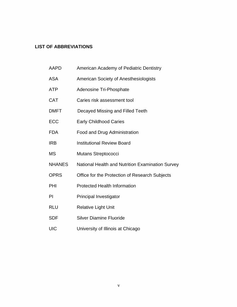
primary teeth by age five.5 A study in 2016 showed improvement in prevalence of dental
caries in a certain age group; documenting only 18% of children 2-5 years old
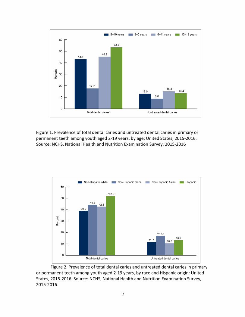
experiencing dental caries (Figure 1). In terms of demographics of children most
affected, Figure 2 shows prevalence across different ethnicities ages 2-19 years:
Hispanic 52.0%, Non-Hispanic Asian 42.6%, Non-Hispanic Black 44.3%, Non-Hispanic
White 39.0%.6
2
Figure 1. Prevalence of total dental caries and untreated dental caries in primary or permanent teeth among youth aged 2-19 years, by age: United States, 2015-2016. Source: NCHS, National Health and Nutrition Examination Survey, 2015-2016
Figure 2. Prevalence of total dental caries and untreated dental caries in primary or permanent teeth among youth aged 2-19 years, by race and Hispanic origin: United States, 2015-2016. Source: NCHS, National Health and Nutrition Examination Survey, 2015-2016

3
The detrimental effects for young children as a result of this disease process
include: compromised learning, communication, nutrition, and other activities essential
for proper growth and development.7,8 The disease process is complex and
multifactorial but is mediated by protective factors (fluoride, salivary buffering capacity,
and host immunity) and risk factors such as frequent carbohydrate exposure, poor oral
hygiene, and biofilm formation.9
Traditionally, the treatment of dental caries has been focused on surgical
management of repairing lesions and less centered on the disease process itself.1,10-12
Recently, personalized healthcare and medical management of caries has been
suggested to be a more effective prevention and treatment rather than treatment of
the disease consequences(cavities).1 Some strategies for employing this patient
centered approach are: use of antimicrobials, re-mineralizing agents, salivary
stimulation, and most importantly behavior modification.11 SDF has also been recently
suggested by the Illinois Department of Public Health to be used as an interim
management of caries during the acute stages of COVID outbreak.13
1.2 Microbiology of ECC
The etiology of this disease is often simply defined by four major components:
cariogenic bacteria, fermentable carbohydrates, susceptible teeth and host, as well as
time for the process to develop.1 Dental caries is a disease caused by accumulation of
microbial biofilm on teeth surfaces but can have interactions within the oral cavity with

4
saliva fluids. Previously dental caries was determined to be explained by the Specific
Plaque Hypothesis while the paradigm has shifted in recent years to an Ecological
Plaque Hypothesis. The difference between these two mechanisms is primarily from
being caused by a specific pathogen or select few to a view that there is a large
microbiome with complex interactions that mediates the caries process. However, a
great deal of research points to mutans streptococci (MS) species and Lactobacilli as
having large contributions to the etiology of caries in the Ecological Plaque Hypothesis.
Due to the complex interactions within the oral microbiome, it is extremely difficult to
make conclusions and associations that hold true across all populations as to how
bacteria within the oral cavity contribute to caries.14,15 A common conclusion is that,
when this microbiota shifts to high levels of specific pathogens, it will create dysbiosis
and disease will manifest, in this case dental caries. The bacterial species most often
implicated as major contributors to the caries process are primarily MS and lactobacillus
species. These bacteria form colonies within plaque present on tooth surfaces, and will
metabolize dietary nutrients to produce acidic byproducts that demineralize and
damage the underlying tooth structure.16 High levels of these bacterial species are
associated with increased caries risk.17,18 Research from Caufield et al. has also proposed
further implications from these oral bacteria within the GI microbiota which indicates
interactions from the tooth surface to the carrier, saliva.19 For these reasons, this study
was focused on the interaction with silver diamine fluoride (SDF) and these two
bacterial species.

5
1.3 Silver Diamine Fluoride (SDF)
1.3.1 SDF Overview
Silver Diamine Fluoride has emerged as a new product that has become available
for dentists in the United States after being approved by the Food and Drug
Administration (FDA) as of April 2015 for desensitization. However, it has a frequent
well-accepted use for caries management as an alternative therapy to prevent caries
progression. It is composed of silver ions which act as an antimicrobial, fluoride for re-
mineralization, and ammonia as a stabilizing agent.12 Use of SDF is a simple, non-
invasive method for treatment that has been shown to be effective in arresting active
caries in primary teeth.20 There are a variety of clinical uses for SDF, one of which being
treatment for children at extreme caries risk such as those with early childhood caries
(ECC) and severe early childhood caries (S-ECC) to prevent caries progression.12
1.3.2 SDF Effectiveness
Most studies have evaluated effectiveness of SDF by recording tooth staining and
hardness following SDF treatment. In a recent systematic review including 8 studies
using 38% SDF, the reported average proportion of arrested dentinal carious lesions has
been found to be 81%.21 In vitro and In vivo studies investigating effects of SDF on
bacterial species within plaque and saliva samples have shown varying results. A recent
study by Mitwalli et. al found no significant differences in microbial plaque samples
following single time SDF application.22 This finding is also consistent with results from
studies completed by Milgrom and Horst where no significant differences were noted in
plaque samples following SDF application.11,23 Some evidence has revealed decreases in

6
bacterial concentrations within plaque and saliva samples while others have found no
changes within composition of these samples.11,24-26
1.3.3 SDF Mechanism of Action
Silver ions within the SDF solution contribute to the antimicrobial effect by
breaking membranes of bacteria, denaturing proteins, and inhibiting DNA replication.
Ionic silver has also been shown to deactivate nearly all macromolecule within its
environment which can contribute to the bactericidal role. The SDF solution can delay
caries progression by forming a thin layer of silver-protein conjugate on the decayed
tooth surface which increases resistance to acid dissolution and host enzymatic
digestion. In addition, fluoride within the solution aids in this process by formation of
hydroxyapatite, fluorapatite, silver chloride, and metallic silver. Fluoridated tooth
surface has been extensively shown to be more resistant to acid degradation than
normal tooth structure.23 The treated lesions will increase in mineral density and
hardness which will contribute to decrease in the lesion depth. SDF will also interact
with the host physiology by inhibiting proteins that break down exposed dentin organic
components such as matrix metalloproteinases, cathepsins, and bacterial collagenases.
In-vitro studies have found that lesions treated with SDF are more resistant to
subsequent biofilm formation and progression, presumed to be related to remnant ionic
silver. When bacteria killed by silver ions are added to live bacteria, silver ions are re-
activated and the dead bacteria will then have a transference effect where they will
then contribute to the killing of the live bacteria. This phenomenon explains how silver
deposited on lesions can have sustained antimicrobial effects.12
7
1.3.4 SDF Ideal Application Frequency
Previous studies exploring the topic of the ideal application frequency are limited
and have shown mixed results. The 2017 guideline published by the AAPD following a
systematic review found that one-time SDF application arrest rates ranged from 47-90
percent. However, the effectiveness of lesion arrest decreases over time, which
indicates successive application is necessary. Half of treated lesions had reverted back
to active lesions at 24 months if no further intervention occurred.27 Studies have shown
unanimously that annual application of SDF is more effective at caries arrest when
compared to 5% sodium fluoride varnish.21 Bi-annual SDF application has been shown to
increase the caries arrest rate compared with annual application. Additionally, three
time per year application showed even higher arrest rates.21,28,29 Individuals with higher
plaque indices and lesions covered in plaque displayed lower rates of arrest, therefore
addressing other risk factors such as plaque presence may increase the rate of
successful treatment outcomes.28 The current standard recommended by the AAPD is 6
month application frequency but this is based on limited quantity and quality of
evidence. Most studies have determined application frequency based on clinical caries
arrest but not evaluating more objective measures such as microbiologic effects. Further
research has been proposed to determine the ideal application frequency when
considering all factors involved.
1.4 CariScreen
The current AAPD caries risk assessment tool advocates use of microbiological testing to
be used for pediatric dental patients. Studies have shown that patients with active

8
caries have much higher concentrations of MS and lactobacilli in their saliva and plaque
than do disease free individuals. Longitudinal studies have shown there are increases in
MS and lactobacilli over time with the progression of caries.18 Traditionally, laboratory
culturing methods have been used for quantification of the bacterial counts in plaque or
saliva samples; however, these methods often tend to be laboratory intensive and
expensive. Recently, a commercially available chair side meter known as CariScreen
(Version 1.4, Albany, Oregon) has become available to quantify oral bacteria samples
and assess patients’ caries risk. The diagnostic tool uses adenosine triphosphate (ATP)
bioluminescence to measure visible light release from dental plaque after mixture with
a luciferase enzyme reaction within a swab. Luciferin and luciferase produce light when
activated by ATP, a metabolic by-product of metabolizing bacteria.30 ATP is then
quantified using a bioluminometer which can measure the light output in relative light
units (RLUs). This allows the identification of non-specific oral bacterial load and biofilm
activity levels. A similar method has been used previously in the food production
industry to rule out bacterial contamination prior to human consumption. Previous
studies have used the CariScreen meter in pediatric dental patients to determine plaque
bacteria levels and caries risk assessment.18,31
1.5 Oral Health Care for Children
The most important component to the management of dental caries in children
is prevention of the disease altogether. One aspect of this prevention has been the
American Academy of Pediatric Dentistry (AAPD) recommendation to establish a dental
home by the eruption of the first tooth or 12 months of age, whichever comes first. A

9
typical initial examination allows pediatric dentists to provide recommendations for
diet, oral hygiene, dental prophylaxis, clinical examination, acquiring dental radiographs
if indicated, and topical application of fluoride. Caries risk assessment is an important
measure to determine an individual’s risk of developing caries based on demographic
information, habits, preventative care, genetic factors, and many others. The amount of
mutans streptococci present within a child’s saliva has been shown to be a good
predictive factor of that child’s caries risk in the near future. This evaluation has been
shown by Edelstein in 2016 to be a more predictive measure for caries risk alone than
the entire AAPD Caries Risk Assessment Tool cumulatively.32 Therefore studies
evaluating effects on oral bacteria and their correlation with caries risk are warranted.
1.6 Purpose
This is a prospective, longitudinal pre- and post- interventional study that aims to
evaluate the effects of application of 38% SDF on microbial load within plaque and saliva
samples at 3 months and 6 months intervals.
The objectives of the study are:
• Determine the oral bacterial load in dental plaque of ECC children using the
chairside CariScreen unit at baseline, 3, and 6 months after silver diamine fluoride
(SDF) application
• Determine the oral bacterial load in saliva of ECC children using bacterial culture
method at baseline, 3, and 6 months after silver diamine fluoride (SDF)
application.

10
• The differences in bacterial levels at different sampling time points will be
compared to reveal the efficacy of SDF in reducing oral bacterial load.
1.7 Hypotheses
The Null Hypothesis of the study is:
• H0: There is no statistical difference between the plaque bacterial level when
measured by ATP score in patients receiving a single versus two time application
of SDF over a 6 month period.
• H0: There is no statistical difference between the oral bacterial level in saliva
when measured by traditional bacterial culture in patients receiving a single
versus two time application of SDF over a 6 month period.
• H0: There is no statistical difference in presence of visible plaque in patients
when receiving a single versus two time application of SDF over a 6 month
period.
2. MATERIALS AND METHODS
2.1 Overview
University of Illinois-Chicago (UIC) IRB approval was attained for this study:
protocol # 2017-0342. Pediatric patients were recruited from the University of Illinois
Chicago College of Dentistry Department of Pediatric Dentistry. Patients were screened
for inclusion within the study and if determined to be eligible, the purpose, risks, and
benefits of the study were reviewed with parents. If parents were interested in being
included in the study, informed consent was obtained and signed. This study was a

11
prospective, longitudinal pre- and post- intervention study with randomization into two
treatment groups. A total of 40 participants (20 in each group) were recruited for the
study. The amounts of fluoride or silver used during treatment did not approach the 5
mg/kg probable toxic dose for fluoride or the 380 mg/kg lethal dose for silver
respectively29. One clinical operator conducted the study, and provided all treatment to
patients including application of SDF, collection of saliva, and CariScreen plaque swabs.
This operator reviewed manufacturer protocols for the CariScreen meter and underwent
training and calibration using the device. Baseline data and outcome measures were
recorded electronically on a data collection form within Microsoft® Excel 2016 and
transferred to SPSS for statistical analysis.
2.2 Study Site, Participants and Enrollment Process
2.2.1 Study Site
This study was carried out at the University of Illinois-Chicago College of Dentistry,
Department of Pediatric Dentistry. The clinic provides care for an extensive number of
patients requiring dental treatment under general anesthesia which provided a sufficient
number of patients for inclusion within this study.
2.2.2 Operator
One designated and trained operator, a pediatric dental resident, administered all
oral swabs, saliva collection, as well as SDF applications for the purposes of this study.
These procedures were performed according to manufacturer recommendations and
instructions.
2.2.3 Study Participants

12
Study subjects were recruited from the UIC Post-graduate Pediatric Dental Clinic.
Eligible patients were identified by the clinical operator (capstone candidate, Dr. Austin
LaMay) through the axiUm® electronic health record system. This individual then
evaluated patients during a comprehensive oral examination, if patients were diagnosed
with early childhood caries requiring treatment under general anesthesia, the parents
were informed their children were eligible for recruitment in the study. Inclusion and
exclusion criteria were specified for the purposes of this study. Forty patients were
expected for recruitment in the study based on a power analysis completed using data
from previous studies conducted within the department.
2.2.4 Inclusion Criteria
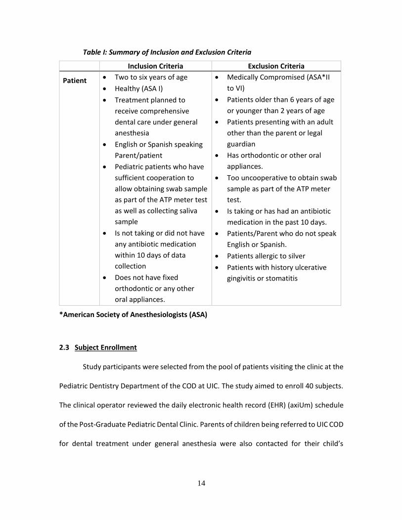
The inclusion criteria for the participants are summarized in Table 1 and were as follows:
1) Children between the ages of 2 and 6 years old. This group represents children
with possible diagnosis of ECC and make up the majority of children requiring
treatment under general anesthesia for complete oral rehabilitation in a
hospital setting.
2) Participants were required to be healthy and classified according to the
American Society of Anesthesiology (ASA) class 1.
3) Participants were required to have extensive caries requiring treatment under
general anesthesia. The study aims to determine the effect of SDF on bacteria
within saliva and plaque samples.
4) Participants needed to be cooperative enough to allow an intra-oral cotton
swab and collection of saliva.

13
5) Participants needed to be cooperative enough for SDF application.
6) Participants were not taking or did not have any antibiotic medication within
10 days of data collection.
7) Participants did not have fixed orthodontic or any other oral appliances.
8) English or Spanish speaking literacy of the parent/guardian was required. The
study documentation, and informed consent form were translated into
Spanish for inclusion of that population as well.
2.2.5 Exclusion Criteria:
The list of the exclusion criteria for this study included:
1) Children younger than 2 years of age and older than 6 years of age were excluded
from the study.
2) Children with medical status categorized as ASA II to VI. Patients with significant
medical history were excluded from study because of possible confounding effects
of their health status.
3) Children with orthodontic or other oral appliances.
4) Children too uncooperative to obtain cotton swab or saliva sample.
5) Children taking or have had an antibiotic medication in the past 10 days.
6) Children allergic to silver.
7) Non-English or Non-Spanish speaking parents/guardians and patients, due to
concerns obtaining informed consent.
14
Table I: Summary of Inclusion and Exclusion Criteria
Inclusion Criteria Exclusion Criteria
Patient • Two to six years of age
• Healthy (ASA I)
• Treatment planned to
receive comprehensive
dental care under general
anesthesia
• English or Spanish speaking
Parent/patient
• Pediatric patients who have
sufficient cooperation to
allow obtaining swab sample
as part of the ATP meter test
as well as collecting saliva
sample
• Is not taking or did not have
any antibiotic medication
within 10 days of data
collection
• Does not have fixed
orthodontic or any other
oral appliances.
• Medically Compromised (ASA*II
to VI)
• Patients older than 6 years of age
or younger than 2 years of age
• Patients presenting with an adult
other than the parent or legal
guardian
• Has orthodontic or other oral
appliances.
• Too uncooperative to obtain swab
sample as part of the ATP meter
test.
• Is taking or has had an antibiotic
medication in the past 10 days.
• Patients/Parent who do not speak
English or Spanish.
• Patients allergic to silver
• Patients with history ulcerative
gingivitis or stomatitis
*American Society of Anesthesiologists (ASA)
2.3 Subject Enrollment
Study participants were selected from the pool of patients visiting the clinic at the
Pediatric Dentistry Department of the COD at UIC. The study aimed to enroll 40 subjects.
The clinical operator reviewed the daily electronic health record (EHR) (axiUm) schedule
of the Post-Graduate Pediatric Dental Clinic. Parents of children being referred to UIC COD
for dental treatment under general anesthesia were also contacted for their child’s

15
possible inclusion within the study. Following these procedures, a group of 40 participants
were obtained to be followed throughout the study period.
Patients presenting to the UIC Department of Pediatric Dentistry had the following
performed at their initial visit: the pediatric dental resident (Dr. Austin LaMay)(AL) would
perform a comprehensive dental exam, complete medical and dental history, caries risk
assessment, extra-oral and intra-oral exam, dental prophylaxis, and fluoride application.
He would then determine each child’s caries risk based on the caries-risk assessment tool
(CAT) which has been developed by the AAPD.33 This tool takes into account:
demographics, oral hygiene practices, dietary habits, and protective factors. If any
negative factors were identified with their child’s routine practices the pediatric dental
resident (AL) would provide recommendations and home care instructions to prevent
caries initiation or progression. If possible and when indicated, the pediatric dental
resident would take radiographs as outlined by AAPD guidelines for radiographic
exposure.34 Following this procedure, a dental prophylaxis was performed. Based on all
findings of the chief complaint, radiographic examination, and clinical examination, a
treatment plan would be developed by the clinical operator. In addition to optimal
treatment to be performed, the mechanism of which treatment would be carried out
would also be discussed with the parent including pharmacologic (nitrous, conscious oral
sedation, general anesthesia) and non-pharmacologic behavior management techniques
(tell-show-do, distraction, imagery, etc).
Due to the extensive need within the public aid system in Illinois and limited
number of dental providers offering access to general anesthesia services for children on

16
public aid dental insurance, the current wait time for dental treatment under general
anesthesia at UIC is approximately 1 year. SDF has been used in the past as an interim
therapeutic treatment regimen to delay the progression of caries in patients awaiting
general anesthesia at the UIC postgraduate pediatric dental clinic. The AAPD recommends
recall status be determined based on a child’s caries risk.33 Children with high caries risk
are recommended for 3 month recall dental appointments, while low risk patients only
require appointments every 6 months. Children with special healthcare needs are more
commonly determined to be at higher risk based on their needs and functional abilities.33
Based on caries burden and behavioral indicators it will be determined whether
the child would best be treated under general anesthesia, the subject was evaluated for
involvement in this study. The clinical operator would review purposes of the study
through a patient information leaflet (PIL), potential risks and benefits involved were
discussed with parents to determine their enrollment in the study. Adverse outcomes
were explained to the parents specifically as it pertains to, expected black stains to carious
tooth structure once SDF is applied. If parents agreed to participate in the study, the study
consent was completed and signed by the parent and clinical operator (parental
permission, Appendix B. Patients and parents were also informed they would be
compensated $10 for each follow-up visit required during participating with the study.
Once patient enrollment was confirmed, baseline measurements were obtained at the
initial appointment and SDF was applied thereafter. Most patients’ dental insurances
covered SDF treatment, however if the procedure was not covered it was not billed to the
patient or insurance company. Participants were assigned a subject number and a master

17
list of participants linked to the patient’s EHR which was kept safely by the clinical
operator. The enrolled participants were then randomized to treatment groups in a
paired manner by coin flip to determine the intervention group. Participants that were to
have SDF applied twice over the 6 month period were designated as Group 2, while
participants receiving only a single SDF application were designated at Group 1.
Participants that did not meet the inclusion criteria were not enrolled in the study.
Treatment recommendations based on their individual needs were determined on a case
by case basis and an optimal treatment plan was reviewed with the parents for their care.
2.4 Armamentarium
Delivery of SDF required SDF, microbrush, and 2x2 cotton gauze followed by
application of 5% fluoride varnish. Silver diamine fluoride 38% sold by Advantage Arrest®
(West Palm Beach, FL) was used for this study. Plaque swab and bioluminescent
evaluation was completed using the chairside Cariscreen® meter. Sterile 10mL collection
vials were used for collection of saliva via the drool method for microbiological culture of
bacterial species.
2.4.1 SDF
Elevate® is the manufacturer of Advantage Arrest® which is 38% SDF. SDF can
safely be used in children without a silver allergy.
2.4.2 ATP Bioluminometer
The CariScreen protocol involves carefully swabbing the mid-lingual surface of
the lower anterior teeth. One firm swipe is required, without contact from the gingiva
or any soft tissue. The swab is placed back in its tube, and the snap valve is broken by

18
bending the bulb forward and backward. The bulb must then be squeezed to expel all
liquid down the swab shaft. The tube is gently agitated for 5-10 seconds prior to
inserting the swab into the CariScreen Meter, which will provide the result in RLUs from
1-9999.
2.5 Procedure
Prior to completing the dental prophylaxis and SDF application, the investigator
carried out the ATP bioluminescence swab test as outlined by the CariScreen collection
protocol. ATP scores were determined immediately after that swab was collected and
documented in the data Excel sheet. Following the bioluminescent swab, unstimulated
saliva was collected over 5 minutes via the drool method. This method involved
participants placing the sterile collection vial up to their lips and allowed saliva to
“drool” into the vial over the course of 5 minutes. This protocol pertained to the initial
visit as well as any follow up appointments required based on the group designation of
the participant. Thereafter, the Pediatric Dentistry Resident completed the dental
prophylaxis and applied the SDF on the dental carious lesions. Participants and parents
were provided with oral hygiene recommendations consistent with AAPD guidelines.9
Standardized oral hygiene instructions included: twice daily parental brushing of child’s
teeth with fluoridated toothpaste, and daily flossing. Dietary advice included: drinking
tap water and milk during the day, no milk or sugar sweetened beverages at night, and
limiting juice to a daily intake of 4-6 ounces.
2.6 Data Collection

19
Information was recorded into an Excel file at the initial exam, three month, and six
month follow up if applicable:
1. Subject ID number
2. SDF application date
3. Group randomization designation
4. ATP bioluminescence score of the patient
5. Salivary culture data
a. Brain heart infusion (BHI) agar – Total bacteria level
b. Mitis salivarius bacitracin agar- Streptococci species
c. Rogosa agar- Lactobacillus species
6. Visible plaque recording (Y/N)
Dates/Experimental
group designation
Subject
ID#
Visible plaque
present (Y/N)
ATP bioluminescence
score
Salivary Culture
data
@ Initial
1
@ 3-months
@ 6-months
@ Initial
2
@ 3-months
@ 6-months
@ Initial
3
@ 3-months
@ 6-months

20
Figure 3: Data Colllection Spreadsheet
Enrolled parents/children consisted of two groups who received the following:
1. Group 1: Exam, caries risk assessment, anticipatory guidance including
standardized oral hygiene instructions, ATP swab testing, saliva sample, dental
prophylaxis, dental radiographs if possible and when indicated, SDF application,
and six month recall dental appointment.
2. Group 2: Exam, caries risk assessment, anticipatory guidance including
standardized oral hygiene instructions, ATP swab testing, saliva sample, dental
prophylaxis, dental radiographs if possible and when indicated, SDF application,
and three and six month recall dental appointment.
If the child became eligible for general anesthesia treatment prior to research
completion (six months), the child would be excluded from the remaining research
procedures in order to receive their definitive treatment. This procedure occurred for
one of the study participants and they were subsequently excluded from the study.
2.7 Chairside steps of SDF application
1. Visible plaque removed prior to SDF application by dental prophylaxis
2. Teeth dried with air syringe or cotton gauze, whichever was feasible
3. Microbrush dipped into SDF drop until saturated

21
4. Microbrush applied to carious lesions and allowed to be absorbed
5. 5% Fluoride varnish applied to dentition
2.8 Statistical Analysis
Data gathered through all study forms were transferred into Microsoft Excel
Spreadsheet (Microsoft Inc., Redmond, WA, USA). The data file was stored on a password-
protected computer. The Excel data file was then transferred to the IBM SPSS statistical
(version 25, IBM corporation, Armunk, NY) software program for statistical analysis. All
data were assigned a numerical value in order to complete statistical analysis.
3. RESULTS
3.1 Number of Participants
Participant enrollment took place over the course of approximately 6 months. The
follow up period for both groups took place over the following 6 months, therefore the
study period took place over a total of 12 months. Forty participants were recruited for
the study, 32 participants completed the necessary follow up appointments entirely. Eight
participants (3 Group-1, 5 Group-2) were lost to follow up for a few primary reasons: loss
of contact, unexpected comprehensive treatment prior to study completion, and study
interruption related to the COVID-19 pandemic. There were no adverse events identified
related to the study treatment regimen noted for either group.
3.2 Demographics and DMFT Scores

22
Table II shows the demographic distribution for study participants and their group
designations. The table represents data for participants that completed study follow up
and were used for complete data analysis N=32. Average ages of participants were 3.85
(SD=.9) overall, 3.55 (SD=.6) for Group-1 and 4.15 (SD=1.2) for Group-2 with a range of
ages 2-6 years old. Gender distribution for the study was overall 62% male (n=20), 37.5%
female (n=12). A similar gender distribution was seen for Group-1: 63.7% male, 35.3%
female and Group-2: 60% male, 40% female. Average dmft scores were also calculated
for each group independently which were 11.5 (SD=3.17) for Group-1, 12.05 (SD=3.22)
for group 2 for an overall average of 11.77 (SD=3.17).
Table II: Participant Demographics
Group 1 n=17
Group 2 n= 15
Total N=32
Age (mean in years)
3.55 (SD=.6) 4.15 (SD=1.2) 3.85 (SD=.9)
Gender (count and
percentage)
M= 11 (63.7%) F= 6 (35.3%)
M= 9 (60%) F= 6 (40%)
M= 20 (62.5%) F= 12 (37.5%)
Average DMFT score
11.5 (SD=3.17)
12.05 (SD=3.22)
11.775 (SD=3.17)
3.3 ATP Bioluminescence Scores
The baseline mean initial ATP bioluminescence score was 7777 RLU for Group-1
(SD= 1570). Using a related samples t-test a statistically significant decrease for Group-1
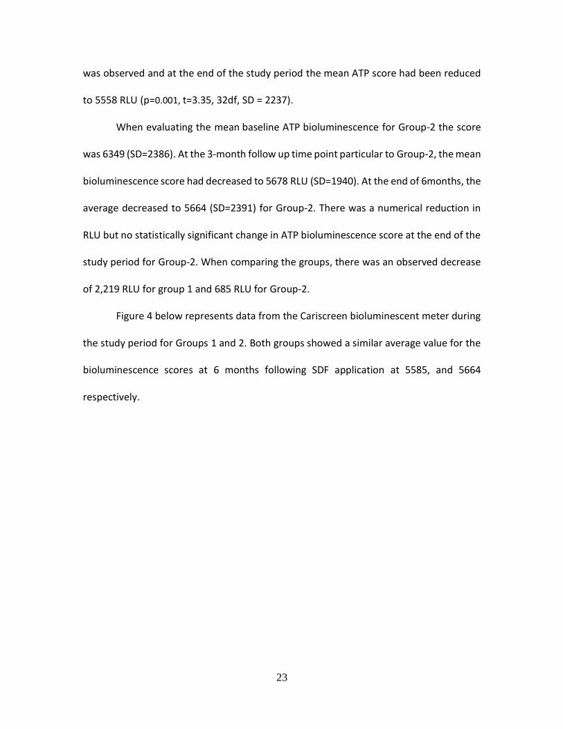
23
was observed and at the end of the study period the mean ATP score had been reduced
to 5558 RLU (p=0.001, t=3.35, 32df, SD = 2237).
When evaluating the mean baseline ATP bioluminescence for Group-2 the score
was 6349 (SD=2386). At the 3-month follow up time point particular to Group-2, the mean
bioluminescence score had decreased to 5678 RLU (SD=1940). At the end of 6months, the
average decreased to 5664 (SD=2391) for Group-2. There was a numerical reduction in
RLU but no statistically significant change in ATP bioluminescence score at the end of the
study period for Group-2. When comparing the groups, there was an observed decrease
of 2,219 RLU for group 1 and 685 RLU for Group-2.
Figure 4 below represents data from the Cariscreen bioluminescent meter during
the study period for Groups 1 and 2. Both groups showed a similar average value for the
bioluminescence scores at 6 months following SDF application at 5585, and 5664
respectively.

24
Figure 4: Bioluminescent meter readings following SDF application to measure relative bacteria in plaque on tooth surface
3.4 Visible Plaque Presence
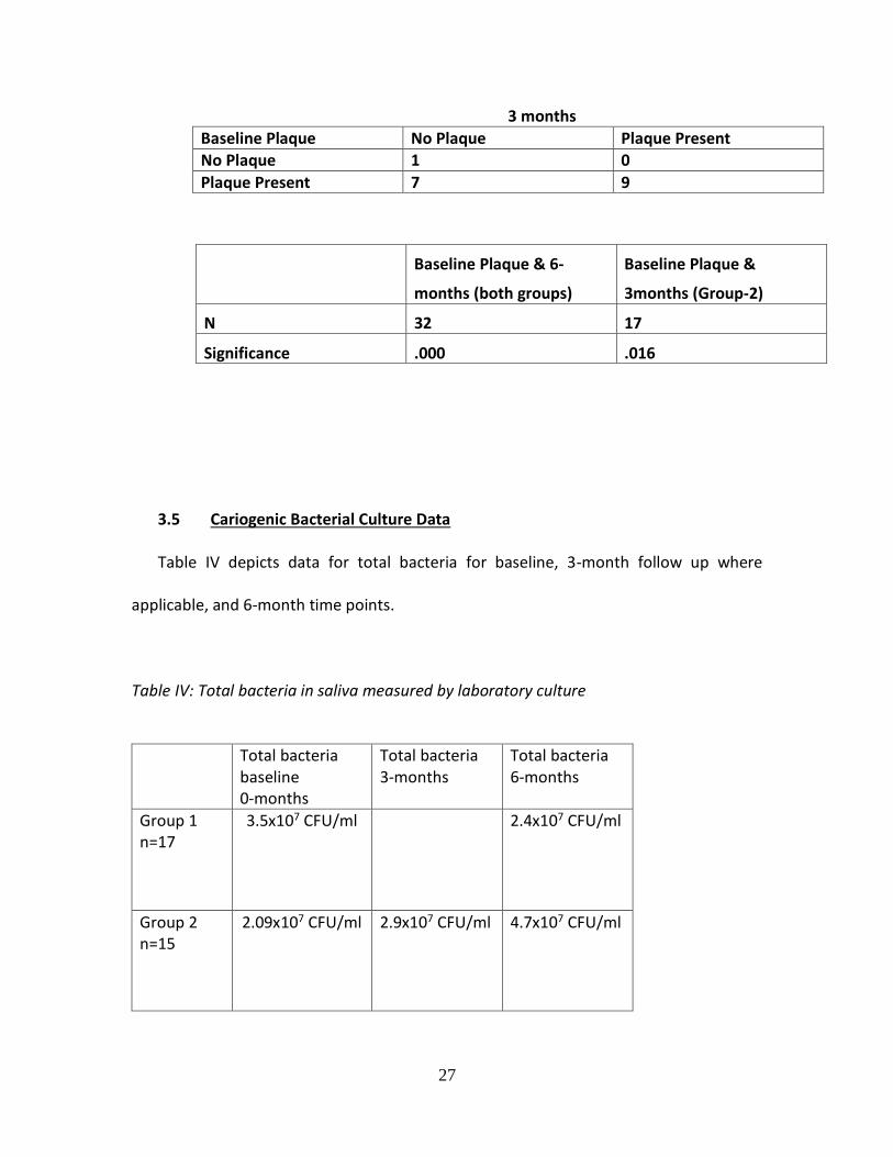
At baseline measurement, in Group-1 there were two participants with no
plaque present and 18 participants with plaque present. For Group-2 there was one
subject with no plaque and 19 participants with plaque present. After interventions
throughout the study respective to each study group, using McNemar’s Chi-Square for
repeated measurements, there were significant decreases in the presence of plaque at
the end of the study period for both groups when compared with baseline
measurement (p=.000 Group-1, p=.016 Group-2). When comparing the effect of study
base line 3M 6M
Group 1 7777.47 5558.35
group2 6349.07 5678.33 5664.53
7777.47
5558.35
6349.07
5678.33 5664.53
0
1000
2000
3000
4000
5000
6000
7000
8000
9000
10000
RLU
Cariscreen Meter ATP Readings

25
interventions with Pearson Chi-Square analysis between Group-1 and Group-2 on
presence of plaque before and after SDF application, there was a numerical difference
that approached statistical significance (x2=3.46, p=.063). All participants were utilized
when determining baseline measurements (n=40) however only 32 participants
returned for complete follow up and were used for complete data analysis.
Figure 5: Visible plaque presence at baseline

26
Figure 6: Visible plaque presence at 6 months
Table III: Visible plaque presence at 6 months following SDF application compared to baseline
Value df Significance
Pearson Chi-Square 3.463 1 .063
McNemar’s Chi Square:
Baseline Plaque vs. 6month (pooled)
N 32
Significance .000
Group-1 & 2 6 months
Baseline Plaque No Plaque Plaque Present
No Plaque 2 1
Plaque Present 19 10
Group-2
27
3 months
Baseline Plaque No Plaque Plaque Present No Plaque 1 0
Plaque Present 7 9
Baseline Plaque & 6-
months (both groups)
Baseline Plaque &
3months (Group-2)
N 32 17
Significance .000 .016
3.5 Cariogenic Bacterial Culture Data
Table IV depicts data for total bacteria for baseline, 3-month follow up where
applicable, and 6-month time points.
Table IV: Total bacteria in saliva measured by laboratory culture
Total bacteria
baseline 0-months
Total bacteria 3-months
Total bacteria 6-months
Group 1 n=17
3.5x10⁷ CFU/ml 2.4x10⁷ CFU/ml
Group 2 n=15
2.09x10⁷ CFU/ml 2.9x10⁷ CFU/ml 4.7x10⁷ CFU/ml

28
Table V depicts data for Lactobacilli species for baseline, 3 month follow up where
applicable, and 6 month time points.
Table V: Lactobacilli measured in saliva by laboratory culture
Lactobacilli baseline 0-months
Lactobacilli 3-months
Lactobacilli 6-months
Group 1 N=17
1.10x10⁵ CFU/ml 1.03x10⁵ CFU/ml
Group 2 N=15
1.22x10⁵ CFU/ml 2.2x10⁴ CFU/ml 3.18x10⁴ CFU/ml
Table VI depicts data for Streptococci species for baseline, 3 month follow up
where applicable, and 6 month time points.
Table VI: Streptococci measured in saliva by laboratory culture
Streptococci Baseline 0-months
Streptococci 3-months
Streptococci 6-months
Group 1 N=17
4.79x10⁵ CFU/ml 5.79x10⁵ CFU/ml
Group 2 N=15
1.01x10⁵ CFU/ml 3.4x10⁵ CFU/ml 1.79x10⁵ CFU/ml

29
Figure 7 illustrates the compilation of data for both groups and all bacterial
species cultured from saliva over the study time points. Overall there is a trend that
bacterial concentrations did not change significantly over time for any of the species
cultured: total bacteria, lactobacilli, nor streptococci.
Figure 7: Viable bacteria measured in saliva by laboratory culture following SDF application for Group-1 participants
0
1
2
3
4
5
6
7
8
9
Baseline 6M Baseline 6M Baseline 6M
Total Bacteria Latobacilli Streptococci
Via
ble
Bac
teri
a (l
og (
CFU
/ml))
Group-1, after 6 months

30
Figure 8: Viable bacteria measured in saliva by laboratory culture following SDF application for Group-2 participants
Statistical analysis to compare the means before and after interventions at each
time point was completed using a paired sample t-test. There was no statistically
significant differences found when comparing time points between groups or within
groups. However, when comparing the proportion of lactobacilli that contributed to the
total bacteria, we observed that this proportion decreased over time for both groups
after SDF application. The decrease for proportion of lactobacillus species within the
total bacteria was more notable for Group-2 compared to that of Group-1. Figure 12
illustrates this change in proportion as a percentage for the study groups.
0
1
2
3
4
5
6
7
8
9
Baseline 3M 6M Baseline 3M 6M Baseline 3M 6M
Total Bacteria Latobacilli Streptococci
Via
ble
Bac
teri
a (l
og (
CFU
/ml))
Group-2, after 3 & 6 months

31
Figure 9: Lactobacilli level in total bacteria (%) measured in saliva following application of SDF
4. DISCUSSION
4.1 Bioluminometer readings to determine 3 month vs 6 month SDF application frequency
The null hypothesis is that there is no difference in CariScreen meter readings,
which detects relative amounts of bacteria within plaque swabs, when SDF is applied on
a 3-month frequency vs 6-month basis. We reject the null hypothesis, since there is a
statistically significant difference for Group-1. When evaluating the data, we see a trend
of a decrease in bioluminescent readings for both groups, with the larger decrease coming
from Group-1. This may be due to the higher average reading at baseline which allowed
for a greater numerical decrease overall. At the 6-month time point the average
bioluminescence scores are very similar for Groups-1 and -2 which shows a trend towards
a value ~5600 at that time. If the mean bioluminescence scores would have been more
similar for both groups at baseline, it is possible there may have been significance for the
-200
-100
0
100
200
300
400
Baseline 3M 6M
Lact
ob
acill
i/To
tal B
acte
ria
(%)
Lactobacilli level in total bacteria (%)
Group-1
Group-2

32
decrease in Group-2 as well. The other possibility for the results we obtained could be
due to error in protocol while obtaining plaque swabs for the bioluminescent meter that
created error in the results we obtained. It is difficult to control for the environment
despite a provider’s best attempts to do so in a young child during a clinical scenario.
Similar studies evaluating the reliability of the CariScreen meter have had conflicting
conclusions. Hallet et al suggested in 2013 that the test was not a reliable caries risk
assessment tool when compared with other more validated measures. The readings they
obtained were three times higher than those suggested by the manufacturer as well as
notably inconsistent.31 Fazilat et al determined in 2010 that the meter was reliable to
determine oral bacteria in plaque based on data from their study correlating ATP readings
with oral streptococci concentrations with high correlation.18 There is widely accepted
evidence that patients involved in research may be particularly diligent with their
behavior because they are aware they are being monitored as participants within study.
This may contribute as a confounding variable to changes we notice in our outcome
measures.35
4.2 Microbial culture of cariogenic salivary bacteria to determine 3 month vs 6
month SDF application frequency
Evaluating results from bacterial culture of salivary bacteria at baseline and each
time point within the study, we did not observe any significant changes. There was a large
diversity and variance of oral bacteria between different subjects and varying
amounts of bacteria over time even within the same subject. This could be attributed to
differing behaviors in diet, oral hygiene, or other activities of daily living. When the data

33
from all subjects was pooled, there was no significant trend indicating SDF played any role
to attribute for differences in the salivary bacteria overall. However, when looking closely
at the data, there was an interesting trend that indicated the proportion of lactobacilli
compared to total bacteria decreased in both treatment groups following SDF application.
This may indicate that SDF may have specific effects on lactobacillus species within saliva.
This trend was not observed for the data obtained from streptococcal species.
4.3 Plaque Presence
When evaluating results for presence of plaque over the course of the study, we
observed a significant change in frequency of plaque present in subjects throughout the
study period. Most subjects presented with plaque present at enrollment for the study,
however during subsequent recalls we observed a significant decrease in the frequency
of plaque present in the study subjects. This shows our data is consistent with evidence
that counseling on proper oral hygiene and instruction by dental professionals can be
effective for behavior modification in these patients. It also provides insight that there
could be possible changes in the microbiota related to the decrease of food debris and
plaque accumulation on tooth surfaces. We might expect to see a decrease in species of
cariogenic bacteria within plaque or saliva that might be attributed to this decrease in
plaque. Past research has shown a decrease in biofilm formation on the surfaces of teeth
treated with SDF.36 This is consistent with our results which may have been attributed to
this mechanism or a combination of changes in oral hygiene in addition to this proposed
mechanism. It is important that future studies evaluating this topic should control for
differences in hygiene that could affect results of oral bacteria measurements.
34
4.4 Comparison to Past Studies
There have been other studies published recently evaluating the effects of silver
diamine fluoride on cariogenic bacteria. Many of these studies were laboratory studies
to validate the antibacterial activity of SDF on various media or simulated tooth structure.
In vitro studies by Savas et al and Lou et al have established that SDF possesses
strong inhibitory and bactericidal properties against a wide range of bacterial species.25,26
However, a few published in-vivo studies on the antibacterial effects of SDF have
shown conflicting results. One study by Mitwalli et al. investigated the effects of SDF on
root/cervical carious lesions in adults. At baseline and following application of SDF, plaque
samples were obtained from the surfaces of the lesions and DNA sequencing was
completed to obtain profiles of bacteria within the plaque samples. There were no
significant differences within the bacterial profiles identified pre- and post-intervention.
However, there were some changes in the relative abundances of some acidogenic
species but none of these were identified as S. mutans or Lactobacillus species.22
The second recent publication is from Garrastazu et al. evaluating effects on
salivary levels of S. mutans following application of SDF compared with those of
chlorhexidine in children. Children were randomized to have SDF or chlorhexidine applied
to their teeth and followed up at 1, 30, and 90 days post intervention for saliva samples.
Saliva samples were cultured on differential media for evaluation of S. mutans levels. The
study found that 30% SDF had similar antimicrobial effects as 1% chlorhexidine in creating

35
a statistically significant reduction of salivary levels of S. mutans at all time points. One of
the main controversies with this study is that the authors did not specify if any of these
children had caries or any mention of the methods in which they applied these
antimicrobials to the children’s teeth.
A well-designed study by Milgrom et al. published in 2018 measured levels of
bacteria on the surfaces of carious lesions and unaffected tooth structure in 66 children
before and 2-3 weeks after SDF application. They utilized RNA sequencing methods to
determine changes in the abundances of microbes sampled from the tooth surfaces.
Contrary to their hypothesis, there was very little difference in the abundances of most
bacterial species. Specifically, there were no significant changes in cariogenic bacteria
including S. mutans and Lactobacilli which were nearly universally found in all samples
pre- and post-intervention despite SDF treatment. Surprisingly, most changes were
modest increases as opposed to decreases as their hypothesis predicted. The authors also
used genetic sequence evaluation to confirm that there were also no significant changes
in expression of antibiotic resistance or metal resistance genes within the samples
obtained during the study. These data suggest that SDF is safe and does not promote the
development of resistance to its bactericidal mechanisms.
Despite conflicting results on the bactericidal effects of SDF in clinical settings,
some studies suggested that it has minimal effect on bacteria within saliva or plaque. Our
data supports the concept: SDF has minimal effect on bacterial concentrations within
plaque and saliva. The exception to this theory is that there may be some reduction of
bacterial species within plaque when measured with the bio luminometer, however this

36
was only found in 1 treatment group. It is unlikely that these results are reliable because
it is very counter-intuitive that a single application of SDF would result in less bacteria in
plaque than 2 applications considering many studies have confirmed the bactericidal
nature of the compound.
4.5 Study Strengths
The study was completed with a single operator and protocol for SDF placement,
examination, oral hygiene instruction, and diet counseling. This provided uniformity
across participants and treatment groups to ensure the same protocols as much as
possible between participants and avoidance the need for inter-operator calibration. The
participants were also randomized to the corresponding treatment group which serves as
a strength by limiting bias within the study.
The participants were all selected from the same subject group within the
pediatric dental clinic. Although a convenient sample and single center study, UIC
pediatric patients are considered diverse and they represented a population that was
consistent between treatment groups in terms of age, extent of decay, and gender.
The study also utilized several independent outcome variables to measure
differences in bacteria throughout the study period. This variation in measurement
methods help to establish more consistent conclusions overall because we would expect
uniformity across the data across the different methods of measurement.
4.6 Study Limitations
One of the study limitations was the relatively small sample size compared to some similar

37
studies. There were 40 participants initially recruited for the study, of which 32 completed
the study. A power analysis was completed prior to the study based on previous
comparable research protocols completed at the department utilizing the
bioluminometer in detecting differences in bacteria after SDF application. The previous
study found a 2382RLU decrease in CariScreen reading following SDF application when
evaluated at 3months. Anticipating sample sizes of 20 in each group achieved >80%
power when considering mean difference of 2382RLU using the CariScreen ATP
device. The previous research found a standard deviation of 861 at baseline and 2237 at
the second time point. Our results were not consistent with those from the previous study
and ultimately resulted in a deficiency of subjects to determine conclusive results. This is
supporting evidence from other studies that the CariScreen meter may not be a reliable
measurement tool.
The second limitation of the study was the related to the study period of 6 months.
Since the study group was those of a remarkably high disease burden waiting for many
months for treatment under general anesthesia, a subset of the study population had
teeth with carious lesions that developed abscesses throughout the study period
despite study interventions. It is possible that the latter affected the bacterial levels due
to the abscesses. This had occurred in a relatively small percentage of the study subjects,
however nearly all of these subjects had no differences in total bacteria measured from
their saliva despite the presence of the chronic abscess.
Despite the study being conducted in a randomized fashion where participants
were placed into groups serially in pairs following enrollment, there were differences in
38
baseline values between the groups. The conditions of the study did not allow the
operator to be blinded as to which treatment group each patient was placed into. This
may have introduced some amount of artificial bias related to how the study was
conducted or what results were expected.
There was intention to provide a control group that did not receive SDF
throughout the study, however due to ethical concerns this was ultimately not possible.
Only one parent had declined SDF throughout the enrollment period which was not
enough to provide an effective control group. The ethical concern would be if SDF were
not offered as a treatment option to a parent and child that would benefit from it’s use
otherwise. For that reason, there are limitations when interpreting the results of the
study without a control group to compare.
4.7 Future Studies
To date, studies evaluating the microbiologic effects of SDF in children have been
limited. In addition, available data has been conflicting, thus making the conclusive
determination as to whether SDF affects microflora of plaque and saliva of upmost
importance. Numerous studies report the successful outcomes when SDF was used as a
treatment agent against ECC. However, the exact mechanism of SDF leading to the
positive clinical benefits remains unclear. Once the understanding of the mechanisms of
SDF are understood, this should enable clinicians to more accurately prescribe its use and
promote effective treatment regimens. Future studies investigating the antimicrobial
effects of SDF could be focused on differences within bacterial profiles within the carious

39
lesions themselves using traditional culture methods or genetic sequencing of bacteria.
Most past research has focused on bacteria on the surfaces of the carious lesions or saliva,
but the mechanism of arrest may be restricted to the carious lesions themselves with little
effects on the plaque found on surfaces of the lesion or surrounding saliva.
5. Study Conclusions
The following conclusions can be made based on the results of this study:
• There is no benefit to applying SDF more frequently than the current 6 month
standard when measuring differences in bacteria within plaque using a chairside
bioluminometer.
• There is no benefit to applying SDF more frequently than the current 6month
standard when measuring differences in salivary bacteria using traditional culture
methods.
• There is a significant reduction in frequency of visible plaque for when SDF is
applied at both 3 and 6 month time intervals in conjunction with recall
appointments.

40
6. Cited Literature
1. Slayton RL, Fontana M, Young D, et al. Dental caries management in children
and adults. NAM Perspectives. 2016;6(9). doi: 10.31478/201609d.
2. Policy on early childhood caries (ECC): Classifications, consequences, and
preventive strategies. Pediatric dentistry. 2016;38(6):52.
https://www.ncbi.nlm.nih.gov/pubmed/27931420.
3. Fleming E, Afful J. Prevalence of total and untreated dental caries among
youth: United states, 2015-2016. NCHS data brief. 2018(307):1.
https://www.ncbi.nlm.nih.gov/pubmed/29717975.
4. Policy on oral health care programs for infants, children, and adolescents.
Pediatric dentistry. 2018;40(6):27-28.
https://search.proquest.com/docview/2359415551.
5. Crall JJ. Development and integration of oral health services for preschool-age
children. Pediatric Dentistry. 2005;27(4):323-330.
http://www.ingentaconnect.com/content/aapd/pd/2005/00000027/00000004/art00
010.
6. Fleming E, Afful J. Prevalence of total and untreated dental caries among
youth: United states, 2015-2016. NCHS data brief. 2018(307):1.
https://www.ncbi.nlm.nih.gov/pubmed/29717975.
41
7. Fisher-Owens SA, Lukefahr JL, Tate AR. Oral and dental aspects of child
abuse and neglect. Pediatrics. 2017;140(2):e20171487.
https://www.ncbi.nlm.nih.gov/pubmed/28771417. doi: 10.1542/peds.2017-1487.
8. Casamassimo PS, Thikkurissy S, Edelstein BL, Maiorini E. Beyond the dmft:
The human and economic cost of early childhood caries. The Journal of the
American Dental Association. 2009;140(6):650.
http://jada.ada.org/cgi/content/abstract/140/6/650.
9. Periodicity of examination, preventive dental services, anticipatory
guidance/counseling, and oral treatment for infants, children, and adolescents.
Pediatric dentistry. 2018;40(6):194.
https://www.ncbi.nlm.nih.gov/pubmed/32074888.
10. Policy on the use of silver diamine fluoride for pediatric dental patients.
Pediatric dentistry. 2017;39(6):51.
https://www.ncbi.nlm.nih.gov/pubmed/29179318.
11. Horst JA. Silver fluoride as a treatment for dental caries. Advances in Dental
Research. 2018;29(1):135-140.
http://journals.sagepub.com/doi/full/10.1177/0022034517743750. doi:
10.1177/0022034517743750.
12. Horst JA, Ellenikiotis H, Milgrom PL. UCSF protocol for caries arrest using
silver diamine fluoride: Rationale, indications and consent. Journal of the
42
California Dental Association. 2016;44(1):16-28.
https://www.ncbi.nlm.nih.gov/pubmed/26897901.
13. COVID-19 interim guidance: Routine oral and dental care. State of Illinois
Illinois Department of Public Health.
14. Lamont RJ, Koo H, Hajishengallis G. The oral microbiota: Dynamic
communities and host interactions. Nature reviews. Microbiology.
2018;16(12):745-759. https://www.ncbi.nlm.nih.gov/pubmed/30301974. doi:
10.1038/s41579-018-0089-x.
15. Banas JA, Drake DR. Are the mutans streptococci still considered relevant to
understanding the microbial etiology of dental caries? BMC oral health.
2018;18(1):129. https://www.ncbi.nlm.nih.gov/pubmed/30064426. doi:
10.1186/s12903-018-0595-2.
16. Qin M, Li J, Zhang S, Ma W. Risk factors for severe early childhood caries in
children younger than 4 years old in beijing, china. Pediatric Dentistry.
2008;30(2):122-128.
http://www.ingentaconnect.com/content/aapd/pd/2008/00000030/00000002/art00
006.
17. Duangthip D, Chen KJ, Gao SS, Lo ECM, Chu CH. Managing early childhood
caries with atraumatic restorative treatment and topical silver and fluoride agents.
International journal of environmental research and public health.

43
2017;14(10):1204. https://www.ncbi.nlm.nih.gov/pubmed/28994739. doi:
10.3390/ijerph14101204.
18. Fazilat S, Sauerwein R, McLeod J, et al. Application of adenosine
triphosphate-driven bioluminescence for quantification of plaque bacteria and
assessment of oral hygiene in children. Pediatric Dentistry. 2010;32(3):195-204.
http://www.ingentaconnect.com/content/aapd/pd/2010/00000032/00000003/art00
004.
19. Caufield PW, Schön CN, Saraithong P, Li Y, Argimón S. Oral lactobacilli and
dental caries. Journal of Dental Research. 2015;94(9_suppl):110S-118S.
https://journals.sagepub.com/doi/full/10.1177/0022034515576052. doi:
10.1177/0022034515576052.
20. Chu CH, Lo ECM, Lin HC. Effectiveness of silver diamine fluoride and
sodium fluoride varnish in arresting dentin caries in chinese pre-school children.
Journal of Dental Research. 2002;81(11):767-770.
https://journals.sagepub.com/doi/full/10.1177/0810767. doi: 10.1177/0810767.
21. Gao SS, Zhao IS, Hiraishi N, et al. Clinical trials of silver diamine fluoride in
arresting caries among children. JDR Clinical & Translational Research.
2016;1(3):201-210.
https://journals.sagepub.com/doi/full/10.1177/2380084416661474. doi:
10.1177/2380084416661474.
44
22. Mitwalli H, Mourao MA, Dennison J, Yaman P, Paster B, Fontana M. Effect of
silver diamine fluoride treatment on microbial profiles of plaque biofilms from
root/cervical caries lesions. Caries Research. 2019;53(5):555-566.
https://www.karger.com/Article/FullText/499578. doi: 10.1159/000499578.
23. Milgrom P, Horst JA, Ludwig S, et al. Topical silver diamine fluoride for dental
caries arrest in preschool children: A randomized controlled trial and
microbiological analysis of caries associated microbes and resistance gene
expression. Journal of Dentistry. 2018;68:72-78.
http://dx.doi.org/10.1016/j.jdent.2017.08.015. doi: 10.1016/j.jdent.2017.08.015.
24. Three-month effect of silver diamine fluoride (SDF) in salivary levels of
streptococcus mutans in children. an exploratory trial. Oral health & preventive
dentistry. 2019:1-5. https://search.proquest.com/docview/2324917793. doi:
10.3290/j.ohpd.a43360.
25. Savas S, Kucukyılmaz E, U. Celik E, Ates M. Effects of different antibacterial
agents on enamel in a biofilm caries model. Journal of Oral Science.
2015;57(4):367-372. https://jlc.jst.go.jp/DN/JLC/20018534410?from=SUMMON.
doi: 10.2334/josnusd.57.367.
26. Lou Y, Darvell BW, Botelho MG. Antibacterial effect of silver diammine
fluoride on cariogenic organisms. The journal of contemporary dental practice.
2018;19(5):591. https://www.ncbi.nlm.nih.gov/pubmed/29807972.

45
27. Yee RTF, Holmgren CJ, Mulder J, Lama D, Walker D, Palenstein Helderman,
W. H. van. Efficacy of silver diamine fluoride for arresting caries treatment.
Journal of Dental Research. 2009;88(7):644-647.
https://www.narcis.nl/publication/RecordID/oai:repository.ubn.ru.nl:2066%2F8081
9. doi: 10.1177/0022034509338671.
28. Fung MHT, Duangthip D, Wong MCM, Lo ECM, Chu CH. Randomized
clinical trial of 12% and 38% silver diamine fluoride treatment. Journal of Dental
Research. 2018;97(2):171-178.
http://journals.sagepub.com/doi/full/10.1177/0022034517728496. doi:
10.1177/0022034517728496.
29. Duangthip D, Fung MHT, Wong MCM, Chu CH, Lo ECM. Adverse effects of
silver diamine fluoride treatment among preschool children. Journal of Dental
Research. 2018;97(4):395-401.
https://journals.sagepub.com/doi/full/10.1177/0022034517746678. doi:
10.1177/0022034517746678.
30. Turner DE, Daugherity EK, Altier C, Maurer KJ. Efficacy and limitations of an
ATP-based monitoring system. Journal of the American Association for
Laboratory Animal Science. 2010;49(2):190-195.
http://www.ingentaconnect.com/content/aalas/jaalas/2010/00000049/00000002/a
rt00011.

46
31. Hallett KB, O'Rourke PK. Baseline dental plaque activity, mutans streptococci
culture, and future caries experience in children. Pediatric dentistry.
2013;35(7):523. https://www.ncbi.nlm.nih.gov/pubmed/24553276.
32. Edelstein BL, Ureles SD, Smaldone A. Very high salivary streptococcus
mutans predicts caries progression in young children. Pediatric dentistry.
2016;38(4):325. https://www.ncbi.nlm.nih.gov/pubmed/27557922.
33. Caries-risk assessment and management for infants, children, and
adolescents. Pediatric dentistry. 2018;40(6):205.
https://www.ncbi.nlm.nih.gov/pubmed/32074889.
34. Prescribing dental radiographs for infants, children, adolescents, and
individuals with special health care needs. Pediatric dentistry. 2017;39(6):205.
https://www.ncbi.nlm.nih.gov/pubmed/29179358.
35. Feil PH, Grauer JS, Gadbury-Amyot CC, Kula K, McCunniff MD. Intentional
use of the hawthorne effect to improve oral hygiene compliance in orthodontic
patients. Journal of Dental Education. 2002;66(10):1129-1135.
http://www.jdentaled.org/cgi/content/abstract/66/10/1129. doi: 10.1002/j.0022-
0337.2002.66.10.tb03584.x.
36. Zhao IS, Gao SS, Hiraishi N, et al. Mechanisms of silver diamine fluoride on
arresting caries: A literature review. International Dental Journal. 2018;68(2):67-
76. https://onlinelibrary.wiley.com/doi/abs/10.1111/idj.12320. doi:
10.1111/idj.12320.

47
APPENDIX A

48
APPENDIX B

49

50

51

52
53

54

55
APPENDIX C

56
APPENDIX D Permission for use of NHANES publications from HHS:
Copyright information
All material appearing in this report is in the public domain and may be
reproduced or copied without permission; citation as to source, however, is
appreciated.

57
VITA
Austin LaMay, DMD
________________________________________________________________________________________________________________________________________________________________________________________________________________________
Education:
2018 – Present University of Illinois at Chicago – College of Dentistry
Pediatric Dentistry Residency, PGY2
Masters in Oral Sciences
Projected Completion: June 2020
2014 – 2018 Southern Illinois School of Dental Medicine
Doctor of Dental Medicine
2011 – 2014 Southern Illinois University-Edwardsville
Major: Biological Sciences
Board Examinations:
NBDE Part I – Pass
NDBE Part II – Pass
Licensure:
CRDTS Licensure Exam – Pass
Illinois State Dental License
Illinois State Controlled Substance License
Work Experiences:
2018 – 2020 University of Illinois at Chicago
PGY-2 Pediatric Dental Resident
Program emphasizes behavioral management, sedation, medically
compromised individuals, hospital protocols, fourhanded dentistry,
and orthodontics
Chicago, IL
Presentations:
2020 Evaluation of Silver Diamine Fluoride in Reduction of Plaque and
Salivary Oral Bacteria in Children with Early Childhood Caries
(ECC)

58
Presented at the UIC Clinic and Research Day, Chicago, IL
2019 Antibacterial Effects of Silver Diamine Fluoride in Patients with
Early Childhood Caries
Presented at the UIC Clinic and Research Day, Chicago IL
Research:
2018 - 2020 Evaluation of Silver Diamine Fluoride in Reduction of Plaque and
Salivary Oral Bacteria in Children with Early Childhood Caries
(ECC)
Mentor: Christine Da Wu PhD
University of Illinois at Chicago Department of Pediatric Dentistry
Chicago, IL
Honors and Awards:
2018 Inductee to Omicron Kappa Upsilon Dental Honor Society
2018 American Academy of Pediatric Dentistry Certificate of Merit
2017 Southern Illinois School of Dental Medicine Dean’s Scholarship
2014, 2018 Inductee to Phi Kappa Phi National Honor Society
2012-2014 Dean’s List Southern Illinois University Edwardsville
2012 Alpha Kappa Lambda – Beta Tau chapter Scholarship Award
2011 Distinguished Writers Award – Rock Valley College English
Department
Affiliations:
2014-Present American Dental Association
2014-Present Illinois State Dental Society
2014-Present Chicago Dental Society
2018-Present Illinois Society of Pediatric Dentistry
2017-Present American Academy of Pediatric Dentistry
2018-Present Omicron Kappa Upsilon Dental Honor Society