Embed Size (px)
Citation preview

EVALUATION OF PATIENTS WITH SWALLOWING DIFFICULTIES BY
MODIFIED BARIUM SWALLOW
DISSERTATIONSUBMITTED FOR M.S(ENT)
BRANCH IVAPRIL – 2014
THE TAMILNADU DR. M.G.R. MEDICAL UNIVERSITYCHENNAI – TAMILNADU

CERTIFICATE
This is to certify that this dissertation
titled “EVALUATION OF PATIENTS WITH SWALLOWING
DIFFICULTIES BY MODIFIED BARIUM SWALLOW” is the original
andbonafide work done by Dr.B.MUTHUKUMARunder the guidance of
Prof.Dr.T.RAMANATHAN,M.S,DLO, HOD and Professor, Department
of Oto rhino laryngology at the Thanjavur medical college and Hospital,
Thanjavur,during the tenure of his course in M.S(ENT) from May-11 to
April-14 held under the regulation ofthe TamilnaduDr.M.G.R.
Medical University,Guindy, Chennai- 600032.
PROF.,Dr.T.RAMANATHAN,M.S,DLO PROF.Dr.K.MAHADEVAN,M.S
Professor and Head DEAN
Department of Otorhinolaryngology, Thanjavur Medical college
Thanjavur Medical college Thanjavur- 613 004
Thanjavur- 613004.
Place: Thanjavur Place: Thanjavur
Date: .12. 2013 Date: .12.2013

CERTIFICATE BY THE GUIDE
This is to certify that this dissertation titled
“EVALUATION OF PATIENTS WITH SWALLOWING
DIFFICULTIES BY MODIFIED BARIUM SWALLOW ”is the
original and bonafide work done by Dr.B.MUTHUKUMAR under
my guidance and supervision at the Thanjavur Medical college &
Hospital, Thanjavur- 613 004, during the tenure of his course in
M.S(ENT) from May-2011 to April 2014 held under the regulation of
the TamilnaduDr.M.G.R. Medical University, Guindy, Chennai-
600032.
PROF.Dr.T.RAMANATHAN,M.S,DLO
Professor and Head
Department of Otorhinolaryngology, Thanjavur Medical college Thanjavur- 613 004.
Place: ThanjavurDate :

ACKNOWLEDGEMENT
I would like to express my most sincere thanks to the
following belovedand respected persons who went the extra mile to help me in
completing this dissertation.First of all, I thank our DEAN, for granting me the
permission to carry out this work.
I wish to express my sincere and profound
gratitude to our PROF .DR. T. RAMANATHAN, M.S, DLO, HOD,
Dept.ofotorhinolaryngology,ThanjavurMedical CollegeHospital,forhis valuable
guidance, constant encouragement, words of advice and judicious help during
the course of this project.
I take immense pleasure in thanking
ourProf.DR.A.RAVINDRAN,M.S,DLO, DR.SEENIVASAN,M.D(Radiology)
for their valuable suggestions, encouragement and guidance throughout my
study.
I would also like to express my sincere thanks to my Assistant Professors DR.
B. GANESH KUMAR,M.S,DLO,& DR.K.RAMESH BABU,
M.S(ENT),DR.M. KAVITHA,M.S(ENT),DGO DR.D.PRINCE PETER
DHAS ,M.S(ENT), and Speech language pathologist Mrs.SajeeSridhar,for
their help and cooperation throughout my study.
I am highly indebted to all technicians , all staffs of ENT and Radiology
Department and Librarians for their timely help throughout my study.I thank my
colleagues & friends who helped me in this work.I wish to record my sincere
respect & to all those patients who despite their agony and suffering have
helped in moulding this study.

ABSTRACT
EVALUATION OF PATIENTS WITH SWALLOWING DIFFICULTIES
BY MODIFIED BARIUM SWALLOW TECHNIQUE
Patients with swallowing difficulties are commonly
encountered in the ENT OPD.Swallowing disorders can occur at any age but it is
more common in the elderly patients.Neurological causes and malignancy account
for the main reason in most of the cases.The modified barium swallow provides
information about the complex split second events underlying the swallowing
process. We can evaluate the effectiveness of swallowing manouveres by which
we can prevent aspiration in patients with pharyngeal retention.
AIMS
1.To study the various phases of swallowing in patients with dysphagia by
modified barium swallow
2.To identify the patients with risk of aspiration during swallowing
3.To study the effectiveness of swallowing manouveres by the modified barium
swallow technique
Materials and methods:
200 patients who attended ear, nose, and
throat out patient department with history of swallowing difficulties for more than

3 months were included in the study.Modified barium swallow was done in all
these patients.
Patients with risk of aspiration were detected by the amount
of pharyngeal retention in the pyriform sinuses and vallecula after swallowing.The
patients with grade 3 severe form of pharyngeal coating were given training for
postural technique and swallowing manoeuvres to prevent aspiration.
Results:
Out of the 200 patients, 92 patients were found to have
abnormal findings during the study. Other 108 patients had normal findings on
videofluoroscopy.Out of the 92 patients with the abnormal videofluoroscopic
findings,28 patients have pharyngeal retention alone with no other structural
abnormalities.We focussed mainly on the patients with pharyngeal retention.16 of
them were found to have grade 3 severe pharyngeal retention.These patients had
history of stroke in the past.They were given training on swallowing manouveres
to prevent aspiration.We found that these manouveres are effective in doing
so.Another 12 patients had pharyngeal retention with no aspiration risk and they
were in between the 61-80 yrs.
Keywords:Swallowing difficulty,modified barium swallow,pharyngeal
retention,aspiration,swallowing manouveres

TABLE OF CONTENTS
S.NO CONTENTS PAGE NO
1. INTRODUCTION 1
2. AIMS 6
3. MATERIALS AND METHODS 7
4. ANATOMY OF SWALLOWING APPARATUS 15
5. PHYSIOLOGY OF SWALLOWING 20
6. EVALUATION OF DYSPHAGIA 46
7. REVIEW OF LITERATURE 53
8. RESULTS AND ANALYSIS 66
9. DISCUSSION 76
10. SUMMARY 90
11. CONCLUSION 93
12. BIBILIOGRAPHY 94
13. ANNEXURES 98

EVALUATION OF PATIENTS
WITH
SWALLOWING DIFFICULTIES
BY
MODIFIED BARIUM
SWALLOW TECHNIQUE

INTRODUCTION
INTRODUCTION :

The very purpose of living beings in this earth is to live
and to transfer the genes to the next generation. All invertebrates and
vertebrates take in nutrition in one or other ways such as diffusion,absorption,
and eating depending on their evolutionary level for their fittest survival in this
earth. All vertebrates without any exception eat and swallow their foods. As a
well evolved species among the vertebrates we humans face with difficulties in
swallowing,coined as a term “dysphagia”,derived from greek word “dys”
meaning “with difficulty” and “phagia” meaning “to eat”.
DEFINITION :
It is the subjective sensation of swallowing difficulty during the
passage of a solid or liquid bolus from mouth to stomach.
HISTORICAL ASPECTS:Generally, a healthy adult swallows between 800 and 2400 times a
day.Exploration into the physiology of swallowing were tried very well
before the invention of the modern investigations and the muscles and
nerves responsible for the act of swallowing were defined.Magendie was
the physiologist who first studied the phases of swallowing and the anatomic
regions traversed by the “bolus” to be swallowed.
EPIDEMIOLOGY :

Swallowing disorders can occur at any age resulting from
congenital abnormalties or structural problems. It is common among the older
age group, especially among the elders. Patients with stroke,acute and chronic
illnesses suffer the most.
A careful history and clinical examination can point to
the phase and site of dysphagia.
CLASSIFICATION :
Depending on the phase of difficulty, it is classified into
1.Oropharyngeal dysphagia and
2.Esophageal Dysphagia
OROPHARYNGEAL DYSPHAGIA:
The main complaints of patient with oropharyngeal dysphagia are
difficulty in swallowing especially in the initial phase,cough,choking and
regurgitation through the nose.Local anatomical defects like diverticulum or
pathological lesions like web or tumour and CNS disorders like cerebrovascular
accident, Parkinson’s disease, Myasthenia gravis,Alzheimer’s disease and
muscular dystrophy may be the cause.
ESOPHAGEAL DYSPHAGIA: Here the main complaints of the patients will be sensation of
blockage and a feeling that food “sticks” after swallowing. However, the
patient’s subjective assessment of the site of dysphagia does not always
correlate with the site of the actual pathology. The abnormalities of the middle

one third or distal one third of esophagus can cause dysphagia which may be
referred to the pharynx or superior part of the thorax.
OTHERS: Mechanical obstruction of the esophagus is classically with
difficulty in swallowing for solids rather than liquids. Peptic stricture,
carcinoma and a lower esophageal ring (Schatzki’s ring) are the common
obstructive causes. In patients with on and off difficulty in swallowing for
solids, the cause will be an esophageal mucosal ring in most of the
occasions.Ineosinophilicesophagitis,the complaints will be difficulty in
swallowing for solids only and the patient will be young in age. In patients with
a progressive nature of difficulty in swallowing and weight loss in the narrow
period of time suggests dysphagia due to malignancy.
Patients with motility disorders commonly presents with difficulty in
swallowing for both solid foods and liquids which is gradually progressive.
Mechanical changes to swallowing related anatomical structures following
surgery ,cancer, inflammation ,trauma are possible causes of dysphagia. Any
anatomical abnormalities in the oral or nasal cavity,pharynx,larynx,trachea or
oesophagus that one is born with can also lead to dysphagia.Moreover,a variety
of neurological impairments such as stroke, cerebral palsy, Parkinson’s
disease,general brain injury,trauma and insult commonly disrupt the
neurophysiology of normal swallowing.
MODIFIED BARIUM SWALLOW TECHNIQUE:

It is also known as video deglutition study or videofluroscopic
study. Other names are cine swallow study,cookie swallow study,oropharyngeal
motility study and video fluoroscopic oropharyngealswallow study.
‘Logemann’ is the pioneer of this studyand he has done several studies in the
field of this investigation. It is the ‘gold standard investigation’ for the patients
with any disorders of swallowing for the assessment of swallowing mechanism
because we can visualize the bolus flow in real time starting from the mouth to
the lower oesophagealsphincter.We can also visualize the presence and timing
of aspiration and we can identify the causative factor for aspiration.The effects
of various bolus volumes and bolus textures can also be studied.Based on these
findings, we can plan the treatment strategies by which aspiration can be
prevented. Recommendations regarding diet type, advice on oral or non oral
intake can also be made.
POSTURAL TECHNIQUES AND SWALLOWING MANOUVERES: Changing the positioning of body,head and neck before the onset of
pharyngeal phase of deglutition while maintaining that position till the entire
swallow is completed is defined as postures.Manual movements of oral,
pharyngeal or laryngeal structures which are under voluntary control before the
onset or during the pharyngeal phase of deglutition which is capable of
increasing the force of swallowing and protecting the airway also are called as
manouveres. Postural techniques like chin tuck or

headflexion,head rotation and swallowing manouveres like Mendelsohn’s
manouvere,supraglottic swallow technique and super supraglottic swallowing
technique can influence the swallowing physiology. So, these techniques and
manouveres can be used as therapeutic methods in patients with risk of
aspiration during swallowing.

AIMS
1.To study the various phases of swallowing in patients with dysphagia by modified barium swallow
2.To identify the patients with risk of aspiration duringswallowing
3.To study the effectiveness of swallowing manouveresbythe modified barium swallow technique

MATERIALS AND METHODS
PATIENTS:

200 patients who attended ear, nose, and throat out patient
department of our hospital with history of difficulty in swallowing ,feeling of
lump in the throat , foreign body sensation of throat and patients with history of
cough during swallowing for more than 3 months were included in the
study.Critically ill and unco-operative patients were excluded from the study.
The study period was from August 2011 to August 2013.Out of these 200
patients, 109 were males and 91 were females. The age range of patients were
20 to 80 yrs.The Chief complaints of patients for difficulty in swallowing for
solids only in 128 patients,solids and liquids in 68 patients and liquids only in 4
patients. Co-existing illnesses which are known to cause swallowing
difficulties like stroke were noticed in 16 individuals of our study population.
Inclusion criteria Patients in the age between 20-80 yrs with the chief complaints
of difficulty in swallowing,feeling of lump in the throat,foreign body sensation
in throat and patients with history of cough during swallowing
Exclusion criteria
Critically ill patients
Un co-operative patients
CLINICAL EVALUATION: Prior to modified barium swallow examination, a detailed
medical history obtained.History on limitations of certain type of diet,manner

with which the food is swallowed,and history of any choking or coughing
during swallowing was enquired. Detailed clinical examination of oral cavity
and throat was carried out. Bed side evaluation of swallowing was done with
various consistencies of foods.
MODIFIED BARIUM SWALLOW TECHNIQUE:
It is defined as dynamic X-ray images of swallowingthat combine traditional
fluoroscopy with video technology to capture the motion andrecord it. Using
this video, the swallowing process is reviewed in realtime, frame-by-frame, or
in slow motion. To capture a fluoroscopic video of theswallowing process, the
subject sits upright in front of an X-ray machine while thecamera records a
lateral view of the head and neck. The X-ray machine is turned on onlyduring
the swallow to limit the radiation dose. Modified barium swallow techniques is
done with patient in erect position. Lateral and antero posterior directions

viewed in real time as the patients swallows the radio opaque barium
suspension. Initially patients are given with 15ml of barium suspension in the
lateral view. Oral cavity and pharynx are concentrated in the centre of view.If
there is no difficulty in swallowing with 15 ml of barium ,patient is given
another 30 ml of barium and the centre of view is concentrated over the pharynx
and oesophagus.Then another 15 ml of barium is given to view the pharynx in
antero posterior direction. If we expect patients with possible aspiration during
swallowing,initially 3ml of water soluble non- ionic thin barium is given.Then it
is gradually increased to 5ml,10ml and 15ml.The recorded images are then
reviewed with the help of the Radiologist and Speech language pathologist of
our hospital. Pharyngeal mucosal coating is a finding which was taken as the
predictor of aspiration.It is graded as grade1 ,2 and 3
Grade 1 – mild form – residue of barium is below 25% of the height of the
vallecula or pyrifom sinuses
Grade 2 – moderate form - residue of barium is below 50% of the height of the
vallecula or pyrifom sinuses
Grade 3 – severe form - residue of barium is below 75% of the height of the
vallecula or pyrifom sinuses.
The patients with grade 3 severe form of pharyngeal coating were given
training for postural technique and swallowing manoeuvres to prevent

aspiration.Patients with grade 1 and 2 of pharyngeal mucosal coating were
given advice on diet modification as suggested by the videofluroscopic images
recorded with various consistencies of boluses.
RADIOLOGICAL FINDINGS DURING ORAL PHASE AND THEIR INFERENCE
Unable to hold barium in mouth anteriorly
Reduced lip closure
Unable to chew Reduced tongue lateralisaionHesitation initiating swallow Neurological disordersForward movement of tongue at onset of swallow
Forward tongue thrust
Stasis in bucco labial sulci Reduced labial or buccal tensionStasis in floor of mouth Reduced tongue shapingReduced tongue to palate contact Reduced tongue elevation
RADIOLOGICAL FINDINGSDURING THE PHARYNGEAL PHASE AND THEIR INFERENCE
Absent or decreased hyoid movement Weakness of suprahyoid musclesResidue in vallecula or pyriform sinus Impairment of pharyngeal peristalsisLaryngeal penetration or aspiration Premature spill,grade 3 pharyngeal
retentionAbnormal opening of upper oesophageal sphincter
Impaired relaxation of upper oesophageal sphincter
VIDEOFLUOROSCOPIC SWALLOWING STUDY REPORT
Findings during oral phase• Oral drooling : yes/no• Oral stasis : yes/no• Premature oral leakage : yes/no• Tongue – palate contact :normal/abnormal• Bolus holding :normal/abnormal• Piecemeal swallowing : yes/no

Findings during pharyngeal phase:
Vallecular stasis : yes/no
Velar elevation :normal/abnormal
Hyoid bone elevation : complete/not
Pharyngeal mucosal coating :normal/abnormal
\Pharyngeal nasl reflux : yes/no
Pyriform sinus stasis : yes/no
Pharyngeal contraction :normal/decreased
Epiglottic movement : normal/fixed
Pharyngeal oral reflux : yes/no
Retropharyngeal soft tissue : normal/abnormal

MODIFIED BARIUM SWALLOW MEASUREMENT TOOL FOR SWALLOW IMPAIRMENT(MBSIMP)
1. LIP CLOSURE
2. HOLD POSITION/TONGUE CONTROL
3. BOLUS PREPARATION/LINGUAL MOTION
4. BOLUS TRANSPORT/LINGUAL MOTION
5. ORAL RESIDUE
6. INITATION OF THE PHARYNGEAL SWALLOW
7. SOFT PALATE ELEVATION
8. LARYNGEAL ELEVATION
9. ANTERIOR HYOID MOTION
10. EPIGLOTTIC MOVEMENT
11. LARYNGEAL CLOSURE
12. PHARYNGEAL STRIPPING WAVE
13. PHARYNGEAL CONTRATION
14. PHARYNGOESOPHEAL SEGMENT OPENING
15. TONGUE BASE RETRACTION
16. PHARYNGEAL RESIDUE
17. OESOPHAGEAL CLEARANCE IN THE UPRIGHT POSITION

ANATOMY
OF
SWALLOWING APPARATUS

ANATOMY:
The anatomic components of the swallowing
apparatus in head and neck includes
(1) cartilages, bones
(2) muscles,
(3)cranial nerves and its branches.
The hard palate opposes the tongue during the oral phase. The temporo
mandibular joint permits grinding and chewing in the initial preparatory phase
and the joint will be in a closed position when the patient swallows. The
anchor of the pharynx to the skull base is pharyngo basilar fascia which
originates from the basiocciput. The cervical spine is a flexible posterior support
for the pharynx. Membranous planes ensheathing the pharynx allow free axial
movements.The hyoid bone is an intermediate support for larynx and platform
of the tongue. It is suspended by the suprahyoid musculature. The larynx and
the hyoid bone are interconnected by strap muscles and thyrohyoid membrane.

The anterior wall of the hypopharynx is formed by laryngeal structures and the
anterior wall of the oropharynx is formed by the base of the tongue.
MUSCLES AND CRANIAL NERVES:
A total of 31 paired striated muscles are involved during
the first phases of swallowing.The muscles of mastication are supplied by the
mandibular branch (V3) of the CN V. The facial muscle movement, supplied by
the CN (VII), has animportant role during chewing. Movements of the tongue is
the main event in first two phases of deglutition. Tongue movements is by the
four intrinsic and extrinsic muscles. All the muscles of tongue are supplied by
the hypoglossal nerve CN(XII), and except palatoglossus which is supplied
by the Vagus nerve CN(X).All the muscles of the soft palate are supplied by
vagus nerve CN (X) , except tensor velipalatini, which is supplied by
mandibular branch of Trigeminal nerve(V3). All the pharyngeal muscles are
innervated by the Vagus nerve CN (X) except the stylopharyngeus, which is
supplied by the Glossopharyngeal nerve CN (IX). The muscles of larynx are
supplied by the recurrent laryngeal nerves except the cricothyroids which is
innervated by the superior laryngeal nerves through its external branch.

Suprahyoid andinfrahyoid muscle groups are innervated by CN V3,CN VII,
and the ansacervicalis (C1-C2).
NEURAL CONTROL:The regulation of the swallowing is controlled by the following components
of the nervous system:
(1) Motor fibers fromcentres(efferent) and the C1,C2(ansacervicalis)
(2) sensoryfibres from periphery to centres ( afferent)
(3) cerebral, midbrain, and cerebellar fibers synapsing within the midbrain
swallowing centers
(4)Brain stem swallowing centers .
The swallowing centres in the brainstem receive the signals from the sensory
fibres and processing the information. From the CNS , output signals are sent
to the muscles of swallowing.
AFFERENT PATHWAY:
Sensory CN pathway goes directly to the brainstem swallowing centres and is
provided mainly by Glossopharyngeal(CN IX) and Vagus( X) nerves,along with
some part by the branch of trigeminal CN (V2) and facial (VII) nerves.Taste
sensation is carried from the anterior two thirds of tongue by facial (CN

VII)nerve. Coarse touch sensation from the lips and face is also carried by the
facial nerve which also contains sensory fibers from the posterior part of the
larynx, tongue base, and hypopharynx get the sensory supply from the superior
laryngeal branch of theVagus( CN X)nerve. Optimal stimuli has regional
variations.Even light touch at the anterior pillars, liquids in hypopharynx and
the laryngeal inlet are most sensitive.Touch sensations from the oral cavity,
tongue and pharynx is a main stimulus for swallowing whereas taste is the poor
one for the same. Sensory fibers from the pharynx are responsible for gag
reflex,cough or other protective reflexes .
BRAINSTEM SWALLOWING CENTERS:
Broad areas of nucleus tractussolitarius and reticular
formation which are poorly defined form the paired swallowing centers in the
hindbrain . Sensory signals from the centers transmitted within the reticular
formation or nucleus tractussolitarius. The network of interneurons in the
swallowing centres which processes input signals thereby triggering the
swallowing response and sends appropriate signal to CNS motor nuclei ,their
axons and then the signals to the muscles involved in swallowing.
Two major hypotheses for execution of first 2 phases

(1) reflex-chain hypothesis
According to this hypothesis, sensory receptors are stimulated by the bolus
passing through the mouth and pharynx thereby triggering the next phase of the
swallowing . The posterior movement of the tongue is believed to stimulate the
swallowing in the absence of the bolus.
(2) central pattern generator hypothesis.
As per this theory,the independently functioning brainstem swallowing centers
act in a stereotyped manner when the swallowing is initiated.Both the
hypotheses are supported by several studies now. The initial two phases of
swallowing are under voluntary control and it is a basic medullary process that
can be modified by volume and consistency of the boluses .

PHYSIOLOGY
OF
NORMAL SWALLOWING

PHYSIOLOGY OFNORMAL SWALLOWING
Swallowing is a complex pattern that involves different levels of brain
control andsequential activation and deactivation of various muscle groups
(oral,pharyngeal, laryngeal, and esophageal) .
This complex pattern isdivided into 4 phases
1) oral preparatory,
2)oral transit,
3) pharyngeal, and
4) esophageal phases.
Figure 1: Body parts involved in the swallowing process

ORAL PREPARATORY PHASE:
In oral preparatory phase,food is chewed and mixed with the saliva andformed
into a bolus. The bolus is kept inside the mouth, as seen in Figure 2,under the
hard palate between the front of the tongue, which is elevated to the lip and the
back of the tongue which is elevated against the depressed soft palate.
Figure 2: Oral Preparatory phase
ORAL TRANSIT PHASE:
During this phase, the bolus is propelled by the tongue
by backward rolling motion . The back of the tongue is depressed as the front
is elevated against the hard palate as shown in Figure 3. Theoral transit phase

ends when the bolus passes the anterior of the throat and touches the posterior
wallof the pharynx.
Figure3 : Oral phase
PHARYNGEAL PHASE:
During the pharyngeal phase, the airway is closed to prevent aspiration.
The soft palate is elevated to open the upper pharynx.The nasal aperture is
closed as a result of the elevation of the soft palate to prevent the bolus from
entering into the nasopharynx region. In addition,laryngeal aperture is closed by

the epiglottis to prevent the food from entering into the airway, as shown
inFigure 4.
Figure 4. Pharyngeal phase
LARYNGEAL SUB-PHASE:
The pharyngeal phase hasa sub-phase called the laryngeal sub-phase.
First, the larynx is pulled upward and forward to enlarge the pharynx, which
creates a vacuum and pulls the bolus down. Second, the vocal folds adduct.
Third, the epiglottis closes the top of the larynx to force the bolus to enter the
esophagus.

ESOPHAGEAL PHASE:
Finally, during the esophageal phase, the bolus travels down the esophagus to
the stomach, propelled by a squeezing action of the throat muscles, as shown in
below.
Figure 5
The upper digestive tract, made up of the oral cavity, pharynx, and larynx,
supports the physiology of deglutition in addition to respiration, phonation, and
articulation. The area is well vascularized and has a consistent architecture of

lymphatic vessels and consistent drainage into regional lymph nodes . To
accomplish each of these functions and the rapid transitions between them, the
upper aerodigestive tract contains a series of valves that are adjusted differently
for each function. During deglutition, the valves in the oral cavity and pharynx
are adjusted to direct the flow of food safely and efficiently from the mouth
through the pharynx and into the esophagus. At the same time, pressure is
exerted on the food or liquid to move it rapidly and cleanly, leaving minimal
residue in the mouth or pharynx when the swallow is completed. Thus, valve
operation and pressure generation are the critical components of normal,
efficient, and safe operation of the upper aerodigestive tract during deglutition.
Valves may function differently for swallow, speech, and respiration.
VALVE FUNCTION FOR DEGLUTITION:
There are six valves that operate during swallow within the upper
digestive tract: lips, tongue, velum to back of tongue (the glossopalatal valve),
velopharynx (velum to posterior pharyngeal wall), larynx, and upper esophageal
(cricopharyngeal) sphincter.
The lips (orbicularis oris muscle) form the first and most anterior valve, closing
around a spoon, fork, cup, or straw to capture food and liquid in the oral cavity,
preventing drooling. Lip closure is maintained throughout the oropharyngeal
stages of swallow. Lip closure permits mastication without loss of food from the
mouth and the generation of pressure in the oral cavity to propel the bolus

posteriorly during the oral stage of swallow. Reduction in lip closure results in
loss of food or saliva from the mouth anteriorly.
The tongue is the second and most mobile valve within the upper digestive tract
and is involved in very different ways in oral preparation and the oral and
pharyngeal phases of deglutition. The tongue is composed almost entirely of
muscle fibers going in all directions. The oral portion of the tongue is under
volitional cortical control and is largely responsible for movement of the food
during chewing and oral preparation. During mastication, the tongue positions
food on the teeth, picks it up as it is crushed by the teeth, mixes the food with
saliva, and replaces it onto the teeth in a rotary lateral action. Facial tone
prevents food from falling into the lateral sulci during chewing. At the end of
oral preparation, the tongue rapidly brings the food particles together into a
cohesive ball or bolus, in preparation for initiation of the oral stage of swallow.
If a large amount of food is placed in the mouth after chewing, the tongue
subdivides the material, forming a bolus that is the right volume in relation to
the food's viscosity. The thicker the food viscosity, the smaller the bolus. Excess
food is put to the side by the tongue for later swallowing.
The edges of the tongue then begin the oral stage by making anterior and lateral
contact with the anterior and lateral alveolar ridge. Vertical midline tongue-to-
palate contact progresses antero-posteriorly, propelling the bolus ahead of it
toward the pharynx . The pressure of the tongue against the palate increases as
the bolus viscosity increases. All of this lingual control during oral preparation

and the oral stage of swallow is thought to be under voluntary cortical control.
When there is no peripheral seal of the sides of the tongue to the alveolar ridge
or reduced range of tongue motion or damage to fine motor control of the
tongue, the person may experience difficulty in chewing, controlling food in the
mouth, forming a bolus, or propelling the bolus posteriorly.
Lateral schematic view of the oral cavity and pharynx with the six valves identified: 1, lips; 2, tongue;
3, soft palate to tongue; 4, velopharynx; 5, larynx, including the true vocal folds (c), the false vocal
folds and the arytenoid to epiglottic base (b), and the epiglottis (a); and 6, the cricopharyngeal (upper
esophageal) sphincter.
As the bolus passes the faucial arches, back of the tongue, or the point where
the ramus of the mandible crosses the base of tongue, the pharyngeal swallow
should trigger. This is accomplished by peripheral afferent input to the cortex
and the brainstem swallowing centers (the nucleus solitarius and the nucleus
ambiguus) and a motor program output. As we age, the pharyngeal stage
triggers later. The later triggering of the pharyngeal stage with age is thought to
result from slowing in neural processing.

When the pharyngeal swallow triggers, a number of physiologic events occur in
the pharynx. When the bolus tail reaches the valleculae during the pharyngeal
swallow, the base or pharyngeal portion of the tongue moves rapidly backward
to contact the inward moving posterior and lateral pharyngeal walls. This base
of tongue action applies pressure to the tail of the bolus, propelling it cleanly
through the pharynx. Neurologically, this base of tongue retraction during the
pharyngeal swallow is under brainstem (medullary) control. If there is reduced
base of tongue retraction, such that the tongue base does not contact the
pharyngeal walls, there will be residue in the valleculae after the swallow,
which may ultimately fall into the airway and be aspirated.The soft palate
contact to the back of the tongue makes up the third or glossopalatal valve,
which is operative while holding a bolus in the mouth before beginning the oral
stage of swallow . As food is held in the mouth or gently manipulated before
swallow, the soft palate is actively pulled down and forward by the
palatoglossus muscle to contact the back of the tongue, which is elevated
slightly. Closure of this posterior glossopharyngeal valve prevents premature
loss of food or liquid into the pharynx and widens the nasal airway to ensure
easy nasal breathing during chewing and oral manipulation of food. During
active chewing, this valve does not operate so that premature spillage of food
into the pharynx while chewing is quite normal in individuals of all ages. While
sucking liquid into the mouth from a straw, the soft palate is also normally
pulled down in contact with the back of the tongue so that suction is created

intraorally. After the liquid bolus is drawn into the mouth through the straw, the
oral swallow is initiated, and the soft palate elevates away from the back of the
tongue. It is also possible to suck liquid from a straw using an inhalation with an
open airway and an elevated and retracted palate. Sucking in this manner can
increase the risk of inhaling food into the airway.The fourth valve, the
velopharyngeal port, closes during the pharyngeal phase of swallow to prevent
the entry of food or liquid into the nasal cavity. Velopharyngeal closure may be
accomplished using velar elevation (levator muscle) and retraction
(palatopharyngeal muscle) alone or in combination with anterior movement of
the posterior pharyngeal wall(Passavant’s ridge) or medial movement of the
lateral pharyngeal walls (superior constrictor). The adenoid pad may also
contribute to velopharyngeal closure. If complete velopharyngeal closure is not
attained during swallow, nasal regurgitation may occur.The fifth valve, the
larynx, closes at three different levels during swallow to prevent the entry of
food or liquid into the airway. The true vocal folds close first, followed by the
false vocal folds with anterior tilting of the arytenoid cartilage to contact the
base of the epiglottis and close the laryngeal entrance. Finally, the epiglottis is
folded over the top of the airway biomechanically. The larynx, including the
cricoid cartilage, is suspended in the neck by muscles (thyrohyoid) and
ligaments attached to the hyoid bone. The hyoid, in turn, is suspended in the
neck by the suprahyoid muscles (anterior digastric, mylohyoid, and geniohyoid
anteriorly and the stylohyoid and posterior belly of digastric posteriorly).

Contraction of these muscles can move the hyoid in various directions
depending on the function underway.
During the normal pharyngeal swallow, the hyoid and larynx elevate and move
forward together. As the larynx and hyoid elevate, the epiglottis is folded to a
horizontal position. Tongue base retraction brings the tip of the epiglottis
backward and continues to squeeze the epiglottis down against the posterior
pharyngeal wall, bringing the epiglottis to its most inferior position. As the
bolus envelops the epiglottis, the downward pressure of the bolus contributes to
this epiglottal descent. As the bolus passes through the pharynx and the tongue
base moves anteriorly toward its rest position, the epiglottis elevates and moves
anteriorly with the tongue base. When the tip of the epiglottis is no longer in
contact with the posterior pharyngeal wall, the elasticity in the cartilage causes
it to spring back to its vertical position within 0.03 to 0.06 second.
If the airway fails to lift well during the swallow, part of the bolus is usually
captured at the entrance to the airway as it passes through the pharynx. This
residual food at the top of the airway may be aspirated after the swallow. If the
laryngeal entrance (arytenoid tilting anteriorly to contact the epiglottic base and
contracted false vocal folds) fails to close, food can penetrate into the airway
entrance and may be aspirated after the swallow. If the entire larynx fails to
close, food or liquid passes through the larynx into the trachea during the
pharyngeal swallow.

The cricopharyngeal valve or upper esophageal sphincter (UES) is the sixth
valve and serves to prevent the entry of air into the esophagus during
respiration. During swallow, the UES opens to allow bolus passage into the
esophagus. The cricopharyngeal muscle is attached to the lateral aspects of the
cricoid cartilage. Therefore, the cricopharyngeal muscle is comprised of the
posterior and lateral walls of the UES. The anterior wall of the sphincter is the
cricoid lamina. The cricopharyngeal sphincter, then, is a musculoskeletal
sphincter, that is, the cricopharyngeal muscle and the cricoid
cartilage.Thecricopharyngeal muscle varies in its degree of contraction at rest.
When stressed (such as when a nasogastric or manometric tube passes through
the UES), the degree of muscle contraction is greater. At other times, the level
of contraction may be minimal. In sleep, the cricopharyngeal muscle is
completely relaxed. During the pharyngeal swallow, as the bolus head (leading
edge) leaves the valleculae, the UES opens.

Fig.No.6 Lateral view diagram of the hyolaryngeal suspension system and itsrelationship to the cricopharyngeal sphincter.
Opening of the UES is a complex event. First, the cricopharyngeal muscle
relaxes. However, this muscle relaxation does not open the sphincter. Rather,
muscle relaxation is considered to be an enabling event, allowing the larynx to
move up and forward. One tenth of a second after the cricopharyngeal muscle
relaxes, opening of the UES is observed radiographically. Opening occurs as the
larynx and hyoid move anteriorly and the cricoid lamina is jerked anteriorly
away from the posterior pharyngeal wall. Thus, the UES is opened by anterior
movement of the hyolaryngeal complex. As the bolus passes through the
sphincter, the pressure of the bolus increases the width of UES opening . If the
cricopharyngeal sphincter (UES) fails to open long or wide enough, there will
be residue remaining in the piriform sinus after the swallow. Most often, failure

of the UES to open long or wide enough is caused by disorders of hyolaryngeal
elevation and anterior movement or bolus pressure.
PRESSURE GENERATION:
The second major component of deglutition is the generation of pressure
on the bolus to drive it from the mouth, through the pharynx, and into the
esophagus. Figure 7 present the major sources of pressure generation during the
oral and pharyngeal phases of swallow. The oral portion of the tongue propels
the bolus posteriorly during the oral phase of swallow, lifting the bolus and
exerting pressure against the palate as it rolls the bolus towards the oropharynx.
As the bolus passes from the mouth and the pharyngeal swallow is triggered, the
pharyngeal pressure generators are activated. The larynx elevates approximately
2 cm and the pharynx is shortened by approximately 2 cm, or one third of its
total length during the pharyngeal swallow in younger adults (under age 65). In
older adults, this movement may be reduced by 0.5 cm. The pharynx lifts with
the larynx as it elevates. The pharyngeal constrictors contract, moving the
posterior pharyngeal wall anteriorly and the lateral pharyngeal walls medially
and narrowing the pharyngeal diameter sequentially from top to bottom. As the
pharynx becomes physically smaller in length and width, the bolus tail has
reached the valleculae at the base of the tongue. The tongue base then retracts
over the bolus to contact the posterior pharyngeal wall as it contracts and bulges
anteriorly. Tongue base retraction acts like a piston during the swallow, moving
the bolus through a chamber (the pharynx) of decreasing size and into the

esophagus. If the tongue base fails to retract sufficiently to make complete
contact to the inward moving pharyngeal walls, residual food or liquid will
remain in the valleculae after the swallow. If there is a unilateral pharyngeal
weakness, food will remain in the piriform sinus on the damaged side of the
pharynx after the swallow. If both sides of the pharynx fail to contract, food will
be left on both sides of the pharynx in the piriformsinuses and the chances of
aspiration increases.
Fig.No8 Lateral schematic view of the pressure generators

Fig,No9anterior view of pharyngeal elevators and the directions of pharyngeal contractionCOORDINATION OF OROPHARYNGEAL SWALLOW
A normal oropharyngeal swallow requires that all valves and pressure
generators operate normally and that these physiologic elements occur in the
correct sequence. Oral preparation precedes the oral stage of swallow. Oral
preparation involves lip closure and lingual manipulation of the bolus to break it
down to a consistency ready for swallow. During mastication, the mandible
moves in a lateral rotatory motion in coordination with lateral rolling tongue

movements that bring the bolus onto the biting surfaces of the teeth. During the
act of chewing, the velopharynx is open to facilitate nasal breathing, the larynx
is open, and the cricopharyngeal region is closed. At the termination of oral
preparation, the food or liquid is brought together into a cohesive bolus by rapid
and finely coordinated tongue movements. Most of the pleasure of eating occurs
during oral preparation.
Before the onset of the oral stage of deglutition, the bolus is held on the floor of
the mouth or between the tongue and the palate. If the bolus is held on the floor
of the mouth, the front tongue lifts the bolus into the superior position at the
onset of the oral swallow. At the initiation of the oral phase, the tongue elevates
the bolus against the palate, rolling and squeezing it posteriorly toward the
oropharynx. As the bolus passes the faucial arches and the back of the tongue
and enters the pharynx, the pharyngeal stage of swallow is triggered. The exact
sensory stimulus needed to elicit the onset of pharyngeal swallow events has not
been clearly identified. Both the bolus and tongue movement appear to be
important components of the sensory stimulus. The region stimulated by the
bolus and tongue movement is innervated by the glossopharyngeal cranial nerve
(IX), which sends afferent input to the medullary swallow center. As the leading
edge of the bolus reaches the pit of the valleculae and the oropharynx, sensory
input is carried to the medullary swallow center by the vagus nerve (cranial
nerve X). In normal swallowing, the pharyngeal swallow is triggered as the
head of the bolus passes the point where the ramus of the manidible crosses the

base of the tongue. If the bolus head (leading edge) passes into the valleculae
and the pharyngeal swallow has not been elicited, the pharyngeal swallow is
described as delayed. As long as the pharyngeal swallow is delayed, the airway
is open. The patient is at risk for aspirating the bolus before the pharyngeal
swallow is triggered, especially with thin liquids, which move quickly by
gravity.
When the pharyngeal swallow triggers, the hyoid and larynx begin to elevate
and move anteriorly, the pharynx shortens, and the velopharyngeal valve closes.
The airway closes as the larynx achieves approximately 50% of its elevation.
The bolus is propelled by oral tongue movement into the pharynx. When the tail
of the bolus reaches the valleculae, the tongue base retracts and the pharyngeal
walls contract, applying pressure to the bolus in the pharynx. The
cricopharyngeal valve opens as the bolus is moving under pressure from the
valleculae to the piriform sinuses. Airway closure and cricopharyngeal opening
always occur within 0.03 second of each other unless the airway is closed
voluntarily at an earlier time. Thus, the airway is closed and protected as the
esophagus is opened to receive the bolus that is being driven through the
pharynx under pressure. This entire sequence of pharyngeal events takes place
in less than 1 second.
EFFECTS OF BOLUS CHARACTERISTICS ON THE
OROPHARYNGEAL SWALLOW:

Recent investigations of swallow physiology with calibrated boluses of
various volumes and viscosities have revealed systematic changes in duration of
pharyngeal swallow events and their temporal relationships as these bolus
variables are changed . Airway closure duration and cricopharyngeal opening
duration increase systematically as bolus volume increases and as bolus
viscosity increases. However, these two events generally occur within 0.03
second of each other, regardless of bolus volume. Lingual palatal pressures,
electromyographic activity, and intrabolus pressures in the pharynx increase as
bolus viscosity increases. Oral and pharyngeal transit times of the bolus increase
as bolus volume increases and viscosity increases. The temporal relationship of
the oral and pharyngeal stages of swallow also changes systematically with
bolus volume. On swallows of small volumes (1 to 5 mL), the oral stage
proceeds first, followed by the pharyngeal stage. On swallows of larger volumes
(10 to 20 mL), the oral and pharyngeal stages occur essentially simultaneously.
The neural control underlying these systematic changes in the oropharyngeal
swallow has still not been clearly defined. It has been hypothesized that afferent
input from the oral cavity, particularly from the tongue as it manipulates the
food or liquid and shapes around it to initiate the oral stage of swallow, to the
cortex and medullary swallow center modulates these physiologic changes,
because many of these systematic changes in the swallow begin while the bolus
is still in the oral cavity.

Understanding these systematic changes in normal pharyngeal swallow
physiology with various bolus types helps to explain the difficulties of
dysphagic patients with various types of boluses. Some bolus characteristics
such as taste and texture have not been well studied and may have significant
effects on normal and abnormal swallow.
EFFECTS OF VOLUNTARY CONTROL:
Voluntary control can be exerted over many components of the
pharyngeal swallow. The airway can be closed voluntarily at the vocal folds
(supraglottic swallow) or at the entrance to the airway (super-supra glottic
swallow) The duration of laryngeal elevation can be prolonged voluntarily,
thereby prolonging the duration of cricopharyngeal opening (Mendelsohn’s
manoeuver). The muscle effort used during the oral and pharyngeal phases of
swallow can be modified (the effortful swallow). These voluntary modifications
are used as therapy strategies for specific pharyngeal swallowing disorders and
are sometimes used spontaneously in the course of normal swallows. For
example, before taking a large swallow of liquid from a glass or cup, many
normal individuals inhale and hold their breath, thus closing their airway before
swallowing as an added protection. This action is essentially the supraglottic
swallow.
EFFECTS OF POSTURE:
Changing head position changes pharyngeal dimensions and the direction
of food flow. With the chin tucked, the entire anterior wall of the pharynx (the

tongue base and epiglottis) is pushed posteriorly and the airway entrance is
narrowed. With the head rotated, one side of the pharynx (the side rotated
toward) is closed off from the passage of the bolus. Tilting the head to one side
directs food down that side of the oral cavity and pharynx. Lifting the chin
decreases oral transit time by using gravity to empty food from the mouth.
Lying the patient down changes the direction of gravity on any residual food left
after the swallow. When the patient is lying down, any residual food simply
adheres to the pharyngeal walls rather than falling down the airway after the
swallow when the patient returns to breathing. Postural changes are often used
as compensatory strategies in the management of swallowing disorders .
EFFECTS OF DEVELOPMENT AND AGING:
During infancy, suckle feeding is used. Suck swallow involves multiple tongue
pumping of small amounts of liquid from the nipple, which are collected in the
posterior oral cavity or valleculae. When an adequate amount has collected, the
pharyngeal swallow triggers. Then the pharyngeal swallow proceeds as in
adults. Most normal infants use a particular pattern or number of tongue pumps
with some variation. Usually one to four tongue pumps are used to generate a
bolus. More than eight pumps before a swallow is considered abnormal. When
infants take discrete amounts of liquid, the oral swallow is similar to that of
adults, with a single well co-ordinated tongue action propelling the bolus
backward.

In adults older than 60 years of age, there is a slight increase (approximately 0.4
second) in the time required to trigger the pharyngeal phase of swallow. There is
also a slight prolongation of oral transit time of the bolus and duration of
cricopharyngeal opening. Adults older than age 60 also tend to hold the bolus
on the floor of the mouth before the swallow, requiring them to pick up the
bolus and bring it onto the surface of the tongue as they initiate the swallow.
This added action may be the cause of the prolonged oral transit time. There is
no documented increase in frequency of aspiration or in the amount of residue
in the mouth or pharynx with aging. There is, however, an increase in frequency
of penetration in older individuals, that is, the entry of food or liquid into the
airway entrance during the swallow with the complete clearance of material
from the airway before the swallow is complete. In normal men older than the
age of 80, there is early evidence that the extent of hyoid and laryngeal
movement is reduced. This may relate to the frequency of penetration, because
reduced elevation may allow the penetration to occur.
TYPES OF SWALLOWS:
There are many different swallow types, that is, systematic variations in
the oropharyngeal physiology used to move food from the mouth to the
esophagus in addition to the variations described previously. This range of
swallow types results from changes in bolus volume, viscosity, voluntary

control, and age. During saliva swallowing, the pharyngeal phase may be
elicited without any oral activity. If liquid is chugged down in large amounts,
the airway is voluntarily closed, the cricopharyngeal valve is voluntarily
opened, and the liquid is literally dropped from the mouth into the pharynx and
esophagus in a steady flow. Understanding the normal systematic variations in
swallow physiology is important in understanding the swallowing problems.
HIGHLIGHTS OF SWALLOWING EVENTS:
• The oropharyngeal swallow comprises a series of valves that are adjusted
to direct the flow of food safely and efficiently from the oral cavity
through the pharynx and into the esophagus at the same time that pressure
is actively exerted against the bolus by the oral tongue, tongue base, and
pharyngeal walls.
• Airway closure at the vocal folds and at the airway entrance, that is, the
false vocal folds and arytenoid tilting to epiglottic base, is neurologically
controlled.
• Movement of the epiglottis is the biomechanical result of hyolaryngeal
vertical and anterior movement, bolus pressure, and tongue base
retraction.
• Opening of the UES is controlled biomechanically and involves a
complex series of muscular actions, beginning with relaxation of the

cricopharyngeal muscle, followed by anterior movement of the
hyolaryngeal complex and pressure of the bolus.
• Airway closure and cricopharyngeal opening are time locked so that they
always occur within 0.03 second of each other as the bolus is driven into
the pharynx under pressure. This relationship remains the same at all
bolus volumes.
• Pressure on the bolus tail is generated by the oral tongue, the tongue base,
and the pharyngeal walls.
• Many aspects of oropharyngeal swallow physiology change
systematically with increases in bolus volume, bolus viscosity, and the
voluntary control exerted over selected physiologic components of the
swallow.
• Normal swallow physiology also differs in infants as compared to young
adults, older adults (older than age 60), and the oldest old (80+ years of
age).
• Head posture affects the dimensions of the pharynx and the direction of
food flow.
RESPIRATION DURING SWALLOWING IN NORMAL PERSONS:
Respiration and swallowing must be co-ordinated precisely so as to
avoid airway invasion.The cessation of breathing during swallowing is known
as swallowing apnoea.The onset and offset timings as well as duration of

swallowing apnoea are very critical in airway penetration during swallowing.
Swallowing is preceded and followed by expiration. Respiratory pattern
depends on number of factors such as age,gender,volume,and viscosity of the
bolus.As the size of the bolus increases,the onset of swallowing apnoea occurs
earlier. Respiratory pattern during swallowing in patients with neurogenic
dysphagia is distinct from those in healthy individuals. Neurological
impairments disturb the neurological co-ordination between respiration and
swallowing and it may be sensory loss (afferent) or motor dysfunction(efferent).
Respiratory centre receives impulses from the swallowing centres in the brain
stem in this regards. Sensory impairment is suspected, if the gag reflexes are
diminished in patients with dysphagia. Motor impairment is suspected if there
is multiple swallowing for a small bolus volume and respiratory pattern is
normal.

EVALUATION OF DYSPHAGIA
HISTORY:

1. Age: The common cause of dysphagia are foreignbody,congenital
problemin children. Malignant
growth,neurologicalproblems,refluxoesophagitis,hiatushernia,anemia,
achalasia , globus syndrome are commonin middle age. Malignant
growth,stricture,pharyngealpouch,motility disorders are seen in old
age.2. History of onset and progression of the symptoms should be elicited.3. Type of foodie,solid or liquids or both.4. Associated symptoms such as odynophagia,change of voice,reflux of
food,earpain,cough after swallowing,recurrent chest infections will
give an idea to come to a provisional diagnosis.5. Feeling of a lump while swallowing is one of the commonest
complaint given by middle aged women.It will be more while
swallowing saliva and not affected by the meal times.This is called
Globus pharyngeus.
CLINICAL EXAMINATION:
Complete examination of oral
cavity,oropharynx,hypopharynx larynx ,head and neck and cranial nerves must

be done and we have to look for any swelling or mass in neck which may be
node ,tumour,thyroid mass. Laryngeal contour and laryngeal crepitus should be
seen. The examination should also include general physical status,the other
systems like nervous system,respiratory system and abdomen..
INVESTIGATIONS:
1.BARIUM SWALLOW:
If there is no obvious cause that could be found after a
complete ENT examination, all those patiens should be subjected for a
barium swallow study.This is aimed at diagnosing diseases of oesophagus
mainly and also hypopharynx in special view.
2.MODIFIED BARIUM SWALLOW TECHNIQUE:
It is a modification of barium swallow study for all
phases of swallowing.This is specifically useful in neurological causes.
3.CHEST X RAYS:
To check for any signs of aspiration or pneumonitis
4.COMPUTED TOMOGRAPHY:

Computed tomography of neck,chest and abdomen
may be useful to detect some extraluminal causes
5.DIRECT PHARYNGOSCOPY AND OESOPHAGOSCOPY:
This will help to directly visualize certain intraluminal and mucosal
lesion that may have been missed in barium swallow.
6.FLEXIBLE UPPER OESOPHAGO GASTRODEUODENOSCOPY;
This is an alternative for the rigid endoscpoic techniques.
7.FIBREOPTIC ENDOSCOPIC EVALUTION OF SWALLOWING:
Using a fibreoopticnasoendoscope, the larynx and pharynx are visualized
at rest and during swallowing.
8.MANOMETRY:
The pressure within the oesophagus is measured at rest and
during swallowing using a catheter with pressure transducers.This is more
useful in diagnosing motility disorders.
9. 24HRS OESOPHAGEAL PH MONITORING:
This is the gold standard method for diagnosing
gastro oesopheal reflux disorder.

DYSPHAGIA THERAPY:
1.Diet modification: One of the simplest ways to achieve an improved
swallowing is to change the dietary composition .For most of the patients,
a semisolid diet is the easiest form to swallow. It has less chance for
aspiration.Another modification to add a sour taste to the bolus (50%
lemon juice) .The idea of using this is to stimulate the oral phase and to
reduce the delay in pharyngeal phase.
2.Postural techniques: The basic mechanism behind the postural techniques
is the change in the direction of the flow of food and the dimensions of
food pathway especially pharynx.
[a]Chin down:The vallecula becomes widened and epiglottis pushed
posteriorly thus protecting the airway .Also the tongue base is pushed
towards posterior pharyngeal wall .By extrinsic pressure the vocal cord
closure will be assisted.
[b] Head back: This posture makes use of the gravity to clear oral cavity
[c]Head rotation: Here, head is rotated to the affected side.The extrinsic

pressure on thyroid cartilage increases adduction and so the damaged side
gets eliminated from the food path.
[d] Head tilt:The head is tilted to the stronger side so that the food will get
diverted down to that side.
3.Compensatorymanoeuvres:
(a) Effortful swallow:
The tongue base motion towards posterior pharyngeal wall increased to squeeze
out the residual food in vallecula.
(b) Supra glottic swallow:
The patient is asked to breath in and hold the breath both before and during the
swallow. At the end of swallow, he should cough to clear any residual food
around airway entrance .The aim of this technique is to minimize aspiration.
During this manoeuvre, the true vocal cords & arytenoids become well
approximated and if the same is done effortfully the false cords also get
approximated.
(C) super supra glottic swallow

This is a continuation of the supraglottic swallowing with an added action of
bearingdown.This will tilt the arytenoids forwards towards base of the
epiglottis. Thus, the efficiency of laryngeal valving is further enhanced.
(d) Mendelsohn’s manoeuvre:
The aim is to increase the degree of laryngeal elevation for a
longer duration. The patient is asked to swallow repeatedly and he can feel the
movement of laryngeal skeleton. Now he should hold the larynx in the highest
position for a few seconds. The larynx should be held in the position by the
action of extrinsic muscles of larynx.
(4) Surgical rehabilitation:
Various surgical techniques like
epiglottoplasty,epiglottopexy,cricopharyngealmyotomy and laryngeal
suspension are described to overcome the problems with swallowing.

REVIEW OF LITERATURE
REVIEW OF LITERATURE

Modified barium swallow(MBS) is the radiographic examination of choice
for evaluating deglutition[1]. Unlike a pharyngo-esophagogram, the MBS uses
contrast agents of different consistencies and is performed with the direct input
of a speech pathologist, so that a more complete assessment of swallowing
physiology is obtained[2].
Flexible endoscopic evaluation of swallowing (FEES) is an alternative means of
assessing deglutition [3]. However, the MBS has several advantages over
endoscopic techniques. In an MBS, there is no endoscope to interfere with
swallowing function. The MBS evaluates the upper phases of swallowing in
greater detail than does FEES. Portions of the mucosa that may be hidden from
an endoscope are more easily evaluatedradiographically.
There are three distinct phases of a swallow: the oral phase, the pharyngeal
phase, and the esophageal phase. The oral phase is under voluntary control,
whereas the pharyngeal and esophageal phases are involuntary reflexes.The
understanding of swallowing function rests on the radiographic studies
performed in the 1980s, and the terminology of swallowing reflects this
historical inclination[1].
The oral phase begins with formation of the oral bolus with the tongue,
including mastication and lubrication of the bolus. The second event in the oral
phase is transport of the bolus to the pharynx. This concludes the voluntary
portion of the swallow, and the swallow reflex initiates.

The first event in the pharyngeal phase of swallowing is elevation of the soft
palate, which, in combination with superior pharyngeal muscle constriction,
occludes the nasopharynx to prevent nasopharyngeal reflux . Next, the hyoid
bone elevates, raising the larynx along with it. This causes the epiglottis to
retroflex and cover the vestibule of the larynx. At the same time, the vocal folds
occlude, but this event is not evident radiographically. The next step in the
pharyngeal phase is contraction of the pharyngeal constrictors in a peristaltic
wave that forces the bolus through the pharynx, across the pyriform sinuses, and
into the esophageal verge.
The esophageal phase of deglutition begins with relaxation of the
cricopharyngeus muscle to allow passage of the food bolus. The peristaltic wave
then progresses through the cervical and thoracic esophagus.
The first two phases of the swallow can be evaluated with MBS [4].Formation of
the oral bolus is assessed by ensuring that contrast does not spill into the
valleculae before the tongue formally transfers the bolus. Movement of the soft
palate and hyoid bone can be observed even without the use of oral contrast, but
their timing relative to delivery of the oral bolus is important.
Epiglotticinversion is a rapid event. It is often necessary to replay the video
fluorography to appreciate epiglottic inversion.
Small amounts of contrast may be seen penetrating into the laryngeal ventricle
during the pharyngeal phase of the swallow. As long as this penetration clears
immediately and completely, this is not considered abnormal. Contraction of the

pharynx should strip away almost all of the contrast in a normal individual.
Small amounts of contrast may persist in the valleculae and pyriform sinuses.
The cricopharyngeus muscle should have completely relaxed by the time that
the bolus reaches it, and it is usually not evident radiographically. However, it
may occasionally be seen as a smooth posterior impression at the level of the C6
vertebra. This smooth impression is not, in itself, predictive of symptomatology
(5). A study was conducted by Ekberg and Michel J.feinberg on 56 patients of
age between 72 to 93 yrs who were having no complaints of impaired
swallowing using modified barium swallow. They observed that the swallowing
function was normal in only 16%. In 20% of peoples there was misdirected
swallowing into subepiglotticsegment of laryngeal vestibule. In 45% of patients
a misdirected swallowing into the supraglottic segment of laryngeal vestibule
was observed,. In 16% of patients there was sensory motor inco-ordination of
oral fluids. Pharyngeal retention was observed in 20%. Defective opening of
upper esophageal sphincter was found in 23% of patients. Structural
abnormalities like web were picked up only in 5% of patients.36% of the
asymptomatic elderly patients were having esophageal dysfunction. These
observations lead them to conclude that non dysphagicpatients of old age have
altered swallowing functions.We can get two important points from this
study,one is we can diagnose even a minor pathology of upper digestive tract by
using MBS. Other thing is that it is difficult in elderly patients with dysphagia
to differentiate whether it is due to aging or due to disease. This needs clinical

correlation.[6] M.G .Rugin found that the sensitivity and specificity of the
bedside examination in diagnosing the following disorders was 40% to 50% and
59 to 91% respectively. Based on these findings he concluded that MBS is
essential in diagnosing and planning for rehabilitative treatment of
dysphagia[7].Various studies have been conducted to compare the efficiency and
usefulness of the different modalities of investigations to evaluate dysphagia. In
1997, Wu et al compared MBS and FEES and found that the sensitivity of FEES
was superior to MBS. But an opposite observation was reported by Kaye in the
same year[8]. Aviv conducted a prospective study in 126 patients and his results
didn’t show any significant difference between FEES and MBS. From the above
studies, MBS is not found to be superior to FEES. But it is complimentary and
in the absence of facilities for FEES, it is not inferior to FEES[9].
Swallowing studies by Videofluoroscopic method are the
gold standard for evaluating the swallowing especially pharyngeal phase.
[12,21,22]Videofluoroscopy can precisely assess the sequences of swallowing,
especially the pharyngeal phase of it. Thecausative etiology for aspiration can
often be identified with videofluoroscopy.
In the considerable number of elderly population ,it is
common to have the finding likepharyngeal retention after swallowing during
modified barium swallow examination[29].But,all the patients with this finding

are not having difficulty in swallowing.So, it may be considered as a normal
finding in the elderly individuals unless otherwise they have any complaints of
difficulty in swallowing.This abnormal finding can be attributable to the normal
aging process.This is supported by one study in which the underlying
mechanical events are[30] studied in the older individuals with out any
swallowing difficulties.Therefore, the mild pharyngeal retention may be taken
as a normal finding in the old patients who are nearing or above the age of 80
years and aspiration is not the potential risk in these patients.Videofluoroscopy
is not useful in differentiating pharyngeal retention due to aging process from
pharyngeal retention because of underlyingpathology So, it is hard to find the
patients with underlying pathology when it is concerned with geriatric age
group.When there is impaired transportation of bolus,the risk of aspiration
after swallowing increases in proportional to the amount of residue in the
pharynx that remains after swallowing[31].Aspiration during swallowing is the
most serious complication, which puts the patient at high risk for developing a
variety of pulmonary complications such as aspiration pneumonia [10]. Aspiration
pneumonitis is a major cause of morbidity and mortality, especially among the

older people, and an overall mortality ranging between 20% and 50% 19,20.
Aspiration is often associated with other functional abnormalities of
swallowing. The causes of aspiration include delayed initiation of swallowing,
reduced elevation of hyoid , impaired epiglottictilting,or defective laryngeal
closure [32].So, to differentiate between normal and abnormal airway protection
during swallowing is important.The entry of contrast material into the airway
occurs in 21% of healthy individuals (by Robbins et al with the penetration
aspiration scale)[16]before completion of the swallow,the contrast materialis
almost always ejected from the airway which remains above the vocal cords [16].
We observed from the results that the amount of pharyngeal residue markedly
more with increasing bolus size and repeated swallowing.We found that an
increase in the amount of pharyngeal retention was seen during the examination
after several swallows in more than 80% of patients and these findings shows
the importance of repeated swallowing and increasing bolus size for
development of pharyngeal retention. We also learnt that the risk of aspiration
increases with the amount of pharyngeal residue. The postswallowing
aspiration risk is negligibly low for patients without pharyngeal retention and
very high for patients with moderate to severe pharyngeal residue. So, the risk
of post swallowing aspiration is more in patients with severe pharyngeal
`retention than in patients without pharyngeal retention.Another factor is the
consistency of the bolus which also decides the aspiration following
swallowing. In patients who are likely to aspirate,the water-soluble, non
ionic,low-osmolar contrast material is used[23].Bolus with thick volume
increases the chancesof pharyngeal retention and thereby the aspiration. Some
patients will be comfortable with thicker consistencies food while aspirating
thin liquids.[24] Hence, it isnecessary to test the various consistencies of food on
modified barim swallow techniques and decides on which food consistency the
patient is comfortable with. This helps us to formulate a treatment regime for
the particular patient. We take pharyngeal residue after swallowing as the
predictive factor for aspiration. Pharyngeal retention results from defects in the
mechanical events of swallowing like reducedpropelling force of the tongue,
decreased force of pharyngeal contraction which results in reduced shortening
of the pharynx .[25,26,30]Videomanometryis the investigation of choice for
identifying the cause of pharyngeal retention due to defective mechanical
events of swallowing.[30,25] Aspiration which is clinically apparent may not
always be detected by modified fluoroscopic examination .Reduced duration
of the study and inability to use the various consistensies of the material may
be the reason behind this[32].It is frequently said that the patients are exposed to
harmful radiation during the study. Any how, one study revealed that the
radiation dose during videofluroscopic study examinations is significantly lower
when compared with other radiological procedures such as lumbar spine x ray.
Even with all these limitations, videofluoroscopy is still considered to be the
investigation of choice in evaluating dysphagia[27,28].
Approximately about 20% of the population of
the elderly individuals have dysphagia with an abnormal finding on modified
barium swallow technique of pharyngeal mucosal coating or residue after
swallowing is completed. According to one study, elderly individuals without
any swallowing difficulties have abnormal swallowing function caused by
ageing process but there is no impairment of swallowing function. Another
group studied the mechanisms involved in pharyngeal residue of the swallowing
in the non symptomatic elderly individuals and the conclusion of the study was

mild pharyngeal retention must be considered as normal in very old patients of
mean age of 80yrs.There is no risk of aspiration in these patients. However, the
risk of aspiration is more when the pharyngeal retention increases after
swallowing. Study by using mechanomyography(MMG)[39] and
electromyography (EMG)[38] stated that no significant effect of ageing on
dysphagia found. Dysphagia can lead to aspiration and there by can result in
grave complications, it is necessary to identify and treat this patients as early as
possible.The role of compensatory strategies like change of posture and
positioning,consistency of the food,size of the bolus or swallowing manoeuvres
such as Mendelsohn’s manoeuvere and supraglottic swallowing. According to
another study, compensatory strategies are able to eliminate aspiration risks in
75 to 80% of all patients with risk of aspiration.
Evaluation of Therapeutic Manoeuvers
Videofluoroscopy is not only a useful method for the diagnostic evaluation of
oral-pharyngeal swallowing disorders but can be used to search for a “safe
swallow” without aspiration accompanied by satisfactory pharyngeal bolus
transport [32]. Therapeutic manoeuveres include

(1) manipulations of bolus variables such as volume and consistency,
(2) variation of head position, and
(3) use of specific therapeutic maneuveressuch as supra glottic swallow [24].
Some patients exhibit aspiration with a small barium bolus(e.g., 2 ml), while
others require a larger bolus (e.g., 10 ml). For this reason, at least several
volumes should be used during the diagnostic examination. Examination with
severalbolus consistencies is critical. In some patients, aspiration occurs most
commonly with thin barium (e.g., 30% wt/vol), but may not occur at all with
thick barium on paste material (e.g., barium paste). Head positioncan be very
important in assisting swallowing.Chin tuck otherwise called as head flexion is
a postural technique followed in patients with dysphagia in which tilting of head
below onto the sternum without any forward extension of the head.This
postural technique significantly increases the pressure of pharyngeal contraction
and its duration.Distance between larynx and hyoid bone,hyoid to mandible,the
angle betweenthe mandible and the posterior pharyngeal wall,the angle
between anterior wall of trachea and epiglottis ,distance between the epiglottis
and posterior pharyngeal wall and the width of airway entrance are all

decreased.These changes were supported by studies by Bullow.Evidences
indicate that the changes of anatomical relationships between pharyngeal
structures involved in swallowing during chin tuck. Head flexion often prevents
aspiration by reducing premature spill from the mouth and also by tucking the
larynx anteriorly beneath the epiglottis .Head rotation is achieved by asking the
patient to simply rotate the head to left or right during swallowing. In patients
with disorders of swallowing head rotated to the weakend side. Rotation of the
head to the left or right increases the pharyngeal contraction pressure at the
level of vallecula and pyriform sinus on the side of rotationwhen compared with
neutral head position . Tilting the head backward can enhance oral transit.
Turning the head directly to the contralateral side may compensatefor unilateral
pharyngeal weakness . Bending the headlaterally directs swallowed liquid to the
ipsilateral side and may also be useful therapeutically to enhance
pharyngealbolus transport and prevent aspiration. Two special manoeuveres that
may be evaluated during videofluoroscopyinclude the supraglottic swallow and
the Mendelsohn’smanoeuvere. The supraglottic swallow is done for patients
with aspiration before or during a pharyngeal swallow.

RESULTS AND ANALYSIS
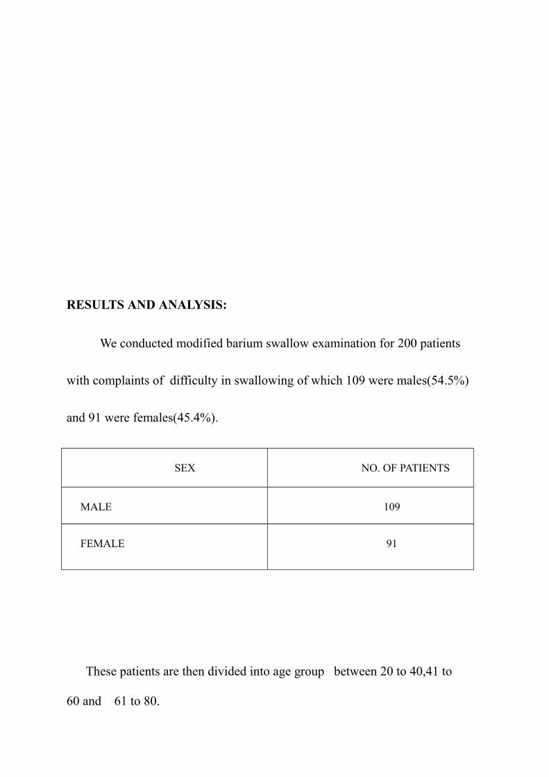
RESULTS AND ANALYSIS:
We conducted modified barium swallow examination for 200 patients
with complaints of difficulty in swallowing of which 109 were males(54.5%)
and 91 were females(45.4%).
SEX NO. OF PATIENTS
MALE 109
FEMALE 91
These patients are then divided into age group between 20 to 40,41 to
60 and 61 to 80.

AGE NO. OF PATIENTS
MALES FEMALES
20 TO 40 YRS 31 42
41 TO 60 YRS 48 43
61 T0 80 YRS 30 6
AGE DISTRIBUTION
About 92 patients were found to have abnormal findings during the study.
Other 108 patients had normal findings on videofluoroscopy.
NORMAL VS ABNORMAL
NORMAL 108
ABNORMAL 92

Abnormal pharyngeal mucosal coating was found in 28 patients,and 16 of
them had past history of stroke for which neurological causes may be
attributed.Another 12 patients however have no significant illnesses.
DISEASE DISTRIBUTION:
Out of the 92 patients with the abnormal videofluoroscopic findings,28 patients
have pharyngeal retention alone with no other structural abnormalities.10
patients were found to have growth in the hypopharynx.8 patients were found to
have growth in the larynx.4 patients were found to have
oesophagealgrowth.Postcricoid web was found in 30 patients.2 patients had
achalasia cardia.4 patients had unilateral vocal cord palsy.
DISEASE No. OF PATIENTS
PHARYNGEAL RETENTION 28
GROWTH HYPOPHARYNX 12
GROWTH LARYNX 8
GROWTH OROPHAYNX 4
GROWTH OESOPHAGUS 4
POSTCRICOID WEB 30
ACHALASIA 2
UNILATERAL VOCAL CORD PALSY
4

This pharyngeal mucosal coating after swallowing is graded as
mild,moderate and severe according to the height of residue in the pyriform
sinuses or vallecule seen in the videofluroscopic images.
SEX PREDILECTION FOR DISEASE:
DISEASE MALES FEMALES
PHARYNGEAL RETENTION
23 5
GROWTH HYPOPHARYNX
9 3
GROWTH LARYNX 8 0
GROWTH OROPHAYNX 3 1
GROWTH OESOPHAGUS 3 1
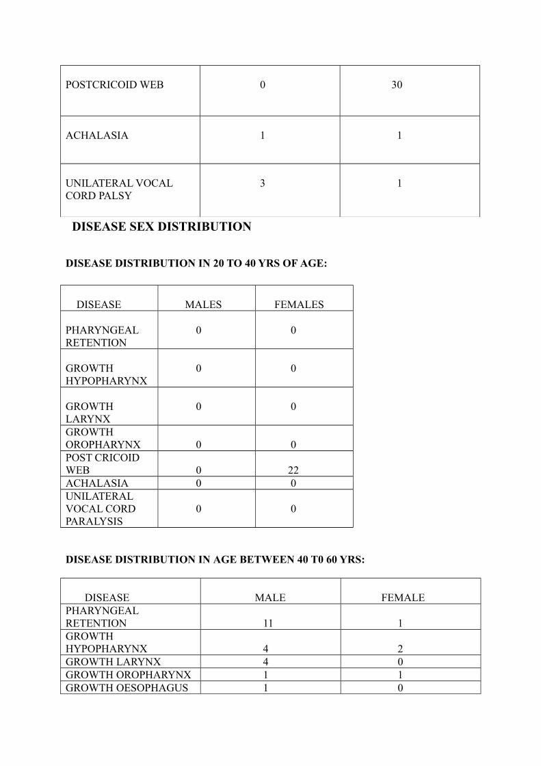
POSTCRICOID WEB 0 30
ACHALASIA 1 1
UNILATERAL VOCAL CORD PALSY
3 1
DISEASE SEX DISTRIBUTION
DISEASE DISTRIBUTION IN 20 TO 40 YRS OF AGE:
DISEASE MALES FEMALES
PHARYNGEAL RETENTION
0 0
GROWTH HYPOPHARYNX
0 0
GROWTH LARYNX
0 0
GROWTH OROPHARYNX 0 0POST CRICOID WEB 0 22ACHALASIA 0 0UNILATERAL VOCAL CORD PARALYSIS
0 0
DISEASE DISTRIBUTION IN AGE BETWEEN 40 T0 60 YRS:
DISEASE MALE FEMALEPHARYNGEAL RETENTION 11 1GROWTH HYPOPHARYNX 4 2GROWTH LARYNX 4 0GROWTH OROPHARYNX 1 1GROWTH OESOPHAGUS 1 0

POSTCRICOID WEB 0 8ACHALESIA 1 1UNILATERAL VOCAL CORD PALSY 1
1
Postcricoid web was found in 30 female patients of which 22 patients fall in the
age group between 20-40 yrs.8 were in between 40-60 yrs of age.
DISEASE DISTRIBUTION IN AGE GROUP 60 T0 80 YRS:
DISEASE MALE FEMALEPHARYNGEAL RETENTION 12 4
GROWTH HYPOPHARYNX 3 1GROWTH LARYNX
4 0GROWTH OROPHAYNX 2 0GROWTH OESOPHAGUS 2 1POSTCRICOID WEB 0 0ACHALASIA 0 0UNILATERAL VOCAL CORD 2 0
AGE WISE DISEASE DISTRIBUTION
Postcricoid web was found in 30 females with anaemia and no males were
found to have this finding.108 patients who are radiologically and clinically
normal were treated appropriately. There was a good response in the patients
who were treated with proton pump inhibitors and domperidone.They were
diagnosed as GERD and managed conservatively.Some patients showed
improvement with placebo treatment itself.In this study, I concentrate mainly on

the patients who were likely to aspirate while swallowing.We identified 28
patients with pharyngel retention and 16 of the patients were given training on
swallowing manouveres as they had severe pharyngeal retention.We studied the
effectiveness of these manouveres again by the videofluoroscopy and we found
that these are very useful in preventing aspiration.
The base of the tongue and the soft palate close the oral cavity posteriorly (arrow) to prevent spill of food into the open larynx.
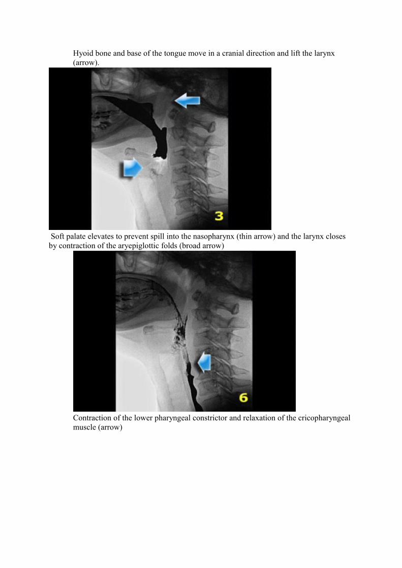
Hyoid bone and base of the tongue move in a cranial direction and lift the larynx (arrow).
Soft palate elevates to prevent spill into the nasopharynx (thin arrow) and the larynx closes by contraction of the aryepiglottic folds (broad arrow)
Contraction of the lower pharyngeal constrictor and relaxation of the cricopharyngeal muscle (arrow)

Epiglottis elevates to regain its resting position and the larynx opens.
Epiglottis in resting position and larynx is open (arrow).

LEFT: Lateral view.Hyoid (H) and tongue base (T) move anteriorly. Left and right piryform sinuses are projected on top of each other. Tip of soft palate (SP) is seen.RIGHT: Valleculae (V) and pyriform sinuses (P).
Asymmetric swallowing due to head turn. The head is turned to the left and contrast is only seen in the Rt.piriform sinus

left asymmetry is seen on the fluorographic study
grade 3 pharyngeal retention

Stasis of contrast at the level of the pyriform sinuses (blue arrows) with subsequent aspiration (yellow arrows)
Grade 3 Pharyngeal retention with aspiration into trachea

mild pharyngeal retention in a case of Lt.sided hemiplegia

Grade 2 pharyngeal retention

Residue in both valleculae and pyriform sinuses
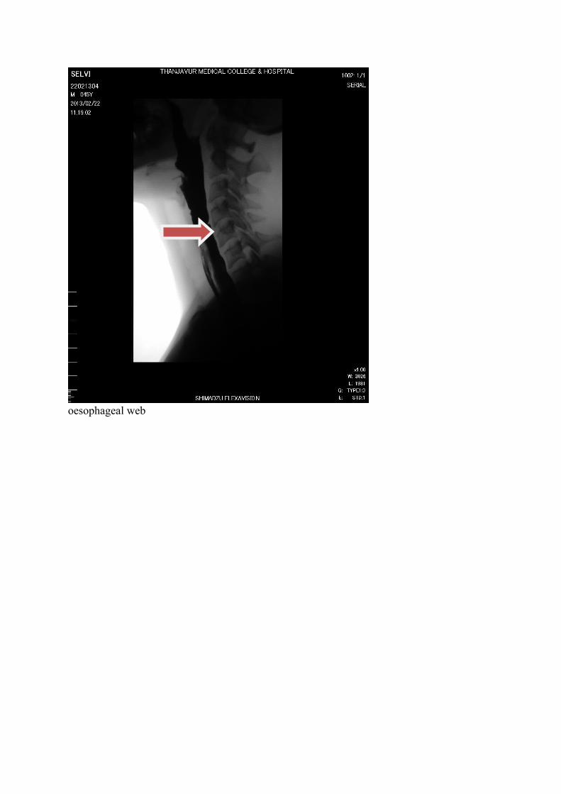
oesophageal web

oesophageal web

DISCUSSION
DISCUSSION:
In our study, we learnt the various phases of swallowing by
modified barium swallow. We found 28 patients with pharyngeal retention
alone.Out of these 28 patients, 16 had past history of stroke and had grade 3
pharyngeal retention.Another 12 patients had grade 1& 2 pharyngeal retention
and they fall between 61-80 yrs of age.We had given training for swallowing
manoeuveres for the patients with severe pharyngeal retention so as to prevent
the aspiration.Another finding we got from our study was postcricoid web
which was typically found in females.Out of the 30 patients,22 were in the age
group between 20-40 and 8 were in the 41-80 yrs of age. These patients were
managed with deworming, iron supplementation,and rigid
oesophagoscopy.They showed significant improvement following this line of
management.
Laryngeal penetration is differentiated from tracheal aspiration by the
involvement of the true vocal cords . Contrast that contacts the true cords, if it is
not immediately removed by a cough reflex, is considered aspiration.
When evaluating laryngeal penetration and tracheal aspiration, it is critical to
assess the timing of the abnormality relative to the swallow reflex[17]. For
example, poor control of the oral bolus will lead to early vallecular spill. If the
vallecula fills, contrast will spill into the laryngeal vestibule and eventually
contact the vocal folds. Because this type of aspiration occurs during the oral

phase, before the initiation of the swallow reflex, it is termed as pre prandial
aspiration.
If the epiglottis fails to invert, or the larynx is otherwise unprotected, the
contrast bolus may enter the laryngeal vestibule during the pharyngeal phase of
the swallow. This type of aspiration is termed prandial aspiration.
If there is a failure of pharyngeal contractility, or the cricopharyngeus muscle
fails to relax, contrast may be retained in the pharynx after the completion of the
entire swallow. This contrast may then drip into the larynx, causing
postprandial aspiration. A diagnosis of postprandial aspiration is sometimes
applied presumptively, when contrast is seen in the trachea late in the
examination but the patient was not seen to aspirate during the videotaped
portions of the swallow.
Whenever laryngeal penetration or tracheal aspiration is identified, comment
should be made about the presence or absence of a cough reflex. Patients with
normal sensation will cough to remove the aspirated material. Silent aspiration
(aspiration without a cough reflex) is a particularly worrisome event, but if it is
identified, it can be addressed with specific swallowing manoeuvers. Silent
aspiration is a radiographic diagnosis.
During the MBS, the patient attempt protective
manoeuvers to determine whether aspiration can be minimized or prevented.
Such manoeuvers include chin tuck and neck rotation. In general, radiographic
images obtained with the patient using protective manoeuvers will not be

diagnostic because of overlapping structures, but the degree of aspiration can
usually be assessed.
Because barium suspension may concrete in the lungs and form a nidus for
infection, patients with copious aspiration should have their studies terminated
as soon as the aspiration is identified. It is thus important for the first swallow to
be properly filmed in case no further swallows are performed.
COMMON DISEASES:
Dysphagia is the most frequent symptom to merit fluoroscopic evaluation
of swallowing. A pharyngo-esophagogram is the examination of choice in this
setting. The most common causes of dysphagia are gastroesophageal reflux
disease (GERD) and its complications, so a pharyngo-esophagogram for
dysphagia must include an evaluation of lower esophageal sphincter
competency. Endoscopy and pH monitoring are more reliable for this
diagnosis[33].Fluoroscopy plays an important role in patients who present with
globus. About half of these patients can be shown to have GERD on endoscopy
or 24-hour pH monitoring. Although an esophagogram will usually be normal,
occasionally a mucosal lesion or extrinsic mass will be identified as the source
of the globussensation. Such lesions may be overlooked if only endoscopy or
pH monitoring are used to evaluate globus.
GERD traditionally causes inflammation in the thoracic esophagus, but in
severe cases, the pharynx may become involved. Laryngopharyngeal reflux, in

which inflammatory changes extend into the laryngeal tissues, is better
evaluated with endoscopy than with fluoroscopy.A frequent finding on
swallowing studies in older individuals is a wavy configuration to the posterior
pharynx caused by anterior osteophytes from the cervical spine. Although this
finding is not usually associated with symptomatology, osteophytes may
interfere with the inversion of the epiglottis, resulting in prandial aspiration.
Large osteophytes may displace the esophagus and simulate an extrinsic mass.
Cricopharyngeal achalasia is a failure of the cricopharyngeus muscle to relax in
response to a food bolus. It appears as a smooth impression on the posterior
pharynx at the level of the C6 vertebral body. Unlike most retropharyngeal
masses, the cricopharyngeus muscle is rarely greater than 1 cm in superoinferior
dimension.
There are many causes of cricopharyngeal achalasia, including amyotrophic
lateral sclerosis, poliomyelitis, pseudobulbar palsy, nasopharyngeal carcinoma,
thyroid myopathy, surgical vagotomy, dermatomyositis, and hiatus hernia.
However, most patients do not have an identifiable cause[34].Other
cricopharyngeal dysfunctions can also be diagnosed radiographically.
Cricopharyngealachalasia is complete incompetence of the cricopharyngeus
muscle and is seen in myotonicdystrophy[35]. Familial dysautonomia manifests
as a delayed opening of the cricopharyngeusmuscle .

There are several types of diverticular outpouchings that arise in the pharynx.
Best known are Zenker’s diverticula, which arise from the posterior wall of the
pharynx just superior to the cricopharyngeus muscle. These lesions are
presumed to represent pulsion diverticula resulting from
cricopharyngealachalasia.Radiographically, a Zenker’s diverticulum appears as
an outpouching from the posterior surface of the pharynx. The diverticulum
may retain contrast between swallows, or it may completely disgorge its
contents. The size of the diverticulum and the caliber of its neck can be assessed
on fluoroscopy. Large Zenker diverticula may compress the
hypopharynx.Pharyngeal pouches are the result of chronically increased
pharyngeal pressure, as in trumpet players and glass blowers. These pouches
arise on the lateral surface of the oropharynx and are thus best viewed in the
frontal projection[36]. Pharyngeal pouches are generally asymptomatic, but large
pouches may cause dysphagia.
Pharyngeal hypocontractility is a frequent finding in elderly patients. It is often
idiopathic, but brainstem diseases (e.g., stroke, demyelination), cranial nerve
dysfunction (particularly IX and X), and diseases of the neuromuscular
junctions or pharyngeal musculature have been implicated.
Diseases that affect the pharyngeal constrictors include myasthenia gravis,
dermatomyositis, systemic lupus erythematosus, myotonic dystrophy, systemic
sclerosis, and oculopharyngeal myopathy. These diseases are usually not

distinguishable radiographically, except myasthenia gravis, which is
characterized by worsening findings over consecutive swallows.
Unilateral pharyngeal hypocontractility can be a confusing radiographic finding.
Asymmetry in the pyriform sinus, with preferential flow into the affected sinus,
may be confused with obstruction of the contralateral pyriform sinus. The
distinction can usually be made with careful analysis of multiple swallows in
both frontal and lateral projections (2).
Although cross-sectional modalities (CT and magnetic resonance imaging) are
the mainstays of pretreatment cancer imaging, fluoroscopy is useful in a few
specific clinical settings. When there is uncertainty about invasion of the
prevertebral fascia from a hypopharyngeal tumor, an esophagogram can
document decreased laryngeal motion to indicate
prevertebralinvasion[37].Pharyngo-esophagography can also document the
mucosal extent of tumors across the postcricoidhypopharynx.
Fluoroscopy plays a specific role in the evaluation of the treated neck. The
Radiologist must be familiar withthe expected appearance of the pharynx after
surgery. For example, after laryngectomy, it is normal for a ridge of soft tissue
to separate the anterior pouch from the remainder of the pharynx. This tissue is
called a pseudo epiglottis because of its radiographic appearance .Jejunal
interposition grafts can create an unusual appearance because the plicacircularis
of the jejunum are identified in the neck . The interposed segment may even
demonstrate peristalsis on video fluorography.

Benign strictures are a common complication of pharyngeal surgery. These
strictures tend to occur at anastomotic sites and are readily evaluated with
fluoroscopy. The strictures that result from radiotherapy tend to affect a longer
segment of the alimentary tract. Strictures generally become symptomatic when
they are less than 13 mm in cross-sectional diameter. If the precise caliber of the
stricture is in question, we must administer a 13-mm barium tablet to see
whether this critical threshold has been breached.
Basedon the findings we got from our study,patients with dysphagia were
divided in to
1.patients with neurogenic dysphagia
2.patients with dysphagia due to head and neck cancer
3.patients with dysphagia due to aging.
We take the pharyngeal residue after swallowing as the risk factor for
aspiration in these patients.We found this factor from 12 patients and it is
observed from our study that the residue in the pharynx is common among
elderly individuals and in patients with history of neurological disorders.The
risk of aspiration is more and is directly proportionate to the amount of
pharyngeal retention in these patients. To recommend a suitable dietary regime,
these patients are tested with various consistencies of barium. Because, we
learnt from several studies that aspiration is more with thickened fluids which

may also aggravate swallowing difficulties.Post swallowing aspiration is
frequently seen in patients with decreased contractions or inadequate opening of
lower oesophagealsphincter.So, these patients are recommended thin liquids
only and the patients who cough on thin liquids may be advised thick
fluids.One study disproved the role of viscosity in pharyngeal retention . They
found no correlation between the viscosity and retention.
Aspiration is the most serious life threatening
complication as it may lead to aspiration pneumonia .We learnt that prior full
bed side examination of the patients to predict aspiration must be carried out
before going for videofluroscopic studies. It includes clinical observation of
swallowing, assessment of intra oral sensibility and functional examination of
the musculature involved in swallowing. One report stated that thorough clinical
examination before study can reach a sensitivity for aspiration upto
80%,specificity to near 70%. Aspiration(food getting into the airway and
lungs)occurs because of incomplete laryngeal closure when the bolus passes
through the larynx which leads to the entry of food or fluid into the larynx
before it is sufficiently closed.This type of aspiration is known as

predeglutitionaspiration.It is frequently found in patients with dysphagia after
stroke and patients with other neurological disorders.Thickened fluids are the
option of dietary modification for this patients because more time is allowed to
establish complete laryngeal closure when the speed of bolus is decreased. A
large trial by Logemenn et al stated that thickened fluids will cause less
aspiration in patients with neurological disorders like Parkinson’s disease,and
dementia.
Laryngeal penetration is defined as the presence of
barium contrast in the laryngeal vestibule that does not pass below the level of
true vocal cords into the airway. Silent aspiration means the aspiration in the
absence of reflexive patients response. An eight point penetration and aspiration
scale developed by Rosenbek Robbins,&Roecker is commonly used for
diagnosing aspiration or penetration. They found that 20 % of normal
individuals have contrast in the laryngeal vestibule during the swallow and on
the penetration aspiration scale they received a score of 2. This implies that the
further entry of the material was restricted before the completion of the
swallow.

TIMING OF THE PHARYNGEAL SWALLOW ONSET:
It is important in determining the interaction of bolus flow
in relation to airway protection. During the pharyngeal stage of swallowing,
there is a period of apnoea in which the respiration stops providing further
protection of airway. Onset of hyolaryngeal elevation is the first radiographic
indication for the onset of laryngeal swallow. It occurs about 500 milliseconds
after the oral bolus starts to move into the pharynx.This is followed by the onset
of laryngeal closure which occurs very soon after hyoid bone began to elevate.
Delayed onset of pharyngeal swallow response is usually the result of decreased
pharyngeal or laryngeal sensation. It is caused by number of conditions
including stroke, Parkinson’sdisease, amyotrophic lateral sclerosis, prior
radiotherapy,post endotracheal intubation or tracheaostomy. If there is more
delay in the pharyngeal swallow initiation response after the bolus is in the
pharynx, the risk of aspiration into the airway will be more. Volume ,texture
and viscosity of the bolus alter the timing of pharyngeal swallow onset with the

begining of vocal cord adduction. Research in normal adults has revealed that
when large liquid boluses are placed in the oral cavity,vocal cord adduction with
the onset of hyolaryngealelevation begins. Average normal pharyngeal delay
time for oral liquid bolus was 0.6 seconds in adults of age between 18 to 80
yrs.Pharyngeal delays in relation to bolus flow is calculated by various
anatomical landmarks by various researches. The onset of pharyngeal swallow
is defined as the time when head of the bolus passes the ramus of mandible and
the tongue base. Due to the reduced sensory awareness of the bolus,pharyngeal
delay time is increased in the old age. Before the closure of the airway closure
and the elevation of hyolarynx, the vallecula is occupied by the bolus.
ORAL AND PHARYNGEAL TRANSIT:
The consistency of food or liquid being swallowed ,the age and sex of the
patient as well as the variation the propulsive forces decide this duration.
Swallowing efficiency is generally calculated by the duration of bolus
transportation through the oral cavity and pharynx, the number of swallows
needed to clean the bolus whether it is solid or liquid. It gives a rough guide for
efficiency of swallow. Normally, a 5 to 30ml of bolus can be cleaned in a single

swallow and may be done by a second cleaning swallow.Several swallows are
required to clean boluses with hard textures.Rademaker et al developed a
measure that depicts the interaction between the speed of flow of bolus and
efficiency with which the bolus is swallowed from the oropharynx taking
aspiration into account.This precisely assesses the oropharyngeal swallow
efficiency.
THERAPEUTIC STRATEGY:
Major advantage of videofluroscopic study is that
the effectiveness of the therapeutic strategies which could be either
rehabilitative or compensatory can be assessed. The aim of compensatory
strategies is to correct the disorder of swallowing by altering the flow of bolus.
Head postion changes can alter the axis of oral,pharyngeal and laryngeal
subsites and it can reduce the angle of opening of laryngeal inlet.Hyolaryngeal
excursion and propulsive forces of the muscles of the pharynx can be increased
by chin down(chin tuck)position.Anyhow,the risk of aspiration is increased
when the pyriform sinuses are filled with barium before the onset of pharyngeal
swallow.Because, the emptying of pyriform sinuses and elevation of larynx

coincides. Testing with various consistencies of boluses permits one to
formulate a treatment plan.The effectiveness of these trials were tested by
modified barium technique in 12 patients of our study.During the study, we
trained the patients on how to accurately perform the maneouvres and postural
techniques.We found as disadvantages of this maneouvres are it requires full
co operation of the patient who must have the mental and cognitive ability to
understand the importance and method of procedures.
Summary:
In a normal modified barium swallow technique, the
images are viewed in the lateral aspect first. There should be co ordinated
tongue movements for manipulation of liquid and semisolid boluses. The bolus

should be in contact with dorsum of the tongue.The base of the tongue and the
soft palate must be ready for action before the onset of pharyngeal
swallow.When the tongue moves upwards and backwards, the soft palate
contacts the posterior pharyngeal wall and shuts off the nasopharynx.Once the
bolus reach the tongue base,it will contact the posterior pharyngeal wall and the
pharyngeal phase of swallowing is initiated.Downward and forward movement
of the hyolaryngeal complex followed by downward tilting of epiglottis and
adduction of arytenoids will close the laryngeal vestibule.When the bolus
reaches the hypopharynx,cricopharynx will open and there should be no
indendation in the posterior part of it.It will be closed once the peristalsis in the
cervical oesophagusstarted.There should be no pharyngeal residue after this
event. Solid residue if present, should be cleaned on subsequent swallowing of
saliva.Residue in the pharynx is usually seen when there is limited motion of
hyoid with limited forward and upward movement of pharynx with swallowing.
As a result, the suprahyoid portion of the epiglottis does not move forward
against the base of tongue and any residual material in the vallecula cannot be
squeezed out. This can occur in neurological disorders like Parkinson’s disease

or after surgey or radiotherapy . Residue in pyriform sinus can also occur when
there is decreased pharyngeal motion as seen in vagal neuropathies. The
decreased constrictions which limits the pharyngeal pressure which normally
helps to push the bolus past the cricopharyngeal spasm can also lead to
pyriformresidue.These type of residue can lead to secondary aspiration or
penetration.The bolus should occupy the lateral pharyngeal walls symmetrically
which can be seen in the anteroposterior view of modified barium swallow
images. The bolus then passes into the cervical oesophagus.Epiglottic deflection
may be viewed as an arc shaped as its dense folding displaces the barium .The
fully adducted vocal folds may be seen when the barium eventually enters
into the cervical oesophagus.In our study, retention of the barium in the
pharynx was a single factor that was taken in to an account for predicting
aspiration and planning for compensatory therapeutic strategies.
CONCLUSION:
From my study ,I came to the conclusion of understandingthe normal and
abnormal biomechanical events of the swallowing that drive the bolus from the
oral cavity to the pharynx and into the oesophagus is necessary for the accurate

diagnosis of oropharyngeal swallowing disorders.Modified Barium Swallow
plays a critical role in the evaluation of swallowing and it provides extensive
information about the swallowing events.Mild and moderate pharyngeal
retention should be considered as normal in older individuals provided other
causes are ruled out.Swallowingmanoueveres are effective in preventing
aspiration in patients with severe pharyngeal retention. Help of expertise of the
Radiologist and Speech language pathologist permit the better evaluation of
therapeutic strategies aimed at improving the chance that the patient with
dysphagia can eat safely and effectively.
BIBILIOGRAPHY:
1. Gustafson-Yoshida N, Maglinte DD, Hamaker RC, et al. Evaluation of swallowing
disorders: the modified barium swallow. Indiana Med 1990;83:892–895.
2. Ekberg O, Olsson R. Dynamic radiology of swallowing disorders. Endoscopy
1997;29:439–446.

3. Aviv JE. Prospective, randomized outcome study of endoscopy versus modified barium
swallow in patients with dysphagia. Laryngoscope 2000;110:563–574.
4. Jones B, Donner MW. Normal and abnormal swallowing: imaging in diagnosis and
therapy. New York: Springer-Verlag, 1991:xvi, 235.
5.Leonard R, Kendall K, McKenzie S. UES opening and cricopharyngeal bar in
nondysphagic elderly and nonelderly adults. Dysphagia 2004;19:182
6.Olle Ekberg,Feinberg11. Cheryl K. Chamberlain, John E. Cornell, Michèle J.
Saunders, John P. Hatch, Rosemary S. Shinkai, Chih-KoYeh. 2007.
7.Intra-oraltactile sensation and aging in a community-based population. Aging
Clinical and Experimental Research 19:2, 85-90. [CrossRef]
8. Karen Kost, Richard PayneLaryngopharyngeal Reflux in the Elderly 383-394. [CrossRef]
9. Bernhard L. Partik, Martina Scharitzer, GerdSchueller, Martin Voracek, Wolfgang
Schima, EwaldSchober, Michael R. Mueller,Ann N. Leung, Doris-Maria Denk, Peter
Pokieser. 2003. Videofluoroscopy of Swallowing Abnormalities in elderly
6.66ccxxcxcxcxccxcxxcxcxcsdsdscxzc6ede66666 gvef
10.Langmore SE, Terpenning MS, Schork A, et al. Predictors of aspiration pneumonia:
how important is dysphagia?Dysphagia 1998; 13:69-81
11. Feinberg MJ, Ekberg O. Videofluoroscopy in elderly patients with aspiration:
importance of evaluating both oral and pharyngeal stages of deglutition. AJR 1991; 156:293-296
12. Dodds WJ, Stewart ET, Logemann JA. Physiology and radiology of the normal oral
and pharyngeal phases of swallowing. AJR 1990; 154:953-963
13. Olsson R, Nilsson H, Ekberg O. Simultaneous videoradiography and computerized
pharyngealmanometry: videomanometry. ActaRadiol 1994; 35:30-34
14. Ekberg O. The normal movements of the hyoid bone during swallow. Invest
Radiol 1986; 21:408-410
15. Ekberg O, Nylander G. Cineradiography of the pharyngeal stage of deglutition in

150 individuals without dysphagia. Br J Radiol 1982; 55:253-257
16. Robbins J, Coyle J, Rosenbek J, Roecker E, Wood J. Differentiation of normal and
abnormal airway protection during swallowing using the penetration—aspiration
scale. Dysphagia 1999; 14:228-232
17. Friedman B, Frazier JB. Deep laryngeal penetration as a predictor of
aspiration. Dysphagia 2000; 15:153-158
18.Frederick MG, Ott DJ, Grishaw EK, Gelfand DW, Chen MYM. Functional
abnormalities of the pharynx: a prospective analysis of radiographic abnormalities
relative to age and symptoms. AJR 1996; 166:353-357
19. Pick N, McDonald A, Bennett N, et al. Pulmonary aspiration in a long-term care
setting: clinical and laboratory observations and an analysis of risk factors. J Am
GeriatrSoc 1996; 44:763-768
20. Bryan CS, Reynolds KL. Bacteremic nosocomial pneumonia: analysis of 172
episodes from a single metropolitan area. Am Rev Respir Dis 1984; 129:668-671
21. Jones B, Donner MW. Examination of the patient with dysphagia. Radiology 1988; 167:319-326
22. Levine MS, Rubesin SE. Radiologic investigation of dysphagia. AJR 1990; 154:1157-1163
23. Auffermann W, Geisel T, Wohltmann D, Gunther RW. Tissue reaction following
endobronchial application of iopamidol and ioxithalamate in rats. Eur J Radiol 1988; 8:13-17
24. Jones B, Donner M. The tailored examination. In: Jones B, Donner M, eds. Normal
and abnormal swallowing. New York: Springer, 1991: 33-50
25. Kahrilas PJ, Logemann JA, Lin S, Ergun GA. Pharyngeal clearance during \
swallowing: a combined manometric and videofluoroscopic
study. Gastroenterology 1992; 103:128-136
26. Olsson R, Castell J, Johnston B, Ekberg O, Castell DO. Combined
videomanometric identification of abnormalities related to pharyngeal retention. AcadRadiol 1997; 4:349-354

27. Groher ME. The detection of aspiration and videofluoroscopy.
(editorial) Dysphagia 1994; 9:147-148
28. Langmore SE, Schatz K, Olson N. Endoscopic and videofluoroscopic evaluations
of swallowing and aspiration.AnnOtolRhinolLaryngol 1991; 100:678-681
29. Ekberg O, Feinberg MJ. Altered swallowing function in elderly patients without
dysphagia: radiologic findings in 56 cases. AJR 1991; 156:1181-1184
30. Dejaeger E, Pelemans W, Ponette E, Joosten E. Mechanisms involved in
postdeglutition retention in the elderly.Dysphagia 1997; 12:63-67
31. Dodds WJ, Logemann JA, Stewart ET. Radiologic assessment of abnormal oral and
pharyngeal phases of swallowing. AJR 1990; 154:965-974
32. Perlman AL, Booth BM, Grayhack JP. Videofluoroscopic predictors of aspiration
in patient with oropharyngeal dysphagia. Dysphagia 1994; 9:90-95
33. Richter JE. Typical and atypical presentations of gastroesophageal reflux disease.
The role of esophageal testing in diagnosis and management.GastroenterolClin North
America 1996;25:75
34. Eisenberg RL. Gastrointestinal radiology: a pattern approach Philadelphia:
Lippincott-Raven Publishers, 1996:xiv, 1194.
35. Marcon M, Briani C, Ermani M, et al. Positive correlation of CTG expansion and
pharyngoesophageal alterations in myotonic dystrophy patients. Ital J NeurolSci
1998;19:75–80.
36. Rubesin SE, Yousem DM. Structural abnormalities of the pharynx. In: Levine MS,
Gore RM, eds. Textbook of gastrointestinal radiology Philadelphia: W.B. Saunders, 2000
37. Ekberg O. Functional abnormalities of the pharynx. In: Freeny PC, Stevenson GW,
Margulis and Burhenne's alimentary tract radiology St. Louis: Mosby-Year Book, 1994.
38 M. Klahn and A. Perlman, “Temporal and durational patterns
associating respiration and swallowing,"Dysphagia, vol. 13, pp. 131-138, 1999.
39.Investigation of Accelerometry, Mechanomyography, and
Nasal Airflow Signals for Abnormal Swallow Detection
byJoonwu tspace.library.utoronto.ca/bitstream/1807/26467/3/ Lee_Joonwu_200911

S.NO
NAME AGE/SEX
I.P.NO Radiological diagnosis
1 THAYAMMA 45/F 1462348
POST CRICOID WEB
2 DANIEL 66/M 016483 GROWTH LARYNX3 GOVINDARAJ 24/M 16327 NORMAL STUDY4 REVATHY 49/F 146232
8GROWTH HYPOPHARYNX
5 RAJENDRAN 34/M 16417 NORMAL STUDY6 BANUMATHY 45/F 16180 UNILATERAL VOCAL CORD
PALSY7 MANORAMMAL 29/F 145918
2POST CRICOID WEB
8 SATHIYAPPAN 33/M 12097 NORMAL STUDY9 SUNDRAMBAL 28/F 145871
6POST CRICOID WEB
10 MOORTHY 34/M 1458121
NORMAL STUDY
11 KRISHNAMOORTHY 39/M 13847 NORMAL STUDY12 MOHAMMED
HANIFA65/F 145636
9PHARYNGEAL RETENTION
13 MARIMUTHU 25/M 12634 NORMAL STUDY14 KALIYAPERUMAL 28/M 11157 NORMAL STUDY15 JEYAPRATHA 49/F 12499 POST CRICOID WEB16 TAMILSELVAN 70/M 14372 GROWTH LARYNX17 MANIKKAM 33/M 3575 NORMAL STUDY18 MURUGESAN 32/M 3578 NORMAL STUDY19 MUTHUSELVI 55/F 4677 GROWTH HYPOPHARYNX20 THIYAGARAJAN 34/M 2605 NORMAL STUDY21 PONNAMMAL 33/F 4773 POST CRICOID WEB22 RAJENDRAN 28/M 6030 NORMAL STUDY23 KALIYAMOORTHY 44/M 5075 GROWTH HYPOPHARYNX24 CHINNACHI 36/F 4209 POST CRICOID WEB25 ABDUL 23/M 4341 NORMAL STUDY26 GANESAN 35/M 5222 NORMAL STUDY27 SELVAM 36/M 6113 NORMAL STUDY28 IRUDHYAMARY 72/F 5395 PHARYNGEAL RETENTION29 SHANMUGAM 23/M 30759 NORMAL STUDY30 RAJENDRAN 36/M 144019
5NORMAL STUDY
31 INDIRANI 38/F 29672 POST CRICOID WEB32 MOHAMMED RAFIQ 39/M 14372 NORMAL STUDY33 PERIYASAMY 54/M 12499 PHARYNGEAL RETENTION34 SURESH 40/M 11157 NORMAL STUDY35 PARIMALAM 50/F 12634 POST CRICOID WEB36 BALAGANESH 31/M 145636
9NORMAL STUDY
37 SELVI 66/f 013847 GROWTH HYPOPHARYNX38 BATCHABEEVI 39/F 145871 POST CRICOID WEB

LIST OF ABBREVIATIONS
CN - Cranial Nerve
MBS - Modified Barium Swallow
FEES - Fibreoptic endoscopic evaluation of swallowing
MMG - MechanoMyoGraphy
EMG - Electro MyoGraphy