Embed Size (px)
Citation preview
Evaluation of NGS and other methods for ALK translocation detection in NSCLC patients in Europe: results of the European Thoracic Oncology Platform (ETOP) Lungscape Project Stephen Finn University of Dublin Trinity College and St. James’s Hospital, Dublin, Ireland

Disclosures
• Commercial research support: Janssen and Astellas
• Honoraria: Merck, Pfizer, Astra Zeneca, Roche, Boehringer Ingelheim, BMS

Current ALK testing • ALK translocation: 2–7%
– inversion or translocation of chromosome 2p – resulting in transforming fusion gene – targetable by TKI
• Reported prevalence varies across studies as a consequence of patient-selection criteria and detection methodology
• FISH and, now, IHC are widely used • However, RT-PCR is possible, and NGS is the emerging major testing platform because
of the possibility to detect multiple biomarkers using both DNA and RNA (EGFR, KRAS, BRAF, HER2 mutation, ALK, ROS1, RET, NTRK1–3)
• Question of LDTs vs CE-IVD also in the mix for laboratories to consider ALK, anaplastic lymphoma kinase; BRAF, B-Raf proto-oncogene; CE-IVD, Conformité Européene in vitro diagnostics; DNA, deoxyribonucleic acid; EGFR, epidermal growth factor receptor; FISH, fluorescent in-situ hybridization; HER2, human epidermal growth factor receptor 2; IHC, immunohistochemistry; KRAS, Kirsten rat sarcoma viral oncogene homologue; LDTs, laboratory developed tests; NGS, next generation sequencing; NTRK1–3, neurotrophic receptor tyrosine kinase 1–3; RET, rearranged during transfection; RNA, ribonucleic acid; ROS1, reactive oxygen species 1; RT-PCR, reverse transcription-polymerase chain reaction; TKI, tyrosine kinase inhibitor.
Soda M et al. Nature. 2007;448:561-6.
Wojas-Krawczyk K, et al. Contemp Oncol. 2013;17:484-92.

Lungscape • Translational research program • Designed, implemented, and conducted
by ETOP • Involving leading hospitals across Europe
and beyond • Multiple decentralized biobanks • Virtual central iBiobank • EQA • Follow-up
EQA, external quality assessment; ETOP, European Thoracic Oncology Platform. Available from: www.etop-eu.org/index.php?option=com_content&
view=article&id=115523&Itemid=197. Accessed August 2017.
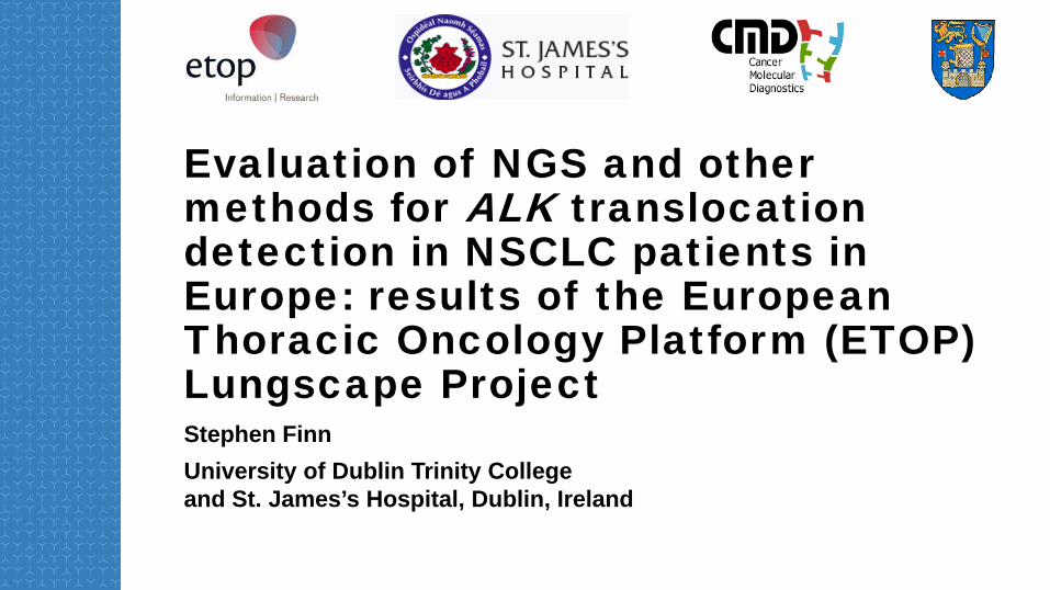
• Retrospective surgical cohort • 16 European centers • 1,281 patients screened (adenocarcinoma only) • 6.2% positive by IHC • 2.2% (at least) positive by FISH • IHC is proposed as a screening tool for further FISH assessment • H-score cut-off of 120 for 5A4 clone
Prevalence and clinical outcomes for patients with ALK-positive resected stage 1 to 3 adenocarcinoma: results from the European Thoracic Oncology Platform Lungscape Project
Blackhall FH, et al. J Clin Oncol. 2014;32:2780-7.

Current study
• A comparison of multiple technologies for assessment of ALK status in the cohort published in 2014
• A unique opportunity to assess in a large European cohort
• Refine and optimize diagnostic strategies
Finn S, et al. Presented at WCLC 2016. P1.02–025.

Methods
NSCLC, non-small-cell lung cancer. *IVD.For In vitro Diagnostc Use
• 96 cases selected from ETOP Lungscape iBiobank – 1,772 screened by IHC – stage 1–3 – based on any degree of IHC staining
– 5A4 (Novocastra, UK) – decentralized, EQA-controlled
• FISH on all cases with positive staining – decentralized, EQA-controlled
• RT-PCR for most frequent fusions – centralized
• Targeted NGS – centralized – Oncomine™ Solid Tumour Fusion Transcript kit* screens for
– 70 ALK, RET, and ROS1-specific fusion transcripts – novel ALK translocations using 3′–5′ ALK gene expression “
assay”
• Macro-dissection for enrichment
• The same RNA used for both NGS and RT-PCR
Finn S, et al. Presented at WCLC 2016. P1.02–025.

3′–5′ assay for targeted NGS • The assay measures the difference in expression between the
5′ assay and the 3′ assay of the ALK gene
• Samples that do not contain a fusion are expected to have similar expression of the 5′ assay compared with the 3′ assay of the driver gene
• Samples that contain a fusion are often expected to have elevated expression of the 3′ assay compared with the 5′ assay
• The 3′–5′ assay is therefore included for 2 alternate purposes – to confirm a specific fusion – to screen for a fusion other than those specifically sequenced
Finn S, et al. Presented at WCLC 2016. P1.02–025.
No evidence Uncertain Strong evidence ALK ≤ 0.001 0.001–0.015 ≥ 0.015

Results 1 • IHC screen
– primary selection criterion (n = 96)
• IHC H-score 120 positive (n = 23)
• FISH – 88 cases: 24 positive, 64 negative
• Tumour cellularity – median 70% (range 10–90) – RNA yield: median 47 ng/μL
(range 9–300)
• RT-PCR – successful in 77 of 96 cases (80.2%)
– B-actin-amplified – ALK rearrangement detected
in 16 cases
Negative: 61 (79.2%) Positive: 16 (20.8%)
Not available: 19 (19.8% of 96)
Negative: 67 (74.4%) Positive: 23 (25.6%)
Not available: 6 (6.3% of 96) 70 cases with
available results for all 4 methods
Negative: 64 (72.7%) Positive: 24 (27.3%)
Not available: 8 (8.3% of 96)
1+: 58 (60.4%) 2+: 16 (16.7%) 3+: 22 (22.9%)
< 120: 69 (75.0%) ≥ 120: 23 (25.0%)
Not available: 4 (4.2% of 96)
96 ETOP Lungscape cases
RT-PCR NGS
FISH IHC level H-score
Finn S, et al. Presented at WCLC 2016. P1.02–025.

Results 2 Targeted NGS
• 95 cases analysed and passed initial QC
• Specific ALK rearrangement detected in 15 cases
• Imbalance assay performed on remaining cases – 6 cases: strong evidence of imbalance – 11 cases: uncertain (considered failed assays) – 63 cases: no evidence – finally 77 cases accepted
– 21 positive and 56 negative
QC, quality control.
3′–5′ Assay
No evidence Uncertain Strong evidence
≤ 0.001 0.001–0.015 ≥ 0.015
63 11 6
Specific ALK assay Positive For imbalance
15 80
Finn S, et al. Presented at WCLC 2016. P1.02–025.

Box-plots of 3′–5′ assay scores by ALK status
Note 1: ALK status is defined by the concordant result of at least 2 of the 3 other methods: RT-PCR, FISH, IHC H-scoring. Note 2: 4 extreme outliers (with values of −1.96, −0.82, −0.71, −0.21) are excluded from the display of negative group.
Negative Positive
3′–5
′ass
ay s
core
s
0.05
0
−0.05
−0.10
−0.15
Finn S, et al. Presented at WCLC 2016. P1.02–025.

Concordance of methodologies FISH positive FISH negative FISH not tested
NGS-positive NGS-positive (strong imb. only) NGS-negative NGS failed (uncertain imb.) NGS failed NGS not tested for imb.
FISH-positive FISH-negative FISH not tested
RT-PCR-positive RT-PCR-negative RT-PCR not tested
H-score not tested
H-s
core
300
250
200
150
100
50
0 3 3 3 3 3 3 3 3 3 3 3 3 3 3 3 3 2 3 3 3 3 2 2 2 1 1 1 1 1 1 1 1 3 2 2 2 2 2 2 2 2 2 3 1 1 1 1 1 1 1 1 1 1 1 1 1 1 1 1 1 1 1 1 1 1 1 1 1 1 1 1 1 1 1 1 1 1 1 1 1 1 1 1 1 1 1 1 1 1 1 1 1 1 2 2 2
FISH RT-PCR IHC
Finn S, et al. Presented at WCLC 2016. P1.02–025.

Variants detected
6
4
4
1
3
1
4
0 1 2 3 4 5 6 7
Strong imbalance
E6(a/b/b)A20
E6(a/b)A20
E6bA20
E6aA20
E20A20
E13A20
Finn S, et al. Presented at WCLC 2016. P1.02–025.

Summary: defining a gold standard • Joint concordance of all 4 methods
– evaluable in 60 cases – 55 cases showed full agreement
• “Gold standard” to determine sensitivity and specificity – gold standard positivity was defined as positive using both IHC-positive threshold
(H-score > 120) and FISH positivity – evaluable in 82 cases
“Gold standard” positive Sensitivity (%) Specificity (%)
NGS 85 87.1
RT-PCR 70 79
Finn S, et al. Presented at WCLC 2016. P1.02–025.

Conclusions
• NGS is a useful screening tool for ALK rearrangement status, superior to RT-PCR when RNA yield is limited
• When using NGS, it is critically important to integrate the 3′–5′ assay and to confirm with one or more additional methods in the “imbalance” cases
• These data further highlight the possibility of missing actionable rearrangements when only one screening methodology is available
Finn S, et al. Presented at WCLC 2016. P1.02–025.

Conclusions (cont.)
• If maximal sensitivity and specificity for ALK rearrangement detection are sought, confirmation of ALK status using 2 techniques or more in discordant cases is mandatory
• Accepted and recommended IHC should be the first step (inexpensive and easy technique for screening out negative cases); further confirmation by a molecular technique FISH (cytogenetics) or RT-PCR vs NGS (RNA-based techniques) should ideally follow
• In discordant cases, a third type of assay could be applied
Finn S, et al. Presented at WCLC 2016. P1.02–025.

Proposal
ALK assessment
2 techniques of: NGS, IHC, and/or FISH
Concordant− No ALK rearrangement
Concordant+ ALK
rearrangement present
Discordant Third technique
Panel testing
• More information – DNA
– BRAF, HER2 exon 20, MET exon 14, KRAS, EGFR – RNA
– RET, ROS1, NTRK, MET exon 14
• Quality assurance
• Optimal use of scant material
• Access to clinical trials

ETOP NGS ALK I. Letovanec1 *, S. Finn2 *, P. Zygoura3, P. Smyth2, A. Soltermann4, L. Bubendorf5, E.-J. Speel6, A. Marchetti7, D. Nonaka8, K. Monkhorst9, H. Hager10, M. Martorell11, A. Sejda12, R. Cheney13, J. Hernandez-Losa14, E. Verbeken15, W. Weder4, S. Savic5, A. Di Lorito7, A. Navarro11, E. Felip16, A. Warth17, P. Baas18, P. Meldgaard10, F. Blackhall19, A.-M. Dingemans6, H. Dienemann17, R. Dziadziuszko20, J. Vansteenkiste15, Cathal O'Brien2, T. Geiger21, J. Sherlock22, J. Schageman23, U. Dafni24, R. Kammler21, K.M. Kerr25, E. Thunnissen26, R. Stahel27, S. Peters1 on behalf of the ETOP Lungscape Consortium21 1Centre Hospitalier Universitaire Vaudois CHUV, Lausanne/Switzerland, 2St James's Hospital and Trinity College, Dublin/Ireland, 3Frontier Science Foundation-Hellas, Athens/Greece, 4University Hospital Zurich, Zurich/Switzerland, 5University Hospital Basel, Basel/Switzerland, 6Maastricht University Medical Center, Maastricht/Netherlands, 7Ospedale Clinicizzato, Chieti/Italy, 8The Christie NHS Foundation Trust, Manchester/United Kingdom, 9The Netherlands Cancer Institute, Amsterdam/Netherlands, 10Aarhus University Hospital, Aarhus/Denmark, 11Consorcio Hospital General Universitario de Valencia, Valencia/Spain, 12Medical University Gdansk, Gdansk/Poland, 13Roswell Park Cancer Institute, Buffalo, NY/United States of America,14Vall d'Hebron University Hospital, Barcelona/Spain, 15University Hospital KU Leuven, Leuven/Belgium, 16Vall D'Hebron University Hospital, Barcelona/Spain, 17Heidelberg University Hospital, Heidelberg/Germany, 18The Netherlands Cancer Institute-Antoni Van Leeuwenhoek Hospital, Amsterdam/Netherlands, 19Christie Hospital NHS Foundation Trust, Manchester/United Kingdom, 20Medical University of Gdansk, Gdansk/Poland, 21European Thoracic Oncology Platform, Bern/Switzerland, 22Thermo Fisher Scientific, Paisley/United Kingdom, 23Thermo Fisher Scientific, Austin, TX/United States of America, 24Frontier Science Foundation-Hellas & University of Athens, Athens/Greece, 25Aberdeen University Medical School, Aberdeen/United Kingdom, 26VU University Medical Center, Amsterdam/Netherlands, 27University Hospital Zurich, Zurich/Switzerland.
Finn S, et al. Presented at WCLC 2016. P1.02–025.

Thank you for your attention!

Thermo Fisher Scientific and its affiliates are not endorsing, recommending, or promoting any use or application of Thermo Fisher Scientific products presented by third parties during this seminar. Information and materials presented or provided by third parties are provided as-is and without warranty of any kind, including regarding intellectual property rights and reported results. Parties presenting images, text and material represent they have the rights to do so.