Embed Size (px)
Citation preview
EVALUATION OF MANDIBULAR RECONSTRUCTION TECHNIQUES
FOLLOWING RESECTION OF BENIGN & MALIGNANT TUMOURS IN
ORAL REGION
Dissertation submitted to
THE TAMILNADU Dr. M.G.R. MEDICAL UNIVERSITY
In partial fulfillment for the Degree of
MASTER OF DENTAL SURGERY
BRANCH III
ORAL AND MAXILLOFACIAL SURGERY
APRIL 2015
CERTIFICATE
This is to certify that this dissertation titled “EVALUATION OF
MANDIBULAR RECONSTRUCTION TECHNIQUES FOLLOWING
RESECTION OF BENIGN & MALIGNANT TUMOURS IN ORAL REGION” is
a bonafide record of work done by Dr. M.SUGANTHI under my guidance during her
postgraduate study period between 2012-2015.
This dissertation is submitted to THE TAMILNADU Dr. M.G.R.
MEDICAL UNIVERSITY, in partial fulfillment for the degree of MASTER OF
DENTAL SURGERY in Branch III – ORAL AND MAXILLOFACIAL
SURGERY.
It has not been submitted (partially or fully) for the award of any other degree
or diploma.
Professor, HOD and Guide Principal
Dr.L.DEEPANANDAN, M.D.S., Dr.V.PRABHAKAR, M.D.S.,
Department of Oral & Maxillofacial surgery, Sri Ramakrishna Dental College & Hospital,
Sri Ramakrishna Dental College & Hospital, Coimbatore.
Coimbatore.
Candidate
Dr.M.SUGANTHI
Department of Oral & Maxillofacial surgery,
Sri Ramakrishna Dental College & Hospital,
Coimbatore.
Date:
Place: Coimbatore
ACKNOWLEDGEMENT
This thesis became a reality due to the efforts of a large number of people to
whom I am greatly indebted.
First and foremost I would like to express my sincere gratitude and thanks to my
teacher and guide Dr.L.Deepanandan, M.D.S., Professor and Head Of the Department
for his continuous support, patience, motivation and immense knowledge. His valuable
guidance has helped me throughout my postgraduate study. I am indebted to him.
I would like to thank my teachers Dr.M.S.Senthil kumar, M.D.S., Associate
Professor and Dr.R.Kannan, M.D.S., Reader for their constant encouragement,
guidance and support throughout my course.
I avail this opportunity to express my deep sense of gratitude to my teacher
Dr.R.S.Karthik, M.D.S., Reader for his motivation, immense help and guidance
throughout the study. Without his help this study would not have been complete.
I also would like to thank my teachers Dr.M.A.I.Munshi, M.D.S., Reader,
Dr.V.Sundararajan M.D.S., and Dr.R.Vijay, M.D.S., Senior Lecturers for their
everlasting guidance, cooperation and support throughout my course.
I thank Dr.Guhan, MD, DM, Director, Dr.Karthikesh, Mch and Dr.Bhargavi,
Mch, Surgical Oncologists, Sri Ramakrishna Institute of Oncology for their guidance
and encouragement throughout the course.
I express my gratitude and heartfelt thanks to Dr.V.Prabhakar, M.D.S., Principal
and Dr.Muralimohan, M.D.S., Director for giving me the opportunity to utilize the
facilities in the institution for the study.
It would be ungrateful of me if I do not thank my batch mate Dr.V.R.Rajinikanth,
my seniors and my juniors for their timely help and support.
This acknowledgement would be incomplete without the mention of my parents
and sister for their love, understanding, support and innumerable sacrifices without
whom my education would have been just a dream.
Above all, I bow to the ALMIGHTY!
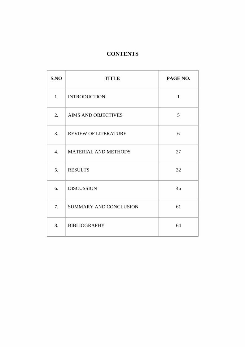
CONTENTS
S.NO TITLE PAGE NO.
1. INTRODUCTION 1
2. AIMS AND OBJECTIVES 5
3. REVIEW OF LITERATURE 6
4. MATERIAL AND METHODS 27
5. RESULTS 32
6. DISCUSSION 46
7. SUMMARY AND CONCLUSION 61
8. BIBLIOGRAPHY 64

Introduction
Introduction
1
Mandibular defects occur as a result of loss of continuity of bone due to
resection of benign or malignant tumours, trauma or inflammatory disease.7 The
mandible has to be reconstructed efficiently as unrepaired defects lead to severe
facial disfigurement, loss of functions such as speech, chewing and swallowing and
ultimately affect the patient’s quality of life.44
But attaining good outcomes in mandibular reconstruction is usually
challenging to a surgeon despite the huge developments in reconstruction techniques
over a century. The reasons for the same are many. The mandible is the only load-
bearing bone of skull and needs to withstand the forces transmitted through
mastication. The goals of mandibular reconstruction are not only to re-establish the
continuity of the mandible but also to restore function. The return of function
includes speech, swallowing, and chewing.47
In case of malignancy, the resection
not only involves mandibular bone but also the adjacent soft tissues. This
complicates the attempts at reconstruction which is further complicated by
radiotherapy which is often necessary in cases of malignant tumours.7
The techniques of mandibular reconstruction have come a long way starting
from the free bone grafts first used by German pioneers7, through pedicled grafts,
reconstruction plates, microvascular free flaps, particulate cancellous marrow grafts,
modular endoprosthesis to the present day distraction osteogenesis22
and tissue
engineering techniques.4
Introduction
2
The free bone grafts first used by Sykoff to reconstruct mandibular defect are
still a good option for defects that are not bigger than approximately 5 cm, provided
the soft tissues are in good condition.7
The reconstruction plates first used by Spiessl in 1979 which were made with
the intention of bridging defects while stabilizing remaining segments and
maintaining occlusion and facial contour are currently used to fix corticocancellous
blocks or vascularized bone grafts to the remaining mandible.7
The pectoralis major myocutaneous flap introduced into head and neck
reconstruction by Ariyan in 1979 raised the bar for head and neck reconstruction and
still remains one of the most commonly used pedicled flap along with reconstruction
plates for mandibular reconstruction in India owing to the advantages that this
method of reconstruction offers such as lesser cost, simplicity of harvesting,
proximity to head and neck or as a salvage surgery when free flap failure occurs.5
The bulk of flap provides good contour when needed in reconstruction of massive
soft tissue defects in cases of locally advanced disease. Several disadvantages of the
flap such as reduced neck mobility, thickness of flap due to excess subcutaneous fat,
complications like partial or complete flap necrosis, fistula formation, dehiscence,
infection following radiotherapy had led to replacement of this workhorse flap by
free flap reconstruction.5
The introduction of microvascular surgery by McKee through the use of a
microvascular free rib graft for mandibular reconstruction in 1971 brought about a
revolution in mandibular reconstructive surgery. At present, the donor sites used
Introduction
3
most commonly for mandibular reconstruction are the radial forearm, scapula, iliac
crest and fibula.7
Though the microvascular free flaps have a high success rate with advantages
such as durable reconstruction, lengthy bone segment with possibility of placing
implants and are usually unaffected by radiation therapy the certain disadvantages
such as high cost, technique sensitivity, requirement of special armamentarium and
lack of bone height in certain free flaps with donor site morbidity has made surgeons
to think over use of free flaps in certain situations.13
Though the above mentioned techniques are widely used in practice and the
techniques of tissue engineering and distraction osteogenesis are slowly developing,
no ideal solution for replacing form and function of mandible through mandibular
reconstruction has been found.7
Earlier studies on different techniques of mandibular reconstruction namely
microvascular free flap with reconstruction plate, pectoralis major myocutaneous
flap used with reconstruction plate or use of reconstruction plate alone have shown
that each of the techniques have their own advantages and disadvantages in terms of
physical and functional outcome. Only few of the studies have been conducted to
analyse the outcome of reconstruction on patients based on the various day to day
activities performed by them.
Although the primary intended outcome of surgery to treat head and neck
tumours is disease-free survival of the patient, health-related quality of life is now
seen as an essential outcome. It is becoming increasingly important and is a global
Introduction
4
construct that reflects the patient’s general sense of well-being. It is by definition
multi-dimensional and reflective of the patient’s point of view.24
It is particularly
important for head and neck patients because social interaction largely depends on
integrity of head and neck region.17

Aims & Objectives
Aims & Objectives
5
The aim of this study is to retrospectively analyze the patients who
underwent different mandibular reconstruction techniques like reconstruction plate
only, reconstruction plate with pectoralis major myocutaneous flap and
reconstruction plate with microvascular free flap following resection of benign and
malignant tumours at our institution and to evaluate their quality of life based on
important factors such as facial appearance, swallowing, tolerance of diet, speech
and activity and to analyse the postoperative complications associated with these
reconstruction techniques.
The study also analyses the associated factors such age, type of tumour,
type of mandibular defect and adjuvant radiotherapy in influencing the postoperative
complications and quality of life of patients undergoing mandibular reconstruction.

Review of literature
Review of Literature
6
Jewer et al (1989)23
reviewed 60 patients who underwent orofacial and
mandibular reconstruction with iliac crest free flap and proposed a classification of
mandibular defects known as the HCL classification to reflect complexity of
reconstructive problem rather size of the reconstruction alone. ‘C’ defects involve
entire symphyseal area including both lower canines, ‘L’ defects are lateral defects
not including condyle and ‘H’ defects are lateral defects which include condyle.
Boyd JB et al (1993)8 modified the classification given by Jewer et al to
overcome difficulties in classifying the mandibular defects when there was a skin or
mucosal defect. The classification is based on 3 upper case and 3 lower case
characters: H, C, L and o, m, s. H are lateral defects of any length including condyle
but not significantly crossing midline; L defects are the same but without the condyle
and Cdefects consists of entire central segment containing 4 incisors and 2 canines.
Combination of these letters are also possible. The letters ‘o’ indicate neither a skin
nor mucosal component,‘s’ for skin, ‘m’ for mucosa and ‘sm’ for skin plus mucosa.
Donald A.Curtis et al (1997)15
compared the oral function in terms of bite
force assessed at first molar and incisor region, tongue and cheek function and
patient reports of tolerance of diet among 10 patients with reconstructed mandible,
10 patients without reconstruction of mandibular defects and 10 controls. The
reconstruction group patients had decreased biting force, more restricted diet and
Review of Literature
7
compromised cheek and tongue function than the control group but had better results
for the same than the non-reconstructed group.
K.R.Spencer et al (1999)25
retrospectively analyzed 21 patients who
underwent primary mandibular reconstruction with titanium reconstruction plates
following ablative surgery for advanced malignant tumours where sophisticated
reconstruction techniques were deemed appropriate. They found the overall success
rate to be 71% over a follow-up period of 7 to 53 months. The failure rate was high
in patients who were subjected to radiotherapy (63%) and in patients with large
central (100%) and combined central and lateral defects of mandible (100%) . They
concluded that the reconstruction plates can be palliatively used for bridging lateral
segmental mandibular defects in patients unsuitable for other reconstruction
techniques.
David A.Hidalgo et al (2002)12
retrospectively analyzed 20 patients who
underwent free flap reconstruction after mandibular resection and at 10 year follow
up found that the functional and esthetic results remain stable (95%) with minimal
bone resorption (8%) even in cases of postoperative radiation therapy with most of
the patients tolerating regular diet(70%) and had dental rehabilitation(55%) with
acceptable speech and appearance. They concluded that the functional and esthetic
results correlate more with extent of soft tissue defect than with the extent of bone
defect.
Review of Literature
8
Raphael Lopez et al (2004)45
retrospectively analyzed 34 patients who
underwent mandibular reconstruction with titanium functionally dynamic bridging
plate system and found that at the end of mean follow-up period of 19 months, the
success rate was 53% with 1 plate exposure (2.9%) and 1 plate fracture (2.9%)
requiring surgical management. The esthetic results were good or acceptable in 79%
cases while the functional results were satisfying. They concluded that the
reconstruction plating technique still remains a viable and acceptable option for
patients who are unable to undergo other complex reconstruction techniques.
Masaya Okura et al (2005)32
retrospectively analyzed 100 patients who
underwent immediate bridging plate reconstruction mandible with a median follow-
up of 70 months. Soft tissues defects were closed with various microvascular
myocutaneous flaps in 34 cases and primary closure was obtained in 29 cases. The 5
year plate survival rate was 62.2% with complications in 34 cases (34%). Intraoral
exposure(6%) was early complication while screw loosening (7%) and plate fracture
(6%) were late complications with extraoral exposure(14%) being intermediate.
Anterolateral defects and preoperative radiotherapy were found as adverse factors
for patients with lateral mandibular defects and no preoperative irradiation in whom
longer operating time and blood transfusion is not feasible.
P.Salvatori et al (2007)38
retrospectively analyzed 27 patients who
underwent mandibular reconstruction with locking-screw titanium plates and
pectoralis major myocutaneous flaps. Over a follow-up period of 13 months, they
found plate exposure occurred in 6 of the 12 patients who were alive (22%). 2
Review of Literature
9
patients required plate removal and 2 patients underwent successful recoverage while
2 patients died with plate exposure. The overall success rate was 85%. Though the
esthetic outcome was found acceptable by most patients, the inability to have dental
rehabilitation , left the patients unsatisfied. Plate exposure was greater in symphyseal
defects(40%) followed by posterolateral defects (12%). They concluded that bridging
plates can be used for reconstructing mandible provided plate is adequately covered
by viable tissue preferably of muscular nature and can be offered to patients
contraindicated for more invasive procedures or with limited functional needs or
have poor prognosis.
Zubing Li et al (2007)56
retrospectively reviewed 242 patients who
underwent mandibular reconstruction by 6 grafting techniques namely free
autogenous bone transplant, frozen autogenous lesioned mandible, frozen autogenous
lesioned mandible – iliac/rib compound, vascularized auotgenous bone transplant,
homologous bone transplant and hydroxyapatite /titanium reconstruction plate. The
functional and esthetic results were found to be good in 83.8% of patients with
serious postoperative complications occurring in 10 patients (4.13%) and no
statistically significant difference between groups. They concluded that autogenous
bone graft was the best reconstruction technique for smaller defects while frozen
autogenous lesional mandible plus autogenous iliac or rib graft can be recommended
for larger defects. Strict patient selection, careful surgical procedure with good
perioperative nursing care were found to be key factors for success.
Review of Literature
10
A.C.Hundepool et al (2008)1 evaluated 24 patients who underwent
segmental mandibular resection and reconstruction with osteocutaneous free fibula
flap and dental rehabilitation for clinical and functional assessment, quality of life
and denture satisfaction. The most frequent reason for a lower rate of dental
rehabilitation(25.7%) was found to be poor survival rate of patients (62.8%). The
benefits of dental rehabilitation either with implant retained denture or fixed
appliance was more in terms of cosmesis than oral function.
Alan S.Herford et al (2008)4 prospectively analyzed 14 patients who
underwent reconstruction of body and angle of mandible with 4-8 mg of Bone
Morphogenic Protein (rhBMP-2) in concentration of 1.5 mg per cc of defect
delivered to surgical site in a collagen carrier and found that the bone formation was
clinically and radiographically appreciable at 4 months and 6 months respectively.
They concluded that the cytokines especially rhBMP-2 can be used for
reconstruction of critically sized mandibular defects without concomitant use of bone
grafting materials.
David D. Vu et al (2008)13
performed quality of life evaluation on 18
patients who underwent mandibular reconstruction with vascularised free fibula flap
and non-vascularized iliac crest bone graft to conclude that the patients with iliac
crest bone graft had better function such as chewing (P= 0.04), swallowing
(P=0.049) and taste (P=0.067). The comparison between irradiated and non-
irradiated patients showed that non-irradiated patients had improved swallowing
(P=0.07) and chewing (P=0.094) with significant difference in salivary flow
Review of Literature
11
(P=0.038). They suggest that the iliac crest reconstruction should be considered when
there is appropriate defect size and no radiotherapy.
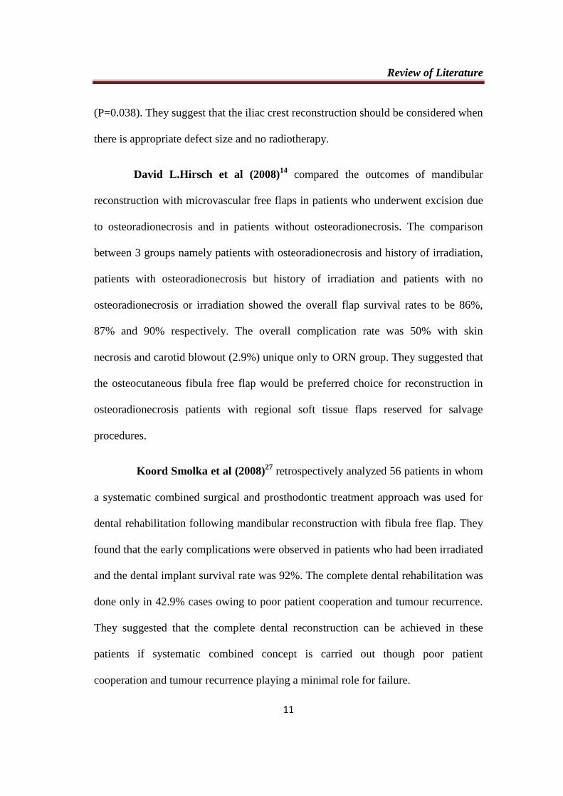
David L.Hirsch et al (2008)14
compared the outcomes of mandibular
reconstruction with microvascular free flaps in patients who underwent excision due
to osteoradionecrosis and in patients without osteoradionecrosis. The comparison
between 3 groups namely patients with osteoradionecrosis and history of irradiation,
patients with osteoradionecrosis but history of irradiation and patients with no
osteoradionecrosis or irradiation showed the overall flap survival rates to be 86%,
87% and 90% respectively. The overall complication rate was 50% with skin
necrosis and carotid blowout (2.9%) unique only to ORN group. They suggested that
the osteocutaneous fibula free flap would be preferred choice for reconstruction in
osteoradionecrosis patients with regional soft tissue flaps reserved for salvage
procedures.
Koord Smolka et al (2008)27
retrospectively analyzed 56 patients in whom
a systematic combined surgical and prosthodontic treatment approach was used for
dental rehabilitation following mandibular reconstruction with fibula free flap. They
found that the early complications were observed in patients who had been irradiated
and the dental implant survival rate was 92%. The complete dental rehabilitation was
done only in 42.9% cases owing to poor patient cooperation and tumour recurrence.
They suggested that the complete dental reconstruction can be achieved in these
patients if systematic combined concept is carried out though poor patient
cooperation and tumour recurrence playing a minimal role for failure.
Review of Literature
12
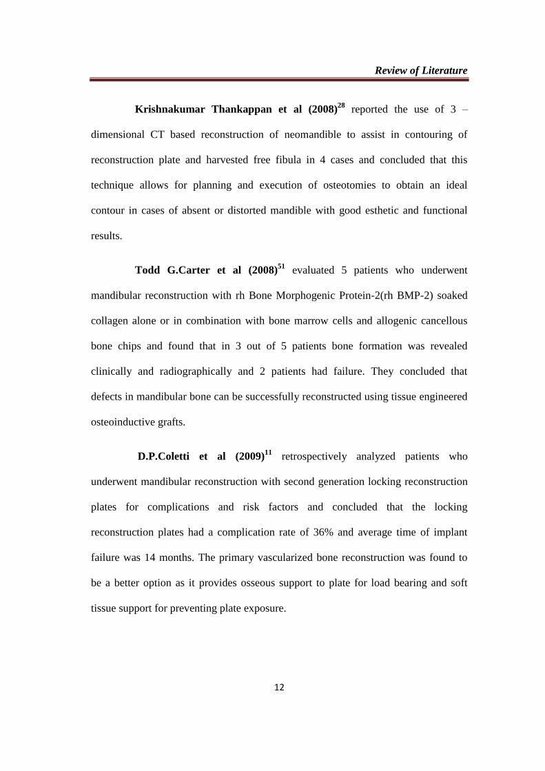
Krishnakumar Thankappan et al (2008)28
reported the use of 3 –
dimensional CT based reconstruction of neomandible to assist in contouring of
reconstruction plate and harvested free fibula in 4 cases and concluded that this
technique allows for planning and execution of osteotomies to obtain an ideal
contour in cases of absent or distorted mandible with good esthetic and functional
results.
Todd G.Carter et al (2008)51
evaluated 5 patients who underwent
mandibular reconstruction with rh Bone Morphogenic Protein-2(rh BMP-2) soaked
collagen alone or in combination with bone marrow cells and allogenic cancellous
bone chips and found that in 3 out of 5 patients bone formation was revealed
clinically and radiographically and 2 patients had failure. They concluded that
defects in mandibular bone can be successfully reconstructed using tissue engineered
osteoinductive grafts.
D.P.Coletti et al (2009)11
retrospectively analyzed patients who
underwent mandibular reconstruction with second generation locking reconstruction
plates for complications and risk factors and concluded that the locking
reconstruction plates had a complication rate of 36% and average time of implant
failure was 14 months. The primary vascularized bone reconstruction was found to
be a better option as it provides osseous support to plate for load bearing and soft
tissue support for preventing plate exposure.
Review of Literature
13
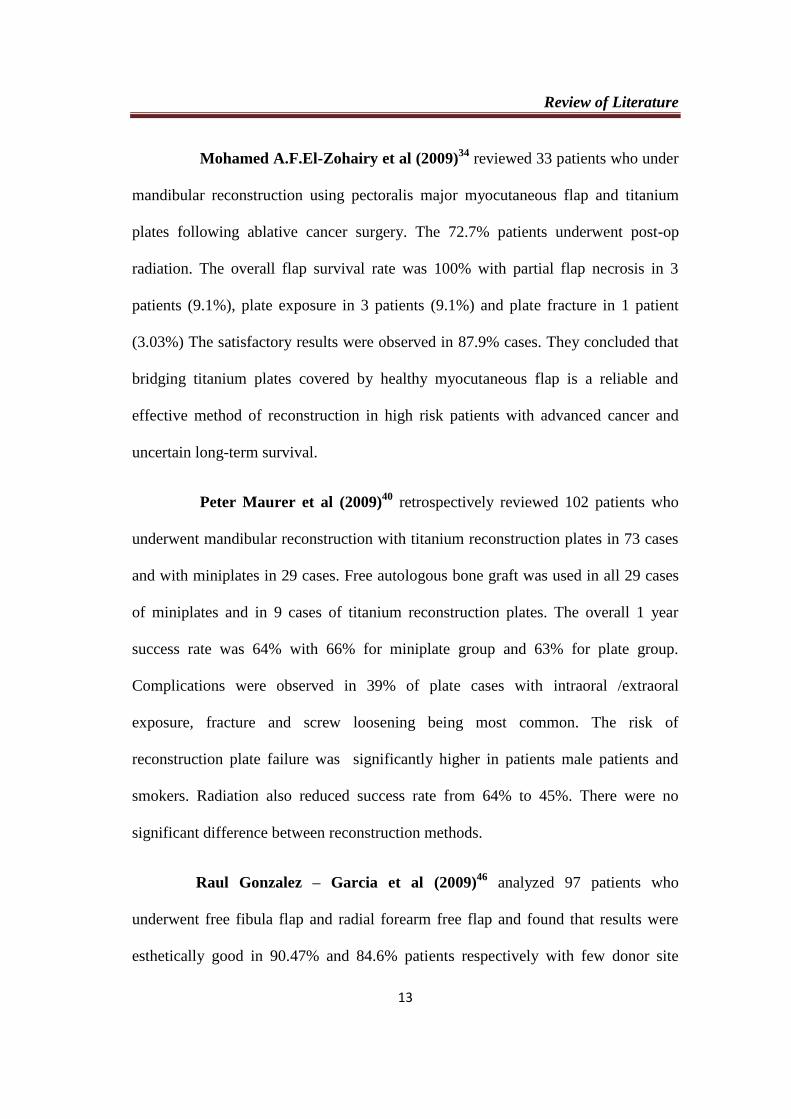
Mohamed A.F.El-Zohairy et al (2009)34
reviewed 33 patients who under
mandibular reconstruction using pectoralis major myocutaneous flap and titanium
plates following ablative cancer surgery. The 72.7% patients underwent post-op
radiation. The overall flap survival rate was 100% with partial flap necrosis in 3
patients (9.1%), plate exposure in 3 patients (9.1%) and plate fracture in 1 patient
(3.03%) The satisfactory results were observed in 87.9% cases. They concluded that
bridging titanium plates covered by healthy myocutaneous flap is a reliable and
effective method of reconstruction in high risk patients with advanced cancer and
uncertain long-term survival.
Peter Maurer et al (2009)40
retrospectively reviewed 102 patients who
underwent mandibular reconstruction with titanium reconstruction plates in 73 cases
and with miniplates in 29 cases. Free autologous bone graft was used in all 29 cases
of miniplates and in 9 cases of titanium reconstruction plates. The overall 1 year
success rate was 64% with 66% for miniplate group and 63% for plate group.
Complications were observed in 39% of plate cases with intraoral /extraoral
exposure, fracture and screw loosening being most common. The risk of
reconstruction plate failure was significantly higher in patients male patients and
smokers. Radiation also reduced success rate from 64% to 45%. There were no
significant difference between reconstruction methods.
Raul Gonzalez – Garcia et al (2009)46
analyzed 97 patients who
underwent free fibula flap and radial forearm free flap and found that results were
esthetically good in 90.47% and 84.6% patients respectively with few donor site
Review of Literature
14
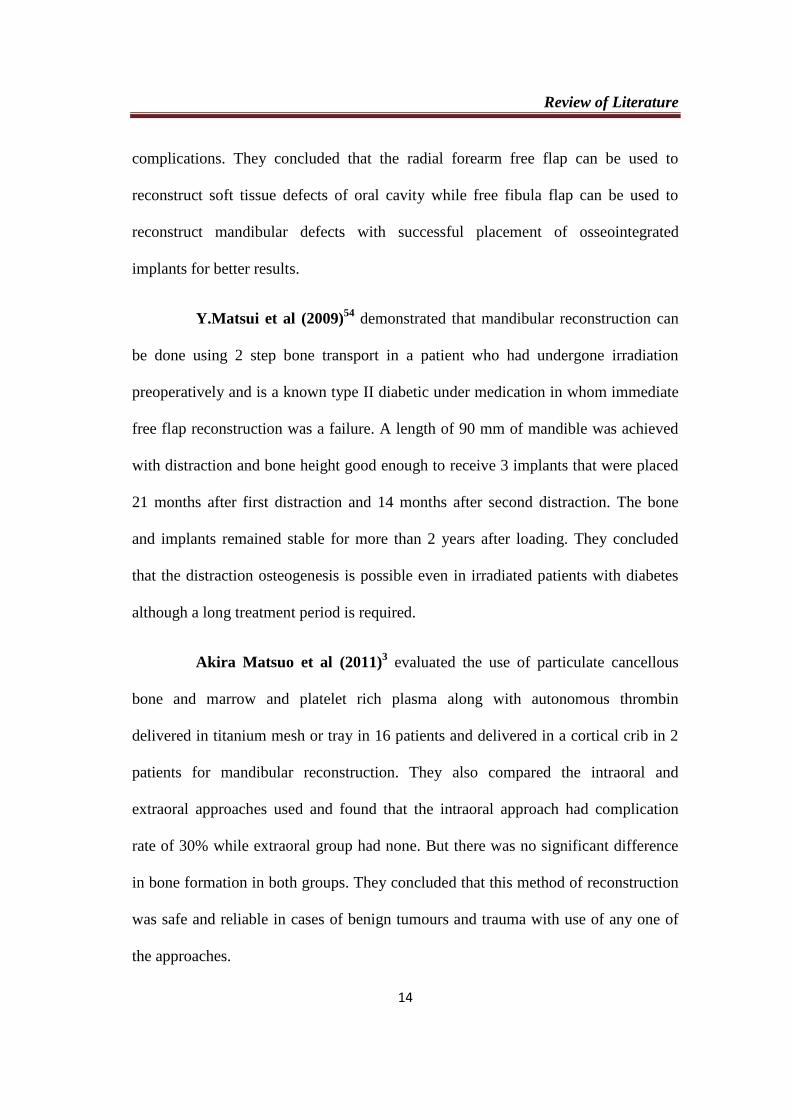
complications. They concluded that the radial forearm free flap can be used to
reconstruct soft tissue defects of oral cavity while free fibula flap can be used to
reconstruct mandibular defects with successful placement of osseointegrated
implants for better results.
Y.Matsui et al (2009)54
demonstrated that mandibular reconstruction can
be done using 2 step bone transport in a patient who had undergone irradiation
preoperatively and is a known type II diabetic under medication in whom immediate
free flap reconstruction was a failure. A length of 90 mm of mandible was achieved
with distraction and bone height good enough to receive 3 implants that were placed
21 months after first distraction and 14 months after second distraction. The bone
and implants remained stable for more than 2 years after loading. They concluded
that the distraction osteogenesis is possible even in irradiated patients with diabetes
although a long treatment period is required.
Akira Matsuo et al (2011)3 evaluated the use of particulate cancellous
bone and marrow and platelet rich plasma along with autonomous thrombin
delivered in titanium mesh or tray in 16 patients and delivered in a cortical crib in 2
patients for mandibular reconstruction. They also compared the intraoral and
extraoral approaches used and found that the intraoral approach had complication
rate of 30% while extraoral group had none. But there was no significant difference
in bone formation in both groups. They concluded that this method of reconstruction
was safe and reliable in cases of benign tumours and trauma with use of any one of
the approaches.
Review of Literature
15
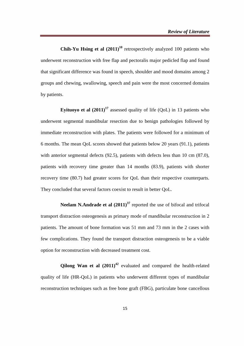
Chih-Yu Hsing et al (2011)10
retrospectively analyzed 100 patients who
underwent reconstruction with free flap and pectoralis major pedicled flap and found
that significant difference was found in speech, shoulder and mood domains among 2
groups and chewing, swallowing, speech and pain were the most concerned domains
by patients.
Eyituoyo et al (2011)17
assessed quality of life (QoL) in 13 patients who
underwent segmental mandibular resection due to benign pathologies followed by
immediate reconstruction with plates. The patients were followed for a minimum of
6 months. The mean QoL scores showed that patients below 20 years (91.1), patients
with anterior segmental defects (92.5), patients with defects less than 10 cm (87.0),
patients with recovery time greater than 14 months (83.9), patients with shorter
recovery time (80.7) had greater scores for QoL than their respective counterparts.
They concluded that several factors coexist to result in better QoL.
Neelam N.Andrade et al (2011)37
reported the use of bifocal and trifocal
transport distraction osteogenesis as primary mode of mandibular reconstruction in 2
patients. The amount of bone formation was 51 mm and 73 mm in the 2 cases with
few complications. They found the transport distraction osteogenesis to be a viable
option for reconstruction with decreased treatment cost.
Qilong Wan et al (2011)42
evaluated and compared the health-related
quality of life (HR-QoL) in patients who underwent different types of mandibular
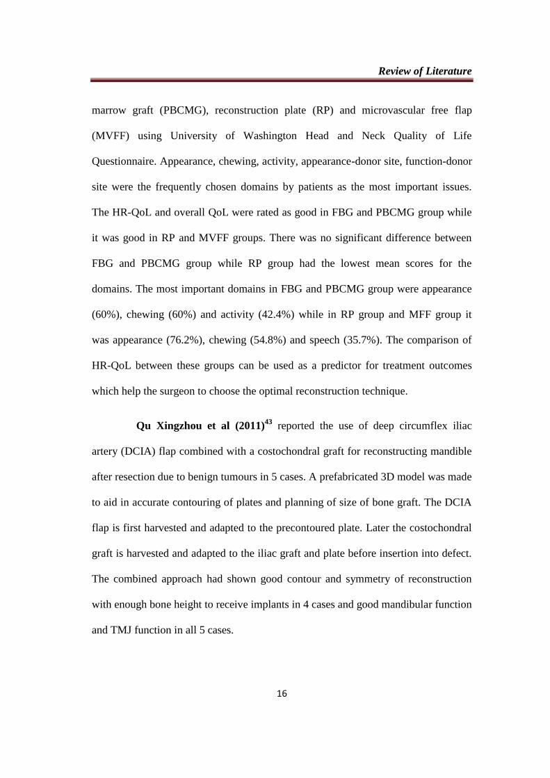
reconstruction techniques such as free bone graft (FBG), particulate bone cancellous
Review of Literature
16
marrow graft (PBCMG), reconstruction plate (RP) and microvascular free flap
(MVFF) using University of Washington Head and Neck Quality of Life
Questionnaire. Appearance, chewing, activity, appearance-donor site, function-donor
site were the frequently chosen domains by patients as the most important issues.
The HR-QoL and overall QoL were rated as good in FBG and PBCMG group while
it was good in RP and MVFF groups. There was no significant difference between
FBG and PBCMG group while RP group had the lowest mean scores for the
domains. The most important domains in FBG and PBCMG group were appearance
(60%), chewing (60%) and activity (42.4%) while in RP group and MFF group it
was appearance (76.2%), chewing (54.8%) and speech (35.7%). The comparison of
HR-QoL between these groups can be used as a predictor for treatment outcomes
which help the surgeon to choose the optimal reconstruction technique.
Qu Xingzhou et al (2011)43
reported the use of deep circumflex iliac
artery (DCIA) flap combined with a costochondral graft for reconstructing mandible
after resection due to benign tumours in 5 cases. A prefabricated 3D model was made
to aid in accurate contouring of plates and planning of size of bone graft. The DCIA
flap is first harvested and adapted to the precontoured plate. Later the costochondral
graft is harvested and adapted to the iliac graft and plate before insertion into defect.
The combined approach had shown good contour and symmetry of reconstruction
with enough bone height to receive implants in 4 cases and good mandibular function
and TMJ function in all 5 cases.
Review of Literature
17
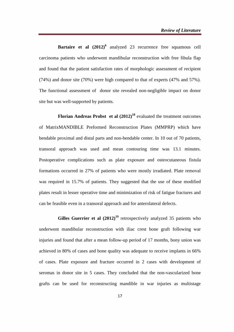
Bartaire et al (2012)6 analyzed 23 recurrence free squamous cell
carcinoma patients who underwent mandibular reconstruction with free fibula flap
and found that the patient satisfaction rates of morphologic assessment of recipient
(74%) and donor site (70%) were high compared to that of experts (47% and 57%).
The functional assessment of donor site revealed non-negligible impact on donor
site but was well-supported by patients.
Florian Andreas Probst et al (2012)18
evaluated the treatment outcomes
of MatrixMANDIBLE Preformed Reconstruction Plates (MMPRP) which have
bendable proximal and distal parts and non-bendable center. In 10 out of 70 patients,
transoral approach was used and mean contouring time was 13.1 minutes.
Postoperative complications such as plate exposure and osteocutaneous fistula
formations occurred in 27% of patients who were mostly irradiated. Plate removal
was required in 15.7% of patients. They suggested that the use of these modified
plates result in lesser operative time and minimization of risk of fatigue fractures and
can be feasible even in a transoral approach and for anterolateral defects.
Gilles Guerrier et al (2012)19
retrospectively analyzed 35 patients who
underwent mandibular reconstruction with iliac crest bone graft following war
injuries and found that after a mean follow-up period of 17 months, bony union was
achieved in 80% of cases and bone quality was adequate to receive implants in 66%
of cases. Plate exposure and fracture occurred in 2 cases with development of
seromas in donor site in 5 cases. They concluded that the non-vascularized bone
grafts can be used for reconstructing mandible in war injuries as multistage
Review of Literature
18
procedures provided the soft tissues are in good condition and in absence of
infection.
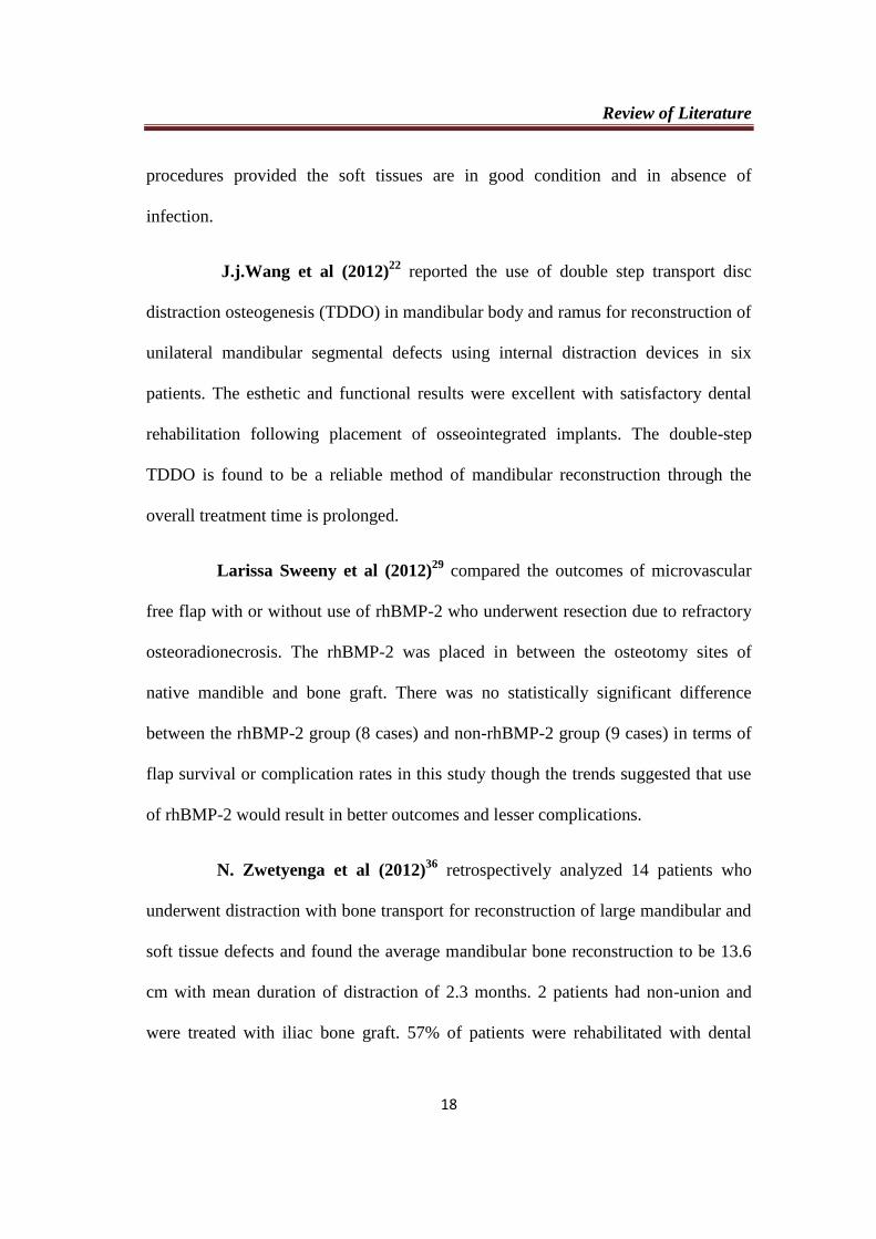
J.j.Wang et al (2012)22
reported the use of double step transport disc
distraction osteogenesis (TDDO) in mandibular body and ramus for reconstruction of
unilateral mandibular segmental defects using internal distraction devices in six
patients. The esthetic and functional results were excellent with satisfactory dental
rehabilitation following placement of osseointegrated implants. The double-step
TDDO is found to be a reliable method of mandibular reconstruction through the
overall treatment time is prolonged.
Larissa Sweeny et al (2012)29
compared the outcomes of microvascular
free flap with or without use of rhBMP-2 who underwent resection due to refractory
osteoradionecrosis. The rhBMP-2 was placed in between the osteotomy sites of
native mandible and bone graft. There was no statistically significant difference
between the rhBMP-2 group (8 cases) and non-rhBMP-2 group (9 cases) in terms of
flap survival or complication rates in this study though the trends suggested that use
of rhBMP-2 would result in better outcomes and lesser complications.
N. Zwetyenga et al (2012)36
retrospectively analyzed 14 patients who
underwent distraction with bone transport for reconstruction of large mandibular and
soft tissue defects and found the average mandibular bone reconstruction to be 13.6
cm with mean duration of distraction of 2.3 months. 2 patients had non-union and
were treated with iliac bone graft. 57% of patients were rehabilitated with dental
Review of Literature
19
implants with 95.5% success rate. They recommended the transport distraction
osteogenesis for patients with severe lower face defect to achieve acceptable
appearance and reasonable quality of life.
VN Okoje et al (2012)52
retrospectively analyzed 47 patients who
underwent iliac crest bone graft reconstruction of mandibular defects due to resection
of benign tumours or trauma and found that the appearance was satisfactory in 89.4%
of patients and graft infection (21.3%) occurred in 10 patients. The comparison
between methods of fixation such as transosseous wires and titanium plates revealed
that infection occurred only in wire group. Six (60%) out of ten infected cases
required graft removal while 4 were successfully treated for infection. They
concluded that the non-vascular iliac crest bone graft can be used as successful,
affordable and less technical choice of reconstruction in less economic patients and
defects due to benign tumour or trauma.
Yi Shen et al (2012)53
retrospectively analyzed 10 patients who
underwent extensive mandibular reconstruction in the symphysis region with or
without condylar prosthesis using partial double-barrel vascularized fibula graft and
found that bony union and wound healing was achieved in all patients during 43
months. The preoperative and postoperative chin-labial angle and bone height were
not significantly different at end of 2 year follow-up and facial appearance was found
to be excellent or good in 8 patients. They concluded that partial double-barrel
vascularized fibula graft can be used for reconstruction of large mandibular defects
Review of Literature
20
in symphysis region to achieve good facial appearance and function with good
stability of soft and hard tissue.
Zachary S.Peacock et al (2012)55
described a novel technique using
custom prostheses to repair fractured mandibular reconstruction plates in 3 patients
who were unable to undergo autogenous bone grafting procedures or replacement of
entire plate due to medical or socioeconomic factors. The custom prosthesis is
designed by 3D virtual planning software. Initially the portion of reconstruction plate
on native mandible is subtracted and later a custom prosthesis is constructed to adapt
to the buccal surface of mandible with an extension of female part which receives the
end of old titanium plate. The fixation is done by locking screws in between the
plates and by screws inserted into radial patterned slots in the distal segment of the
prosthesis. They found that this method served as permanent solution to the problem
of plate fracture.
Emeka Nkenke et al (2013)16
demonstrated that the bony microvascular
reconstruction following segmental mandibulectomy due to ameloblastoma can be
achieved using an intraoral microvascular anastomosing technique. The arterial and
venous anastomoses was achieved using intraoral vertical incision of buccal mucosa
placed taking parotid duct as a guide. They recommended intraoral approach for
microvascular flap reconstruction for segmental defects should be considered always
if feasible.
Review of Literature
21
Hitoshi Yoshimura et al (2013)21
reported the use of iliac crest bone
graft and greater auricular nerve graft for reconstructing mandible after segmental
resection due to ossifying fibroma using temporal, submandibular and intraoral
approaches. The greater auricular nerve graft was obtained from same side using
submandibular approach. The nerve was sutured to the proximal and distal cut ends
of inferior alveolar nerve using 10-0 nylon under surgical microscope. The iliac crest
graft was fixed using miniplates to native mandible. The postoperative follow up
showed that there was sufficient consolidation of grafted bone to receive two
implants at 7 months postoperatively. There was return of sensation to lower lip and
chin with pulpal sensitivity of teeth on surgical side. The patient had good esthetic
outcome and functional recovery.
Juanfang Zhu et al (2013)24
retrospectively analyzed 25 young patients
with mean age of 35.5 years who underwent primary mandibular reconstruction with
free fibula flap for assessing qulatiy of life and found that among various domains in
University of Washington QoL questionnaire, appearance (72%) was the most
concerning for most patients with best scores. Chewing (56%) and anxiety (52%)
domains had lowest scores. In Medical Outcomes Study short form- 36
questionnaire, the best scoring domain was physical functioning (77.3 points)
followed by bodily pain (74.56 points) and general health (72.56 points). They
concluded that the postoperative facial appearance was the most concerning factor
for young patients and it should be considered in surgical planning.
Review of Literature
22
K.Yagihara et al (2013)26
prospectively evaluated the stability and
viability of mandibular bone regeneration using a poly L-lactide (PLLA) mesh tray
and autogenous particulate cancellous bone and marrow (PCBM) in 62 patients who
underwent mandibular resection due to benign and malignant tumours, cysts,
Osteomyelitis or trauma and found the success rate to be 84% with a mean follow-up
period of 88.2 months. They concluded that this method was stable and effective due
to favourable morphological and functional recovery with low invasiveness. They
proposed the technique as an alternative procedure for mandibular reconstruction as
the regenerated bone showed low incidence of resorption over long term follow-up.
M.W.Ho et al (2013)31
introduced a method for intraoperative temporary
fixation for primary reconstruction of composite mandibular ablative defects. The
technique involves use of a long (40 hole) miniplate which is bent into the shape of
bucket handle and fixed with 2-3 screws on both sides of the bony resection margins.
Marker sutures were placed to mark orientation of plate. The shape of the plate gives
greater room for fashioning the free flap to reconstruct the defect and fixing the free
flap by use of miniplates. The temporary long miniplate can then be removed. The
advantages of this technique are minimal periosteal stripping of flap since miniplates
are used and the shape of the temporary plate allows use of reconstruction plate in
cases with ballooning of buccal or labial cortex.
N.Parbo et al (2013)35
retrospectively analyzed 36 patients who
underwent mandibular reconstruction with free fibula flap and found that the survival
rate of graft was 97% over a mean follow-up period of 22 months and the rate of
Review of Literature
23
dental rehabilitation was about 50% with implant survival rate of 96%. Non-severe
complications were seen in 50% of patients. Death was the main reason for lack of
prosthetic rehabilitation. They concluded that fibula graft with implant-supported
prosthesis had high survival rates and few complications.
Praveen Sharma et al (2013)41
reported spontaneous mandibular
regeneration in 4 children who were treated with resection of mandibular bone due to
benign tumours. The spontaneous regeneration was detected clinically and
radiographically between 3 and 5 months after resection eliminating the need or
atleast decreasing the size of the bone graft needed for reconstruction. The
spontaneous regeneration was thought to be due to the intact periosteal layer which
could provide osteogenic progenitor cells with good vascular supply and also
preventing soft tissue prolapse. The age of the patients (6 – 12 years) was also
thought to be influential.
T.J.Verhoeven et al (2013)50
introduced a new method to quantify soft
tissue facial asymmetry in patients who underwent mandibular reconstruction using
3D photographs obtained using stereophotogrammatrical camera. The comparison
between 3D photographs of 5 patients and 5 controls revealed a significant difference
of 1.19 mm in asymmetry between patients and controls. They concluded that this
method to be a valid, fast and clinically acceptable technique for assessing facial
asymmetry.
Review of Literature
24
A.M.Fry et al (2014)2 developed a new technique for creating
intermaxillary splint and positioning stents to guide mandibular reconstruction. The
positioning stent is formed by by using thermoforming plastic vacuum-formed over
cast made from impression made after prebent reconstruction plate was adapted to
3D model by wax. The intermaxillary splint is formed from preoperative upper and
lower models and bite registration done in wax to record occlusion. The splint holds
the remaining mandibular segments in correct occlusal relationship with maxilla
while the stent is used as guide to place the plate in desired position.4
Carlos Navarro Cuellar et al (2014)9 described a mandibular
reconstructive technique used in 12 patients which consisted of iliac crest free flap,
nasolabial flap and osseointegrated implants for bone augmentation, soft tissue defect
closure and dental rehabilitation respectively performed as a single procedure. The
functional and esthetic results were excellent with 95.2% success rate for implants.
Failure was associated with irradiated patients only.
Harry R.F.Powell et al (2014)20
retrospectively analyzed 10 patients who
underwent free fibula flap reconstruction following resection due to
osteoradionecrosis. The amount of bone resorption or formation was measured at 25,
50 and 75% of distance along bone graft in series of rotational radiographs taken
from 5 months to 20 months. Reduction of bone height was seen in 8 cases with
mean value of 1.5 mm while increase in bone height was seen in 2 cases. It has been
suggested that radiation before surgery causes increased resorption of fibular bone
after reconstruction. The increase in bone height was explained by two theories. First
Review of Literature
25
as a result of periosteal thickening along full length of bone due to periosteal
stripping and subsequent inflammation. Second as a result of the potential for callous
to form at the osteotomy sites.
Lidiya Zavalishina et al (2014)30
retrospectively analyzed 11 patients who
underwent free fibula flap reconstruction after segmental resection for assessing their
quality of life using questionnaire and simultaneously evaluated the esthetic
outcomes using patients’ photographs which were assessed by two dental
professionals using visual analog scale. They found that though there was a low
correlation between patient and expert assessment, most of the patients rated their
overall QoL as outstanding, very good or good (72.7%).
S. Arun Paul et al (2014)48
assessed the outcome of 32 patients who
underwent mandibular reconstruction with titanium reconstruction plate following
resection due to jaw pathologies and found that the success rate was 94% with plate
exposure occurring in 2 cases(6.3%) requiring its removal(6.3%). They concluded
that the titanium reconstruction plates can be used for mandibular reconstruction
provided the soft tissue provides sufficient bulk.
Si-Lian Fang et al (2014)49
reviewed 12 instances of exposure of
reconstruction plates which were treated with extended vertical lower trapezius
island myocutaneous flaps to cover exposed areas of plate intraorally, extraorally or
intra and extraorally. The flap was found healthy in all cases over mean follow-up
period of 22.8 months with exposure of plate extraorally in only one patient. They

Review of Literature
26
concluded that extended vertical lower trapezius island myocutaneous flaps can be
used reliably to cover plates exposed intraorally, extraorally or both intra and
extraorally.

Material & Methods
Materials & Methods
27
STUDY DESIGN:
The data of 18 patients who underwent mandibular reconstruction using
reconstruction plate, reconstruction plate and pectoralis major myocutaneous flap
and reconstruction plate and microvascular free flap following resection of benign
and malignant tumours were analyzed. The quality of life and postoperative
complications of these patients were assessed. All patients were treated at Sri
Ramakrishna Dental College and Hospital, Coimbatore.
MATERIAL:
The records of all patients who underwent mandibular reconstruction
between October 2009 to April 2014 were systematically reviewed. 82 patients were
treated with resection of mandible due to benign and malignant tumours. Out of 82,
32 patients underwent reconstruction with reconstruction plate. Of these only 18
patients were taken up for study as the others were either deceased or unavailable for
follow-up. These patients had undergone mandibular reconstruction with
reconstruction plate only or reconstruction plate and pectoralis major myocutaneous
flap or reconstruction plate with microvascular free flap.
Materials & Methods
28
INCLUSION CRITERIA:
1. Patients who underwent mandibular reconstruction with reconstruction plate
only with primary closure following resection due to benign and malignant
tumours.
2. Patients who underwent mandibular reconstruction with reconstruction plate
covered with pectoralis major myocutaneous flap following resection due to
benign and malignant tumours.
3. Patients who underwent mandibular reconstruction with reconstruction plate
covered with microvascular free flap following resection due to benign and
malignant tumours.
4. Isolated mandibular resection.
5. Patients who underwent neoadjuvant and adjuvant radiotherapy.
EXCLUSION CRITERIA:
1. Patients who developed locoregional recurrence of the tumour.
2. Patients who developed secondary tumours.
3. Patients who were medically compromised.
4. Patients unwilling to participate in the evaluation.
Materials & Methods
29
METHODS OF EVALUATION:
The patients taken up for study were asked to fill the subjective Quality of life
Questionnaire which was prepared by modifying University of Washington- Quality
of life questionnaire. The patients had a minimum of 6 months postoperative
recovery period before participating in the study. The quality of life was assessed
using questionnaire in terms of facial appearance, swallowing, tolerance of diet,
speech and activity.
Facial appearance was the major concern for patients and was classified as:
1. Good
2. Satisfied
3. Acceptable
4. Dissatisfied
Difficulty of patients to swallow liquid and solid foods was classified as:
1. Good
2. Mild difficulty
3. Moderate difficulty
4. Severe difficulty
Materials & Methods
30
The type of diet tolerated by patient was classified as:
1. Normal diet
2. Semisolid diet
3. Liquid diet
The ability of patient to speak was classified as:
1. Normal
2. Easily understandable
3. Difficult to understand
4. Poorly understood
The ability of patients to carry out their daily activities was classified as:
1. Normal
2. Moderately active
3. Minimally active
The quality of life of patient was given as good, fair, acceptable and poor based on
the total score obtained from the questionnaire.
Materials & Methods
31
The incidence of postoperative complications following reconstruction were also
noted:
1) Infection – recipient site, donor site
2) Wound dehiscence
3) Flap necrosis
4) Fistula formation
5) Plate exposure
6) Plate removal
7) Derangement of occlusion
8) Pain/tenderness in TMJ
9) Deviation in mouth opening
STASTICAL ANALYSIS:
Statistical analysis was done using Chi-square test, students‘t’ test, Mann
Whitney U test and Kruskal Wallis test. Statistical significance was defined as P
<0.05.

Figures
Figures
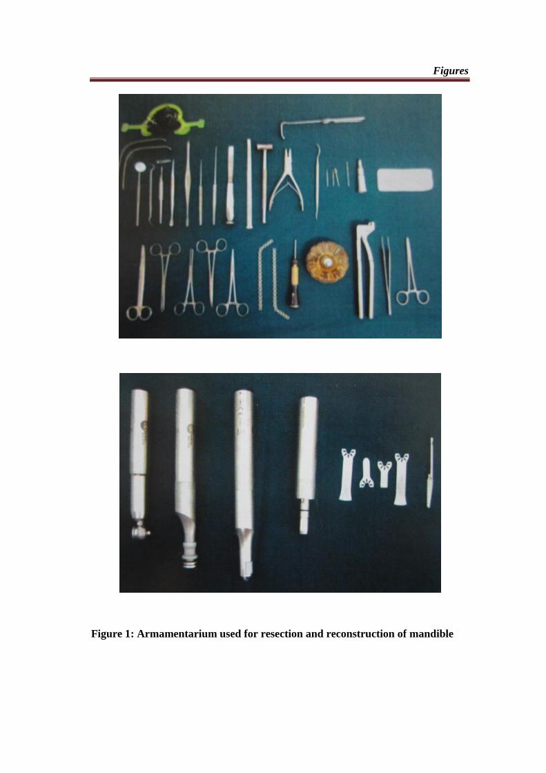
Figure 1: Armamentarium used for resection and reconstruction of mandible
Figures
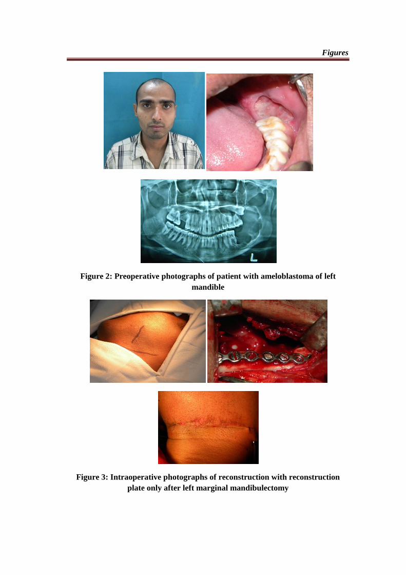
Figure 2: Preoperative photographs of patient with ameloblastoma of left
mandible
Figure 3: Intraoperative photographs of reconstruction with reconstruction
plate only after left marginal mandibulectomy
Figures
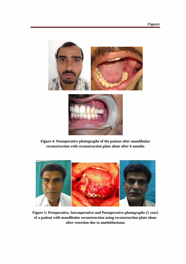
Figure 4: Postoperative photographs of the patient after mandibular
reconstruction with reconstruction plate alone after 6 months
Figure 5: Preoperative, Intraoperative and Postoperative photographs (1 year)
of a patient with mandibular reconstruction using reconstruction plate alone
after resection due to ameloblastoma
Figures
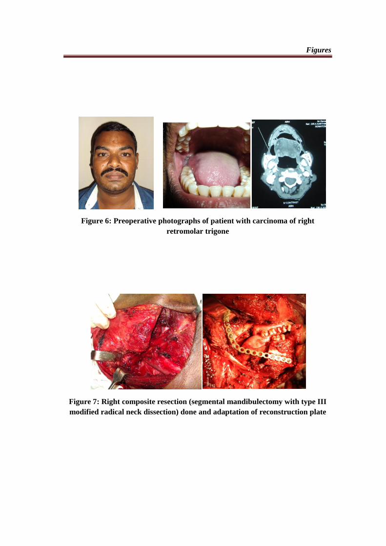
Figure 6: Preoperative photographs of patient with carcinoma of right
retromolar trigone
Figure 7: Right composite resection (segmental mandibulectomy with type III
modified radical neck dissection) done and adaptation of reconstruction plate
Figures
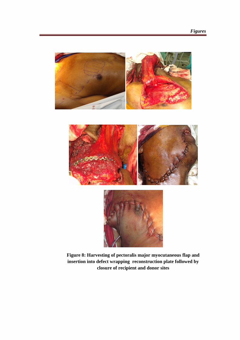
Figure 8: Harvesting of pectoralis major myocutaneous flap and
insertion into defect wrapping reconstruction plate followed by
closure of recipient and donor sites
Figures
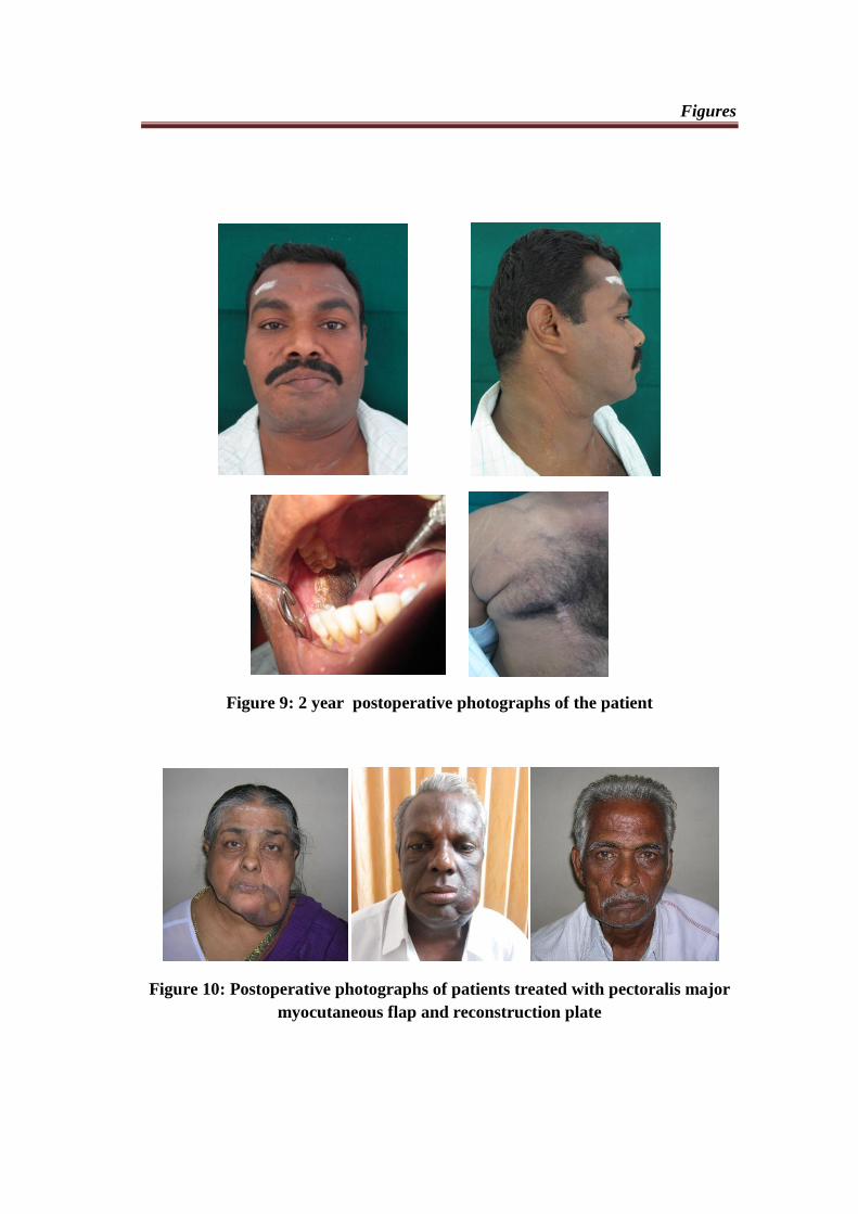
Figure 9: 2 year postoperative photographs of the patient
Figure 10: Postoperative photographs of patients treated with pectoralis major
myocutaneous flap and reconstruction plate
Figures
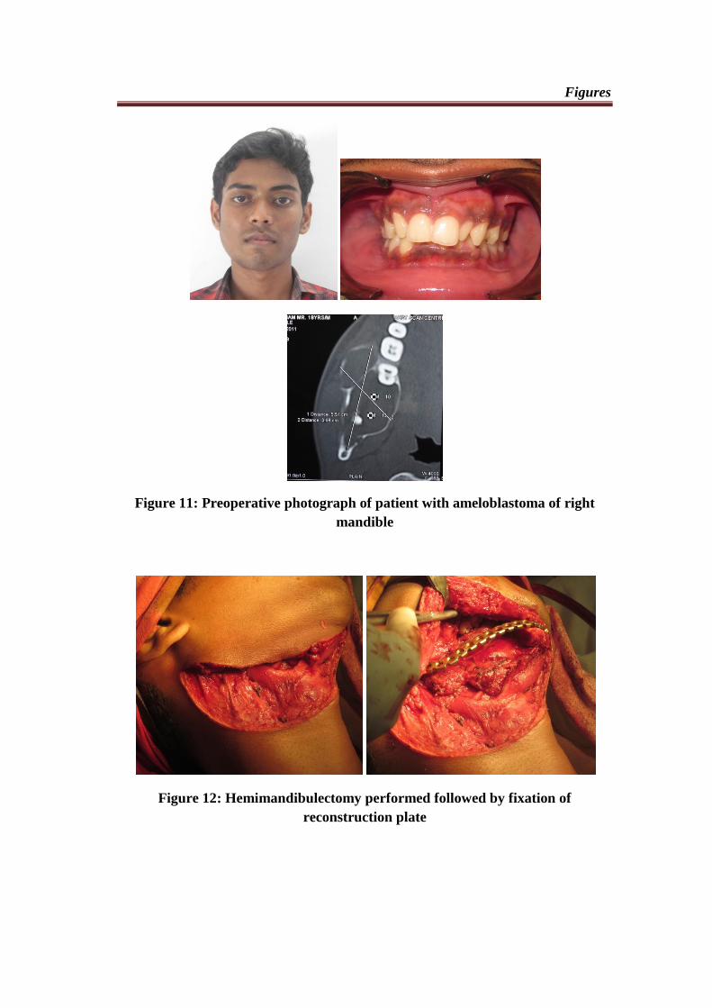
Figure 11: Preoperative photograph of patient with ameloblastoma of right
mandible
Figure 12: Hemimandibulectomy performed followed by fixation of
reconstruction plate
Figures
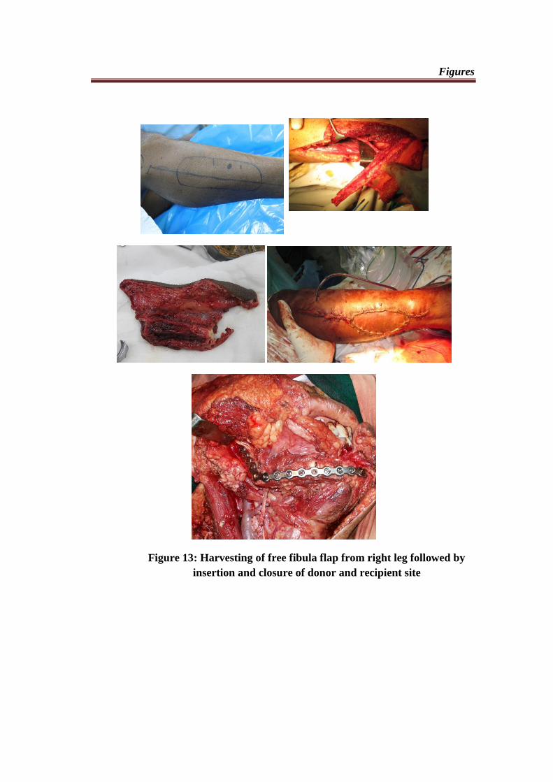
Figure 13: Harvesting of free fibula flap from right leg followed by
insertion and closure of donor and recipient site
Figures
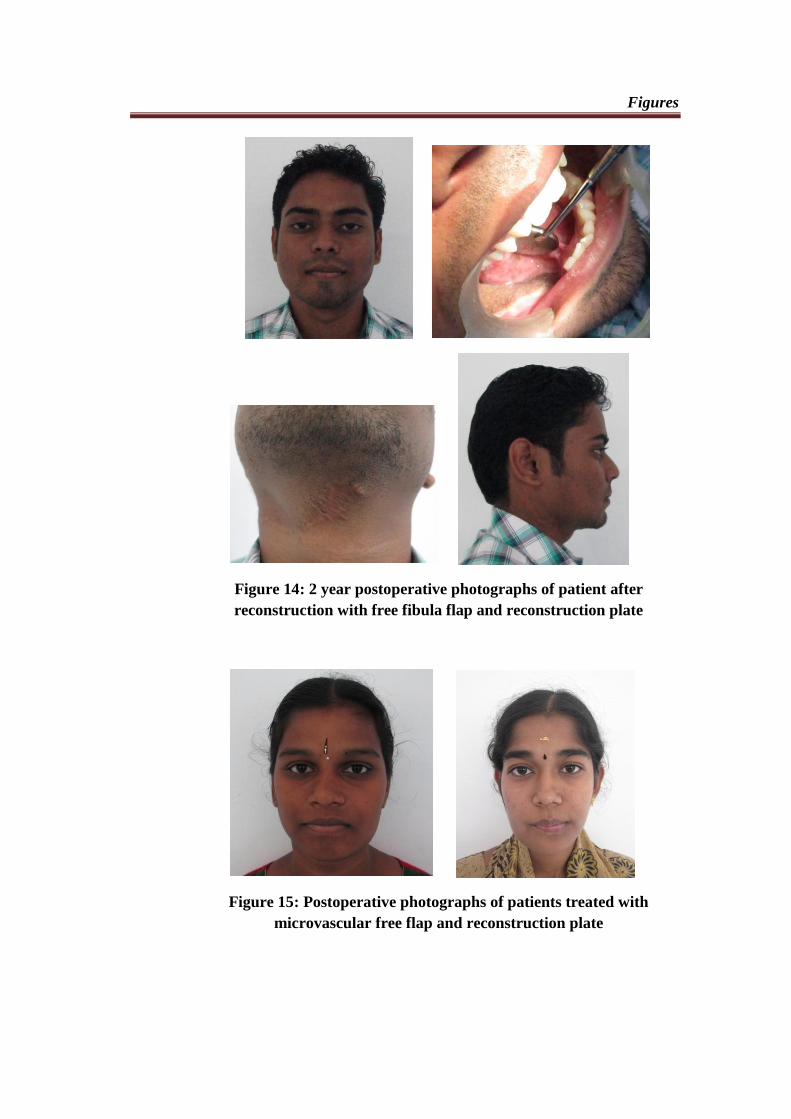
Figure 14: 2 year postoperative photographs of patient after
reconstruction with free fibula flap and reconstruction plate
Figure 15: Postoperative photographs of patients treated with
microvascular free flap and reconstruction plate
Figures
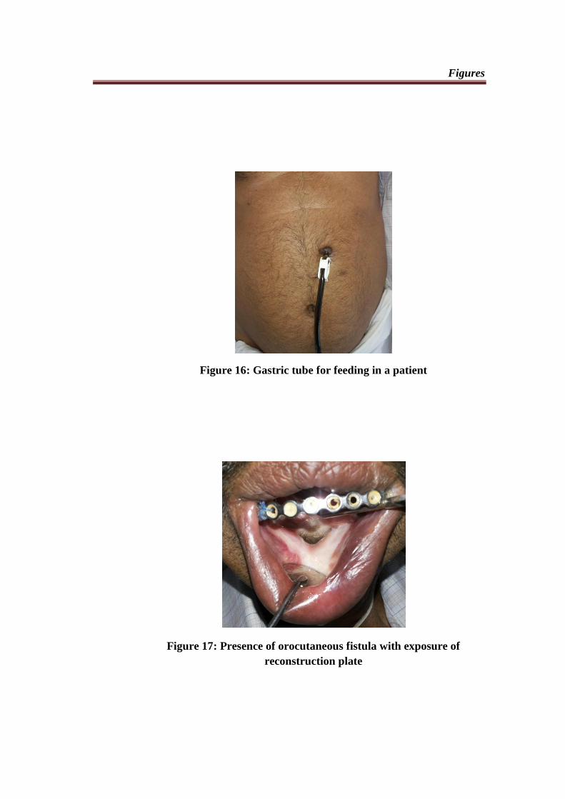
Figure 16: Gastric tube for feeding in a patient
Figure 17: Presence of orocutaneous fistula with exposure of
reconstruction plate

Figures
Figure 18: Hematoma formation followed by infection in recipient site
Figure 19: Wound dehiscence

Results
Results
32
A retrospective study was conducted on quality of life and postoperative
complications in 18 patients who underwent mandibular resection due to benign and
malignant tumours followed by reconstruction with reconstruction plate only,
reconstruction plate with pectoralis major myocutaneous flap and reconstruction
plate with microvascular free flap. The patients included in this study were operated
in the time interval of October 2009 to April 2014 in Department of Oral and
Maxillofacial Surgery at Sri Ramakrishna Hospital, Coimbatore.
The results of this study are shown under following subheadings:
1) Age and gender distribution
2) Side of tumour
3) Type of tumour
4) Type of resection
5) Type of mandibular defect
6) Type of reconstruction
7) Adjuvant radiotherapy
8) Facial appearance
9) Swallowing
10) Tolerance of diet
11) Speech
12) Activity
Results
33
13) Overall quality of life
14) Postoperative complications
- Recipient site infection
- Donor site infection
- Wound dehiscence
- Flap necrosis
- Fistula
- Plate exposure
- Plate removal
AGE AND GENDER DISTRIBUTION:
In 18 cases with mandibular resection and reconstruction, the gender
distribution showed 10 male patients and 8 female patients underwent reconstruction.
The age distribution showed that 50% of the study population were in the 30 – 60
years age group and that micro vascular free flap were preferred by the <30 years
group followed by 30 – 60 years age group. The mean age of the study group was
49.6 years. The overall quality of life scores were higher in patients of younger age
(< 30 years) than in older age groups.
SIDE OF THE TUMOUR:
In the 18 cases, 44.4% of patients had resection and reconstruction performed
on right side of mandible while 44.4% had involvement of left side. 11.1% had
Results
34
resection and reconstruction due to lesions located in central portion of mandible.
There was no significant association between side of resection and associated
complications (P>0.05).
TYPE OF TUMOUR:
In the 18 cases included in the study, 5 patients underwent resection due to
benign tumours and were reconstructed with reconstruction plate alone (n=2) or with
micro vascular free flap (n=3). The patients with malignant tumour underwent
resection and reconstruction with either reconstruction plate and pectoralis major
myocutaneous flap (n=11) or reconstruction plate with microvascular free flap (n=2).
There was significant association between speech domain and the type of tumour
(P<0.05). The overall quality of life scores also had statistical significance with type
of tumour (P<0.05).
TYPE OF RESECTION:
In the 18 cases of resection, 15 patients had segmental mandibulectomy done
while 2 patients underwent hemimandibulectomy.1 patient had undergone marginal
mandibulectomy.
TYPE OF MANDIBULAR DEFECT:
Out of 18 patients, 12 patients had lateral defects without involving condyle
(L), 2 patients had lateral mandibular defects including condyle (H) while 4 patients
had combination defects of mandible (LC or CL or LCL). There was no significant
Results
35
association between type of mandibular defect and associated complications (P
>0.05).
TYPE OF RECONSTRUCTION:
Of the 18 cases included in the study, 11 patients had reconstruction with
reconstruction plate and pectoralis major myocutaneous flap, 5 patients had
reconstruction with micro vascular free flap reconstruction with reconstruction plate
and 2 patients received only reconstruction plate to maintain continuity of mandible.
There was no statistically significant association between type of reconstruction and
quality of life scores and associated complications (P >0.05).
ADJUVANT RADIOTHERAPY:
Of the 18 patients who underwent mandibular resection and reconstruction, 5 of
the patients received adjuvant radiotherapy while 13 patients were confined only to
surgical management. There was statistically significant association between
radiotherapy and facial appearance, swallowing, speech and overall quality of life (P
<0.05). The associated complications like recipient site infection and fistula
formation were statistically significant in irradiated patients.
FACIAL APPEARANCE:
50% of reconstruction plate group (1/2), 9.1% of reconstruction plate with
pectoralis major myocutaneous flap group (1/11), 40% of reconstruction plate with
microvascular free flap group (2/5), 30.8% of non-irradiated patients (4/13), 60% of
Results
36
benign tumour group (3/5) and 7.7% of malignant tumour group (1/13) reported their
facial appearance as good.
54.6% of reconstruction plate with pectoralis major myocutaneous flap group
(6/11), 20% of reconstruction plate with microvascular free flap group(1/5), 20% of
irradiated patients (1/5), 53.9% of non-irradiated patients (7/13), 20% of benign
tumour group (1/5) and 53.9% of malignant tumour group (7/13) were satisfied with
their facial appearance.
50% of reconstruction plate group (1/2), 9.1% of reconstruction plate with
pectoralis major myocutaneous flap group (1/11), 20% of reconstruction plate with
microvascular free flap group (1/5), 60% of irradiated patients (3/5), 7.7% of non-
irradiated patients (1/13), 20% of benign tumour group (1/5) and 23% of malignant
tumour group (3/13) had acceptable appearance.
9.1% of reconstruction plate with pectoralis major myocutaneous flap group
(1/11), 20% of reconstruction plate with microvascular free flap group (1/5), 20% of
irradiated patients (1/5),7.7% of non-irradiated patients (1/13) and 15.4% of
malignant tumour group (2/13) were dissatisfied with their facial appearance. There
was statistically significant association between facial appearance and radiotherapy
(P <0.05) while type of tumour and the type of reconstruction did not have statistical
significance (P >0.05).
Results
37
SWALLOWING:
The swallowing was found to be good in all patients (100%) in reconstruction
plate only group (2/2), 63.6% of patients in reconstruction plate with pectoralis major
myocutaneous flap group (7/11), 60% of patients in reconstruction plate with
microvascular free flap group (3/5), 92.3% of non-irradiated patients (12/13), 100%
of benign tumour group (5/5) and 53.9% of malignant tumour group (7/13).
Mild difficulty with swallowing was reported in 18.2% patients in
reconstruction plate with pectoralis major myocutaneous flap group (2/11), 20% of
patients in reconstruction plate with microvascular free flap group (1/5), 40% of
irradiated patients (2/5) and 15.4% of malignant tumour group (2/13).
18.2% of patients in reconstruction plate with pectoralis major myocutaneous
flap group (2/11), 20% of patients in reconstruction plate with microvascular free
flap group (1/5), 60% of irradiated patients (3/5), 7.7% of non-irradiated patients
(1/13) and 30.8% of malignant tumour group(4/13) reported moderate difficulty.
There was statistically significant association between swallowing and radiotherapy
(P <0.05) while type of tumour and the type of reconstruction did not have statistical
significance (P >0.05).
TOLERANCE OF DIET:
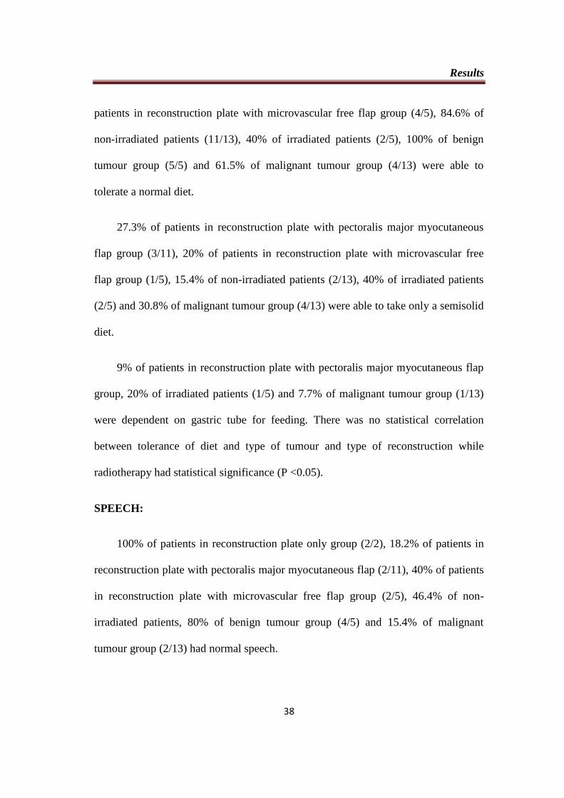
All the patients (100%) in reconstruction plate only group, 63.6% patients in
reconstruction plate with pectoralis major myocutaneous flap group (2/2), 80%
Results
38
patients in reconstruction plate with microvascular free flap group (4/5), 84.6% of
non-irradiated patients (11/13), 40% of irradiated patients (2/5), 100% of benign
tumour group (5/5) and 61.5% of malignant tumour group (4/13) were able to
tolerate a normal diet.
27.3% of patients in reconstruction plate with pectoralis major myocutaneous
flap group (3/11), 20% of patients in reconstruction plate with microvascular free
flap group (1/5), 15.4% of non-irradiated patients (2/13), 40% of irradiated patients
(2/5) and 30.8% of malignant tumour group (4/13) were able to take only a semisolid
diet.
9% of patients in reconstruction plate with pectoralis major myocutaneous flap
group, 20% of irradiated patients (1/5) and 7.7% of malignant tumour group (1/13)
were dependent on gastric tube for feeding. There was no statistical correlation
between tolerance of diet and type of tumour and type of reconstruction while
radiotherapy had statistical significance (P <0.05).
SPEECH:
100% of patients in reconstruction plate only group (2/2), 18.2% of patients in
reconstruction plate with pectoralis major myocutaneous flap (2/11), 40% of patients
in reconstruction plate with microvascular free flap group (2/5), 46.4% of non-
irradiated patients, 80% of benign tumour group (4/5) and 15.4% of malignant
tumour group (2/13) had normal speech.
Results
39
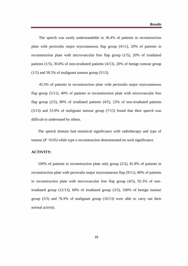
The speech was easily understandable in 36.4% of patients in reconstruction
plate with pectoralis major myocutaneous flap group (4/11), 20% of patients in
reconstruction plate with microvascular free flap group (1/5), 20% of irradiated
patients (1/5), 30.8% of non-irradiated patients (4/13), 20% of benign tumour group
(1/5) and 38.5% of malignant tumour group (5/13).
45.5% of patients in reconstruction plate with pectoralis major myocutaneous
flap group (5/11), 40% of patients in reconstruction plate with microvascular free
flap group (2/5), 80% of irradiated patients (4/5), 23% of non-irradiated patients
(3/13) and 53.9% of malignant tumour group (7/13) found that their speech was
difficult to understand by others.
The speech domain had statistical significance with radiotherapy and type of
tumour (P <0.05) while type o reconstruction demonstrated no such significance.
ACTIVITY:
100% of patients in reconstruction plate only group (2/2), 81.8% of patients in
reconstruction plate with pectoralis major myocutaneous flap (9/11), 80% of patients
in reconstruction plate with microvascular free flap group (4/5), 92.3% of non-
irradiated group (12/13), 60% of irradiated group (3/5), 100% of benign tumour
group (5/5) and 76.9% of malignant group (10/13) were able to carry out their
normal activity.
Results
40
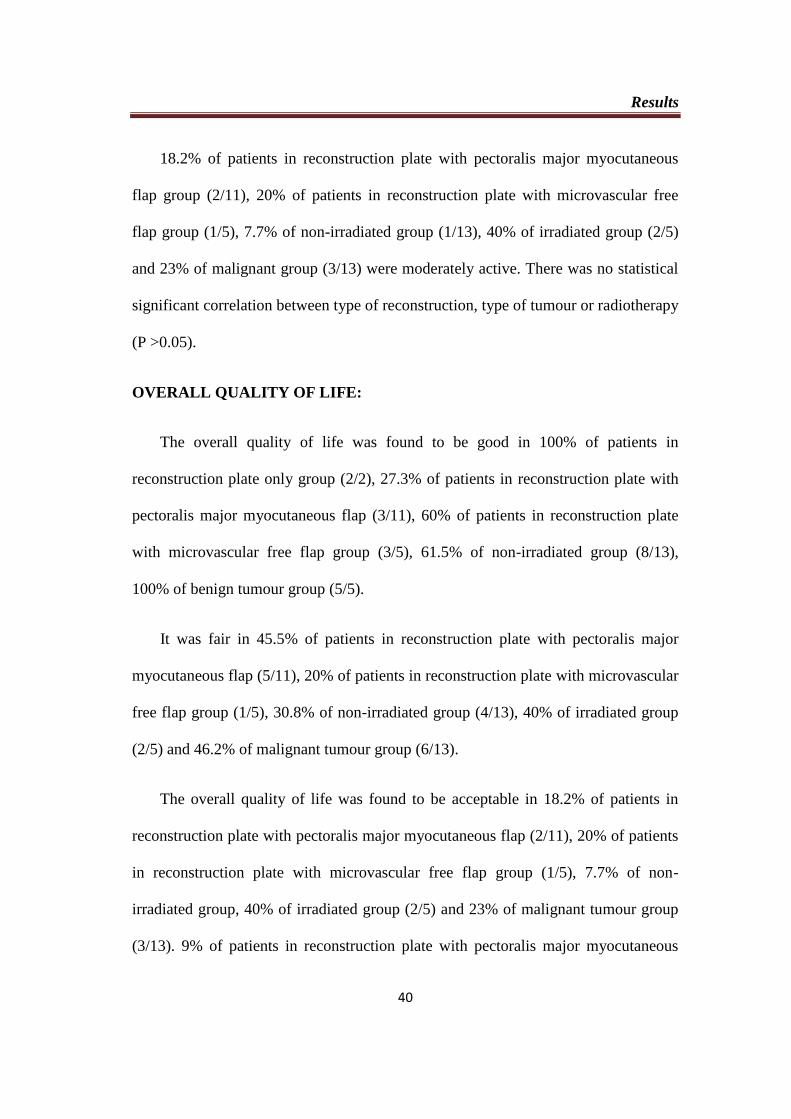
18.2% of patients in reconstruction plate with pectoralis major myocutaneous
flap group (2/11), 20% of patients in reconstruction plate with microvascular free
flap group (1/5), 7.7% of non-irradiated group (1/13), 40% of irradiated group (2/5)
and 23% of malignant group (3/13) were moderately active. There was no statistical
significant correlation between type of reconstruction, type of tumour or radiotherapy
(P >0.05).
OVERALL QUALITY OF LIFE:
The overall quality of life was found to be good in 100% of patients in
reconstruction plate only group (2/2), 27.3% of patients in reconstruction plate with
pectoralis major myocutaneous flap (3/11), 60% of patients in reconstruction plate
with microvascular free flap group (3/5), 61.5% of non-irradiated group (8/13),
100% of benign tumour group (5/5).
It was fair in 45.5% of patients in reconstruction plate with pectoralis major
myocutaneous flap (5/11), 20% of patients in reconstruction plate with microvascular
free flap group (1/5), 30.8% of non-irradiated group (4/13), 40% of irradiated group
(2/5) and 46.2% of malignant tumour group (6/13).
The overall quality of life was found to be acceptable in 18.2% of patients in
reconstruction plate with pectoralis major myocutaneous flap (2/11), 20% of patients
in reconstruction plate with microvascular free flap group (1/5), 7.7% of non-
irradiated group, 40% of irradiated group (2/5) and 23% of malignant tumour group
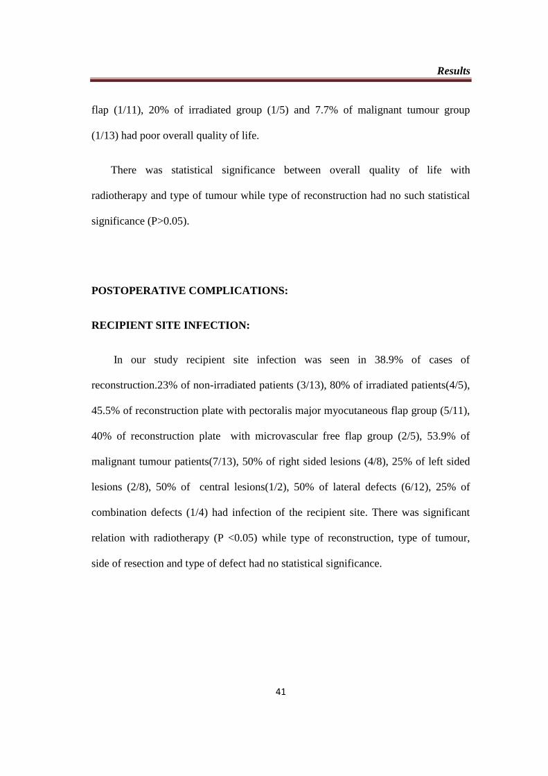
(3/13). 9% of patients in reconstruction plate with pectoralis major myocutaneous
Results
41
flap (1/11), 20% of irradiated group (1/5) and 7.7% of malignant tumour group
(1/13) had poor overall quality of life.
There was statistical significance between overall quality of life with
radiotherapy and type of tumour while type of reconstruction had no such statistical
significance (P>0.05).
POSTOPERATIVE COMPLICATIONS:
RECIPIENT SITE INFECTION:
In our study recipient site infection was seen in 38.9% of cases of
reconstruction.23% of non-irradiated patients (3/13), 80% of irradiated patients(4/5),
45.5% of reconstruction plate with pectoralis major myocutaneous flap group (5/11),
40% of reconstruction plate with microvascular free flap group (2/5), 53.9% of
malignant tumour patients(7/13), 50% of right sided lesions (4/8), 25% of left sided
lesions (2/8), 50% of central lesions(1/2), 50% of lateral defects (6/12), 25% of
combination defects (1/4) had infection of the recipient site. There was significant
relation with radiotherapy (P <0.05) while type of reconstruction, type of tumour,
side of resection and type of defect had no statistical significance.
Results
42
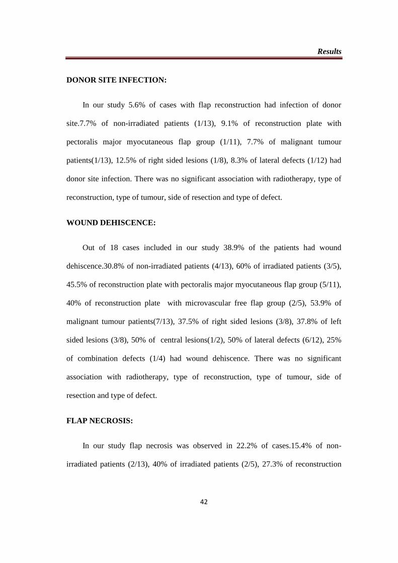
DONOR SITE INFECTION:
In our study 5.6% of cases with flap reconstruction had infection of donor
site.7.7% of non-irradiated patients (1/13), 9.1% of reconstruction plate with
pectoralis major myocutaneous flap group (1/11), 7.7% of malignant tumour
patients(1/13), 12.5% of right sided lesions (1/8), 8.3% of lateral defects (1/12) had
donor site infection. There was no significant association with radiotherapy, type of
reconstruction, type of tumour, side of resection and type of defect.
WOUND DEHISCENCE:
Out of 18 cases included in our study 38.9% of the patients had wound
dehiscence.30.8% of non-irradiated patients (4/13), 60% of irradiated patients (3/5),
45.5% of reconstruction plate with pectoralis major myocutaneous flap group (5/11),
40% of reconstruction plate with microvascular free flap group (2/5), 53.9% of
malignant tumour patients(7/13), 37.5% of right sided lesions (3/8), 37.8% of left
sided lesions (3/8), 50% of central lesions(1/2), 50% of lateral defects (6/12), 25%
of combination defects (1/4) had wound dehiscence. There was no significant
association with radiotherapy, type of reconstruction, type of tumour, side of
resection and type of defect.
FLAP NECROSIS:
In our study flap necrosis was observed in 22.2% of cases.15.4% of non-
irradiated patients (2/13), 40% of irradiated patients (2/5), 27.3% of reconstruction
Results
43
plate with pectoralis major myocutaneous flap group (3/11), 20% of reconstruction
plate with microvascular free flap group (1/5), 30.8% of malignant tumour
patients(4/13), 25% of right sided lesions (2/8), 12.5% of left sided lesions (1/8),
50% of central lesions(1/2), 25% of lateral defects (3/12), 25% of combination
defects (1/4) had necrosis of flap. There was no significant association with
radiotherapy, type of reconstruction, type of tumour, side of resection and type of
defect.
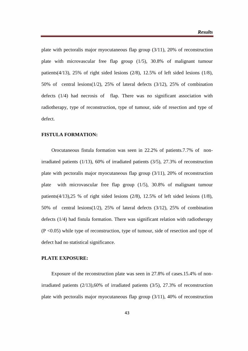
FISTULA FORMATION:
Orocutaneous fistula formation was seen in 22.2% of patients.7.7% of non-
irradiated patients (1/13), 60% of irradiated patients (3/5), 27.3% of reconstruction
plate with pectoralis major myocutaneous flap group (3/11), 20% of reconstruction
plate with microvascular free flap group (1/5), 30.8% of malignant tumour
patients(4/13),25 % of right sided lesions (2/8), 12.5% of left sided lesions (1/8),
50% of central lesions(1/2), 25% of lateral defects (3/12), 25% of combination
defects (1/4) had fistula formation. There was significant relation with radiotherapy
(P <0.05) while type of reconstruction, type of tumour, side of resection and type of
defect had no statistical significance.
PLATE EXPOSURE:
Exposure of the reconstruction plate was seen in 27.8% of cases.15.4% of non-
irradiated patients (2/13),60% of irradiated patients (3/5), 27.3% of reconstruction
plate with pectoralis major myocutaneous flap group (3/11), 40% of reconstruction

Results
44
plate with microvascular free flap group (2/5), 38.5% of malignant tumour
patients(5/13), 37.5% of right sided lesions (3/8), 12.5% of left sided lesions (1/8),
50% of central lesions(1/2), 33.3% of lateral defects (4/12), 25% of combination
defects (1/4) had plate exposure. There was no significant association with
radiotherapy, type of reconstruction, type of tumour, side of resection and type of
defect.
PLATE REMOVAL:
In our study plate removal was necessary in 27.8% of patients. 15.4% of non-
irradiated patients (2/13),60% of irradiated patients (3/5), 27.3% of reconstruction
plate with pectoralis major myocutaneous flap group (3/11), 40% of reconstruction
plate with microvascular free flap group (2/5), 38.5% of malignant tumour
patients(5/13), 37.5% of right sided lesions (3/8), 12.5% of left sided lesions (1/8),
50% of central lesions(1/2), 33.3% of lateral defects (4/12), 25% of combination
defects (1/4) underwent plate removal. There was no significant association with
radiotherapy, type of reconstruction, type of tumour, side of resection and type of
defect.
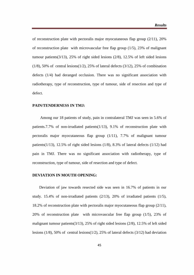
OCCLUSION:
In our study of 18 cases 61.1% of patients had a normal occlusion of the
contralateral side while 16.7% of patients had deranged occlusion. The status of
occlusion could not be applied to 22.2% of population due to their completely
edentulous or partially edentulous state.23% of non-irradiated patients (3/13), 18.2%
Results
45
of reconstruction plate with pectoralis major myocutaneous flap group (2/11), 20%
of reconstruction plate with microvascular free flap group (1/5), 23% of malignant
tumour patients(3/13), 25% of right sided lesions (2/8), 12.5% of left sided lesions
(1/8), 50% of central lesions(1/2), 25% of lateral defects (3/12), 25% of combination
defects (1/4) had deranged occlusion. There was no significant association with
radiotherapy, type of reconstruction, type of tumour, side of resection and type of
defect.
PAIN/TENDERNESS IN TMJ:
Among our 18 patients of study, pain in contralateral TMJ was seen in 5.6% of
patients.7.7% of non-irradiated patients(1/13), 9.1% of reconstruction plate with
pectoralis major myocutaneous flap group (1/11), 7.7% of malignant tumour
patients(1/13), 12.5% of right sided lesions (1/8), 8.3% of lateral defects (1/12) had
pain in TMJ. There was no significant association with radiotherapy, type of
reconstruction, type of tumour, side of resection and type of defect.
DEVIATION IN MOUTH OPENING:
Deviation of jaw towards resected side was seen in 16.7% of patients in our
study. 15.4% of non-irradiated patients (2/13), 20% of irradiated patients (1/5),
18.2% of reconstruction plate with pectoralis major myocutaneous flap group (2/11),
20% of reconstruction plate with microvascular free flap group (1/5), 23% of
malignant tumour patients(3/13), 25% of right sided lesions (2/8), 12.5% of left sided
lesions (1/8), 50% of central lesions(1/2), 25% of lateral defects (3/12) had deviation

Results
46
of jaw during mouth opening. There was no significant association with
radiotherapy, type of reconstruction, type of tumour, side of resection and type of
defect.

Discussion
Discussion
46
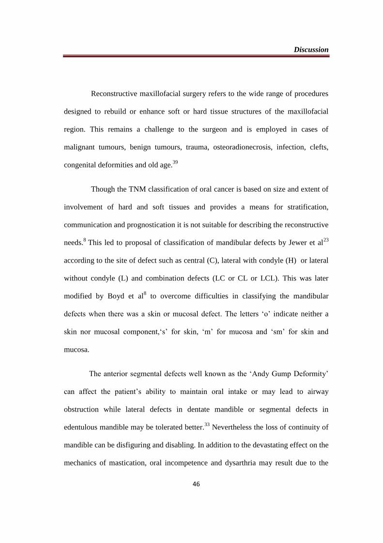
Reconstructive maxillofacial surgery refers to the wide range of procedures
designed to rebuild or enhance soft or hard tissue structures of the maxillofacial
region. This remains a challenge to the surgeon and is employed in cases of
malignant tumours, benign tumours, trauma, osteoradionecrosis, infection, clefts,
congenital deformities and old age.39
Though the TNM classification of oral cancer is based on size and extent of
involvement of hard and soft tissues and provides a means for stratification,
communication and prognostication it is not suitable for describing the reconstructive
needs.8
This led to proposal of classification of mandibular defects by Jewer et al23
according to the site of defect such as central (C), lateral with condyle (H) or lateral
without condyle (L) and combination defects (LC or CL or LCL). This was later
modified by Boyd et al8 to overcome difficulties in classifying the mandibular
defects when there was a skin or mucosal defect. The letters ‘o’ indicate neither a
skin nor mucosal component,‘s’ for skin, ‘m’ for mucosa and ‘sm’ for skin and
mucosa.
The anterior segmental defects well known as the ‘Andy Gump Deformity’
can affect the patient’s ability to maintain oral intake or may lead to airway
obstruction while lateral defects in dentate mandible or segmental defects in
edentulous mandible may be tolerated better.33
Nevertheless the loss of continuity of
mandible can be disfiguring and disabling. In addition to the devastating effect on the
mechanics of mastication, oral incompetence and dysarthria may result due to the
Discussion
47
loss of support and contraction of perioral soft tissues, tethering of lip and tongue.
This is usually further worsened by adjuvant radiotherapy. Most importantly, the
change in facial appearance has a terrible impact on the patient’s feeling of self
confidence and their desire to return to their pre-disease state of life.33
Disfigurement and impaired oral function of patients who underwent
mandibular reconstruction adversely affect the health related quality of life. Quality
of life may be described as the “gap between one’s actual functional level and one’s
ideal standard,” but it is important to keep in mind that a patient’s assessment of their
quality of life is dynamic, changing over time and situations. Patient assessment of
quality of life tends to be the worst in the months after surgery, improving slightly at
1 year, or even approaching pretreatment levels with time.13
Even though evaluation and comparison of different mandibular
reconstructions have already been reported in literature, most of them focus only on
physical outcomes rather than psychological outcome. For surgeons it is important to
understand the patient’s perception of their health related quality of life and their
influencing factors.24,42
This may serve as an important factor for optimizing the
choice of reconstruction. The relatively large number of questionnaires specific for
diseases of the oral cavity reflects that there is no ‘gold standard’.24
In our study, we
modified the University of Washington- Quality Of Life questionnaire so that it can
be easily applied to our Indian population. The concerns of a patient with benign
lesions are clearly different from those of cancer patients. Despite undergoing
surgical resection, the patient’s life expectancy is not adversely at risk17
and they tend
Discussion
48
to expect a more satisfying outcome after surgery than patients treated for
malignancies.
In our study 10 males and 8 females underwent mandibular reconstruction.
The mean age for our study population was 49.61 years. In our study the age
distribution showed that 50% of the study population were in the 30 – 60 years age
group and that micro vascular free flap were preferred by the <30 years group
followed by 30 – 60 years age group. Eyituoyo et al17
stated a significant relation
between age of patient and quality of life while Qilong Wan et al42
found no such
significance. The mean overall quality of life score in our study was 14.6 (fair) with
higher scores in younger patients (< 30 years) than in older age group. This may be
the result of better adaptability of younger age group to changes following resection
and reconstruction than the older age group.
In the 18 cases included in our study 15 patients underwent segmental
mandibulectomy while 2 patients had hemi mandibulectomy done. Marginal
mandibulectomy was performed in 1 patient. In our study 44.4% of patients had
resection and reconstruction performed on right side of mandible while 44.4% had
involvement of left side. 11.1% had resection and reconstruction due to lesions
located in central part of mandible.
Of the 18 cases included in the study, 11 patients had reconstruction with
reconstruction plate and pectoralis major myocutaneous flap, 5 patients had
reconstruction with micro vascular free flap reconstruction with reconstruction plate
Discussion
49
and 2 patients received only reconstruction plate to maintain continuity of mandible.
In our study the leading cause for resection was squamous cell carcinoma followed
by ameloblastoma.
Facial appearance is reported as the most concerning domain in the quality of
life questionnaire from patients’ perspective in various studies especially in younger
patients.24
In our study in reconstruction with pectoralis major myocutaneous flap,
9.1% patients reported their facial appearance as good while 54.6% were satisfied
with their facial appearance. 9.1% of reconstruction plate with pectoralis major
myocutaneous flap group had acceptable appearance and 9.1% were dissatisfied with
their facial appearance. Raphael Lopez et al45
, Chih- Yu Hsing et al10
, P.Salvatori et
al38
and Mohamed A.F.El-Zohairy et al34
found that most of the patients
reconstructed with pectoralis major myocutaneous flap found their appearance as
satisfied or good.
In reconstruction with microvascular free flap group, 40% reported their facial
appearance as good while 20% found the appearance to be fair, 20% found it
acceptable and 20% were dissatisfied. The dissatisfaction was due to the total flap
loss due to hematoma and infection. The findings of Lidiya Zavalishna et al30
, Raul
Gonzalez – Garcia et al46
, Bartaire et al6, Hidalgo et al
12 reported a high incidence of
97% of patient satisfaction with free flap reconstruction.
In our study we found that in reconstruction plate group 50% of patients found
their appearance as good and the other 50% found their appearance to be acceptable.
Discussion
50
S.Arun Paul48
and K.R.Spencer et al25
reported higher incidence of patient
satisfaction upto 94%. The results of our study revealed no statistically significant
difference in facial appearance domain among different types of reconstruction.
T.J.Verhoeven et al50
introduced a new method to quantify soft tissue facial
asymmetry in patients who underwent mandibular reconstruction using 3D
photographs obtained using stereophotogrammatrical camera. This may serve a
useful tool for expert assessment for facial asymmetry and may reveal correlation
between facial appearance among various types of reconstruction as expert
assessment and patient assessment had significant difference in the study reported by
Lidiya Zavalishna et al30
.
In our study, 63.6% of patients in reconstruction plate with pectoralis major
myocutaneous flap group were able to swallow solids and liquids normally while
mild difficulty was noticed in 18.2% of patients and 18.2% patients found
swallowing food moderately difficult. In the study by Lidiya Zavalishna et al30
63.6% of free flap patients had normal swallowing while 9% patients had mild
difficulty and 18.2% patients had moderate difficulty. In our study 60% of patients
in microvascular free flap group reported that they did not have any problem with
swallowing while 20% patients had mild and 20% patients had moderate difficulty.
Juanfang Zhu et al24
reported that swallowing was not a problem of concern to free
flap patients.
Literature states that in patients with pectoralis major myocutaneous flap
reconstruction, 40-90% of them were able to take a normal diet.45,34
In our study
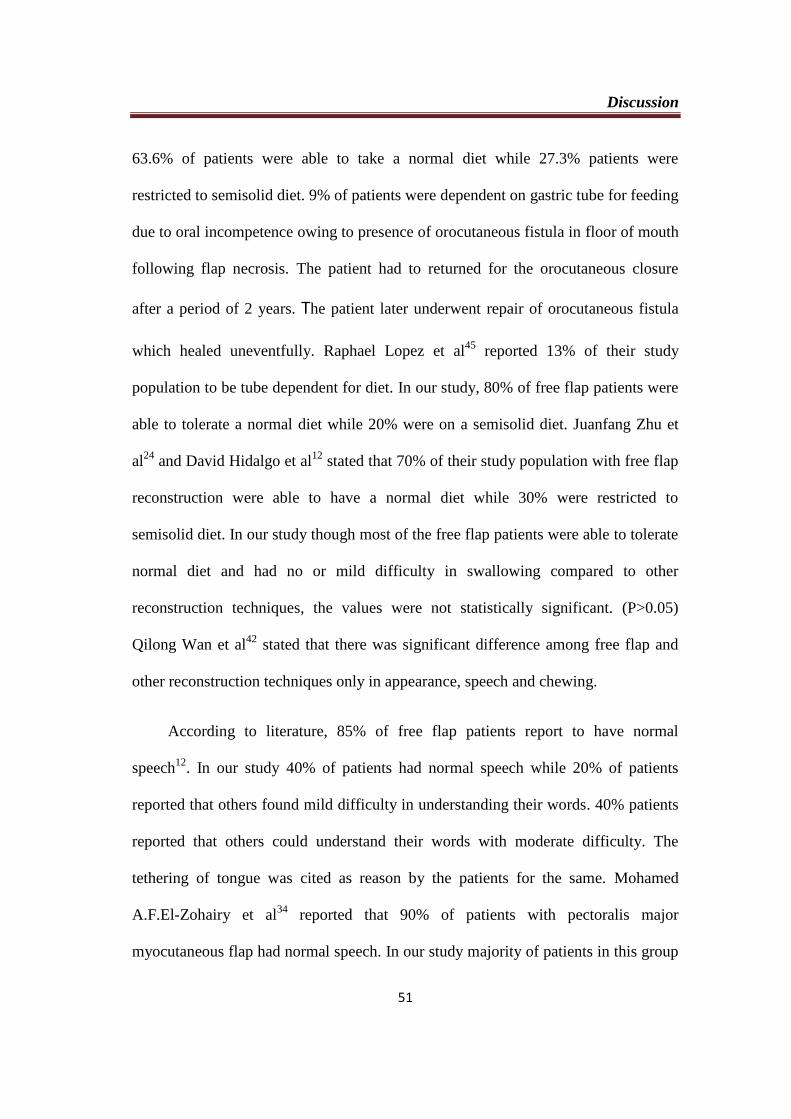
Discussion
51
63.6% of patients were able to take a normal diet while 27.3% patients were
restricted to semisolid diet. 9% of patients were dependent on gastric tube for feeding
due to oral incompetence owing to presence of orocutaneous fistula in floor of mouth
following flap necrosis. The patient had to returned for the orocutaneous closure
after a period of 2 years. The patient later underwent repair of orocutaneous fistula
which healed uneventfully. Raphael Lopez et al45
reported 13% of their study
population to be tube dependent for diet. In our study, 80% of free flap patients were
able to tolerate a normal diet while 20% were on a semisolid diet. Juanfang Zhu et
al24
and David Hidalgo et al12
stated that 70% of their study population with free flap
reconstruction were able to have a normal diet while 30% were restricted to
semisolid diet. In our study though most of the free flap patients were able to tolerate
normal diet and had no or mild difficulty in swallowing compared to other
reconstruction techniques, the values were not statistically significant. (P>0.05)
Qilong Wan et al42
stated that there was significant difference among free flap and
other reconstruction techniques only in appearance, speech and chewing.
According to literature, 85% of free flap patients report to have normal
speech12
. In our study 40% of patients had normal speech while 20% of patients
reported that others found mild difficulty in understanding their words. 40% patients
reported that others could understand their words with moderate difficulty. The
tethering of tongue was cited as reason by the patients for the same. Mohamed
A.F.El-Zohairy et al34
reported that 90% of patients with pectoralis major
myocutaneous flap had normal speech. In our study majority of patients in this group
Discussion
52
(45.5%) stated that their speech was understood with moderate difficulty. Only
18.2% patients were able to maintain normal speech. This can be due to the fact that
resection in this group was extensive involving tongue and other soft tissues as result
of treatment of malignancy. Chih- Yu Hsing et al10
and Qilong Wan et al42
reported
significant difference between free flap and other reconstruction methods only in the
speech domain of quality of life analysis. In our study there was no statistically
significant difference in speech domain between the different types of reconstruction
though the number of free flap patients with normal speech were higher than the
other counterparts.
In our study, 81.8% of pectoralis major myocutaneous flap group and 80% of
free flap group were able to carry out their normal activities while 20% of them
restricted their activity to a moderate level mainly as a result of their dissatisfied
facial appearance and fatigue due to lack of balanced diet. Lidiya Zavalishna et al30
stated that only 45% of their free flap study group reported normal activity while
literature states higher normal activity rates.
In our study we found that in patients reconstructed only with bridging plates had
no problems problems associated with swallowing, tolerance of diet, speech or
activity. The overall quality of life scores were also found to be good. This can be
attributed to the fact that this type of reconstruction was carried out only in cases of
benign tumours which required a lesser extensive resection compared to malignancy
and absence of irradiation. The only problem with facial appearance in a patient in
this group was absence of chin prominence due to anterolateral defect of mandible.
Discussion
53
In our study we found that mean overall quality of life scores was 14.6 (fair).
Overall quality of life scores were in higher side of scale for reconstruction plate
only group followed by free flap group while majority of pectoralis major
myocutaneous flap group patients had fair scores. The differences in quality of life
scores were not statistically significant among the different methods of
reconstruction. Qilong Wan et al42
also found that their study population had good
overall quality of life scores among all groups with significant difference in free flap
group in the appearance, speech and swallowing domains. Lidiya Zavalishna et al30
found that the free flap patients were satisfied with their overall quality of life. Chih-
Yu Hsing et al10
reported that there was no statistical difference between free flap
and pectoralis major myocutaneous flap group in all domains of quality of life except
for speech.
In our study statistical significance was found between irradiated and non-
irradiated patients in terms of facial appearance, swallowing, speech and overall
quality of life score.(P>0.05). Analysis of quality of life scores for benign and
malignant tumours showing that esthetic outcome was less satisfying in patients with
malignancy and irradiation was reported by Lidiya Zavalishna et al.30
David Vu et al13
and Lidiya Zavalishna et al30
reported statistically significant
difference in different domains and overall quality of life among irradiated and non-
irradiated patients. Irradiation is known to have significant effects on various tissues
of the oral cavity. One of the main disadvantages is a marked decrease in salivation
leading to difficulty in swallowing, tolerating a normal diet and speech due to loss of
Discussion
54
lubricating effect of saliva. There are also changes in soft tissues of oral cavity as a
result of fibrosis.
In our study there was significant difference between benign tumours and
malignant tumours in terms of individual domains of quality of life especially in
speech and overall score. Eyituoyo et al17
and Lidiya Zavalishna et al30
found that
patients treated for benign tumours rather than malignancies had better scores for
quality of life domains especially speech and swallowing. This is because phonation
capacities mainly depend on involved soft tissue resection. The amount of soft
tissues resected is greater while treating a malignancy than in benign tumour as the
extent of resection is a result of primary tumour infiltration or for the sake of three
dimensional clearance.
David Vu et al13
states that the quality of life scores tend to change over time
as initially the patients find the changes following resection and reconstruction
difficult to adapt but may later tend to get along with the modification of lifestyle.
Lidiya Zavalishna et al30
found that the quality of life scores given by the patient
itself and given by a expert after assessing the patient tend to vary significantly. The
patient assessment resulted in higher values compared to expert assessment. This
reveals that patients tend to accept the lifestyle modifications over period of time
while experts look out for more realistic reconstruction.
The quality of life analysis stated in literature has been conducted among
different populations in different parts of the world and has given variable results.
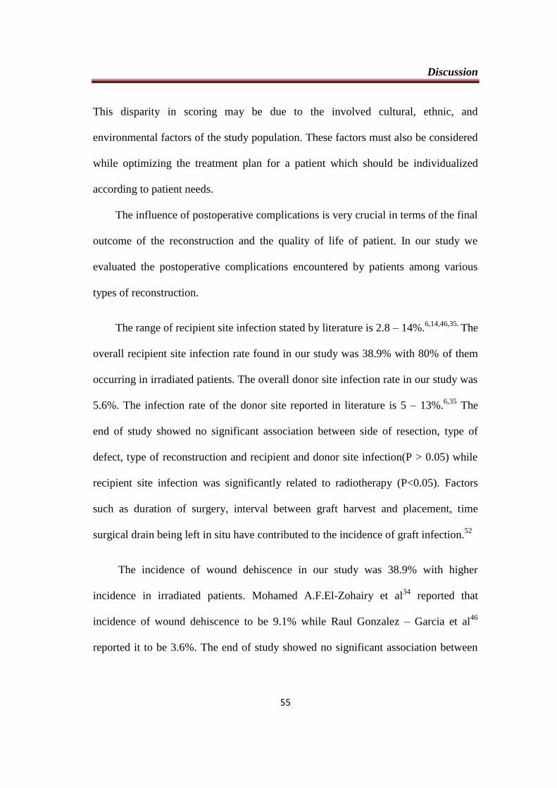
Discussion
55
This disparity in scoring may be due to the involved cultural, ethnic, and
environmental factors of the study population. These factors must also be considered
while optimizing the treatment plan for a patient which should be individualized
according to patient needs.
The influence of postoperative complications is very crucial in terms of the final
outcome of the reconstruction and the quality of life of patient. In our study we
evaluated the postoperative complications encountered by patients among various
types of reconstruction.
The range of recipient site infection stated by literature is 2.8 – 14%.6,14,46,35.
The
overall recipient site infection rate found in our study was 38.9% with 80% of them
occurring in irradiated patients. The overall donor site infection rate in our study was
5.6%. The infection rate of the donor site reported in literature is 5 – 13%.6,35
The
end of study showed no significant association between side of resection, type of
defect, type of reconstruction and recipient and donor site infection(P > 0.05) while
recipient site infection was significantly related to radiotherapy (P<0.05). Factors
such as duration of surgery, interval between graft harvest and placement, time
surgical drain being left in situ have contributed to the incidence of graft infection.52
The incidence of wound dehiscence in our study was 38.9% with higher
incidence in irradiated patients. Mohamed A.F.El-Zohairy et al34 reported that
incidence of wound dehiscence to be 9.1% while Raul Gonzalez – Garcia et al46
reported it to be 3.6%. The end of study showed no significant association between
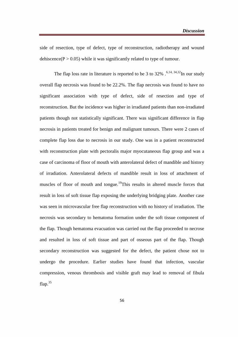
Discussion
56
side of resection, type of defect, type of reconstruction, radiotherapy and wound
dehiscence(P > 0.05) while it was significantly related to type of tumour.
The flap loss rate in literature is reported to be 3 to 32% .6,14, 34,32
In our study
overall flap necrosis was found to be 22.2%. The flap necrosis was found to have no
significant association with type of defect, side of resection and type of
reconstruction. But the incidence was higher in irradiated patients than non-irradiated
patients though not statistically significant. There was significant difference in flap
necrosis in patients treated for benign and malignant tumours. There were 2 cases of
complete flap loss due to necrosis in our study. One was in a patient reconstructed
with reconstruction plate with pectoralis major myocutaneous flap group and was a
case of carcinoma of floor of mouth with anterolateral defect of mandible and history
of irradiation. Anterolateral defects of mandible result in loss of attachment of
muscles of floor of mouth and tongue.19
This results in altered muscle forces that
result in loss of soft tissue flap exposing the underlying bridging plate. Another case
was seen in microvascular free flap reconstruction with no history of irradiation. The
necrosis was secondary to hematoma formation under the soft tissue component of
the flap. Though hematoma evacuation was carried out the flap proceeded to necrose
and resulted in loss of soft tissue and part of osseous part of the flap. Though
secondary reconstruction was suggested for the defect, the patient chose not to
undergo the procedure. Earlier studies have found that infection, vascular
compression, venous thrombosis and visible graft may lead to removal of fibula
flap.35
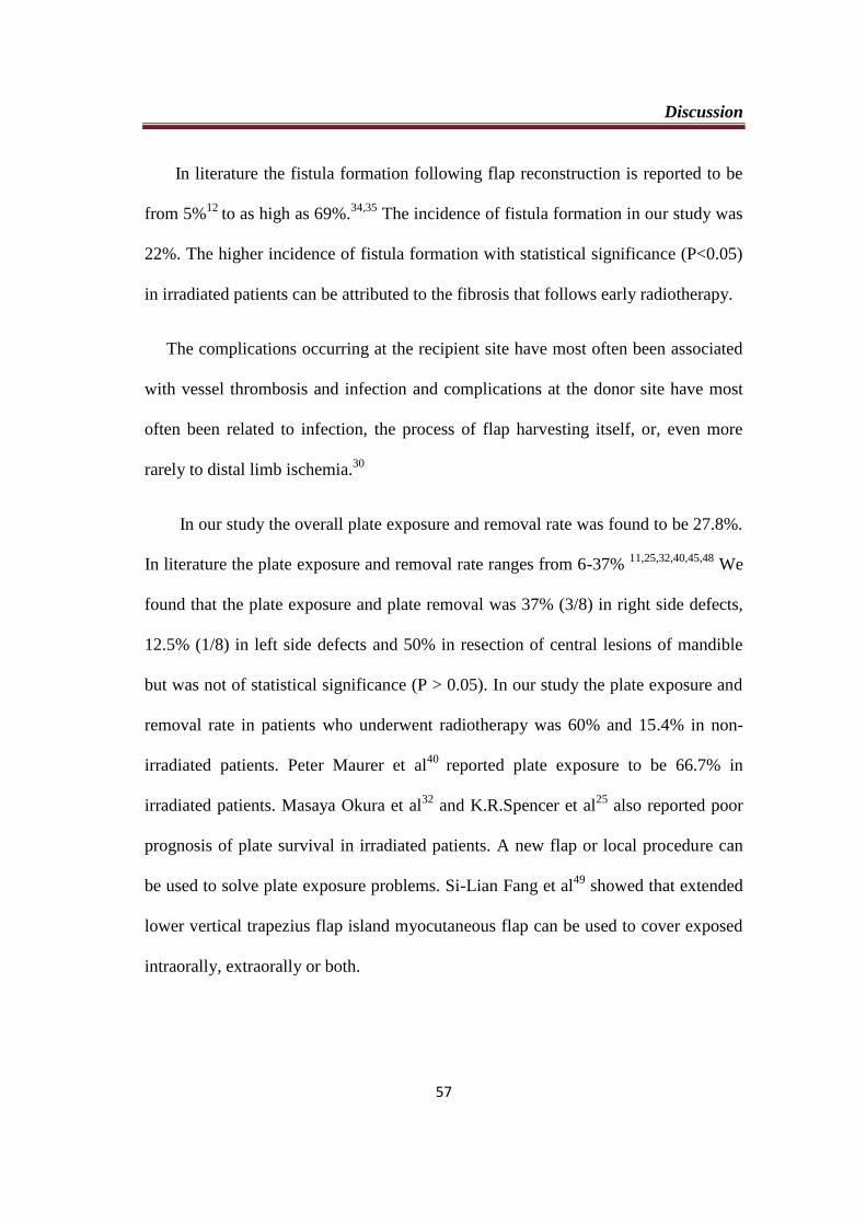
Discussion
57
In literature the fistula formation following flap reconstruction is reported to be
from 5%12
to as high as 69%.34,35
The incidence of fistula formation in our study was
22%. The higher incidence of fistula formation with statistical significance (P<0.05)
in irradiated patients can be attributed to the fibrosis that follows early radiotherapy.
The complications occurring at the recipient site have most often been associated
with vessel thrombosis and infection and complications at the donor site have most
often been related to infection, the process of flap harvesting itself, or, even more
rarely to distal limb ischemia.30
In our study the overall plate exposure and removal rate was found to be 27.8%.
In literature the plate exposure and removal rate ranges from 6-37% 11,25,32,40,45,48
We
found that the plate exposure and plate removal was 37% (3/8) in right side defects,
12.5% (1/8) in left side defects and 50% in resection of central lesions of mandible
but was not of statistical significance (P > 0.05). In our study the plate exposure and
removal rate in patients who underwent radiotherapy was 60% and 15.4% in non-
irradiated patients. Peter Maurer et al40
reported plate exposure to be 66.7% in
irradiated patients. Masaya Okura et al32
and K.R.Spencer et al25
also reported poor
prognosis of plate survival in irradiated patients. A new flap or local procedure can
be used to solve plate exposure problems. Si-Lian Fang et al49
showed that extended
lower vertical trapezius flap island myocutaneous flap can be used to cover exposed
intraorally, extraorally or both.
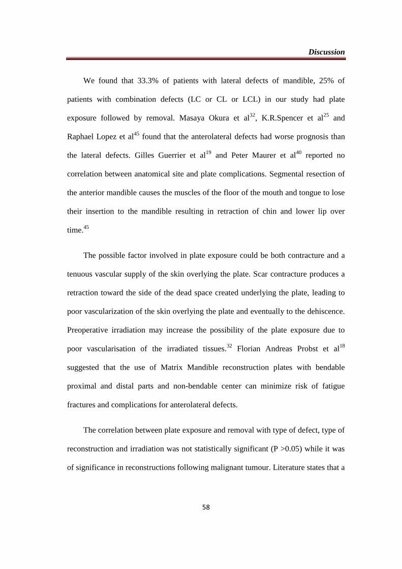
Discussion
58
We found that 33.3% of patients with lateral defects of mandible, 25% of
patients with combination defects (LC or CL or LCL) in our study had plate
exposure followed by removal. Masaya Okura et al32
, K.R.Spencer et al25
and
Raphael Lopez et al45
found that the anterolateral defects had worse prognosis than
the lateral defects. Gilles Guerrier et al19
and Peter Maurer et al40
reported no
correlation between anatomical site and plate complications. Segmental resection of
the anterior mandible causes the muscles of the floor of the mouth and tongue to lose
their insertion to the mandible resulting in retraction of chin and lower lip over
time.45
The possible factor involved in plate exposure could be both contracture and a
tenuous vascular supply of the skin overlying the plate. Scar contracture produces a
retraction toward the side of the dead space created underlying the plate, leading to
poor vascularization of the skin overlying the plate and eventually to the dehiscence.
Preoperative irradiation may increase the possibility of the plate exposure due to
poor vascularisation of the irradiated tissues.32
Florian Andreas Probst et al18
suggested that the use of Matrix Mandible reconstruction plates with bendable
proximal and distal parts and non-bendable center can minimize risk of fatigue
fractures and complications for anterolateral defects.
The correlation between plate exposure and removal with type of defect, type of
reconstruction and irradiation was not statistically significant (P >0.05) while it was
of significance in reconstructions following malignant tumour. Literature states that a
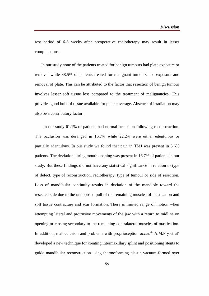
Discussion
59
rest period of 6-8 weeks after preoperative radiotherapy may result in lesser
complications.
In our study none of the patients treated for benign tumours had plate exposure or
removal while 38.5% of patients treated for malignant tumours had exposure and
removal of plate. This can be attributed to the factor that resection of benign tumour
involves lesser soft tissue loss compared to the treatment of malignancies. This
provides good bulk of tissue available for plate coverage. Absence of irradiation may
also be a contributory factor.
In our study 61.1% of patients had normal occlusion following reconstruction.
The occlusion was deranged in 16.7% while 22.2% were either edentulous or
partially edentulous. In our study we found that pain in TMJ was present in 5.6%
patients. The deviation during mouth opening was present in 16.7% of patients in our
study. But these findings did not have any statistical significance in relation to type
of defect, type of reconstruction, radiotherapy, type of tumour or side of resection.
Loss of mandibular continuity results in deviation of the mandible toward the
resected side due to the unopposed pull of the remaining muscles of mastication and
soft tissue contracture and scar formation. There is limited range of motion when
attempting lateral and protrusive movements of the jaw with a return to midline on
opening or closing secondary to the remaining contralateral muscles of mastication.
In addition, malocclusion and problems with proprioception occur.39
A.M.Fry et al2
developed a new technique for creating intermaxillary splint and positioning stents to
guide mandibular reconstruction using thermoforming plastic vacuum-formed over
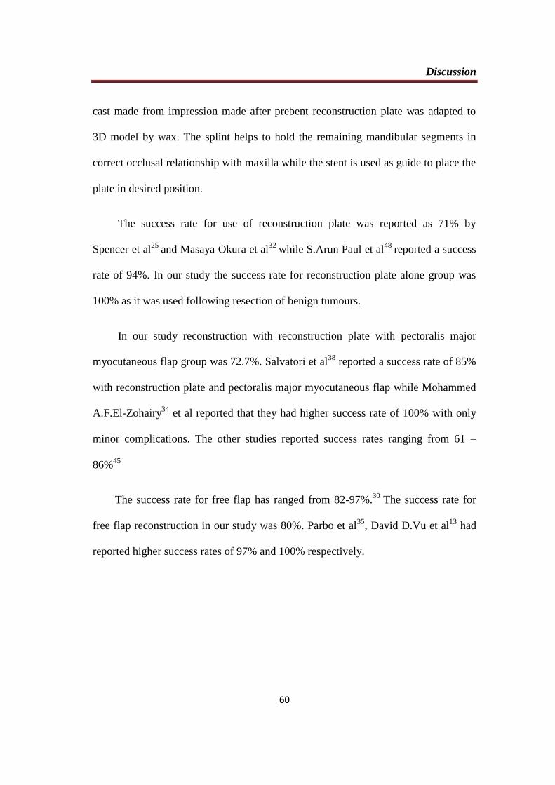
Discussion
60
cast made from impression made after prebent reconstruction plate was adapted to
3D model by wax. The splint helps to hold the remaining mandibular segments in
correct occlusal relationship with maxilla while the stent is used as guide to place the
plate in desired position.
The success rate for use of reconstruction plate was reported as 71% by
Spencer et al25
and Masaya Okura et al32
while S.Arun Paul et al48
reported a success
rate of 94%. In our study the success rate for reconstruction plate alone group was
100% as it was used following resection of benign tumours.
In our study reconstruction with reconstruction plate with pectoralis major
myocutaneous flap group was 72.7%. Salvatori et al38
reported a success rate of 85%
with reconstruction plate and pectoralis major myocutaneous flap while Mohammed
A.F.El-Zohairy34
et al reported that they had higher success rate of 100% with only
minor complications. The other studies reported success rates ranging from 61 –
86%45
The success rate for free flap has ranged from 82-97%.30
The success rate for
free flap reconstruction in our study was 80%. Parbo et al35
, David D.Vu et al13
had
reported higher success rates of 97% and 100% respectively.

Summary & Conclusion
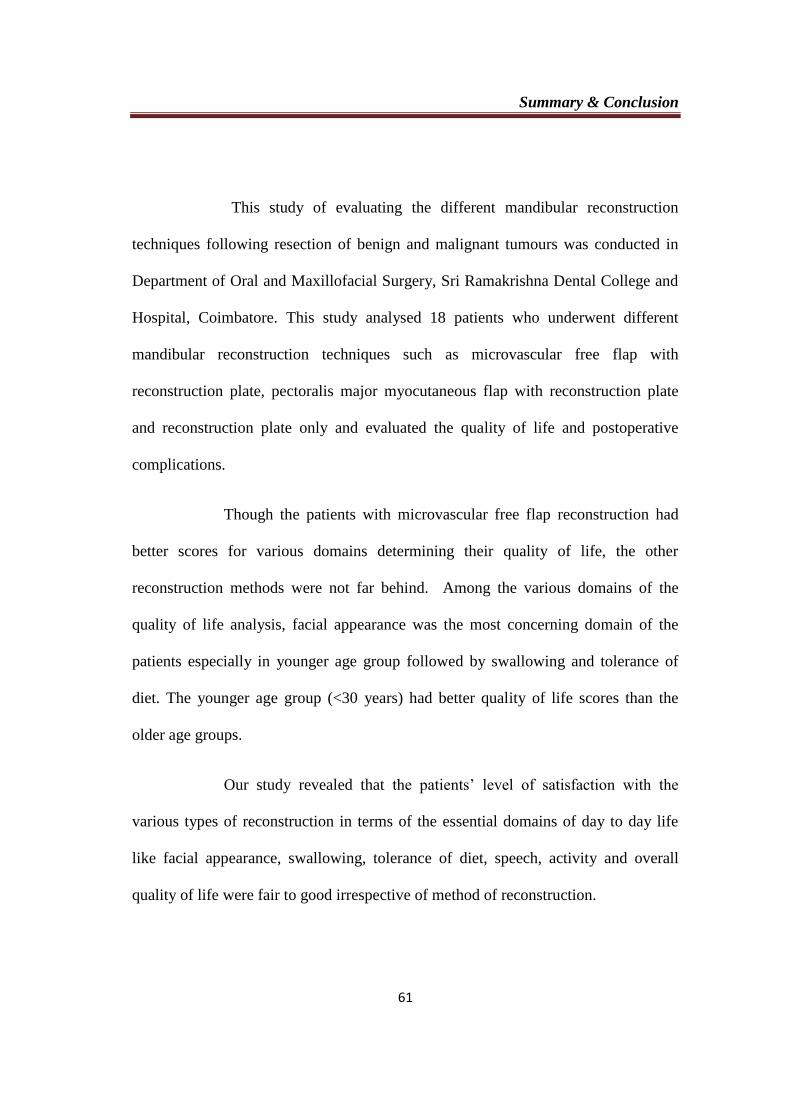
Summary & Conclusion
61
This study of evaluating the different mandibular reconstruction
techniques following resection of benign and malignant tumours was conducted in
Department of Oral and Maxillofacial Surgery, Sri Ramakrishna Dental College and
Hospital, Coimbatore. This study analysed 18 patients who underwent different
mandibular reconstruction techniques such as microvascular free flap with
reconstruction plate, pectoralis major myocutaneous flap with reconstruction plate
and reconstruction plate only and evaluated the quality of life and postoperative
complications.
Though the patients with microvascular free flap reconstruction had
better scores for various domains determining their quality of life, the other
reconstruction methods were not far behind. Among the various domains of the
quality of life analysis, facial appearance was the most concerning domain of the
patients especially in younger age group followed by swallowing and tolerance of
diet. The younger age group (<30 years) had better quality of life scores than the
older age groups.
Our study revealed that the patients’ level of satisfaction with the
various types of reconstruction in terms of the essential domains of day to day life
like facial appearance, swallowing, tolerance of diet, speech, activity and overall
quality of life were fair to good irrespective of method of reconstruction.
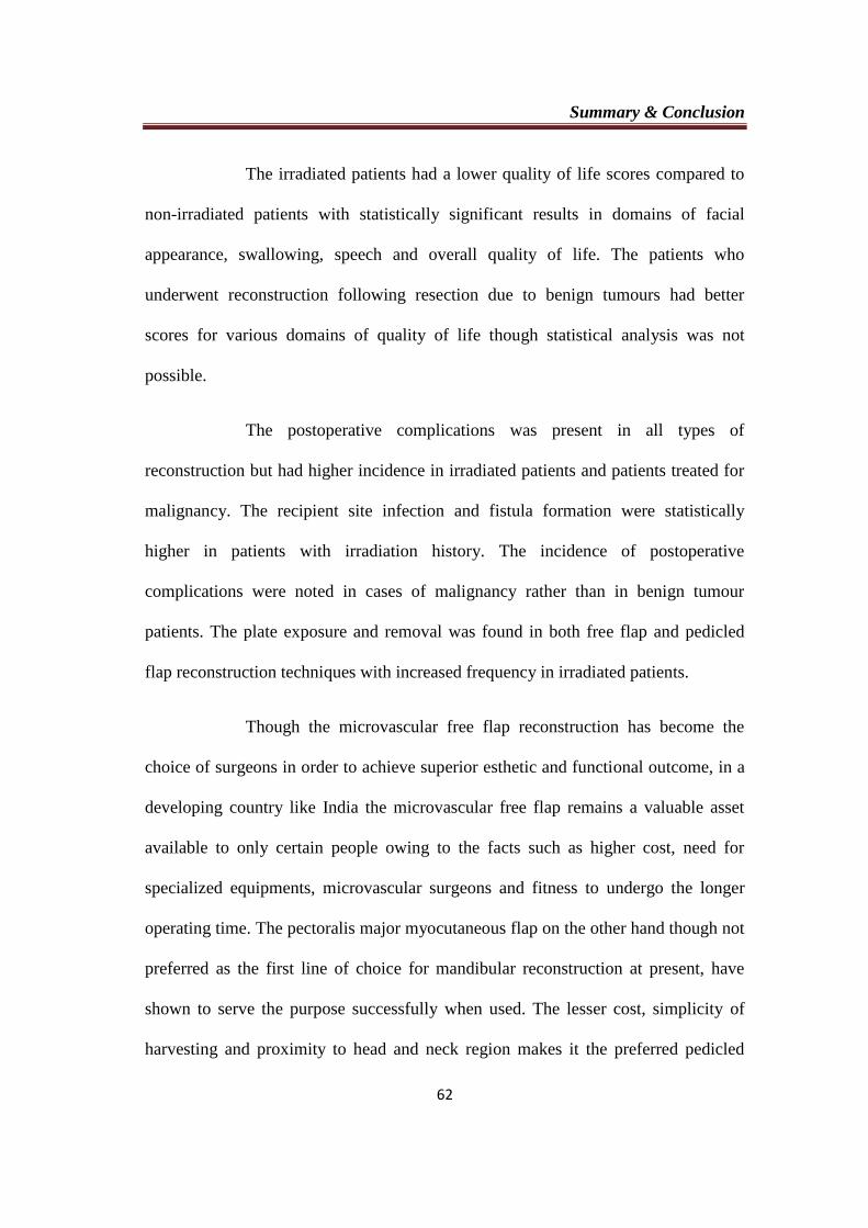
Summary & Conclusion
62
The irradiated patients had a lower quality of life scores compared to
non-irradiated patients with statistically significant results in domains of facial
appearance, swallowing, speech and overall quality of life. The patients who
underwent reconstruction following resection due to benign tumours had better
scores for various domains of quality of life though statistical analysis was not
possible.
The postoperative complications was present in all types of
reconstruction but had higher incidence in irradiated patients and patients treated for
malignancy. The recipient site infection and fistula formation were statistically
higher in patients with irradiation history. The incidence of postoperative
complications were noted in cases of malignancy rather than in benign tumour
patients. The plate exposure and removal was found in both free flap and pedicled
flap reconstruction techniques with increased frequency in irradiated patients.
Though the microvascular free flap reconstruction has become the
choice of surgeons in order to achieve superior esthetic and functional outcome, in a
developing country like India the microvascular free flap remains a valuable asset
available to only certain people owing to the facts such as higher cost, need for
specialized equipments, microvascular surgeons and fitness to undergo the longer
operating time. The pectoralis major myocutaneous flap on the other hand though not
preferred as the first line of choice for mandibular reconstruction at present, have
shown to serve the purpose successfully when used. The lesser cost, simplicity of
harvesting and proximity to head and neck region makes it the preferred pedicled
Summary & Conclusion
63
flap when needed. The flap also provides bulk for reconstruction in cases of
extensive resection following locally advanced diseases but the same bulk may act as
disadvantage in certain cases. The other disadvantages of the flap such as increased
incidence of infection, partial or complete flap necrosis, dehiscence or fistula
formation especially following irradiation can be managed conservatively although
more aggressive procedures may be needed occasionally. The reconstruction plate
alone used to bridge segmental mandibular defects are used only when other popular
reconstruction method are not feasible.
Though our study had a smaller sample size, the results suggest that
the proper planning and execution of the reconstruction technique combined with
important cofactors like type of tumour and irradiation serve as key factors in
determining the quality of life of patients rather than the method of reconstruction
proper. Prospective study with larger sample size and longer follow-up with periodic
analysis may be necessary to reemphasize the results of our study.

Tables
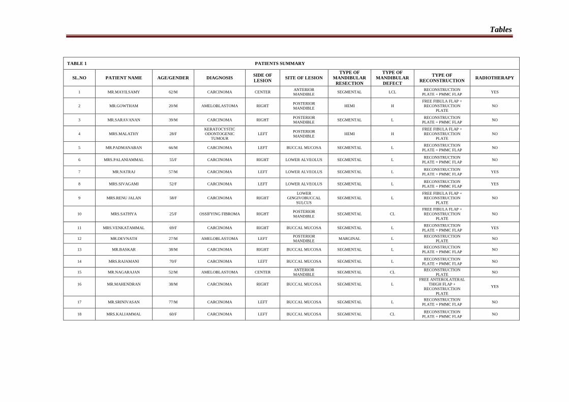
Tables
TABLE 1 PATIENTS SUMMARY
SL.NO PATIENT NAME AGE/GENDER DIAGNOSIS SIDE OF
LESION SITE OF LESION
TYPE OF
MANDIBULAR
RESECTION
TYPE OF
MANDIBULAR
DEFECT
TYPE OF
RECONSTRUCTION RADIOTHERAPY
1 MR.MAYILSAMY 62/M CARCINOMA CENTER ANTERIOR
MANDIBLE SEGMENTAL LCL
RECONSTRUCTION
PLATE + PMMC FLAP YES
2 MR.GOWTHAM 20/M AMELOBLASTOMA RIGHT POSTERIOR
MANDIBLE HEMI H
FREE FIBULA FLAP +
RECONSTRUCTION
PLATE
NO
3 MR.SARAVANAN 39/M CARCINOMA RIGHT POSTERIOR
MANDIBLE SEGMENTAL L
RECONSTRUCTION
PLATE + PMMC FLAP NO
4 MRS.MALATHY 28/F
KERATOCYSTIC
ODONTOGENIC
TUMOUR
LEFT POSTERIOR
MANDIBLE HEMI H
FREE FIBULA FLAP +
RECONSTRUCTION
PLATE
NO
5 MR.PADMANABAN 66/M CARCINOMA LEFT BUCCAL MUCOSA SEGMENTAL L RECONSTRUCTION
PLATE + PMMC FLAP NO
6 MRS.PALANIAMMAL 55/F CARCINOMA RIGHT LOWER ALVEOLUS SEGMENTAL L RECONSTRUCTION
PLATE + PMMC FLAP NO
7 MR.NATRAJ 57/M CARCINOMA LEFT LOWER ALVEOLUS SEGMENTAL L RECONSTRUCTION
PLATE + PMMC FLAP YES
8 MRS.SIVAGAMI 52/F CARCINOMA LEFT LOWER ALVEOLUS SEGMENTAL L RECONSTRUCTION
PLATE + PMMC FLAP YES
9 MRS.RENU JALAN 58/F CARCINOMA RIGHT
LOWER
GINGIVOBUCCAL
SULCUS
SEGMENTAL L
FREE FIBULA FLAP +
RECONSTRUCTION
PLATE
NO
10 MRS.SATHYA 25/F OSSIFYING FIBROMA RIGHT POSTERIOR
MANDIBLE SEGMENTAL CL
FREE FIBULA FLAP +
RECONSTRUCTION
PLATE
NO
11 MRS.VENKATAMMAL 69/F CARCINOMA RIGHT BUCCAL MUCOSA SEGMENTAL L RECONSTRUCTION
PLATE + PMMC FLAP YES
12 MR.DEVNATH 27/M AMELOBLASTOMA LEFT POSTERIOR
MANDIBLE MARGINAL L
RECONSTRUCTION
PLATE NO
13 MR.BASKAR 38/M CARCINOMA RIGHT BUCCAL MUCOSA SEGMENTAL L RECONSTRUCTION
PLATE + PMMC FLAP NO
14 MRS.RAJAMANI 70/F CARCINOMA LEFT BUCCAL MUCOSA SEGMENTAL L RECONSTRUCTION
PLATE + PMMC FLAP NO
15 MR.NAGARAJAN 52/M AMELOBLASTOMA CENTER ANTERIOR
MANDIBLE SEGMENTAL CL
RECONSTRUCTION
PLATE NO
16
MR.MAHENDRAN
38/M
CARCINOMA
RIGHT
BUCCAL MUCOSA
SEGMENTAL
L
FREE ANTEROLATERAL
THIGH FLAP +
RECONSTRUCTION
PLATE
YES
17 MR.SRINIVASAN 77/M CARCINOMA LEFT BUCCAL MUCOSA SEGMENTAL L RECONSTRUCTION
PLATE + PMMC FLAP NO
18 MRS.KALIAMMAL 60/F CARCINOMA LEFT BUCCAL MUCOSA SEGMENTAL CL RECONSTRUCTION
PLATE + PMMC FLAP NO
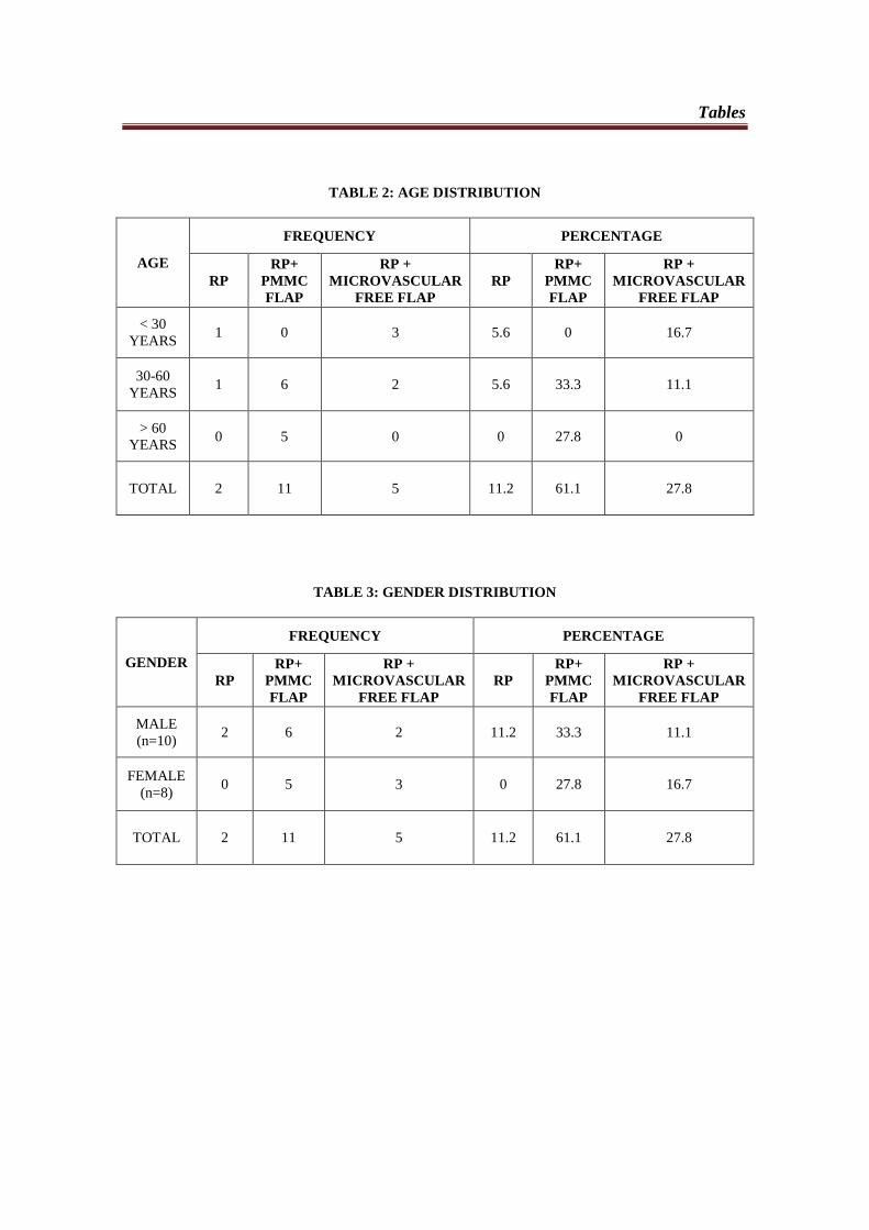
Tables
TABLE 2: AGE DISTRIBUTION
AGE
FREQUENCY PERCENTAGE
RP
RP+
PMMC
FLAP
RP +
MICROVASCULAR
FREE FLAP
RP
RP+
PMMC
FLAP
RP +
MICROVASCULAR
FREE FLAP
< 30
YEARS 1 0 3 5.6 0 16.7
30-60
YEARS 1 6 2 5.6 33.3 11.1
> 60
YEARS 0 5 0 0 27.8 0
TOTAL 2 11 5 11.2 61.1 27.8
TABLE 3: GENDER DISTRIBUTION
GENDER
FREQUENCY PERCENTAGE
RP
RP+
PMMC
FLAP
RP +
MICROVASCULAR
FREE FLAP
RP
RP+
PMMC
FLAP
RP +
MICROVASCULAR
FREE FLAP
MALE
(n=10) 2 6 2 11.2 33.3 11.1
FEMALE
(n=8) 0 5 3 0 27.8 16.7
TOTAL 2 11 5 11.2 61.1 27.8
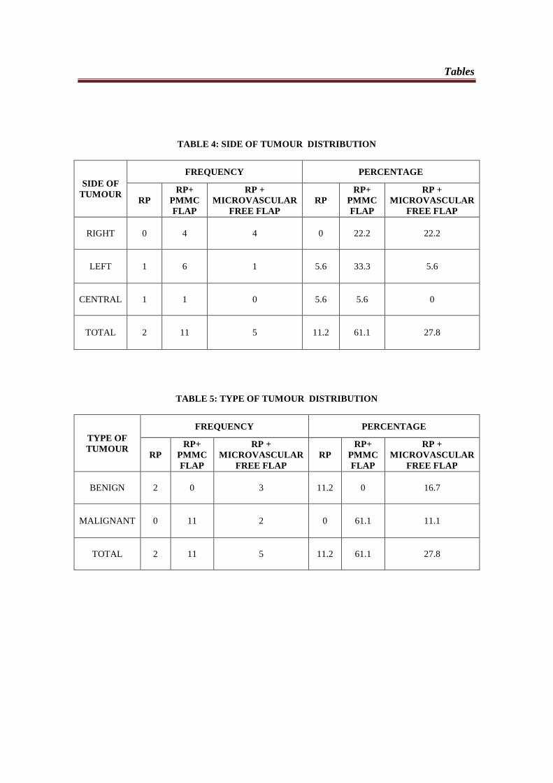
Tables
TABLE 4: SIDE OF TUMOUR DISTRIBUTION
SIDE OF
TUMOUR
FREQUENCY PERCENTAGE
RP
RP+
PMMC
FLAP
RP +
MICROVASCULAR
FREE FLAP
RP
RP+
PMMC
FLAP
RP +
MICROVASCULAR
FREE FLAP
RIGHT 0 4 4 0 22.2 22.2
LEFT 1 6 1 5.6 33.3 5.6
CENTRAL 1 1 0 5.6 5.6 0
TOTAL 2 11 5 11.2 61.1 27.8
TABLE 5: TYPE OF TUMOUR DISTRIBUTION
TYPE OF
TUMOUR
FREQUENCY PERCENTAGE
RP
RP+
PMMC
FLAP
RP +
MICROVASCULAR
FREE FLAP
RP
RP+
PMMC
FLAP
RP +
MICROVASCULAR
FREE FLAP
BENIGN 2 0 3 11.2 0 16.7
MALIGNANT 0 11 2 0 61.1 11.1
TOTAL 2 11 5 11.2 61.1 27.8
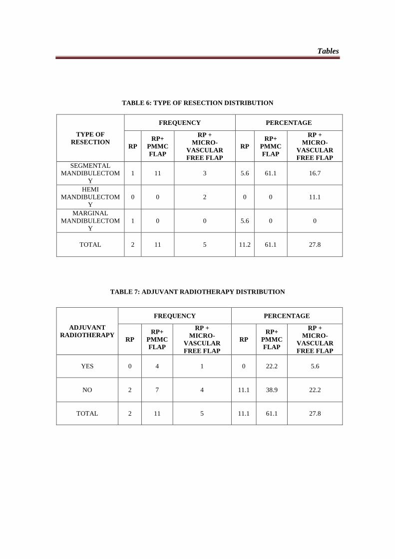
Tables
TABLE 6: TYPE OF RESECTION DISTRIBUTION
TYPE OF
RESECTION
FREQUENCY PERCENTAGE
RP
RP+
PMMC
FLAP
RP +
MICRO-
VASCULAR
FREE FLAP
RP
RP+
PMMC
FLAP
RP +
MICRO-
VASCULAR
FREE FLAP
SEGMENTAL
MANDIBULECTOM
Y
1 11 3 5.6 61.1 16.7
HEMI
MANDIBULECTOM
Y
0 0 2 0 0 11.1
MARGINAL
MANDIBULECTOM
Y
1 0 0 5.6 0 0
TOTAL 2 11 5 11.2 61.1 27.8
TABLE 7: ADJUVANT RADIOTHERAPY DISTRIBUTION
ADJUVANT
RADIOTHERAPY
FREQUENCY PERCENTAGE
RP
RP+
PMMC
FLAP
RP +
MICRO-
VASCULAR
FREE FLAP
RP
RP+
PMMC
FLAP
RP +
MICRO-
VASCULAR
FREE FLAP
YES 0 4 1 0 22.2 5.6
NO 2 7 4 11.1 38.9 22.2
TOTAL 2 11 5 11.1 61.1 27.8

Tables
TABLE 8: TYPE OF MANDIBULAR DEFECT DISTRIBUTION
TYPE OF
RESECTION
FREQUENCY PERCENTAGE
RP
RP+
PMMC
FLAP
RP +
MICRO-
VASCULAR FREE
FLAP
RP
RP+
PMMC
FLAP
RP +
MICRO-
VASCULAR FREE
FLAP
CENTRAL
(C) 0 0 0 0 0 0
LATERAL
WITHOUT
CONDYLE
(L)
1 9 2 5.6 50 11.1
LATERAL
WITH
CONDYLE
(H)
0 0 2 0 0 11.1
LATERAL
WITH
CENTRAL
(LC OR CL
OR LCL)
1 2 1 5.6 11.1 5.6
TOTAL 2 11 5 11.2 61.1 27.8
TABLE 9: TYPE OF RECONSTRUCTION DISTRIBUTION
TYPE OF
RECONSTRUCTION FREQUENCY PERCENTAGE
RP 2 11.1
RP+ PMMC FLAP 11 61.1
RP +
MICROVASCULAR
FREE FLAP
5 27.8
TOTAL 18 100.0
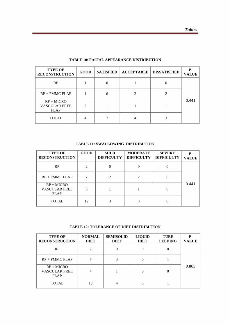
Tables
TABLE 10: FACIAL APPEARANCE DISTRIBUTION
TYPE OF
RECONSTRUCTION GOOD SATISFIED ACCEPTABLE DISSATISFIED
P-
VALUE
RP 1 0 1 0
0.441
RP + PMMC FLAP 1 6 2 2
RP + MICRO
VASCULAR FREE
FLAP
2 1 1 1
TOTAL 4 7 4 3
TABLE 11: SWALLOWING DISTRIBUTION
TYPE OF
RECONSTRUCTION
GOOD MILD
DIFFICULTY
MODERATE
DIFFICULTY
SEVERE
DIFFICULTY P-
VALUE
RP 2 0 0 0
0.441
RP + PMMC FLAP 7 2 2 0
RP + MICRO
VASCULAR FREE
FLAP
3 1 1 0
TOTAL 12 3 3 0
TABLE 12: TOLERANCE OF DIET DISTRIBUTION
TYPE OF
RECONSTRUCTION
NORMAL
DIET
SEMISOLID
DIET
LIQUID
DIET
TUBE
FEEDING
P-
VALUE
RP 2 0 0 0
0.865
RP + PMMC FLAP 7 3 0 1
RP + MICRO
VASCULAR FREE
FLAP
4 1 0 0
TOTAL 13 4 0 1

Tables
TABLE 13: SPEECH DISTRIBUTION
TYPE OF
RECONSTRUCTION NORMAL
EASY TO
UNDER-
STAND
DIFFICULT
TO UNDER-
STAND
VERY
DIFFICULT
TO UNDER-
STAND
P-VALUE
RP 2 0 0 0
0.171
RP + PMMC FLAP 2 4 5 0
RP + MICRO
VASCULAR FREE
FLAP
2 1 2 0
TOTAL 6 5 7 0
TABLE 14: ACTIVITY DISTRIBUTION
TYPE OF
RECONSTRUCTION NORMAL
MODERATELY
ACTIVE
MINIMALLY
ACTIVE PVALUE
RP 2 0 0
0.805
RP + PMMC FLAP 9 2 0
RP + MICRO VASCULAR
FREE FLAP 4 1 0
TOTAL 13 3 0
TABLE 15: OVERALL QUALITY OF LIFE DISTRIBUTION
TYPE OF
RECONSTRUCTION GOOD FAIR ACCEPTABLE POOR P-VALUE
RP 2 0 0 0
0.188 RP + PMMC FLAP 3 5 2 1
RP + MICRO
VASCULAR FREE FLAP 3 1 1 0
TOTAL 8 6 3 1
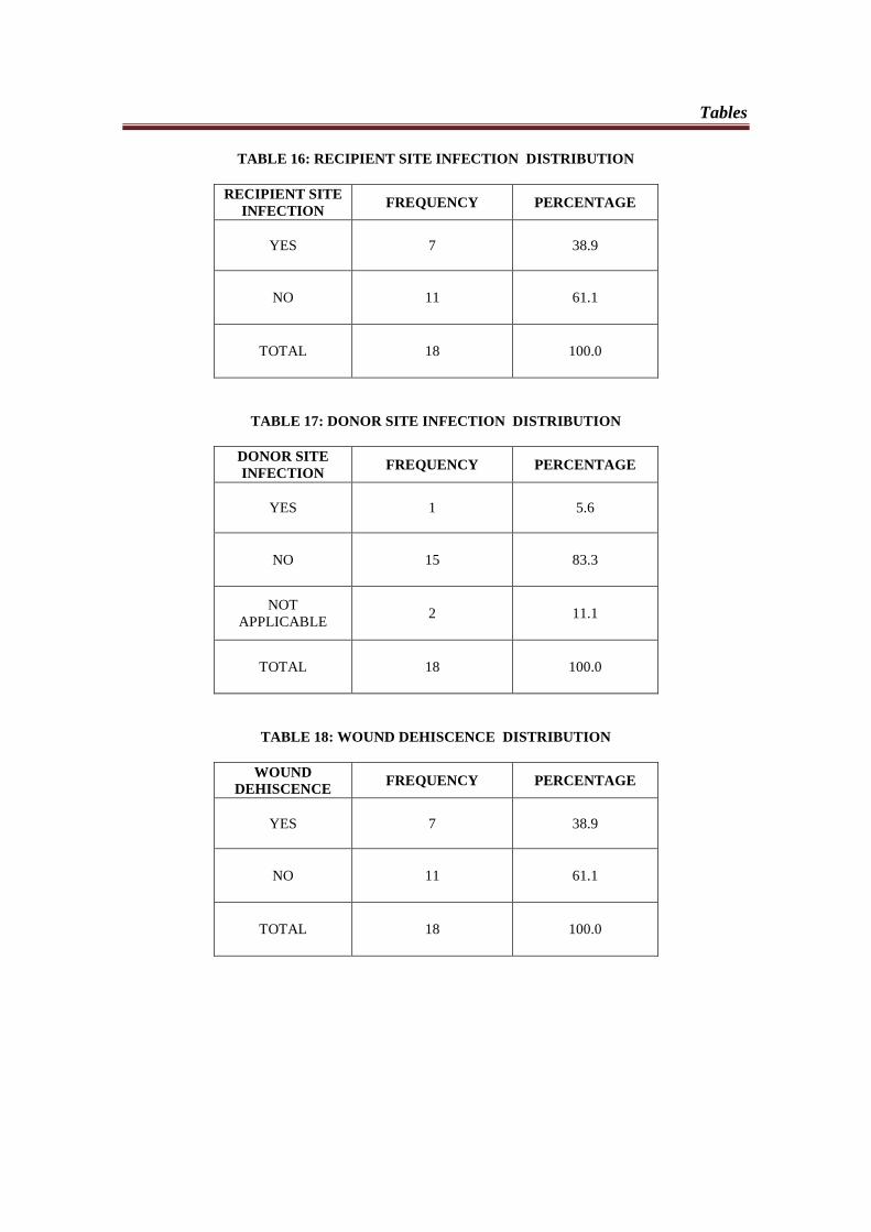
Tables
TABLE 16: RECIPIENT SITE INFECTION DISTRIBUTION
RECIPIENT SITE
INFECTION FREQUENCY PERCENTAGE
YES 7 38.9
NO 11 61.1
TOTAL 18 100.0
TABLE 17: DONOR SITE INFECTION DISTRIBUTION
DONOR SITE
INFECTION FREQUENCY PERCENTAGE
YES 1 5.6
NO 15 83.3
NOT
APPLICABLE 2 11.1
TOTAL 18 100.0
TABLE 18: WOUND DEHISCENCE DISTRIBUTION
WOUND
DEHISCENCE FREQUENCY PERCENTAGE
YES 7 38.9
NO 11 61.1
TOTAL 18 100.0
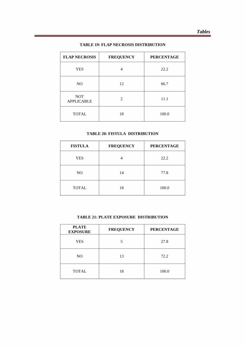
Tables
TABLE 19: FLAP NECROSIS DISTRIBUTION
FLAP NECROSIS FREQUENCY PERCENTAGE
YES 4 22.2
NO 12 66.7
NOT
APPLICABLE 2 11.1
TOTAL 18 100.0
TABLE 20: FISTULA DISTRIBUTION
FISTULA FREQUENCY PERCENTAGE
YES 4 22.2
NO 14 77.8
TOTAL 18 100.0
TABLE 21: PLATE EXPOSURE DISTRIBUTION
PLATE
EXPOSURE FREQUENCY PERCENTAGE
YES 5 27.8
NO 13 72.2
TOTAL 18 100.0
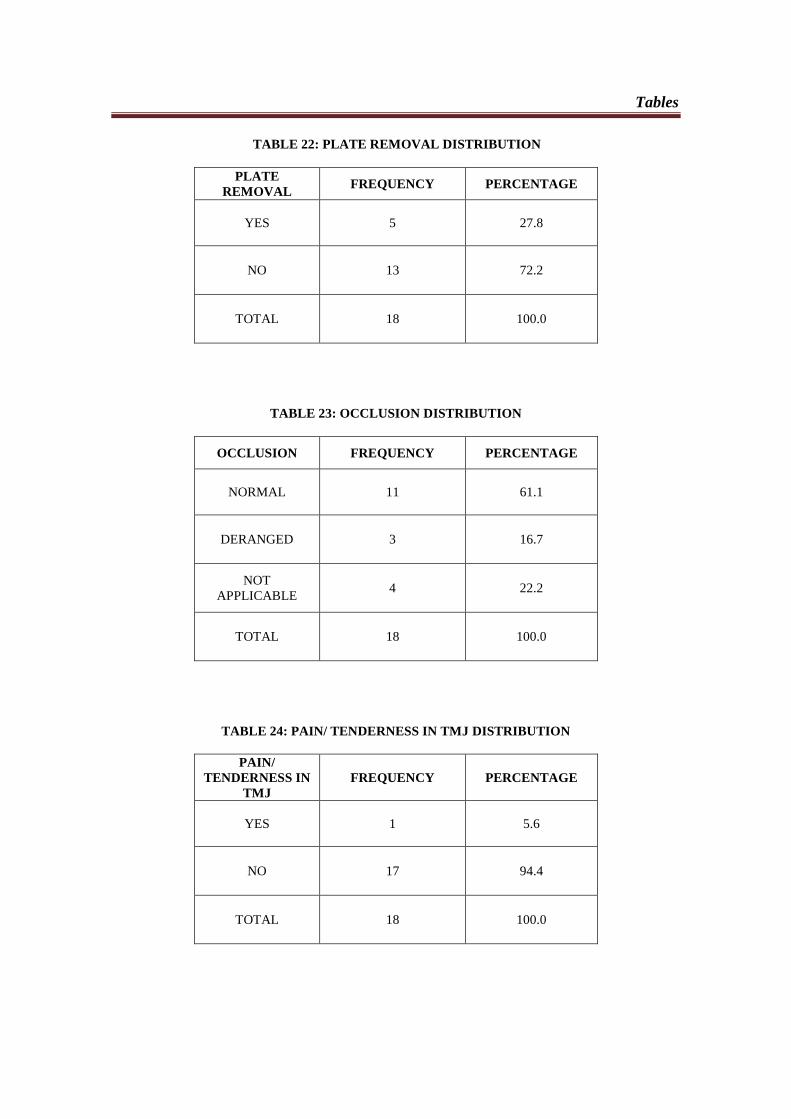
Tables TABLE 22: PLATE REMOVAL DISTRIBUTION
PLATE
REMOVAL FREQUENCY PERCENTAGE
YES 5 27.8
NO 13 72.2
TOTAL 18 100.0
TABLE 23: OCCLUSION DISTRIBUTION
OCCLUSION FREQUENCY PERCENTAGE
NORMAL 11 61.1
DERANGED 3 16.7
NOT
APPLICABLE 4 22.2
TOTAL 18 100.0
TABLE 24: PAIN/ TENDERNESS IN TMJ DISTRIBUTION
PAIN/
TENDERNESS IN
TMJ
FREQUENCY PERCENTAGE
YES 1 5.6
NO 17 94.4
TOTAL 18 100.0
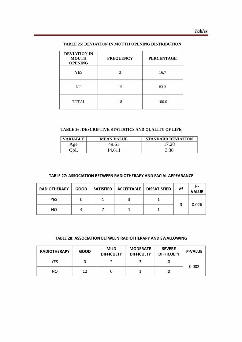
Tables TABLE 25: DEVIATION IN MOUTH OPENING DISTRIBUTION
DEVIATION IN
MOUTH
OPENING
FREQUENCY PERCENTAGE
YES 3 16.7
NO 15 83.3
TOTAL 18 100.0
TABLE 26: DESCRIPTIVE STATISTICS AND QUALITY OF LIFE
VARIABLE MEAN VALUE STANDARD DEVIATION
Age 49.61 17.28
QoL 14.611 3.38
TABLE 27: ASSOCIATION BETWEEN RADIOTHERAPY AND FACIAL APPEARANCE
RADIOTHERAPY GOOD SATISFIED ACCEPTABLE DISSATISFIED df P-
VALUE
YES 0 1 3 1 3 0.026
NO 4 7 1 1
TABLE 28: ASSOCIATION BETWEEN RADIOTHERAPY AND SWALLOWING
RADIOTHERAPY GOOD MILD
DIFFICULTY MODERATE DIFFICULTY
SEVERE DIFFICULTY
P-VALUE
YES 0 2 3 0 0.002
NO 12 0 1 0
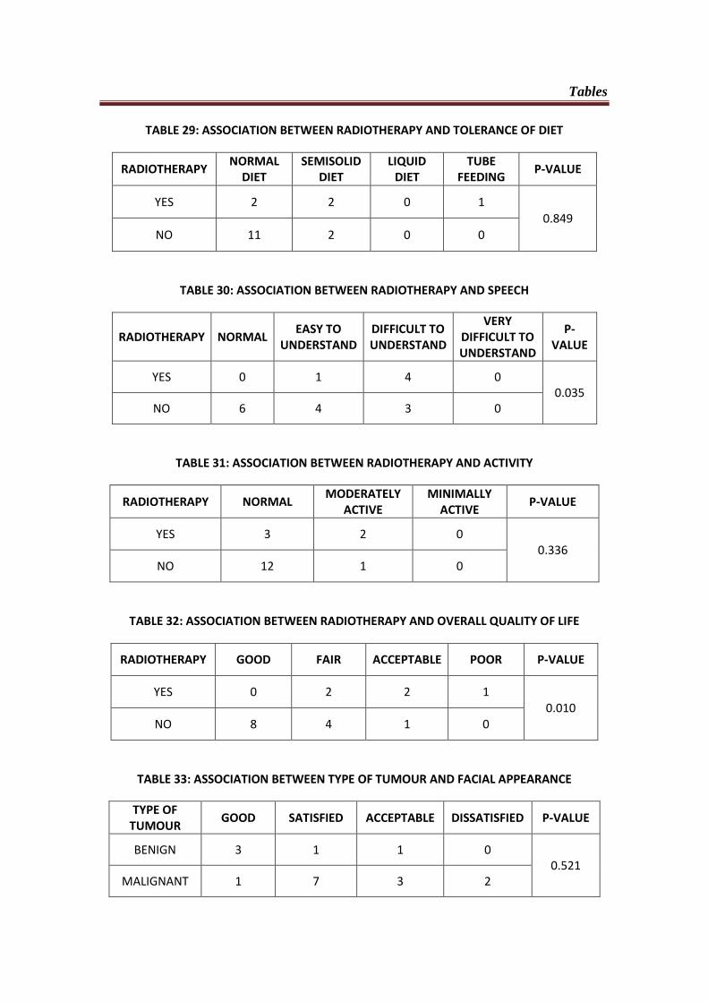
Tables
TABLE 29: ASSOCIATION BETWEEN RADIOTHERAPY AND TOLERANCE OF DIET
RADIOTHERAPY NORMAL
DIET SEMISOLID
DIET LIQUID
DIET TUBE
FEEDING P-VALUE
YES 2 2 0 1 0.849
NO 11 2 0 0
TABLE 30: ASSOCIATION BETWEEN RADIOTHERAPY AND SPEECH
RADIOTHERAPY NORMAL EASY TO
UNDERSTAND DIFFICULT TO UNDERSTAND
VERY DIFFICULT TO UNDERSTAND
P-VALUE
YES 0 1 4 0 0.035
NO 6 4 3 0
TABLE 31: ASSOCIATION BETWEEN RADIOTHERAPY AND ACTIVITY
RADIOTHERAPY NORMAL MODERATELY
ACTIVE MINIMALLY
ACTIVE P-VALUE
YES 3 2 0 0.336
NO 12 1 0
TABLE 32: ASSOCIATION BETWEEN RADIOTHERAPY AND OVERALL QUALITY OF LIFE
RADIOTHERAPY GOOD FAIR ACCEPTABLE POOR P-VALUE
YES 0 2 2 1 0.010
NO 8 4 1 0
TABLE 33: ASSOCIATION BETWEEN TYPE OF TUMOUR AND FACIAL APPEARANCE
TYPE OF TUMOUR
GOOD SATISFIED ACCEPTABLE DISSATISFIED P-VALUE
BENIGN 3 1 1 0 0.521
MALIGNANT 1 7 3 2
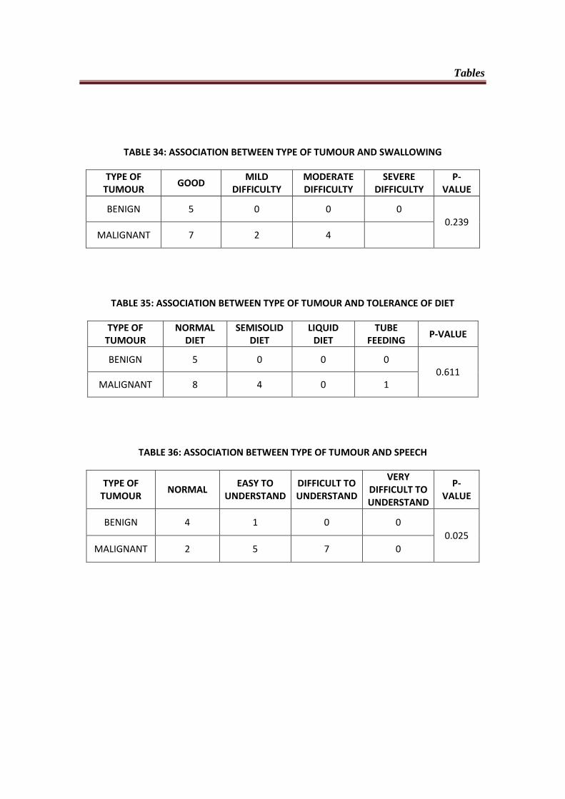
Tables
TABLE 34: ASSOCIATION BETWEEN TYPE OF TUMOUR AND SWALLOWING
TYPE OF TUMOUR
GOOD MILD
DIFFICULTY MODERATE DIFFICULTY
SEVERE DIFFICULTY
P-VALUE
BENIGN 5 0 0 0 0.239
MALIGNANT 7 2 4
TABLE 35: ASSOCIATION BETWEEN TYPE OF TUMOUR AND TOLERANCE OF DIET
TYPE OF TUMOUR
NORMAL DIET
SEMISOLID DIET
LIQUID DIET
TUBE FEEDING
P-VALUE
BENIGN 5 0 0 0 0.611
MALIGNANT 8 4 0 1
TABLE 36: ASSOCIATION BETWEEN TYPE OF TUMOUR AND SPEECH
TYPE OF TUMOUR
NORMAL EASY TO
UNDERSTAND DIFFICULT TO UNDERSTAND
VERY DIFFICULT TO UNDERSTAND
P-VALUE
BENIGN 4 1 0 0
0.025
MALIGNANT 2 5 7 0
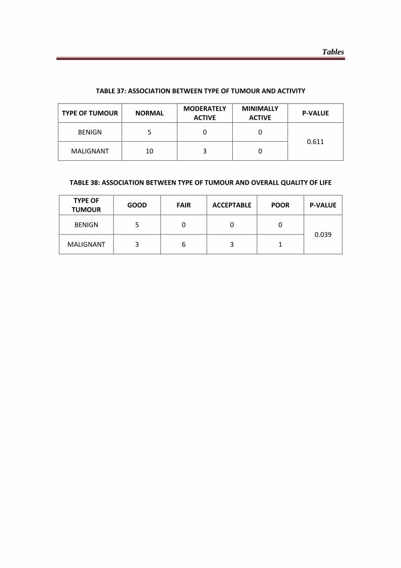
Tables
TABLE 37: ASSOCIATION BETWEEN TYPE OF TUMOUR AND ACTIVITY
TYPE OF TUMOUR NORMAL MODERATELY
ACTIVE MINIMALLY
ACTIVE P-VALUE
BENIGN 5 0 0
0.611
MALIGNANT 10 3 0
TABLE 38: ASSOCIATION BETWEEN TYPE OF TUMOUR AND OVERALL QUALITY OF LIFE
TYPE OF TUMOUR
GOOD FAIR ACCEPTABLE POOR P-VALUE
BENIGN 5 0 0 0
0.039
MALIGNANT 3 6 3 1
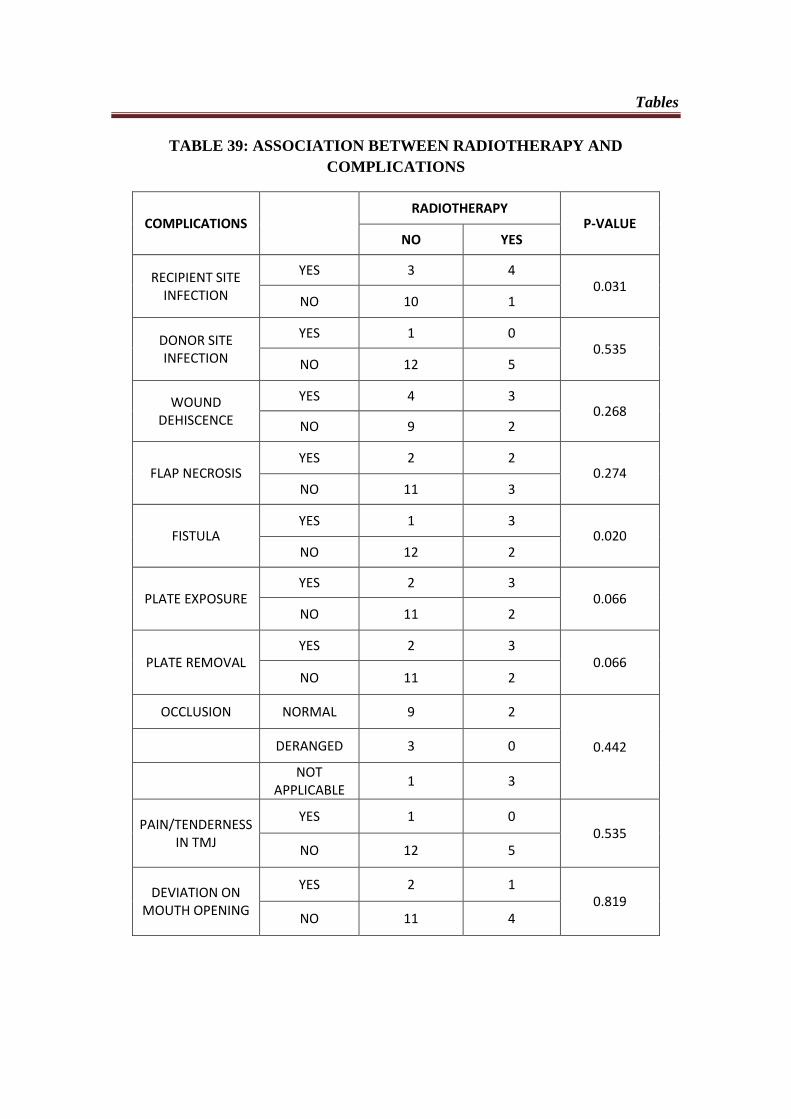
Tables
TABLE 39: ASSOCIATION BETWEEN RADIOTHERAPY AND
COMPLICATIONS
COMPLICATIONS RADIOTHERAPY
P-VALUE NO YES
RECIPIENT SITE INFECTION
YES 3 4 0.031
NO 10 1
DONOR SITE INFECTION
YES 1 0 0.535
NO 12 5
WOUND DEHISCENCE
YES 4 3 0.268
NO 9 2
FLAP NECROSIS YES 2 2
0.274 NO 11 3
FISTULA YES 1 3
0.020 NO 12 2
PLATE EXPOSURE YES 2 3
0.066 NO 11 2
PLATE REMOVAL YES 2 3
0.066 NO 11 2
OCCLUSION NORMAL 9 2
0.442 DERANGED 3 0
NOT
APPLICABLE 1 3
PAIN/TENDERNESS IN TMJ
YES 1 0 0.535
NO 12 5
DEVIATION ON MOUTH OPENING
YES 2 1 0.819
NO 11 4
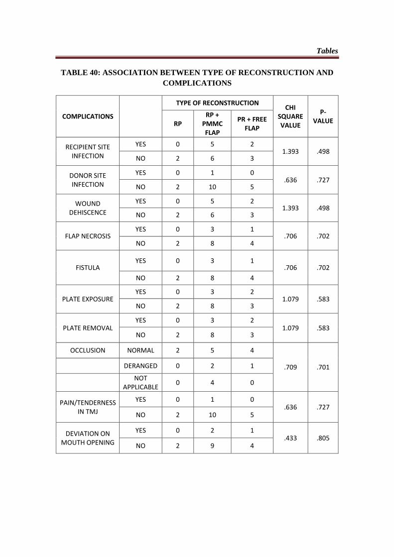
Tables
TABLE 40: ASSOCIATION BETWEEN TYPE OF RECONSTRUCTION AND
COMPLICATIONS
COMPLICATIONS
TYPE OF RECONSTRUCTION CHI
SQUARE VALUE
P-VALUE RP
RP + PMMC FLAP
PR + FREE FLAP
RECIPIENT SITE INFECTION
YES 0 5 2 1.393 .498
NO 2 6 3
DONOR SITE INFECTION
YES 0 1 0 .636 .727
NO 2 10 5
WOUND DEHISCENCE
YES 0 5 2 1.393 .498
NO 2 6 3
FLAP NECROSIS YES 0 3 1
.706 .702 NO 2 8 4
FISTULA YES 0 3 1
.706 .702
NO 2 8 4
PLATE EXPOSURE YES 0 3 2
1.079 .583 NO 2 8 3
PLATE REMOVAL YES 0 3 2
1.079 .583 NO 2 8 3
OCCLUSION NORMAL 2 5 4
.709 .701 DERANGED 0 2 1
NOT
APPLICABLE 0 4 0
PAIN/TENDERNESS IN TMJ
YES 0 1 0 .636 .727
NO 2 10 5
DEVIATION ON MOUTH OPENING
YES 0 2 1 .433 .805
NO 2 9 4
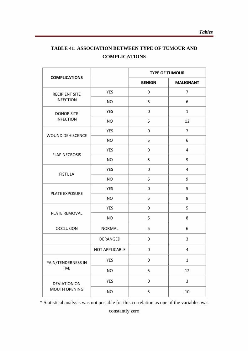
Tables
TABLE 41: ASSOCIATION BETWEEN TYPE OF TUMOUR AND
COMPLICATIONS
COMPLICATIONS TYPE OF TUMOUR
BENIGN MALIGNANT
RECIPIENT SITE INFECTION
YES 0 7
NO 5 6
DONOR SITE INFECTION
YES 0 1
NO 5 12
WOUND DEHISCENCE YES 0 7
NO 5 6
FLAP NECROSIS YES 0 4
NO 5 9
FISTULA YES 0 4
NO 5 9
PLATE EXPOSURE YES 0 5
NO 5 8
PLATE REMOVAL YES 0 5
NO 5 8
OCCLUSION NORMAL 5 6
DERANGED 0 3
NOT APPLICABLE 0 4
PAIN/TENDERNESS IN TMJ
YES 0 1
NO 5 12
DEVIATION ON MOUTH OPENING
YES 0 3
NO 5 10
* Statistical analysis was not possible for this correlation as one of the variables was
constantly zero
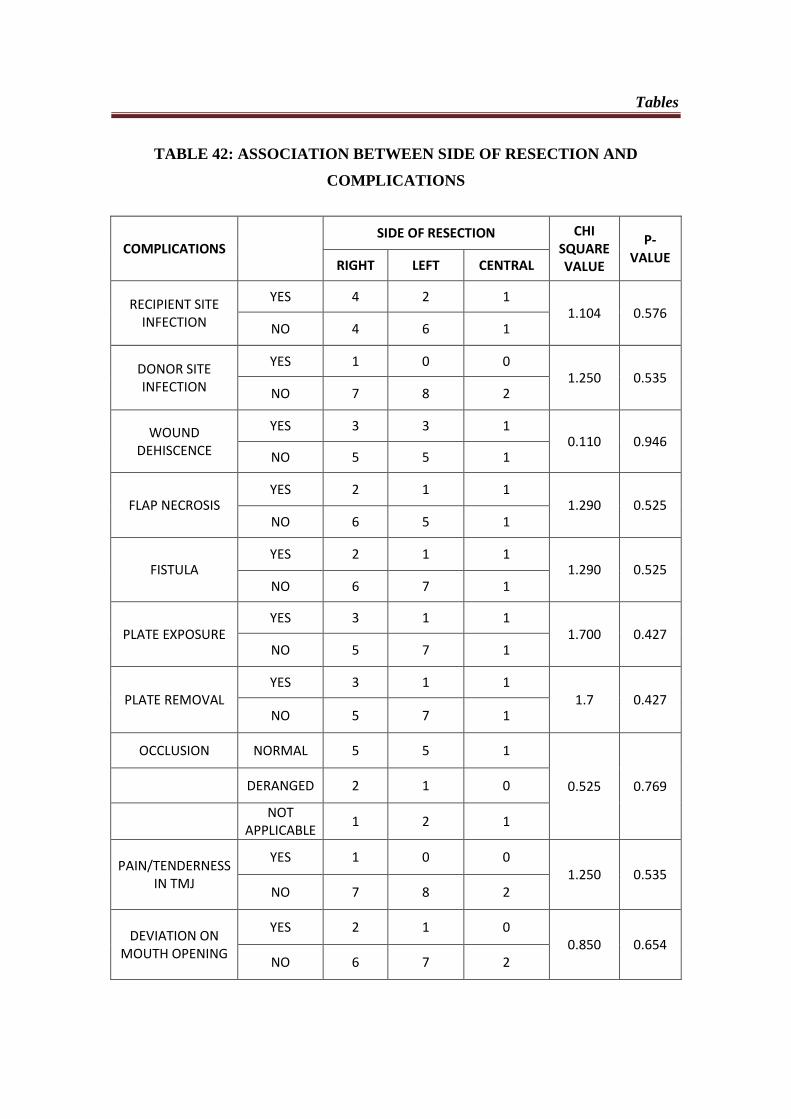
Tables
TABLE 42: ASSOCIATION BETWEEN SIDE OF RESECTION AND
COMPLICATIONS
COMPLICATIONS SIDE OF RESECTION CHI
SQUARE VALUE
P-VALUE
RIGHT LEFT CENTRAL
RECIPIENT SITE INFECTION
YES 4 2 1 1.104 0.576
NO 4 6 1
DONOR SITE INFECTION
YES 1 0 0 1.250 0.535
NO 7 8 2
WOUND DEHISCENCE
YES 3 3 1 0.110 0.946
NO 5 5 1
FLAP NECROSIS YES 2 1 1
1.290 0.525 NO 6 5 1
FISTULA YES 2 1 1
1.290 0.525 NO 6 7 1
PLATE EXPOSURE YES 3 1 1
1.700 0.427 NO 5 7 1
PLATE REMOVAL YES 3 1 1
1.7 0.427 NO 5 7 1
OCCLUSION NORMAL 5 5 1
0.525 0.769 DERANGED 2 1 0
NOT
APPLICABLE 1 2 1
PAIN/TENDERNESS IN TMJ
YES 1 0 0 1.250 0.535
NO 7 8 2
DEVIATION ON MOUTH OPENING
YES 2 1 0 0.850 0.654
NO 6 7 2
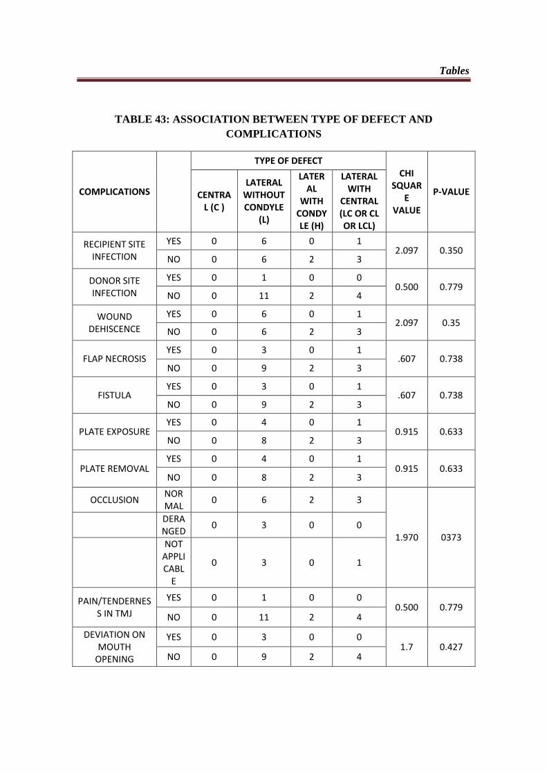
Tables
TABLE 43: ASSOCIATION BETWEEN TYPE OF DEFECT AND
COMPLICATIONS
COMPLICATIONS
TYPE OF DEFECT CHI
SQUARE
VALUE
P-VALUE CENTRAL (C )
LATERAL WITHOUT CONDYLE
(L)
LATERAL
WITH CONDYLE (H)
LATERAL WITH
CENTRAL (LC OR CL OR LCL)
RECIPIENT SITE INFECTION
YES 0 6 0 1 2.097 0.350
NO 0 6 2 3
DONOR SITE INFECTION
YES 0 1 0 0 0.500 0.779
NO 0 11 2 4
WOUND DEHISCENCE
YES 0 6 0 1 2.097 0.35
NO 0 6 2 3
FLAP NECROSIS YES 0 3 0 1
.607 0.738 NO 0 9 2 3
FISTULA YES 0 3 0 1
.607 0.738 NO 0 9 2 3
PLATE EXPOSURE YES 0 4 0 1
0.915 0.633 NO 0 8 2 3
PLATE REMOVAL YES 0 4 0 1
0.915 0.633 NO 0 8 2 3
OCCLUSION NORMAL
0 6 2 3
1.970 0373
DERANGED
0 3 0 0
NOT APPLICABL
E
0 3 0 1
PAIN/TENDERNESS IN TMJ
YES 0 1 0 0 0.500 0.779
NO 0 11 2 4
DEVIATION ON MOUTH
OPENING
YES 0 3 0 0 1.7 0.427
NO 0 9 2 4

Bibliography
Bibliography
64
1. A.C. Hundepool, A.G. Dumans, S.O.P. Hofer, N.J.W. Fokkens, S.S.
Rayat, E.H.Van der Meij, K.P. Schepman: Rehabilitation after mandibular
reconstruction with fibula free flap: Clinical outcome and quality of life
assessment. Int. J. Oral Maxillofacial Surgery 2008; 37: 1009-1013
2. A.M. Fry, R.L. Orr, A. Patterson, P.T. Doyle: Intermaxillary splint and
positioning stents to guidemandibular reconstruction. British Journal of Oral
and Maxillofacial Surgery 52 (2014) 473–474
3. Akira Matsuo, Hiroshige Chiba, Jun Toyoda, Harutsugi Abukawa, Ko
Fujikawa, Masako Tsuzuki and Masato Watanabe: Mandibular
Reconstruction Using a Tray With Particulate Cancellous Bone and Marrow
and Platelet-Rich Plasma by an Intraoral Approach. J Oral Maxillofac Surg
69:1807-1814, 2011
4. Alan S. Herford and Philip J. Boyne: Reconstruction of Mandibular
Continuity Defects with Bone Morphogenetic Protein-2 (rhBMP-2). J Oral
Maxillofac Surg 66:616-624, 2008
5. Aleksandar, Miso Virag, Vedran Uglesic: Pectoralis major flap in head and
neck reconstruction: first 500 patients. Journal of craniomaxillofacial surgery,
2006: 134(6): 340-343.
6. Bartaire, Mouawad, Mallet, Milet, El Bedoui, Ton Van, Chevalier,
Lefebvre: Morphologic assessment of mandibular reconstruction by free
fibula flap and donor-site functional impairment in a series of 23 patients.
Bibliography
65
European Annals of Otorhinolaryngology, Head and Neck diseases. 2012;
129: 230-237
7. Bee Tin Goh, Shermin Lee, Henk Tideman, Paul J. W. Stoelinga:
Mandibular reconstruction in adults: a review. Int. J. Oral Maxillofac. Surg.
2008; 37: 597–605.
8. Boyd JB, Gullane PJ, Rotstein LE, Brown DH, Irish JC: Classification of
mandibular defects. Plast Reconstr Surg. 1993 Dec; 92(7):1266-75.
9. Carlos Navarro Cuellar, Santiago Jose Ochandiano Caicoya, Julio Jesus
Acero Sanz,, Ignacio Navarro Cuellar, Cristina Maza Muela and Carlos
Navarro Vila: Mandibular Reconstruction With Iliac Crest Free Flap,
Nasolabial Flap, and Osseointegrated Implants. J Oral Maxillofac Surg
72:1226.e1-1226.e15, 2014
10. Chih-Yu Hsing, Yong-Kie Wong, Ching Ping Wang, Chen-Chi Wang,
Rong-San Jiang, Fun-Jou Chen, Shih-An Liu: Comparison between free
flap and pectoralis major pedicled flap for reconstruction in oral cavity cancer
patients – A quality of life analysis. Oral Oncology 47 (2011) 522–527
11. D.P.Coletti, R.Ord, X.Liu: Mandibular reconstruction and second
generation locking reconstruction plates: outcome of 110 patients. Int. J. Oral
Maxillofacial Surg. 2009; 38: 960-63
12. David A.Hidalgo, Andrea L. Pusic: Free-Flap Mandibular Reconstruction:
A 10-Year Follow-Up Study. Plastic and reconstructive surgery, August
2002, Vol.110, No. 2
Bibliography
66
13. David D. Vu and Brian L. Schmidt: Quality of Life Evaluation for Patients
Receiving Vascularized Versus Nonvascularized Bone Graft Reconstruction
of Segmental Mandibular Defects. J Oral Maxillofac Surg 66:1856-1863,
2008
14. David L.Hirsch, R. Bryan Bell, Eric J. Dierks, Jason K. Potter and Bryce
E. Potter: Analysis of Microvascular Free Flaps for Reconstruction of
Advanced Mandibular Osteoradionecrosis: A Retrospective Cohort Study. J
Oral Maxillofac Surg 66:2545-2556, 2008
15. Donald A. Curtis, Octavia Plesh, Arthur J. Miller, Thomas A. Curtis,
Arun Sharma, Robert Schweitzer, Raymond L. Hilsinger, Lionel Schour,
Mark Singer: A comparison of masticatory function in patients with or
without reconstructionof the mandible. Head Neck 19: 287–296, 1997.
16. Emeka Nkenke, Abbas Agaimy, Cornelius von Wilmowsky and Stefan
Eitner: Mandibular Reconstruction Using Intraoral Microvascular
Anastomosis Following Removal of an Ameloblastoma. J Oral Maxillofac
Surg 71:1983-1992, 2013
17. Eyituoyo Okoturo, Olabode Ogunbanjo, Aliru Akinleye and Martin
Bardi: Quality of Life of Patients With Segmental Mandibular Resection and
Immediate Reconstruction With Plates. J Oral Maxillofac Surg 69:2253-
2259, 2011
18. Florian Andreas Probst, Gerson Mast, Michael Ermer, Ralf Gutwald,
Rainer Schmelzeisen, Christoph Pautke, Sven Otto, Sebastian Schiel,

Bibliography
67
Michael Ehrenfeld, Carl-Peter Cornelius and Marc Christian Metzger:
MatrixMANDIBLE Preformed Reconstruction Plates—A Two-Year Two-
Institution Experience in 71 Patients. J Oral Maxillofac Surg 70:e657-e666,
2012
19. Gilles Guerrier, Ali Alaqeeli, Ammar Al Jawadi, Nancy Foote,
Emmanuel Baron, Ashraf Albustanji: Reconstruction of residual
mandibular defects by iliac crest bone graft in war-wounded Iraqi civilians,
2006–2011. British Journal of Oral and Maxillofacial Surgery (2012)
YBJOM-3847
20. Harry R.F. Powell, Mustafa Jaafar, Brian Bisase, Cyrus J. Kerawala:
Resorption of fibula bone following mandibularreconstruction for
osteoradionecrosis. British Journal of Oral and Maxillofacial Surgery 52
(2014) 375–378
21. Hitoshi Yoshimura, Seigo Ohba, Mikiko Nakamura and Kazuo Sano:
Mandibular Reconstruction Using Iliac Bone and Great Auricular Nerve
Grafts and Oral Rehabilitation Using Osseointegrated Implants in a Patient
With a Large Ossifying Fibroma: A 10-Year Follow-Up Study. J Oral
Maxillofac Surg 71:2176-2188, 2013
22. J.j. Wang, J. Chen, F.y. Ping, F.g. Yan: Double-step transport distraction
osteogenesis in the reconstruction of unilateral large mandibular defects after
tumour resection using internal distraction devices. Int. J. Oral Maxillofac.
Surg. 2012; 41: 587–595.
Bibliography
68
23. Jewer et al: Orofacial and mandibular reconstruction with the iliac crest free
flap: a review of 60 cases and a new method ofclassification. Plast Reconstr
Surg. 1989 Sep;84(3):391-403
24. Juanfang Zhu, Yan Xiao, Fei Liu, Jing Wang, Wenli Yang and Weihong
Xie: Measures of health-related quality of life and socio-cultural aspects in
young patients who after mandible primary reconstruction with free fibula
flap. World Journal of Surgical Oncology 2013, 11:250
25. K.R.Spencer, A.Sizeland, G.I.Taylor, D.Wiesenfeld: The use of titanium
mandibular reconstruction plates in patients with oral cancer. Int J Oral
Maxillofacial Surgery 1999; 28: 288-290.
26. K.Yagihara, S.Okabe, J.Ishii, T.Amagasa, M.Yamashiro, S.Yamaguchi,
S.Yokoya, T.Yamazaki, Y.Kinoshita: Mandibular reconstruction using a
poly (L-lactide) mesh combined with autogenous particulate cancellous bone
and marrow: a prospective clinical study. Int. J. Oral Maxillofac. Surg. 2013;
42: 962–969.
27. Koord Smolka, Michel Kraehenbuehl, Nicole Eggensperger, Wock
Hallermann, Hanna Thoren, Tateyuki Iizuka, Wenko Smolka: Fibula
free flap reconstruction of the mandible in cancer patients: Evaluation of a
combined surgical and prosthodontic treatment concept. Oral Oncology
(2008) 44, 571– 581
28. Krishnakumar Thankappan, Nirav Pravin Trivedi, Pramod Subash,
Sreekumar Karumathil Pullara, Sherry Peter, Moni Abraham
Bibliography
69
Kuriakose and Subramania Iyer: Three-Dimensional Computed
Tomography-Based Contouring of a Free Fibula Bone Graft for Mandibular
Reconstruction. J Oral Maxillofac Surg 66:2185-2192, 2008
29. Larissa Sweeny, William P. Lancaster, Nichole R. Dean, J. Scott
Magnuson, William R. Carroll, Patrick J. Louis, Eben L. Rosenthal: Use
of Recombinant Bone Morphogenetic Protein 2 in Free Flap Reconstruction
for Osteonecrosis of the Mandible. J Oral Maxillofac Surg 70:1991-1996,
2012
30. Lidiya Zavalishna, Nour Karra, Waleed SulimanZaid and Michel El-
Hakim: Quality of Life Assessment in Patients After Mandibular Resection
and Free Fibula Flap Reconstruction. J Oral Maxillofac Surg 72:1616-1626,
2014
31. M.W. Ho, J.S. Brown, R.J. Shaw: Intraoperative temporary fixation for
primary reconstruction of composite mandibular ablative defects. British
Journal of Oral and Maxillofacial Surgery 51 (2013) 976–977
32. Masaya Okura, Emiko Tanaka Isomura, Seiji Iida, Mikihiko Kogo:
Long-term outcome and factors influencing bridging plates for mandibular
reconstruction. Oral Oncology (2005) 41, 791–79
33. Mathew Bak, Adam S. Jacobson, Daniel Buchbinder, Mark L.Urken:
Contemporary reconstruction of the mandible. Oral Oncology 46 (2010) 71–
76
Bibliography
70
34. Mohamed A.F.El-Zohairy, Ahmed Mostafa, Ayman Amin, Hisham Abd
El-Fattah, Sherouk Khalifa: Mandibular reconstruction using pectoralis
major myocutaneous flap and titanium plates after ablative surgery for locally
advanced tumours of the oral cavity. Journal of the Egyptian Nat Cancer Inst,
vol 21, No 4, December 2009, 299-307
35. N. Parbo, N.T. Murra, K. Andersen, J. Buhl, B. Kiil, S.E. Norholt:
Outcome of partial mandibular reconstruction with fibula grafts and implant-
supported prostheses. Int. J. Oral Maxillofac. Surg. 2013; 42: 1403–1408.
36. N. Zwetyenga, F. Siberchicot, A. Emparanza: Reconstruction of large
mandibular and surrounding soft-tissue defects using distraction with bone
transport. Int. J. Oral Maxillofac. Surg. 2012; 41: 1215–1222.
37. Neelam N. Andrade, Shaikhan M. S. Reshamwala and K. Subburaj:
Mandibular Reconstruction Through Transport Distraction Using Intraoral
Appliance. J Oral Maxillofac Surg 69:2260-2269, 2011
38. P.Salvatori, E.Motto, S.Paradisi, A.Zani, S.Podrecca, R.Molinari:
Oromandibular reconstruction using titanium plate and pectoralis major
myocutaneous flap. Acta Otorhinolaryngologica Italica 2007; 27: 227-232.
39. Padraig OFearraigh: Review of methods used in the reconstruction and
rehabilitation of the maxillofacial region. Journal of the Irish Dental
Association 2009; 56 (1): 32-37.
40. Peter Maurer, Alexander W. Eckert, Marcus S. Kriwalsky, Johannes
Schubert: Scope and limitations of methods of mandibular reconstruction: a
Bibliography
71
long-term follow-up. British Journal of Oral and Maxillofacial Surgery 48
(2010) 100–104
41. Praveen Sharma, Rhodri Williams, Andrew Monaghan: Spontaneous
mandibular regeneration: another option for mandibular reconstruction in
children. British Journal of Oral and Maxillofacial Surgery 51 (2013) e63–
e66
42. Qilong Wan, Roger Arthur Zwahlen, Gu Cheng, Zhi Li, Zubing Li:
Influence of mandibular reconstruction on patients’ Health- related Quality of
life. J Oral Maxillofac Surg 69: 1782-1791, 2011
43. Qu Xingzhou, Zhang Chenping, Zhong Laiping, Ruan Min, Zhou
Shanghui, Wang Mingyi: Deep circumflex iliac artery flap combined with a
costochondral graft for mandibular reconstruction. British Journal of Oral and
Maxillofacial Surgery 49 (2011) 597–601
44. R. C. W. Wong, H. Tideman, L. Kin, M. A. W. Merkx: Biomechanics of
mandibular reconstruction: a review. Int. J. Oral Maxillofac. Surg. 2010; 39:
313–319.
45. Raphael Lopez, Celine Dekeister, Ziad Sleiman and Jean-Roch Paoli:
Mandibular Reconstruction Using the Titanium Functionally Dynamic
Bridging Plate System: A Retrospective Study of 34 Cases. J Oral Maxillofac
Surg 62:421-426, 2004
46. Raul Gonzalez – Garcia, Luis Naval-Gías, Francisco J. Rodríguez-
Campo and Leticia Román-Romero: Reconstruction of Oromandibular
Bibliography
72
Defects by Vascularized Free Flaps: The Radial Forearm Free Flap and
Fibular Free Flap as Major Donor Sites. J Oral Maxillofac Surg 67:1473-
1477, 2009
47. Rui Fernandes: Fibula free flap in mandibular reconstruction. Atlas Oral
Maxillofacial Surg Clin N Am 14 (2006) 143-150.
48. S.Arun Paul, A. Kaneesh Karthik, Rabin Chacko, and Whinny Karunya:
Audit on titanium reconstruction of mandibular defects for jaw lesions. J
Pharm Bioallied Sci. Jul 2014; 6(Suppl 1): S39–S43.
49. Si-Lian Fang, You-yuanWang, Wei-liangChen and Da-ming Zhang: Use
of extended vertical lower trapezius island myocutaneous flaps to cover
exposed reconstructive plates. J Oral Maxillofac Surg 72:2092.e1-2092.e7,
2014
50. T. J. Verhoeven, C. Coppen, R. Barkhuysen, E. M. Bronkhorst, M. A. W.
Merkx, S. J. Berge, T. J. J. Maal: Three dimensional evaluation of facial
asymmetry after mandibular reconstruction: validation of a new method using
stereophotogrammetry. Int. J. Oral Maxillofac. Surg. 2013; 42: 19–25.
51. Todd G. Carter Pardeep S. Brar, Andrew Tolas and O. Ross Beirne: Off-
Label Use of Recombinant Human Bone Morphogenetic Protein-2 (rhBMP-
2) for Reconstruction of Mandibular Bone Defects in Humans. J Oral
Maxillofac Surg 66:1417-1425, 2008
Bibliography
73
52. VN Okoje, OS Obimakinde, JT Arotiba, AO Fasola, SO Ogunlade, AE
Obiechina: Mandibular defect reconstruction with nonvascularized iliac crest
bone graft. Nigerian Journal of Clinical Practice. Apr-Jun 2012, Vol 15,
Issue 2
53. Y. Matsui, T. Shirota, T. Iwai, T. Ozawa, K. Watanuki, M. Hirota, I.
Tohnai: J. Maegawa: Mandibular reconstruction using 2-step bone transport
in an irradiated cancer patient with type 2 diabetes mellitus Int. J. Oral
Maxillofac. Surg. 2009; 38: 1201–1225.
54. Yi Shen, Jian Sun, Jun Li, Jun Shi and Andrew Ow: Long-term results of
partial double-barrel vascularised fibula graft in symphysis for extensive
mandibular reconstruction. J Oral Maxillofacial Surgery 70: 983-991, 2012
55. Zachary S. Peacock, Salim Afshar, Saylan J. Lukas and Leonard B.
Kaban: Customized Repair of Fractured Mandibular Reconstruction Plates. J
Oral Maxillofac Surg 70:e563-e573, 2012
56. Zubing Li, Yifang Zhao, Sheng Yao, Jihong Zhao, Shibin Yu and
Wenfeng Zhang: Immediate Reconstruction of Mandibular Defects: A
Retrospective Report of 242 Cases. J Oral Maxillofac Surg 65:883-890, 2007

Annexure
Annexure
SRI RAMAKRISHNA DENTAL COLLEGE AND HOSPITAL
DEPARTMENT OF ORAL AND MAXILLOFACIAL SURGERY
REVIEW PROFORMA
PATIENT NAME:
AGE/SEX:
DIAGNOSIS:
TREATMENT DONE:
ADJUVANT THERAPY:
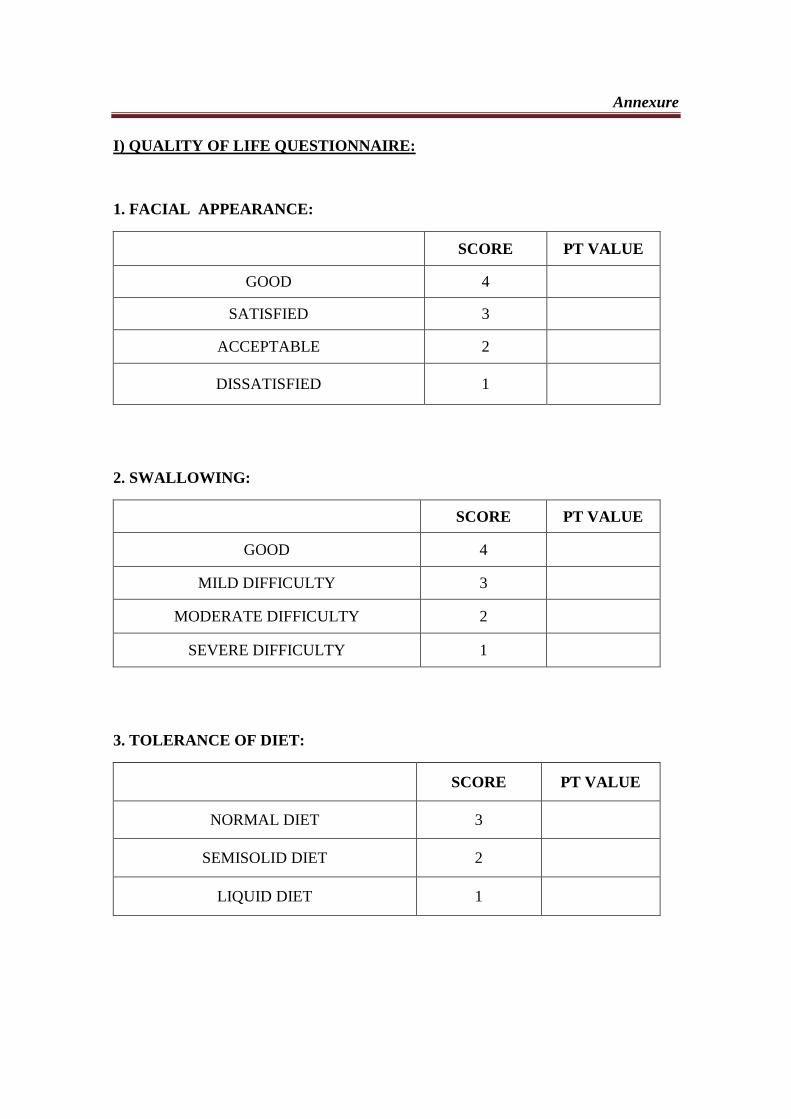
Annexure
I) QUALITY OF LIFE QUESTIONNAIRE:
1. FACIAL APPEARANCE:
SCORE PT VALUE
GOOD 4
SATISFIED 3
ACCEPTABLE 2
DISSATISFIED 1
2. SWALLOWING:
SCORE PT VALUE
GOOD 4
MILD DIFFICULTY 3
MODERATE DIFFICULTY 2
SEVERE DIFFICULTY 1
3. TOLERANCE OF DIET:
SCORE PT VALUE
NORMAL DIET 3
SEMISOLID DIET 2
LIQUID DIET 1
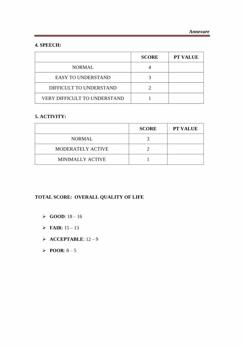
Annexure
4. SPEECH:
SCORE PT VALUE
NORMAL 4
EASY TO UNDERSTAND 3
DIFFICULT TO UNDERSTAND 2
VERY DIFFICULT TO UNDERSTAND 1
5. ACTIVITY:
SCORE PT VALUE
NORMAL 3
MODERATELY ACTIVE 2
MINIMALLY ACTIVE 1
TOTAL SCORE: OVERALL QUALITY OF LIFE
GOOD: 18 – 16
FAIR: 15 – 13
ACCEPTABLE: 12 – 9
POOR: 8 – 5
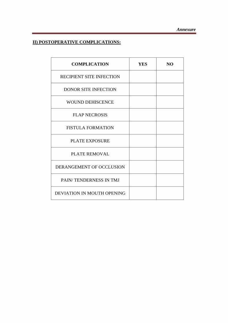
Annexure
II) POSTOPERATIVE COMPLICATIONS:
COMPLICATION YES NO
RECIPIENT SITE INFECTION
DONOR SITE INFECTION
WOUND DEHISCENCE
FLAP NECROSIS
FISTULA FORMATION
PLATE EXPOSURE
PLATE REMOVAL
DERANGEMENT OF OCCLUSION
PAIN/ TENDERNESS IN TMJ
DEVIATION IN MOUTH OPENING