Embed Size (px)
Citation preview
Technical Notes
Evaluation of Indium-111-2-Mercaptopyridine-TV-Oxide for Labeling Leukocytes in Plasma: A Kit Preparation M L Thakur, S. L. McKenney. and C. H. Park
Thomas Jefferson University, Department of Radiation Therapy and Nuclear Medicine,
Philadelphia, Pennsylvania
Pure neutrophils, lymphocytes, and mixed leukocytes have been labeled in vitro with 111ln chelated to a nontoxic, water soluble agent 2-mercaptopyridine-A/-oxide (Merc). Cells were labeled in isotonic salt-balanced medium with preformed [111ln]Merc (yield >95%), or in 0.5 ml autologous plasma by incubation with dry Merc first and then with 111ln (yield 75%). The latter method facilitated a kit procedure that required 2 /xg dry Merc when acid citrate dextrose was used as anticoagulant or 20 /ig when heparin was used. Labeling efficiency was dependent on cell concentration and pH. Labeled cells accumulated avidly in experimental abscesses and provided abscess/blood ratios of >75 at 24 hr postinjection. In dogs, the liver uptake of labeled cells was only 18.8 ± 7.1 % compared to that of 48.5% when cells were labeled with [111ln]oxine.
J Nucl Med 26: 518-523, 1985
D. uring the past few years we have witnessed a dramatic increase in the use of indium-111- ( n l ln) labeled leukocytes for detecting abscesses and other inflammatory processes induced experimentally or occurring clinically deep in the body (1-10). The technique of labeling neutrophils with a gamma-emitting radionuclide, first evaluated in 1976 and further studied for its feasibility in patients since 1977, was based upon the passive diffusion of m l n chelated with 8-hydroxyquinoline (oxine) {] 1,12). In the presence of plasma however, m l n chelated with oxine binds to transferrin and necessitates the removal of blood plasma requiring the cells to be suspended in a suitable balanced salt media for efficient incorporation of radioactivity. Since this is undesirable, newer lipophilic agents such as f'' 'Injacetylacetone (13) and tropolone (14 J5) have been developed. While some investigators have used them efficiently {16,17) others have found them to be less attractive than [ n ' Injoxine {I ft-21).
Received Sept. 21. 1984; revision accepted Feb. 4. 1985. F or reprints contact: M. L. Thakur, PhD. Professor, and Director
Radiopharmaceutical Research. Div. of Nuclear Medicine, Dept. of Radiation Therapy and Nuclear Medicine, Thomas Jefferson University. 11th and Walnut Sts., Philadelphia, PA 19107.
We have recently developed a new agent, namely [,nIn]-2-Mercaptopyridine-/V-oxide ([1HIn]Merc), which allows efficient labeling of human platelets in plasma by a kit procedure (22). The purpose of this investigation was to evaluate the use of [ m In]Merc to label leukocytes in plasma, also by a kit procedure. Various parameters that may optimize labeling yields have been studied and the efficacy of the new agent has been compared with those of oxine and tropolone. The ability of labeled leukocytes to accumulate in experimental abscesses has been compared with gallium-67 (67Ga) citrate in dogs.
MATERIALS AND METHODS
The new agent: Properties, preparation, and stability
The chemical characteristics of Merc, its toxicity and the methods of preparing [,1!In]Merc are described elsewhere (22). It has been shown that Merc with n iIn forms a lipid soluble (1:3) complex at a wide range of pH and is stable under normal laboratory conditions (23,24).
Studies of the influence of some cationic impurities such as Zn2+, Cd2+, Fe2+, and Fe3+ that may exist in [] ]'In]chloride
518 Thakur, McKenney, and Park The Journal of Nuclear Medicine
by on December 19, 2018. For personal use only. jnm.snmjournals.org Downloaded from
solution on the formation of ['' •in] Merc complex, have shown that the presence of Fe3+ interferes most {22).
For investigational use, ['' 'In]Merc was prepared in either 0.9% NaCl, 0.05%A/ phosphate buffer pH 7.4,0.1 Af acetate buffer pH 7.4 or 3.8% sodium citrate. To 1 ml of these solutions were added a known quantity of Merc (1 mg/ml prepared freshly in corresponding medium) and [niln]chloride (50 mCi/ml). Five hundred microliters of this preparation were diluted to 1 ml with a corresponding buffer solution and subjected to chloroform extraction. Percentage extraction efficiency was determined. The other 0.5 ml of [niIn]Merc aqueous solution was used for cell labeling.
Alternatively 1 to 50 ix\ of 1 mg/ml Merc solution in 0.05M phosphate buffer pH 7 were dispensed in sterile polystyrene test tubes, air-dried in a laminar flow hood, and stored for subsequent use. A required quantity of []' 'ln]chloride was also dried to eliminate the acid or buffered with citrate buffer pH 6.5 so that the final strength of the citrate buffer solution was 0.25M and the final total volume of [!' 'In]citrate was not more than 50 ^1 (0.0125 mAf).
Cell separation procedures
Studies, with the exception of three abscess-bearing dogs, were carried out using venous blood obtained from healthy human volunteers. Blood was drawn in a 30-ml syringe containing ~300 units of heparin* or 4 ml of acid citrate dextrose solution. For studies requiring a larger number of cells, multiples of these blood volumes were used. Leukocyte-rich plasma was obtained using our routine procedure in which erythrocytes were allowed to sediment by hanging the syringe upright supported by an U-shaped 19 G needle in a laminar flow hood (24). The sedimentation was aided by adding to the blood 3% (v/v) of 2% (w/v) methylcellulose (Erythrocytes in patients' blood were allowed to sediment spontaneously). Leukocyte-rich plasma was then separated and centrifuged at 450 g for 5 min. A cell button thus obtained was then suspended either in 0.5 ml plasma, or washed once with phosphate buffered saline (PBS, pH 7) and suspended in 1.5 ml PBS. On most occasions mixed leukocytes were used. However, in some studies, pure neutrophils or lymphocytes were employed. These were separated using either Boyum's Lymphoprep {25) or noncontinuous density gradient media of 50, 60, and 65% Percoll.*
Labeling leukocytes with [mIn]Merc
Using [H1In]Merc, leukocytes could be labeled either in phosphate buffered saline or in plasma. Since being able to label cells efficiently in plasma was our primary objective, a major proportion of evaluation studies was carried out with cells labeled in plasma. For labeling cells in PBS, preprepared [mIn]Merc was used and cells were incubated at room temperature for 15 min. Cells were then centrifuged at 450 g X 5 min and the cell button was washed by carefully layering over 0.5 ml leukocyte-poor plasma. Labeled leukocytes were then resuspended in plasma.
In plasma labeling, cells were suspended in 0.5 ml plasma, incubated with dry Merc for 5 min in a polystyrene tube (not polypropylene) and then with m I n either dry or in 0.25M citrate buffer pH 6. The mixture was incubated for 20 min at room temperature, and centrifuged at 450 g for 5 min. The
supernatant was removed, fresh leukocyte-poor plasma was gently layered over the cell button and removed. Finally cells were suspended in 5 ml plasma. A sterility test was performed by adding a 100 p\ aliquot to a Trypticase soy broth tissue culture* and incubating at 37°C for 8 days. A smear was made for observing structural integrity and differential cell counting. The specific conditions evolved from the results of experiments described as follows.
Influence of pH
Since it is difficult to maintain pH of plasma, PBS was used as a suspending medium and pH (3.8-7) was then adjusted using isotonic solution of sodium citrate or citric acid. Pure neutrophils at a concentration of 1.3 to 4.4 million cells/ml and pure lymphocytes at a concentration of 21 million/ml were used. To each test tube was then added [3 nIn] Merc, and the cells were incubated at 22°C for 15 min, centrifuged and labeling efficiency was determined.
Effect of anticoagulant: Heparin compared with acid citrate dextrose (ACD-A)
Ninety milliliters of blood were drawn using either 9001.U. of heparin or 12 ml ACD-A as an anticoagulant and leukocytes were separated as described before. They were then suspended in 3.5 ml plasma. Half a milliliter of this suspension was incubated with an accurately known quantity of dry Merc which ranged between 1 to 100 /zg. Cells were then incubated with ' " In and labeling efficiency was determined in the usual manner.
Effect of cell concentration
Leukocytes separated from 90 ml of blood drawn using heparin as an anticoagulant were employed. These were suspended in 1 ml plasma and cell concentration was determined using a Coulter counter. Contaminating erythrocytes were eliminated by hypotonic lysis.
One hundred to 500 fil aliquot of this suspension was then added to test tubes containing dry Merc. This was followed by the addition of autologous plasma so that the final volume in each test tube was 500 nl. The cells were then incubated with 1 Mln and labeling efficiency was determined.
Comparative use of Merc, oxine, and tropolone
Test tubes containing known quantities of either Merc, oxine, or tropolone were incubated with ~60 million leukocytes suspended in 0.5 ml plasma and then with x u In. The percentage of labeling efficiencies were then calculated for each chelating agent.
Differential radioactivity distribution
The aim of this exercise was to determine the percentage of radioactivity associated with neutrophils, lymphocytes, erythrocytes, and contaminating platelets when leukocytes were labeled with ['' 'InJMerc in plasma using the dry Merc kit procedure carried out for clinical studies. This was accomplished by diluting an aliquot of ~ 100 fi\ labeled cells
Volume 26 • Number 5 • May 1985 519
by on December 19, 2018. For personal use only. jnm.snmjournals.org Downloaded from
(ready for injection) to 3 ml leukocyte-poor plasma and cen-trifuging over Ficoll-Hypaque (1.077 g/cm3) density gradient at 450 g for 30 min, as per Boyum (25).
For quantitative distribution of radioactivity, platelets in the upper plasma layer and lymphocytes at the interphase were separated. Erythrocytes in the cell button were iysed with 1 ml water for 60 sec, centrifuged and the supernatant separated from neutrophils at the bottom of the test tube. Radioactivity in the supernate and neutrophils was measured in an ionization chamber and percentage radioactivity in each fraction was calculated.
In vivo evaluation
Studies were carried out in three dogs in which abscesses were induced by an intramuscular injection of 0.1 ml turpentine (2). Twenty-four hours following this procedure, animals received ~500 jiCi of [''' In] Merc labeled autologous leukocytes as well as 1.5 mCi [67Ga]citrate. The next day, the animals were killed with an overdose of pentobarbital and tissues were dissected. These were weighed and concomitant! n In and 67Ga radioactivity was determined using a 15 cc Ge (Li) detector1
coupled to a multi-channel analyzer.** Suitable m I n and 67GA standards were made and counted similarly to enable us to determine percentage radioactivity of each radionuclide associated with each type of tissue. Abscess to blood, fat, muscle, liver, and spleen ratios were also calculated and compared to those with 1!1In]oxine labeled leukocytes studied previously (2).
RESULTS
Using Merc as a chelating agent, neutrophils or lymphocytes can be labeled when suspended either in PBS (average of 95% labeling efficiency) or in plasma (average of 75% labeling efficiency).
In PBS labeling a preprepared ['J 'In]Merc was used. Such preprepared [ ,nIn]Merc can also be used when plasma is employed as a suspending medium. However, incubating cells in plasma with dry Merc for 5 min at 22°C, followed by a 15-min incubation also at 22°C, with ' " In either dry or in 0.25M citrate buffer pH 6 (total volume less than 50 fi\), made the procedure simple, attractive, and easy to adapt for the preparation of a kit.
Table 1 indicates that the highest incorporation of radioactivity was achieved at neutral pH and that it decreased rapidly as pH of the medium decreased. Labeling efficiency was also dependent on cell concentration and decreased as the
TABLE 1 Influence of pH on Labeling Efficiency (LE)
Neutrophils Lymph 21 mill
ocytes -13 million/ml 44 million/ml
Lymph 21 mill ion/ml
LE LE LE pH (%) pH (%) pH <%)
3.8 14.4 4.5 44.3 4.8 35.8 5.2 45.9 5.8 90.7 5.5 76.5 6.4 91.9 6.5 96.9 6.2 94.6 6.9 94.4 6.6 98.2 6.4 96.0 7.0 93.0 6.8 98.0 6.9 96.7
S 8 0
<r
| 60 o u z > 40
o < 20 o o < 3* 10 8 62 58 5
x I06 LEUKOCYTES / ml PLASMA
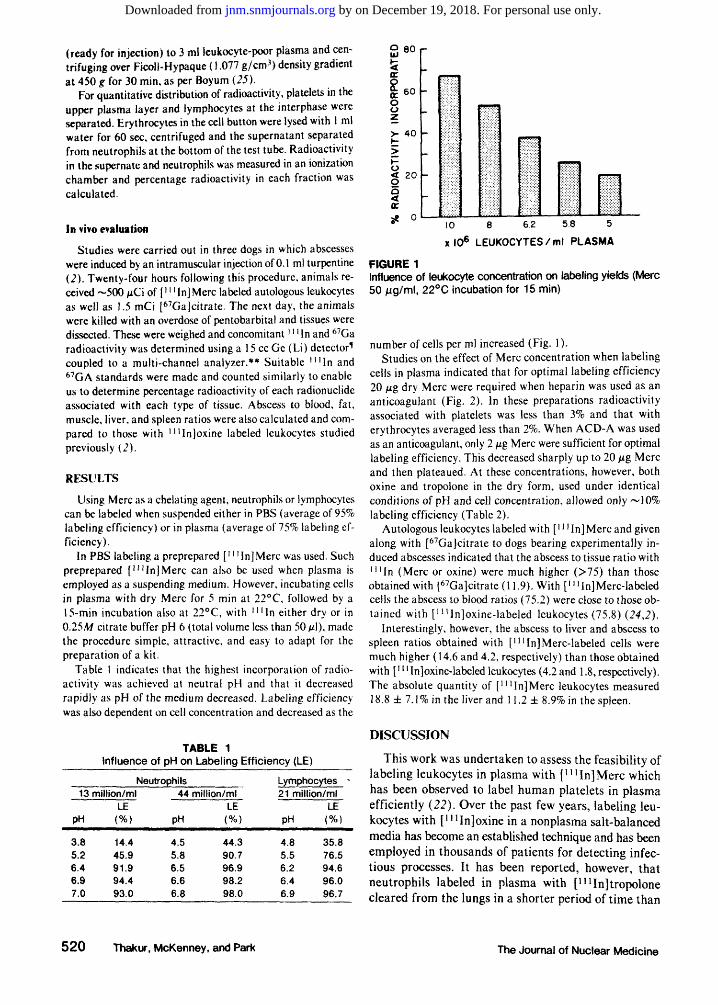
FIGURE 1 Influence of leukocyte concentration on labeling yields (Merc 50 /ug/ml, 22°C incubation for 15 min)
number of cells per ml increased (Fig. 1). Studies on the effect of Merc concentration when labeling
cells in plasma indicated that for optimal labeling efficiency 20 jug dry Merc were required when heparin was used as an anticoagulant (Fig. 2). In these preparations radioactivity associated with platelets was less than 3% and that with erythrocytes averaged less than 2%. When ACD-A was used as an anticoagulant, only 2 /ug Merc were sufficient for optimal labeling efficiency. This decreased sharply up to 20 jug Merc and then plateaued. At these concentrations, however, both oxine and tropolone in the dry form, used under identical conditions of pH and cell concentration, allowed only ~10% labeling efficiency (Table 2).
Autologous leukocytes labeled with [n!In]Merc and given along with [67Ga]citrate to dogs bearing experimentally induced abscesses indicated that the abscess to tissue ratio with i n I n (Merc or oxine) were much higher (>75) than those obtained with [67Ga]citrate (11.9). With [niIn]Merc-labeled cells the abscess to blood ratios (75.2) were close to those obtained with [11!In]oxine-labeled leukocytes (75.8) (24,2).
Interestingly, however, the abscess to liver and abscess to spleen ratios obtained with [mIn]Merc-labeled cells were much higher (14.6 and 4.2, respectively) than those obtained with [' uIn]oxine-labeled leukocytes (4.2 and 1.8, respectively). The absolute quantity of [ lnIn]Merc leukocytes measured 18.8 ± 7.1 % in the liver and 11.2 ± 8.9% in the spleen.
DISCUSSION
This work was undertaken to assess the feasibility of labeling leukocytes in plasma with [ l n In ]Merc which has been observed to label human platelets in plasma efficiently (22). Over the past few years, labeling leukocytes with [n iIn]oxine in a nonplasma salt-balanced media has become an established technique and has been employed in thousands of patients for detecting infectious processes. It has been reported, however, that neutrophils labeled in plasma with [1!1 In]tropolone cleared from the lungs in a shorter period of time than
520 Thakur, McKenney, and Park The Journal of Nuclear Medicine
by on December 19, 2018. For personal use only. jnm.snmjournals.org Downloaded from
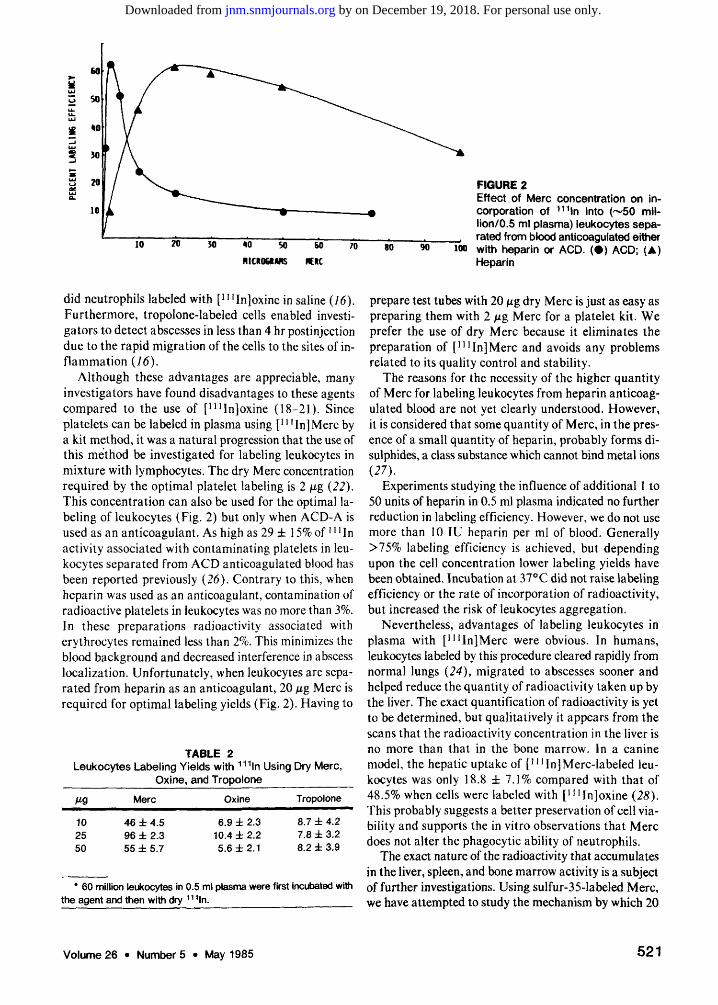
FIGURE 2 Effect of Merc concentration on incorporation of 111ln into (~50 million/0.5 ml plasma) leukocytes separated from blood anticoagulated either with heparin or ACD. ( • ) ACD; (A) Heparin
did neutrophils labeled with [inIn]oxine in saline (76). Furthermore, tropolone-labeled cells enabled investigators to detect abscesses in less than 4 hr postinfection due to the rapid migration of the cells to the sites of inflammation (16).
Although these advantages are appreciable, many investigators have found disadvantages to these agents compared to the use of [mIn]oxine (18-21). Since platelets can be labeled in plasma using [J' 'In]Merc by a kit method, it was a natural progression that the use of this method be investigated for labeling leukocytes in mixture with lymphocytes. The dry Merc concentration required by the optimal platelet labeling is 2 fig (22). This concentration can also be used for the optimal labeling of leukocytes (Fig. 2) but only when ACD-A is used as an anticoagulant. As high as 29 ± 15% of'n In activity associated with contaminating platelets in leukocytes separated from ACD anticoagulated blood has been reported previously (26). Contrary to this, when heparin was used as an anticoagulant, contamination of radioactive platelets in leukocytes was no more than 3%. In these preparations radioactivity associated with erythrocytes remained less than 2%. This minimizes the blood background and decreased interference in abscess localization. Unfortunately, when leukocytes are separated from heparin as an anticoagulant, 20 fig Merc is required for optimal labeling yields (Fig. 2). Having to
TABLE 2 Leukocytes Labeling Yields with 111ln Using Dry Merc,
Oxine, and Tropolone
M9 Merc Oxine Tropolone
10 25 50
46 ± 4.5 96 ± 2.3 55 ± 5.7
6.9 ± 2.3 10.4 ± 2.2 5.6 ±2 .1
8.7 ± 4.2 7.8 ± 3.2 8.2 ± 3.9
* 60 million leukocytes in 0.5 ml plasma were first incubated with the agent and then with dry 111ln.
prepare test tubes with 20 fig dry Merc is just as easy as preparing them with 2 fig Merc for a platelet kit. We prefer the use of dry Merc because it eliminates the preparation of [1HIn]Merc and avoids any problems related to its quality control and stability.
The reasons for the necessity of the higher quantity of Merc for labeling leukocytes from heparin anticoagulated blood are not yet clearly understood. However, it is considered that some quantity of Merc, in the presence of a small quantity of heparin, probably forms di-sulphides, a class substance which cannot bind metal ions (27).
Experiments studying the influence of additional l to 50 units of heparin in 0.5 ml plasma indicated no further reduction in labeling efficiency. However, we do not use more than 10 IU heparin per ml of blood. Generally >75% labeling efficiency is achieved, but depending upon the cell concentration lower labeling yields have been obtained. Incubation at 37°C did not raise labeling efficiency or the rate of incorporation of radioactivity, but increased the risk of leukocytes aggregation.
Nevertheless, advantages of labeling leukocytes in plasma with [3lIIn]Merc were obvious. In humans, leukocytes labeled by this procedure cleared rapidly from normal lungs (24), migrated to abscesses sooner and helped reduce the quantity of radioactivity taken up by the liver. The exact quantification of radioactivity is yet to be determined, but qualitatively it appears from the scans that the radioactivity concentration in the liver is no more than that in the bone marrow. In a canine model, the hepatic uptake of [lnIn]Merc-labeled leukocytes was only 18.8 ± 7.1% compared with that of 48.5% when cells were labeled with [mIn]oxine (28). This probably suggests a better preservation of cell viability and supports the in vitro observations that Merc does not alter the phagocytic ability of neutrophils,
The exact nature of the radioactivity that accumulates in the liver, spleen, and bone marrow activity is a subject of further investigations. Using sulfur-35-labeled Merc, we have attempted to study the mechanism by which 20
Volume26 • Numbers • May 1985 521
by on December 19, 2018. For personal use only. jnm.snmjournals.org Downloaded from
Mg dry Merc allows " Hn to incorporate into the cells. The initial results of these experiments indicated that after cell incubation, greater than 90% of Merc remained in plasma and probably only indium bound Merc entered in cells. Indium-111 was bound to cytoplasmic components and Merc also appeared to remain in association with cytoplasmic components.
When J 00 million leukocytes containing ~30 million lymphocytes are labeled with ~500 fid i n I n , each lymphocyte receives a radiation dose of >8000 rad. Consequently, they may no longer be viable and pose no further risk to the recipient (29). These are probably taken up by the spleen.
Our experiments in which we used technetium-99m (9 9 mTc), 67Ga, or thallium-201 as probable tracers for Merc incubated leukocytes produced lower labeling yields than ' ' ' In. This eliminated the possibility of using f"mTc] Merc which could have been particularly useful since early abscess detection appeared feasible. In comparison with other agents such as oxine or tropolone Merc produced higher labeling efficiency (Table 2). We believe, therefore, that Merc is not only a nontoxic, water soluble, chelating agent, but also allows efficient cell labeling in plasma by a simple kit procedure that could be uniformly performed in any laboratory. It is hoped that investigators will find this agent easier to use and will be able to eliminate the laboratory to laboratory variations that exist in cell labeling procedures today (30).
FOOTNOTES
* Elkin-Sinn, Inc., NJ. t Pharmacia. * Becton Dickinson. § Capintec CRC-5, Capintec Inc., Ramsey, NJ. ^ Canberra Industries, Meriden, CT. ** Tracor Northern, TN-1710.
ACKNOWLEDGMENTS
Part of the work was carried out during MLTs tenure in the Department of Diagnostic Radiology at Yale University School of Medicine.
The support of Medi-Physics, Inc., (a wholly-owned subs. of Hoffman-LaRoche Inc.), and helpful suggestions of Drs. Jim Lamb, Henry Kramer and Ms. Mary Sue Philps are gratefully acknowledged.
Skillful assistance of Carol Seifert, Mohan Suntherligam, and of Michael Barry, Paul Carbo, and Debbie Vivoda of Yale University School of Medicine is appreciated. Thanks are also due to Debbie Bronson who typed the manuscript.
REFERENCES
1. Thakur ML, Coleman RE, Mayhall CG, et al: Preparation and evaluation of n i In-labeled leukocytes as an abscess imaging agent in dogs. Radiology 119(3):731,
2. Thakur ML, Coleman RE, MJ Welch: Indium-111-labeled leukocytes for the localization of abscess: Preparation, analysis, tissue distribution, and comparison with gallium-67 citrate in dogs. J Lab Clin Med 89(1): 217-228,1977
3. Thakur ML, Lavender JP, Arnot RN, et al: Indium-111-labeled autologous leukocytes in man. J Nucl Med 18(10):1014-1021,1977
4. Doherty PW, Bushberg JT, Lipton MJ, et al: The use of indium-111 -labeled leukocytes for abscess detection. Clin AWM£*/3(3):108-110, 1978
5. Ascher NL, Ahrenholz DHR, Simmons LS, et al: Indium-1 11 autologous tagged leukocytes in the diagnosis of intraperitoneal sepsis. Arch Surg 114:386-392, 1979
6. Black RE, Coleman RE, Welch DM, et al: Abscess detection using autologous leukocytes labeled with indium-1 11. Curr Surg 36(4):288-290,1979
7. McAfee JG, Gagne GM, Subramanian G, et al: Distribution of leukocytes labeled with In-111 oxine in dogs with acute inflammatory lesions. J Nucl Med 21: 1059-1068,1980
8. Forstrom LA, Loken MK, Cook A, et al: In-111 -labeled leukocytes in the diagnosis of rejection and cytomegalovirus infection in renal transplant patients. Clin Nucl Med 6(4): 146-148, 1981
9. Rovekamp MH, Hardeman MR, van der Schoot JB, et a l : , n Indium-labelled leukocyte scintigraphy in the diagnosis of inflammatory disease—First results. Br J Surg 68(3):150-153, (1981)
10. Sfakianakis GN, Al-Sheikh W, Heal A, et al: Comparisons of scintigraphy with In-111 leukocytes and Ga-67 in the diagnosis of occult sepsis. J Nucl Med 23(7): 618-626, 1982
11. Thakur ML, Segal AW, Louis L, et al: Indium-111 labeled cellular blood components: Mechanism of labeling and intracellular location in human neutrophils. J Nucl Med 18(10): 1022-1026,1977
12. Zakhireh B, Thakur ML,Malech HL, et al: Indium-111 -labeled human polymorphonuclear leukocytes: Viability, random migration chemotaxis, bacterial capacity, and ultrastructure. J Nucl Med 20(7):741-747, 1979
13. Sinn H, Silvester DJ: Simplified cell labelling with indium-1 11 acetylacetone. Br J Radiol 52(621 ):7 5 8-7 59, 1979
14. Danpure HJ, Osman S, Brady F: The labelling of blood cells in plasma with inIn-tropolonate. Br J Radiol 55(651 ):247-249, 1982
15. Dewanjee MK, Rao SA, Rosemark JA, et al: Indium-111-tropolone, a new tracer for platelet labeling. Radiology 145(1): 149-153, 1982
16. Peters AM, Saverymuttu SH, Reavy HJ, et al: Imaging of inflammation with indium-Ill tropolonate labeled leukocytes. J Nucl Med 24(l):39-44, 1983
17. McAfee JG, Subramanian G, Gagne G: Techniques of leukocytes harvesting and labeling: Problems and perspectives. Semin Nucl Med XIV.83-106,1984
18. Goedemans WT: Simplified cell labelling with indium-111 acetylacetonate and indium-111 oxine. Br N Radiol 54(643) :636-637,1981
19. Mathias CJ, Heaton WA, Welch MJ, et al: Comparison of lllIn-oxine and ulIn-acetylacetone for the labeling of cells: in vivo and in vitro biological testing. IntJAppl Radial hot 32(9):651-656, 1981
20. Datz F, Baker W, Taylor A, et al: No difference in sensitivity to occult infection between oxine and tropolone
522 Thakur, McKenney, and Park The Journal of Nuclear Medicine
by on December 19, 2018. For personal use only. jnm.snmjournals.org Downloaded from
labeled In-Ill leukocytes when imaged early. J Nucl Med 25:P43, 1984 (abst)
21. Gunter KP, Lukens JN, Clanton J A, et al: Neutrophil labelling with In-1111: Tropolone vs. oxine. Radiology 149:563-566, 1983
22. Thakur ML, McKenney SL, Park CH: Simplified and efficient labeling of human platelets in plasma using in-dium-111-Mercaptopyridine-jV-oxide (Merc): Preparation and Evaluation. J Nucl Med 26:570-577, 1985
23. Thakur ML, Barry MJ: Preparation and evaluation of a new indium-111 agent for efficient labeling of human platelets in plasma. / Lab Comp Radiopharm 19: 1410-1412,1982
24. Thakur ML, Seifert CL, Madsen MT, et al: Neutrophil labeling: Problems and pitfalls. Semin Nucl Med XIV: 67-83,1984
25. Boyum A: Isolation of mononuclear cells and granulocytes from human blood. Scand J Clin Lab Invest (Suppl 21)97:77-89,1968
26. Dewanjee MK, Chowchung S, Brown ML, et al: Distribution of In-111 in granulocyte and other cellular ele
ments of blood (CBE) in human In-111 labeled mixed white cells (M WC) and platelets preparations, Fifth Int. Symposium on Radiopharmaceutical Chemistry. (Abstract) July 1984, Tokyo (to be published in J Lab Comp Radiopharm 1985)
27. Albert A, Rees CW, Tomlinson AJH: The influence of chemical constitution on antibacterial activity. Part III. 2-Mercaptopyridine-N-oxide and some general observations of metal binding agents. Br J Exp Pathol 37: 500-511,1956
28. McAfee JG, Gagne GM, Subramanian G, et al: Distribution of leukocytes labeled with In-111 oxine in dogs with acute inflammatory lesions. J Nucl Med 21: 1059-1068, 1980
29. Thakur ML, McAfee JG: Significance of chromosomal aberrations in In-111-labeled lymphocytes. J Nucl Med 25:922-927, 1984
30. Thakur ML, McKenney SL: Cell labeling techniques: An overview. Proceedings of NATO ASI on Radiolabeled Cellular Blood Elements, New York, Plenum, 1983: in press
Volume 26 * Number 5 • May 1985 523
by on December 19, 2018. For personal use only. jnm.snmjournals.org Downloaded from
1985;26:518-523.J Nucl Med. M. L. Thakur, S. L. McKenney and C. H. Park Leukocytes in Plasma: A Kit Preparation
-Oxide for LabelingNEvaluation of Indium-111-2-Mercaptopyridine-
http://jnm.snmjournals.org/content/26/5/518This article and updated information are available at:
http://jnm.snmjournals.org/site/subscriptions/online.xhtml
Information about subscriptions to JNM can be found at:
http://jnm.snmjournals.org/site/misc/permission.xhtmlInformation about reproducing figures, tables, or other portions of this article can be found online at:
(Print ISSN: 0161-5505, Online ISSN: 2159-662X)1850 Samuel Morse Drive, Reston, VA 20190.SNMMI | Society of Nuclear Medicine and Molecular Imaging
is published monthly.The Journal of Nuclear Medicine
© Copyright 1985 SNMMI; all rights reserved.
by on December 19, 2018. For personal use only. jnm.snmjournals.org Downloaded from