Embed Size (px)
Citation preview

Evaluation of Hip Pain in
Adults
Jerry Ahluwalia, M.D.
November 13, 2015

Objectives
► Develop a better understanding of the differential diagnosis of
hip pain in active young adults
► Appreciate key points in patient history
► Learn physical examination pearls for evaluating hip pain
► Understand role for various imaging modalities in patients with
hip pain
► Differentiate between those processes in which observation is
indicated and those diseases that need earlier orthopaedic referral

Hip Pain Differential Diagnosis
►Musculoskeletal
Intra-articular
Extra-articular
Radicular/ Spinal stenosis
►Non musculoskeletal
Hernia
►Abdominal/umbilical, sports, inguinal
Retroperitoneal abscess/ hematoma
Tumor/infection

DDx for Intra-articular Hip Disease
► Osteoarthritis Primary or Idiopathic
Secondary (Degenerative Joint Disease)
► Rheumatoid Arthritis and Juvenile Chronic Arthritis
► The Seronegative Spondyloarthropathies
► Psoriatic Arthritis
► Reiter’s Disease
► Ankylosing Spondylitis
► Inflammatory Bowel Associated Arthropathy
► Connective Tissue Disease
► Crystal Arthropathies
► Gout
► CPPD
► Rapidly Destructive Hip disease
► Chondrolysis
► Pigmented Villonodular Synovitis
► Synovial (Osteo) Chrondromatosis
► Metabolic and Endocrine Disease
► Nutritional Disorders
► Radiation Chrondrosis and Osteonecrosis
► Miscellaneous Disorders

AND…
►Labral tears
►Femoral acetabular impingement (FAI)
►Loose bodies
►Chondral flaps or lesions
►Ruptured ligamentum teres
►Synovial chondromatosis
►Developmental Dysplasia of the Hip (DDH)

Patient History
History of trauma vs atraumatic onset
► Subacute vs acute vs insidious
Mechanical symptoms such as locking, catching, and giving way
Pain only with prolonged weight bearing activities
Pain with out of plane activities only
Systemic symptoms or multiple joint involvement

Patient History ► Childhood conditions
First born?
Female?
Natural birth? C-section?
Bracing as a child?
► Malignancy
► Inflammatory conditions
► ETOH/ Corticosteroid use
► Back Problems

Patient History
►Occupational Risk factors
Diving
Construction/Heavy labor
Farmers
Lifting > 25 -50 Kg on regular basis
►Recreational risk factors
Hockey, Rugby, Martial Arts, Soccer, Football (place kicker, punter), etc.
►Obesity not a consistent risk factor

Patient History
►Location of pain
Groin
Buttock
Lateral
Radiation to knee and past knee

Physical Exam - Gait
►Antalgic or coxalgic gait
►Trendelenburg gait
Fractures, CDH, SCFE, old SCFE and L-5
neuropathy
►Shuffling or ataxia
►Flexed trunk/stenosis

Physical exam

Extra-articular sources of pain
►Trochanteric Bursitis
► Snapping Iliotibial Band
“External snapping hip”
► Iliopsoas tendonitis
Could result in “internal
snapping hip”
► Ischial bursitis

RANGE OF MOTION
►Can depend upon body habitus
►Can also depend upon injuries
Simple things such as swelling can affect ROM
►Joint effusion from an injury
►Post-surgical swelling

Flex 105-135°
Extension 0-20 °

Abduction 30-50 °
Adduction 10-30 °

Internal Rotation 30 °

External Rotation 60 °

Physical Exam
Assess for pelvic obliquity and leg length difference

Special Tests
►Thomas test
►FABER or Patrick’s Test
►Active SLR or Stinchfield test
►Ober test
►Impingement test {Flexion, Adduction, Internal Rotation}
►McCarthy Test for intra-articular hip pathology
►Apprehension {Extension, Abduction, External Rotation}

Thomas test - Hip flexor tightness or hip flexion contractures

Faber or Patrick’s -Pathology of the hip joint and/or sacroiliac joint

Ober Test - IT Band tightness

Tests for labral pathology and hip instability
►Apprehension test
Hip dysplasia
► Impingement test
Femoral acetabular
impingement
►McCarthy test for intra-
articular pathology
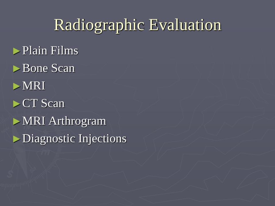
Radiographic Evaluation
►Plain Films
►Bone Scan
►MRI
►CT Scan
►MRI Arthrogram
►Diagnostic Injections

Radiographic Studies
High quality X-rays are critical
►Standing AP pelvis
►AP of the hip
►Frog lateral of the hip
►False Profile

Normal Anatomy

AP pelvis, Frog lateral view

False Profile view

CT scan
►Useful in trauma
►Delineate subtle arthritis
►Clarify unusual anatomy

Bone Scan
►Largely supplanted by
MRI
►Assessing occult lesions/
malignancy
►Determine metabolic
activity of plain film
findings

MRI
►Most sensitive and specific for
Osteonecrosis (AVN)
Stress Fractures
Early arthritis
Certain soft tissue problems about the hip
►Not as good for intra-articular labral or chondral
pathology, without contrast (arthrogram)

OSTEONECROSIS (AVN)

Transient Osteoporosis
or
Bone Marrow edema Syndrome
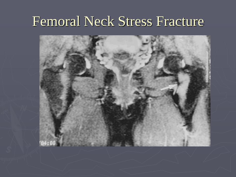
Femoral Neck Stress Fracture

Dysplasia with labral tear (and
subchondral cysts)

MR Arthrogram
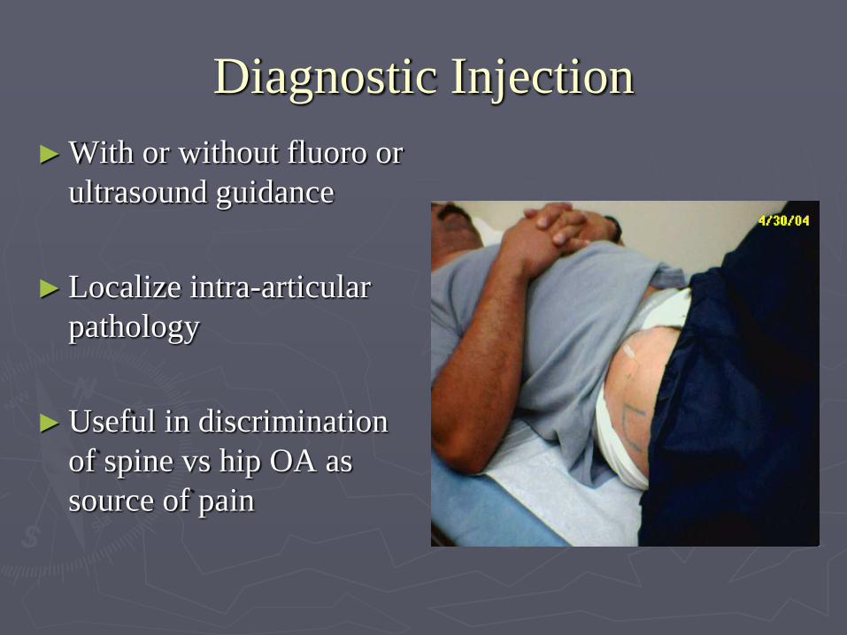
Diagnostic Injection
►With or without fluoro or
ultrasound guidance
►Localize intra-articular
pathology
►Useful in discrimination
of spine vs hip OA as
source of pain

Femoral Acetabular Impingement

FAI
►3 TYPES:
CAM (Primary lesion on the femur)
PINCER (Primary lesion on the acetabulum/pelvis)
COMBINED (Both lesions present)
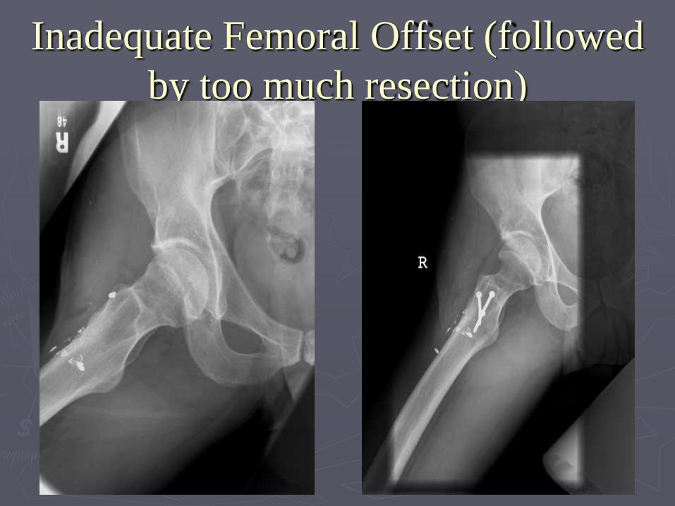
Inadequate Femoral Offset (followed
by too much resection)

Hip Arthroscopy
Minimally invasive means of seeing inside the hip
joint without cutting muscles or using big incisions
much like is done in the knee or shoulder
First done in 1930s but re-introduced in late 1980s
by Dr. Glick of San Francisco
Techniques and indications refined in mid-late 90s
allowing more predictable results

Hip Arthroscopy Becoming much more popular recently

Hip Arthroscopy Becoming much more popular recently
►Hip joint is much less accessible than other
joints
►More technically difficult
Need specialized equipment and expertise
►Conditions warranting its use are more rare
►Use and indications are emerging

Hip Arthroscopy
► Indications
Remove loose bodies such
as cartilage or bone from
hip joint as on right
►Synovial chondromatosis

Hip Arthroscopy
► Indications
Diagnostic
Synovial Biopsy

Hip Arthroscopy
►Indications
As an adjunct to other procedures in order to rule out
problems inside the hip joint or allow other
procedures to be performed less invasively
►Peri-acetabular osteotomy (PAO)

Hip Arthroscopy ►Contraindications (reasons not to do hip arthroscopy)
Advanced arthritis
Arthritis without mechanical symptoms (catching, locking)
Very stiff hips
Fresh fractures or dislocations
Surgical problems in which opening the hip joint is not necessary
Obesity…the instruments are sometimes not long enough
Hip dysplasia

Technique
► In order to view the hip
joint without scuffing
the cartilage it is
necessary to use a
traction device to open
up the hip joint and
allow instruments to be
introduced
►General or spinal
anesthesia is preferred
to allow for complete
muscle relaxation

Technique
► Special instruments have
been designed to aid
entry into hip joint and to
remove damaged tissues

Technique
► In most cases surgery can
be performed through
two small incisions
► In lower picture patient is
draped and fluoroscopy
unit in position to guide
procedure

Hip arthroscopy
Example ► 29 year old woman with
pain and catching after
intense period of exercise
8 months previously
►X-rays were normal but
the MRI arthrogram
showed a tear in the
labrum

Hip Arthroscopy
Example ►At surgery a torn labrum
was diagnosed and
excised
► Patient was back to full
activities at 3 months

Hip Arthroscopy Example
► 41 yo male with
persistent hip pain after
ATV accident 2y
previously


Post debridement….

What needs early referral
►Osteonecrosis
Early stages have higher success rates in saving hip
►Femoral neck stress fractures
Early diagnosis and treatment prevents AVN, non-union
►Sepsis…Tumor
►Early OA
Some hips are amenable to biologic solutions

OA of the Hip
usually has a cause ►DDH – 43%
►LCP – 22%
► SCFE – 11%
► Idiopathic – 12%
►Other – 12%
Aronson 1986
►By age 50 years:
25-50% in DDH
50% in Perthes
20% in SCFE

Saving Hips
►Hip Arthroscopy
►Osteotomy
►Debridement

Periacetabular Osteotomy
Treatment of choice for dysplasia
Hip Arthroscopy Failures
►When hip arthroscopies fail, they fail very
quickly – recent study presented this past
weekend during AAHKS 2015:
Tracking of patients revealed 67/1305 (5.1%)
patients that had a hip arthroscopy went on to a
subsequent ipsilateral THA within the time
constraints of the dataset (2007-2014). Of the
subsequent THA, 37.3% occurred within 6
months of hip arthroscopy and 85.1% had
occurred within 18 months. 100% of
subsequent THA occurred within 48 months of
initial hip arthroscopy.

Conclusions
►Hip Pain is a common presenting complaint
►A thorough history and examination accompanied by appropriate studies generally leads to successful diagnosis
► Improved knowledge and understanding of the causes of hip osteoarthritis has led to earlier diagnosis of hip conditions and emerging methods of treating those conditions
►Early recognition is the key

THANK YOU